Copyright ( 1973 AmericanSociety forMicrobiology Printed in U.S.A.
Nucleoprotein Complexes Containing
Replicating
Simian
Virus
40
DNA:
Comparison
with
Polyoma Nucleoprotein
Complexes
MARK R. HALL, WILLIAM MEINKE, AND DAVID A. GOLDSTEIN
Department ofMicrobiology, Scripps Clinic and Research Foundation, La Jolla, California92037
Receivedfor publication 11April 1973
Procedures for isolating nucleoprotein complexes containing replicating
poly-oma DNA from infected mouse cells were used to prepare short-lived
nucleo-protein complexes (r-SV40 complexes) containing replicating simian virus 40
(SV40) DNA from infected monkey cells. Like thepolyoma complexes, r-SV40 complexes were only partially released from nuclei by cell lysis but could be
extracted from nuclei by prolonged treatment with solutions containing Triton X-100. r-SV40 complexes sedimented faster than complexes containing SV40 supercoiled DNA (SV40 complex) in sucrose gradients, and bothtypes ofSV40 nucleoprotein complexes sedimented ahead of polyoma complexes containing supercoiled polyoma DNA (py complex). The sedimentationratesofpycomplex
and SV40 complex were 56 and 61S, respectively, based on the sedimentation
rate of the mouse large ribosomal subunit as a marker. r-SV40 complexes
sedimented asmultiple peaks between 56 and 75S. Sedimentation and buoyant
densitymeasurementsindicated thatprotein is boundtoall formsofSV40 DNA
at about the sameratio ofprotein to DNA (1-2/1) as wasreported for polyoma
nucleoproteins.
Nucleoprotein complexes containing newly replicated viral DNA can be extracted from
polyoma- and simian virus 40 (SV40)-infected
cells after lysiswith nonionic detergents (8, 10, 23). Complexes isolated after long pulses of
3H-thymidine contain mostly supercoiled viral
DNA, while complexes isolated after short pulses (5-30 min) contain bothsupercoiled and replicating viral DNA. Many propertiesof
poly-oma supercoiled DNA-protein complexes (py
complex) aresimilar tothe corresponding prop-erties of SV40 supercoiled DNA-protein
com-plexes (SV40 complex), yet the reported
sedi-mentation coefficients differby more than 20%
(8, 10, 23).
ReplicatingviralDNA-protein complexes ap-pearto be precursors tosupercoiled viral
DNA-protein complexes (8, 23). In the preceding
communication someproperties of the polyoma
complexes containing replicating DNA (r-py
complex) were described (8). In this study, we have used the same methods to isolate com-plexes containing replicating SV40 DNA
(r-SV40 complexes) from infected CV-1 cells and have compared the properties of these
com-plexes tothose ofr-py complexes.
MATERIALS AND METHODS
Virus and cells. Strain RH 911 SV40 virus was grown intheTC7 subline of CV-1 cells(17).Cellswere propagatedin 9-cmplastic petridishes (A. S. Niinc)
at 37C.MonolayersweregrowntoconfluenceinEagle
mediumsupplementedwith10%calfserum.Cultures were infected 24 to 48 h after confluence, and the viruswasallowedtoabsorb for90min. After
absorp-tion,4 ml of mediumcontaining 5% horseserumwas addedto each culture. The methods forcultivation,
isotope labeling, and purification of polyoma virus have beendescribed (8, 15).
Triton extraction of SV40-infected cells. SV40 nucleoprotein complexes were extracted by proce-duresdescribedintheprecedingpaper(8). Confluent monolayers were pulsed with 3H-thymidine (55 Ci/
mmol)at 40h after infection for2h(10
gCi/ml)
or at 41.75h for15 min (100ACi/ml).
Afterpulses, mono-layerswere washed twice with ice-cold Tris-buffered saline containing magnesium and calcium (TBS)(24).Eachpetriplatewas then treated with 0.9 ml of lysing fluid (TTE) containing 0.25% Triton X-100, 0.01 M Tris-hydrochloride (pH 7.9), and 0.01 M EDTA (10).Cellswereallowedtoswellfor10 min at 22C, and 0.1 ml of 2.0 M NaCl was added to each plate. Although addition ofNaCl caused the cells to lyse, nuclei remained morphologically intact and formed large aggregates. The lysate, containing nu-901
on November 10, 2019 by guest
http://jvi.asm.org/
HALL,MEINKE,AND GOLDSTEIN
clearaggregates,wasdecantedorgentlyscraped from
the plates with a rubber policeman and poured into
ice-cold, 12-ml centrifuge tubes. Nuclear aggregates and large cellular debriswerepelleted by
centrifuga-tionat2,000rpmfor 5minat4C in the HL-4 swinging bucketrotorofaSorvall GLC centrif'uge. The
super-natant fluid (Triton supernatant fraction) was
de-canted and stored at 4C. The SV40 DNA-protein complex in this fractionwasdesignated Triton
super-natant complex (TSC).
Thepellet obtained from low-speed centrifugation oftheTriton cell lysate was resuspended in ice-cold
TTE (0.9ml/petri dish), one-tenth volume of 2.0 M
NaClwasadded, and the solutionwasincubated for3
h on ice with occasional mixing. Nuclei remained
intact and were pelleted at 2,000 rpm as described
above. The supernatant fluid (Triton nuclear
frac-tion) was decanted and stored at 4C. The SV40 nucleoprotein complex in the nuclear fraction was
designated nuclear extraction complex(NEC). 14C-labeledpycomplex. Labeled polyoma
nucleo-protein complex (py complex) was extracted from
cellspulsed for 6 h with '4C-thymidine (1,uCi/ml)22h after infection and lysed with Triton as described
above. Pycomplex in the Tritonsupernatantfraction
was used as a marker for sedimentation coefficients
anddensitydeterminations throughout this investiga-tion. We havepreviously shown that glutaraldehyde-fixedpycomplex hasadensity in CsCl of1.437g/cm3,
and 85 to 90%/ of the DNA in the complex is supercoiled (8).
Velocity sedimentation. Linear 5 to 20% (wt/wt)
sucrose gradients were prepared in TTE buffer
con-taining 0.2 M NaCI (STTE). Samples (0.1or0.2ml) werelayeredonto4.8-ml gradients andcentrifugedat 36,000 rpm for 120 min at 4C in a Spinco SW50.1
rotor (10). Fractionswerecollected, and
trichloroace-tic acid-precipitable radioactivity was determined
(15). For velocity sedimentation in CsCl, a 0.2-ml
sample of DNA was layered on 3.0 ml of a CsCl
solution (density 1.50 g/Cm3in0.01 MEDTA, pH7.5) and centrifugedin aSpincoSW50.1 rotor at 25C for
3.0hat35,000rpm.
Buoyant density centrifugation of glutaralde-hyde-fixed complexes. Peak fractions of SV40 and polyoma nucleoprotein complexes pooled from
su-crose gradients were fixed with glutaraldehyde as
previously described (8). Thedensity of glutaralde-hyde-fixed samples of polyoma and SV40 nucleo-protein complexes was determined by analysis in preformedlinearCsClgradients (density 1.3g/cm3to
1.6 g/cm3), prepared and centrifuged as previously
described (8).
Dye-buoyant density gradient centrifugation. Analysis of DNA by dye-buoyant density centrifuga-tion in CsCl-propidium diiodide (CsCl-PI) density gradientswas described inthepreceding report(8).
Benzoylated naphthoylated DEAE-cellulose
(BND) chromatography.Portionsof 3H-labeled Tri-ton nuclear and Triton supernatant fractions (0.5to 1.0ml) werediluted toa volume of 2.7 ml in 0.2 M
Tris-hydrochloride(pH 8.0), 0.01 M EDTA. A 0.3-ml amountof10%SDSwasadded,and the solutionwas
incubated at50C for 20 min. After coolingto room
temperature,NaClwasaddedtoafinalconcentration
of0.5 M, and the DNA was extracted bymixing the
solution with an equal volume of redistilled phenol saturated with 0.2 M Tris-hydrochloride (pH 8.0), 0.01 M EDTA for15 minat roomtemperature. After
low-speed centrifugation, the phenol phase was dis-carded, yeast tRNAwas added to the aqueousphase ascarrier,and theDNA wasprecipitated by adding2 volofabsoluteethanolat -20Covernight.DNA was
suspended in 1 ml of 0.01 MTris-hydrochloride (pH 7.2), 0.001 M EDTA(Tris-EDTA) in 0.3MNaCl and appliedto a0.9-by30-cmSephadex(Pharmacia)G-50 column equilibrated with the same buffer at room temperature. The 3H-labeled DNA peak was pooled
(6-7 ml) and adsorbed to a 0.9- by 10-cm BND-cel-lulose (Gallard-Schlesinger) column equilibrated in 0.3M NaCl, Tris-EDTAbuffer (5, 14). After washing the column with 2 vol of the same buffer, double-stranded DNA was eluted by passing 1.0 M NaCl, Tris-EDTA buffer throughthecolumnat a rate of 0.5 ml/min. An additional 40 ml of 1.0 M NaCl, Tris-EDTA buffer was passed through the column, and then replicating DNA was eluted with 1 M NaCl, Tris-EDTA buffer containing 2% caffeine. The col-umn waswashed withanadditional20mlofthe 1M NaCl Tris-EDTA buffercontaining 2% caffeine, fol-lowed by a final 20-ml wash with 0.3 M NaCl, Tris-EDTA buffer containing4M urea.
RESULTS
Triton extraction of nucleoproteins from
SV40-infected cells. SV40-infected CV-1 cells were pulsed with 3H-thvmidine for 15 or 120 min about 40h after infection. Cells were lysed with Triton X-100 and separated into
superna-tant and nuclear pellet fractions. Nuclei were
resuspended in buffer containing Triton and
SV40nucleoprotein complex were extracted for
3 h at 0 C. After extraction, nuclei were recen-trifuged and low-molecular-weight DNA was ex-tracted from the resulting pellet by a modi-fication of the Hirt SDS selective extraction
procedure (8, 12). The sum of the
radioactivity
in these three cell fractions was about equal to the radioactivitv obtained by Hirt extraction of total viral DNA (8).
Samples were withdrawn from each
sample,
and the alkali-resistant, trichloroacetic acid-precipitableradioactivity wasmeasured (Table
1).After a 15-minpulse, the Tritonsupernatant
fraction contained only about halfof the total radioactive viralDNA, while the remainderwas
associated with the nuclear fraction. About 70%/
of the viral DNA remaining in the nuclear fraction could be extracted by prolonged incu-bation ofnuclei with Triton lysingbuffer. After
a2-hpulse, however, themajorityofradioactive viral DNA was found in the Triton supernatant
fraction while onlyan additional
14%7c
wasreex-tracted from nuclei.
Portions from each extract were mixed with
4C-thymidine-labeled
pycomplex,
and the902 J. VIROL.
on November 10, 2019 by guest
http://jvi.asm.org/
TABLE 1. Distribution ofSV40nucleoprotein complexes in subcellular fractions"
Fraction (7' ot total) Lengthot
pulse(min) Triton Triton'nuclei SDS pellet supernatant
15 51 30 13
120 78 14 8
The valuesarebasedonalkali-resistant. trichloro-acetic acid-precipitableradioactivity (8).
Nuclei obtained
by
Tritonlvsis
wereextractedbv incubationfor3h in S'I'TE but'l'er.'Nuclei extracted in STTE butter were recen-trit'uged and extracted by a modit'ied Hirt procedure
(8).
samples were analyzed in 5 to 20% sucrose gradients (Fig. 1). In all gradients SV40 nucleo-proteincomplexes sedimented aheadofpy com-plex. SV40 nucleoprotein complex extracted in
the Triton supernatant fraction
(TSC)
after a120-min pulse sedimented as a homogeneous
component only slightly ahead of '4C-py
com-plex (Fig. 1A). A similar pattern was observed
for thenucleoprotein complex obtainedby Tri-ton extraction of nuclei (NEC) after the
120-min pulse (not shown in Fig. 1).
In contrast, TSC prepared from cells pulsed
for 15 min sedimented as two peaks, one
corre-sponding to the TSC peak extracted after the
120-min pulse and a smaller peak sedimenting about
15%
faster (Fig. IB). The SV40 nucleo-protein complex obtained by Triton extraction ofnuclei (NEC) pulsedfor15min(Fig. 1C) alsosedimented as two peaks, but both peaks were faster than the TSC peak from the 120-min
pulse and corresponded more closely with the
fast peak in the 15-min TSC.
The results presented above are similar to
those obtained with polyoma-infected mouse cells (8). Tritonlysisofcells after a shortpulse liberates intothesupernatant fraction amixture of fastsedimenting polyoma nucleoprotein
com-plexes containing replicating viral DNA and
slower sedimenting complexes containing both replicating viral DNA and supercoiled viral
DNA. Complexes extracted from the nuclear fraction are mostly fast sedimenting and con-tain replicating viral DNA. With long pulses,
relatively small amounts of fast sedimenting
complexes containing
replicating
DNA arefoundand most ofthecomplexesextracted into
the Triton supernatant fraction contain super-coiled viral DNA.
Characterization of the DNA in SV40
nu-cleoprotein complexes. To determine whether
SV40 complexes did indeed contain a similar distributionofsupercoiled andreplicatingDNA
as that found after polyoma infection, Triton supernatant and Triton nuclear fractions were
treated with SDS to strip the protein from
complexes, and the DNA was analyzed by
velocity sedimentation, equilibrium centrifuga-tion in CsCl-PI density gradients and by
chro-Io
0
a-u
cf-)
I
(\J
0
x
a_
u~
"IU
10 20 30 40
Fraction Number
FIG. 1. Velocitysedimentation in 5 to20% sucrose gradients ofpolyoma and SV40 nucleoprotein com-plexes. SV40-infected cultures were either labeled with 3H-thymidine (10gCi/ml) for 2 h at 40 h after infection or for 15 min (100 gCi/ml) at 41.75 h after infection. SV40 nucleoprotein complexes were pre-pared from Triton supernatant (TSC) and Triton nuclear(NEC)fractions.Polyomanucleoprotein com-plex(TSC)wasprepared from cells pulsed for 6 h with "C-thymidine (1 gCi/ml) 22 h after infection and lysed with Triton. "C-labeled polyoma TSC mixed with 3H-labeled SV40TSC from a 2-h pulse (A), and 3H-labeled SV40 TSC (B), or NEC from a 15-min pulse (C).
903
on November 10, 2019 by guest
http://jvi.asm.org/
[image:3.495.48.241.100.169.2]HALL,
MEINKE,
ANDGOLDSTEINmatography on BND-cellulose. In velocity gra-dients, the Triton supernatant fraction pre-pared after a 15-min pulse contained a large peak of21SDNA, whereas 25S DNA (replicat-ing SV40 DNA) was distributed about equally between the Triton supematant fraction and the Triton nuclear fraction (Fig. 2A and B, Table 1). In contrast. after a 120-min pulse, most labeled material in both the Triton super-natant fraction and the Triton nuclear fraction was21Sviral DNA (Fig.2C and D). The nuclear extract prepared after a chase (Fig. 2D) does, nevertheless, contain a small amount of' SV40 25S DNA. However, the 25S DNA in this nuclearf'raction only represents about 5%.of' the total labeled viral DNA (see also Table 1).
Analysis of DNA in CsCl-PIequilibrium den-sity gradients clearly shows a differential ex-traction of complexes containing supercoiled andreplicating DNA between the Triton super-natant and Triton nuclear fractions after a 15-min pulse (Fig. 3A and B). About40%. of the DNA in the Triton supernat ant f'ormed a
"heavy" peakcharacteristic ofsupercoiled viral DNAwhile the restbandedat intermediate and light densities characteristic ofreplicating and
10(
I
0
(9)
5
4
2
A -oSV40
* Polyoma
nicked DNA (8, 13, 19). However, very little supercoiled DNA was found in the Triton nu-clear fraction and almost 90% of' the DNA in this fraction had buoyant densities characteris-tic of'replicating DNA (Fig. 3B). Actually, the supercoiled DNA in the Triton nuclear fraction represented only about
9%7(
of the total 3H-labeledsupercoiled DNA (refer also to Table 1). As expected, after a 120-min pulse, most of the DNA in the supernatant as well as the nuclear fraction is supercoiled (Fig. 3C and D).Comparison of'the DNA invelocity gradients (Fig. 2) and inCsCl-PIgradients (Fig. 3) reveals that about half of the DNA sedimenting in the
21S region of the Triton supernatant fraction after a 15-minpulseisactuallvSV40 replicating DNA(SV40 RI DNA). Although a large amount of radioactivity in the Triton nuclear fraction also sediments in the 21S region of' thegradient.
almost all of this DNA is replicating DNA. To showthat most of the DNA banding in the intermediate and light regions of'CsCl-PI gradi-ents was SV40 RI DNA and not breakdown of'
supercoiled
viral
DNA. DNA was extracted fromthe Triton supernatant and Triton nuclear fractions with phenol and analyzed onBND-IC
-30
BP
DA
5
0
x
30 vi
1.5
10 20 30
[image:4.495.119.400.350.626.2]Fraction Number
FIG. 2. Velocitysedimentation inCsCIgradients of SV40 DNAfrom Tritonsupernatant and Triton nuclear fractions. The Triton supernatant (A) and nuclear(B) fractionsfrom cells labeledfor15 min, as wellas the Tritonsupernatant (C)and Triton nuclear(D)fractions fromcells labeledfor120minweretreated withSDS, mixed with '4C-labeled polyomaDNA,andanalyzed byvelocitycentrifugation in CsCl.
904 J. VIROL.
1
10 20 30
on November 10, 2019 by guest
http://jvi.asm.org/
4
I
0U
I
('1
2
4
2
4
2
I
0
x
4
7
2
2
[image:5.495.98.394.55.366.2]Fraction
NumberFIG. 3. Equilibrium centrifugation of SV40 DNA from Triton supernatant and Triton nuclearfractions in cesiumchloride-propidium diiodide gradients.SampleswerepreparedbymixingSDS-treated 3H-labeled SV40 fractions with "C-labeledpolyoma DNAmarker,propidium-diiodide,andCsCI.Solutions were adjusted to a final densityof1.515g/cm3and centrifuged aspreviously described(8). Triton supernatant (A) and Triton
rnuclear(B) fractionsfrom cells labeled for 15 min; Triton supernatant (C) and Triton nuclear (D) fractions from cells labeledfor120min.
cellulose columns. Double-strandedDNAelutes
fromthese columns at 1 M NaCl while
replicat-ing DNA remains bound but can be elutedby
addition of caffeine (5, 14). Table 2 shows a
comparison ofthe percent
radioactivity
which elutes from the two fractions ofthe BND-cel-lulose column with the percentradioactivity
inthe heavy (supercoiled) and intermediate plus light peaks from the CsCl-PI gradients. In all cases, there is a
good
correlation between thepercentage of DNA in the intermediate and light regions of the CsCl-PI gradients and the
percentage of DNA
eluting
as SV40RI from the BND-cellulose column. We found in ourexperi-ments, in agreement with the observations of Levine and co-workers (14), that there is 10 to 20% reciprocal "spillover" of double-stranded
and replicating DNA with these columns. This spillover could account for the differences shown in Table 2.
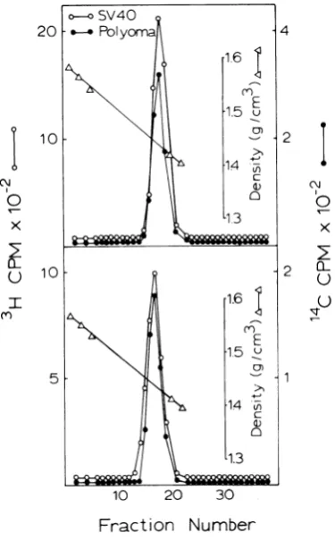
Buoyant densities of glutaraldehyde-fixed
SV40 nucleoprotein complexesinCsCl.Inthe accompanying study (8), r-py complexes as well
as py complexes were shown to have the same ratio ofprotein to DNA asestimatedfromtheir buoyantdensities after fixationwith glutaralde-hydeorformaldehyde.To obtainanestimate of
theratio ofprotein to DNA inthecorresponding
SV40
complexes,
peak fractions of 3H-labeledSV40 TSC and NEC from sucrose gradients
were fixed in glutaraldehyde. Glutaraldehyde-fixed
"4C-labeled
py complex was added asmarker, and a portion of the solution was
layered onto preformed CsCl density gradients and centrifugedto
equilibrium
(1).As showninFig. 4, SV40 TSC and NEC had buoyant densities identical to 14C-labeled py complex.
These results indicatethat protein is bound to all forms of SV40 DNA at the same ratio of protein to DNA.
Theaveragedensity reported for the
glutaral-dehyde-fixed polyoma nucleoprotein complexes was 1.437 0.008g/cm3, whereas the
formalde-hyde-fixed complexes had a slightly higher
density, namely, 1.476 0.01 g/cm3 (8). The reason for thisdensity difference appears to be 905
on November 10, 2019 by guest
http://jvi.asm.org/
HALL, MEINKE,AND GOLDSTEIN 1TABLE 2. Fractionation of3H-labeled DNA from
SV4()nucleoprotein (romplexes F'raction
P'rocedure 15-millpulse 120-minpulse
''1St
NE('
'['S(
NEC',
Propidium diiodide
Heavypeak 40 13 78:I 65
Intermediate light 60 8?4 27 :35 peak
BND-Cellulose
1 MNaCl 57 27 87 I
1 M NaCI +2' cal- 4 78: 1 1 [29
Values represent percentage ofco <ntrecoxered.1
C\x
0
0-10 20 30
Fraction
NumberFIG. 4. Equilibrium centrifugation of glutaralde-hyde-fixed polyoma and SV40 nucleoprotein
com-plexes. SV40 Triton supernatantand Triton nuclear fractions wereprepared from cells labeled for15min at41.75h after infection. SV40 TSC and NECwere
isolatedon5to20%sucrosegradients. Peakfractions
from each gradient were pooled and treated with
glutaraldehyde. Fixed SV40 TSC (top) and SV40 NEC(bottom)weremixed withglutaraldehyde-fixed, "4C-labeledpolyoma TSC andcentrifugedto
equilib-rium inpreformed linearCsCIgradients.
related to the chemical effects of'
binding
and not because the reagents are binding different amounts ofprotein.As estimate of'the ratio (Aprotein to DNA in the complexes can he made by assuming that the relative
density
contributions of DNA and protein moietiesareadditive.SV40 DNA withaguanine plus cytosine content of' 41
%.
(6, 18) has a density in CsCl of 1.700 g/cm3. The origin of the protein component is not known, but since mostproteins donotdif'fergreatly
indensity, an approximate density estimate can be made by using an averagevalue
of' the virion protein.namely.
1.295 g/cm3 (3. 16). Usingthesedensity
values, the ratio of'protein to DNA of'theglu-taraldehyde-fixed
complexes would be about 1.85(65/35'%
) and, of the formaldehyde-f'ixed complexes. 1.24(55/45%).
Assuming
that SV40complex contains one supercoiled DNA mole-cule of about 3 . 106 daltons and only protein. SV40 complex would have a molecular weight
in the range between about 6 and 9 million. Since the protein-to-DNA ratio in the com-plexes containing replicating DNA is the same, these complexes would range in molecular weight f'rom that of SV40 complex to
about
twicethatvalue.Sedimentation ofnucleoprotein complexes in sucrose gradients containing labeled
ribo-somes. When
polyoma
or SV40 DNAwereusedasmarkers, the apparent sedimentation ratesof the nucleoprotein complexes varied with the type of gradient used and with the time of
sedimentation. Furthermore, viral DNA
ap-peared to sediment faster if Triton was not
included in thegradients. Basically, viral DNA is not a suitable marker for the DNA-protein
complexes because of the large differences in sedimentation rates and densities. Since ribo-somal subunits are nucleoproteins with similar sedimentation coefficients and densities (2, 11, 20-22), nucleoprotein complexes were prepared andsedimented
together
with labeled ribosomal subunits used as markers.The sedimentation pattern of 3H-labeled py complex and "C-labeled ribosomal subunits is
shown in Fig. 5. In this gradient, py complex
appears as a single peak sedimenting aheadof
thelarge ribosomal subunit. Inbuffers contain-ingrelativelyhighconcentrationsof EDTAand salt, the large ribosomal subunit is reported to
have a sedimentation coefficient (s20 w) of 47 (20, 21).Assuminglinearityovershortdistances
in the gradients, the sedimentation coefficient ofpy complex is about56S.
Green and co-workers (9, 10)
reported
a sedimentation coefficient of 55S for thepoly-oma DNA-protein
complex containing
super-906 J.VIROL.
on November 10, 2019 by guest
http://jvi.asm.org/
[image:6.495.63.251.260.563.2]coiledpolyomaDNA, whereas White and Eason (23) reported a sedimentationcoefficientof44S for thecorresponding SV40 DNA-protein com-plex. In both cases, these values were appar-ently derived from sedimentation in sucrose using polyoma 20S DNA orSV40 21S DNA as markers. As shown in Fig. 1, when theSV40and the polyoma complexes were sedimented to-gether in the same gradient, the SV40 plexes sedimented ahead of the polyoma
com-plexes. Based on 56S asthe sedimentation rate of py complex, the SV40 complex containing supercoiled DNA(SV40complex) would have a
sedimentation coefficient ofabout61S,whereas
SV40 complexescontainingprimarily 25S repli-cating DNA (r-SV40 complex) would sediment
as broad heterogenous peaks of about 70S (see Fig. 1C).
DISCUSSION
There arestriking similarities between SV40 nucleoproteins and polyoma nucleoproteins ex-tracted from infected cells byTriton lysis. Like polyoma, short-lived SV40 complexes contain-ing replicating viral DNA (r-SV40 complexes) are not as easily extractable as complexes
containing SV40 supercoiled DNA (SV40
com-plexes), suggesting that r-SV40 complexes are bound differently to nuclear sites. Furthermore, protein is bound to both replicating as well as
supercoiled SV40 DNA atabout thesame ratio of protein to DNA as is found in polyoma
nucleoproteins.
Fast sedimenting labeledcomplexes contain-ing predominantly SV40 RI DNA were ex-tracted after short pulses with 3H-thymidine, while mostly slower sedimenting complexes containing SV40 supercoiled DNA were ex-tracted after long pulses. Similar results were reported by White and Eason (23). In addition, we found some slow sedimenting complexes after shortpulsescontaining a mixture of SV40 RI and SV40 supercoiled DNA. Thus, SV40
nucleoprotein complexes, like polyoma
com-plexes,appear tosediment at rates proportional
to the sedimentation rates of the DNA in the
complexes rather than on the basis of whether
the DNA is replicating or supercoiled. Since protein is bound to all forms of SV40 DNA to
thesame extent, the correlation between sedi-mentationrates ofcomplexes and DNA indicate that the relativeconfigurations of various forms
of SV40 DNA are similarly affected by the addition ofprotein.
Considering that the sedimentation rates of SV40complexes were reported to be about 20% less thanthose of polyoma (10, 23), a somewhat surprising finding was that SV40 complexes
I
0IL 8
4
21
I0
()X
1 E
CL
U
u
[image:7.495.255.443.54.230.2]Fraction Number
FIG. 5. Velocity centrifugation of polyoma nucleo-proteincomplex andmouseribosomal subunits.Cells werelabeled with '4C-uridine (0.1 0Ci/ml) from8to 26 h afterinfection.At 26h 14C-uridinewasremoved and the cells labeled for2h with 3H-thymidine (10
LCi/ml).Cellswerelysedwith Triton,and the super-natant fluid was analyzed on 5 to 20% sucrose gradients.
actually sedimented slightly faster than the
corresponding polyoma complexesin our
gradi-ents.Thisdiscrepancy maybe attributedtothe use of viral DNA as markers in the previous
studies. We found viral DNAunsatisfactoryas a
marker for several reasons. First, under sedi-mentation conditions which gave good resolu-tion ofthe nucleoprotein complexes,viral DNA
only sedimented about 20% into the gradient. Furthermore, when the compositionofthe gra-dients were
varied,
the relative sedimentation rates changed dramatically. Even the presence of Triton in the gradients affected the relative sedimentation rates in an unpredictableman-ner. These difficulties are no doubt related to
the fact that DNA and nucleoproteins have
different partial specificvolumes and therefore
the buoyancy forces in sedimentation will be
different dependingonthe densityand compo-sition ofthe solvent solution.
To circumvent these difficulties ribosomal subunits were employed as markers since they
have sedimentation rates and densities compa-rable to those of the SV40andpolyoma
nucleo-proteins (2, 11, 20). The S values ofthe
com-plexes were calculated from the s2o, value reported for the large ribosomal subunit deter-mined under conditions similartothoseusedin
our studies, namely 47S (20). Nonetheless,
values from 44 to 50S have been reported for large subunits in EDTA (2, 11). For these rea-sons, the actual S for py complex may be a value between 53 and 59S. It should be noted
that, as reported (20), ribosomal subunits were 907
on November 10, 2019 by guest
http://jvi.asm.org/
HALL, MEINKE, ANDGOLDSTEIN
not stable in EDTA and were used within 72h after being isolated.
In the preceding publication analysis of the sedimentation and buoyancy data revealed that polyoma nucleoproteins have somewhat more
compact structures than would beexpected for complexes with the DNA in configurations characteristic of free DNA (8). Analogous
argu-ments would apply to SV40 complexes having sedimentation rates and densities like those of polyoma complexes. Previous data concerning the effectsofchemicals and enzymes on SV40
complexes (23)suggestevenfurther similarities
in stability and chemical composition to the polyoma complexes. Ofgreat interest will be a
comparison ofnucleoprotein complexes isolated after infection by "late" mutants from these
twoviruses.
ACKNOWLEDGMENTS
WethankMichele English andHelen Bartlett forexcellent technical assistance.
Thisinvestigationwassupported by Public Health Service
grant CA-11151, postdoctoral fellowship award CA 53413 (M.R.H.), and Research Career Development Award CA 32425(D.A.G.)fromtheNational Cancer Institute.
LITERATURE CITED
1. Baltimore, D.,and A. S. Huang. 1968. Isopycnic
separa-tion ofsubcellularcomponentsfrompoliovirus-infected and normal HeLacells. Science 162:572-574. 2. Bielka,H., H.Welfle,M. B6ttger,andW.Forster.1968.
Strukturveranderungen und dissoziation von
leber-ribosomen inabhangigkeit von der Mg'+ konzentra-tion.Eur.J. Biochem. 5:183-190.
3. Black, P. H., E. M. Crawford, and L. V. Crawford. 1964. The purificationofsimian virus40. Virology 24:381-387.
4. Bourgaux, P., and D. Bourgaux-Ramoisy. 1972. Unwind-ing ofreplicating polyoma virus DNA. J. Mol. Biol. 70:399-413.
5. Bourgaux, P., D. Bourgaux-Ramoisy, andP. Seiler.1971.
Thereplication ofring-shapedDNAofpolyomavirus.
II. Identification of molecules at various stages of
replication.J. Mol. Biol. 59:195-206.
6. Crawford, L.V., and P. H. Black. 1964. The nucleicacid
of simian virus 40.Virology24:388-392.
7. Eason, R., and J. Vinograd. 1971. Superhelical density heterogeneity of intracellular simian virus 40
deoxy-ribonucleic acid. J. Virol. 7:1-7.
8. Goldstein, D. A., M. R. Hall, and W. Meinke. 1973.
Properties of nucleoprotein complexes containing
replicating polyomaDNA.J. Virol. 12:887-900.
9. Green,M. H. 1972.Biosyntheticpropertiesof apolyoma
nucleoprotein complex: evidencefor replication sites.
J.Virol. 10:32-41.
10. Green,M.H.,H.I.Miller,and S.Hendler.1971. Isolation
of a polyoma-nucleoprotein complex from infected
mouse-cell cultures. Proc. Nat. Acad. Sci. U.S.A.68:
1032-1036.
11. Hamilton,M.G.,and M.E.Ruth.1969. The dissociation
of rat liver ribosomes by ethylenediaminetetraacetic acid; molecular weights, chemical composition, and
buoyant densities of the subunits. Biochemistry 8:
851-856.
12. Hirt, B. 1967. Selective extraction of polyoma DNA from infected mousecell cultures.J. Mol. Biol. 26:365-369. 13. Jaenisch,R.,A. Mayer, and A. Levine. 1971. Replicating SV40 molecules containing closed circular template DNA strands. Nature N. Biol. 233:72-75.
14. Levine, A. J., H. S. Kang, and F. E. Billheimer. 1970.
DNAreplication in SV40infected cells. I. Analysis of
replicatingSV40 DNA. J. Mol. Biol. 50:549-568. 15. Meinke, W., andD. A. Goldstein. 1971. Studies onthe
structure and formation of polyoma DNA replicative intermediates.J.Mol. Biol. 61:543-563.
16. Ozer, H. L. 1972.Synthesisandassemblyof simian virus 40.I.Differentialsynthesisof intact virionsandempty
shells.J. Virol. 9:41-51.
17. Robb, J. A., and R. G. Martin. 1972. Geneticanalysisof simian virus 40. III. Characterization of a
tempera-ture-sensitive mutant blocked at an early stage of productive infection in monkey cells. J. Virol. 9:956-968.
18. Schildkraut, C. L., J. Marmur, and P. Doty. 1962. Deter-mination of the basecomposition ofdeoxyribonucleic acid from its buoyant density in CsCl. J. Mol. Biol. 4:430-443.
19. Sebring, E. D., T. J. Kelly, M. M. Thoren, and N. P. Salzman. 1971. Structure of replicating simian virus 40 deoxyribonucleic acid molecules. J. Virol. 8:478-490.
20. Tashiro, Y., and P. Siekevitz. 1965a. Ultracentrifugal studies on the dissociation of hepatic ribosomes. J. Mol. Biol. 11:149-165.
21. Tashiro, Y., and P. Siekevitz. 1965b. Localizationon
he-patic ribosomes ofprotein newlysynthesized in uiuo.
J. Mol. Biol. 11:166-173.
22. Vournakis, J., and A. Rich. 1972. Ribosomal
transforma-tions during protein synthesis, p. 287-299. In R. A.
Coxand A.A. Hadjiolov(ed.), Functionalunits in
pro-teinbiosynthesis. Academic Press, Inc., New York. 23. White, M., and R. Eason. 1971.Nucleoprotein complexes
insimian virus40-infected cells. J. Virol.8:363-371.
24. Winocour,E., 1963. Purification of polyomavirus.
Virol-ogy.19:158-168.
908 J.VIROL.