JOURNALOFVIROLOGY, Feb. 1973, p.198-206
Copyright 01973 AmericanSocietyforMicrobiology
Vol. 11,No. 2 PrintedinU.S.A.
Protein
Synthesis
Directed
by
an
RNA-Temperature-Sensitive
Mutant
of
Sindbis
Virus
MARILYNN R. F. WAITE
Department of Microbiology, Dartmouth MedicalSchool, Hanover,NewHampshire03755
Received for publication 22 March 1972
The structural proteins of wild-type Sindbis viruswereshowntoariseby
post-translational cleavage of larger precursors. The proteins synthesized in
wild-type infectionwerecompared with those specified by ts-11, a
temperature-sen-sitivemutantunabletosynthesize viral RNAatthe restrictivetemperature. Ab-normally large, virus-specific proteins were found in the mutant-infected cells
after the shift from 28 C to 41.5 C. These large polypeptideswere presumably precursorswhich werecleavedtoorapidlytobe detected in the wild-type infec-tion. Thelargest hadamolecularweight of 133,000 andwasthesamesizeasthe
apparent precursor detected during infection with a groupofSindbis mutants
which could not form nucleocapsids at the nonpermissive temperature. The stability of ts-11-specific RNA synthesis, after shift from permissiveto restric-tive conditions, differed from that in cellsinfected by wild-type virus, indicat-ing that the virus hadagenetic lesion inanenzymeinvolved in RNAsynthesis.
This mutation might have caused the precursor to fold incorrectly so that it
could not be cleaved. The possibilitycannot be excluded, however, thata
sec-ond lesion inanuncharacterized viral function, suchas aprotease,wasthecause
of the accumulation oftheprecursors.
In enteroviral infections, functional viral proteins are formed by the post-translational
cleavage of large precursor polypeptides (1).
Precursors large enough to be the gene
prod-uct of the entire viral genome have been
de-tected (10, 12). Ingroup Aarbovirusinfections,
the situation is not as clear. Several reports
have noted the inability to detect a precursor protein in chick embryo fibroblast (CEF) cells infected with either wild-type Sindbis virus
(20) or Semliki Forest virus(9). When Semliki
Forest virus was grown inbabyhamsterkidney
cells, however, definite evidence for the exist-ence of precursors was obtained (5). A recent study by Pfefferkorn and Boyle (16) using in-hibitors of protease activity has suggested that, inSindbis infection, a protein the same size as
thelargestonepresent under normal conditions may function as a precursor of the viral mem-brane and capsid proteins.
Evidence for the existence of a still larger, virus-specified polypeptide in Sindbis-infected
CEF was found by Strauss et al. (23) and by
Scheele and Pfefferkorn (20) who studied pro-teins made by temperature-sensitive (ts)
mu-tants ofSindbis virus. Theyfound that, at the
nonpermissive temperature, cells infected with any of the three members ofcomplementation
group C
(capsid-,
or defective innucleocapsid
formation) didnot contain capsid protein (mo-lecular weight 30,000; reference 24). The pre-dominant protein species in thesemutant-in-fected cells had alower electrophoretic mobil-ity than those seen during normal infection.
This polypeptide was
presumably
a precursor,although itsradioactivelabel didnotchaseinto
smaller proteins when the temperature was
lowered (20). Since these mutants are capable
ofsynthesizingviral RNA whenincubatedfrom the time of infection at the nonpermissive temperature (RNA+), the viral
RNA-synthe-sizing enzyme(s) is presumably freed fromthis precursor.
found among the RNA- mutants which are un-able to makevirus-specific RNA at the nonper-missivetemperature. A mutation which did not permit any virus-specific proteins to be cut from the precursor would probably result in a viral mutant that was phenotypically RNA-,
because it seems unlikelythat an RNA
polym-erasewould be functional while itwasstillpart of a large polypeptide. Accordingly, I screened
198
on November 10, 2019 by guest
http://jvi.asm.org/
Downloaded from
on November 10, 2019 by guest
http://jvi.asm.org/
Downloaded from
on November 10, 2019 by guest
http://jvi.asm.org/
the proteins made by the RNA- mutants of
Sindbis virus before and after the shift from
28 C, the permissive temperature, to 41.5 C,
the nonpermissive temperature. Acrylamide
gel electrophoresis (AGE) revealed a variety of
normal and abnormal protein patterns. The most conspicuous of these was that of ts-11; after a three-hour labeling period, about 60% of the virus-specific proteins had
electropho-reticmobilities less thanthat ofthemembrane
protein. In cells infected with thewild-type vi-rus, less than 25% ofthe viralprotein was this
large. The predominantspecies inthe
mutant-infected cells was larger than any seen during
normal infections. Ts-11 was, therefore, chosen asthe subject for moreintensive study. A
pre-liminary report of this work has already
ap-peared (M. R. F. Waite and E. R. Pfefferkorn, Bacteriol. Proc., p. 233, 1971).
MATERIALS AND METHODS
The isolation and characterization of the ts mu-tantsofSindbis virus and the HRstrain fromwhich they were derived (referred toaswild-type), aswell
asthe conditionsfortheir growth and titration,have been described (2-4, 17). After infection, CEF
cul-tures were incubated in Eagle medium containing
3% rabbit serum and actinomycin D (1 ug/ml). To
deplete the amino acid pools of culturesbeing pre-paredforAGE, the medium wasmodifiedtocontain only 1 ,ug ofleucine per ml (low leucine medium) or
one-tenth the normal amount of amino acids (low
amino acid medium). Radioactive precursors, ob-tained from Schwartz BioResearch, Inc., included
3H-leucine (40 Ci/mmole), reconstituted
"C-pro-tein hydrolysate (500
MCi/ml),
and 3H-uridine (25mCi/mmole). Chase medium contained an excess ofunlabeled leucine (100
4g/ml).
To examine virus-specific RNA, infected cells werepreincubated with actinomycin D, labeled with
3H-uridine, and treated as described previously (4). Briefly, the labeledmonolayerswerewashed and dis-solved inabuffered solution containing sodium dode-cyl sulfate (SDS) and ethylenediaminetetraacetate. Portions of the cell extracts wereanalyzed on 15 to 30%SDS-glycerol gradients(12).
The procedures for preparing extracts of labeled cells and subjectingthem to AGE were identical to
thoseemployed by Scheele and Pfefferkorn (18), ex-cept that 7.5% polyacrylamide gels were employed.
Samples of from 0.05 to 0.2 ml (5,000 to 70,000
counts/minof 3H or1,000to2,000counts/minof14C) wereapplied to the gels. Unless otherwise specified,
a 0.05-ml sample of an extract of purified Sindbis virus labeledwith
"4C-protein
hydrolysate was coelec-trophoresedwith eachsampletoindicate thelocationofthe viral membraneandcapsid proteins.
To determine the molecular weights of the large proteins, theirelectrophoretic mobilitywascompared
withthatofthyroglobulin (molecularweight 160,000;
reference 26) and bovine serum albumin (BSA) (mo-lecular weight 67,000). The thyroglobulin prepara-tion contained traces of two minor protein species,
oneslightly larger and one slightly smaller than thyro-globulin. When boiled prior to electrophoresis, the BSA preparation gave only a single band. If unboiled,
a pronounced band with a molecular weight of 134,000, presumably the dimer, was also observed; this was used as an additional marker. 3H-labeled ts-11-specific viral protein or "4C-labeled ts-13-spe-cific protein were mixed with oneorboth markers and subjected to AGE on 5 or 7.5% gels at 5 mA/gel until thebromophenol blue marker neared the bottom of the tube. The gels were stained with Coomassie blue inmethanol-acetic acid fortwohr and destainedby soaking for two days in several changes of the meth-anol-acetic acid solution (26). The gels were then soaked overnight in 7.5% acetic acid, and the loca-tions of the stained bands were determined. The gels were frozen and sliced with a transverse gel slicer (Misco Scientific). After soaking overnight in NCS-toluene scintillation fluid (Amersham/Searle). the slices were counted to determine the locations of the radioactive peaks. The relative mobilities and molecular weights were determined as described by WeberandOsborn (26).
RESULTS
Protein synthesis directed by wild-type Sindbis.Thepermissivetemperature, atwhich the tsmutants of Sindbis virus growwell, is 28 C. Since the previous studies of protein syn-thesis directed by wild-type arboviruses have been carriedout at37 C(5,9, 20, 23), it seemed advisable to reexamine the pattern of protein synthesis bywild-type Sindbis virusat28C to
provideabasisforcomparison with the
mutant-infected cells. Replicate CEF cultureswere
in-fected andincubatedat28Cfor 8hr inlow
leu-cine medium. They were then exposed for two min to medium containing 3H-leucine. One culture was processed
immediately,
whereas the otherwas washed and incubatedfor anad-ditional 30 min in chase medium to allow the
labeled proteins tobe convertedintotheir final form. The chased culture contained 10% less radioactive protein than the culture dissolved immediately after
labeling,
confirming theef-fectivenessofthe chase conditions. Both prep-arations were
subjected
toAGE,
and acom-parison ofthe two patternsappears in Fig. 1. After the 2-min pulse, the membrane (M)
and capsid (C) protein peaks were present,
although much reduced in comparison with experimentsperformedat 37 C (20, 23) orfrom the30-min chase at 28 C (Fig. 1). Althoughno
proteins with veryhighmolecularweightswere
observed, there was increased label in the re-gion of the large protein (L; moelcular weight
90,000 to 10,000; reference 23) and a
on November 10, 2019 by guest
http://jvi.asm.org/
WAITE
E 300
w L
z
ZOO D
100-RELATIVE MIGRATION
FIG. 1. Pulse-chase experiment with the protein of the wild-type virus at 28 C. Cultures infected with
thewild-type viruswereincubatedat28Cfor8.5 hr in low-leucine medium. They werethen washed and exposed for2 mintomedium containing 3H-leucine
(100 gCi/ml) and no additional unlabeled leucine.
One culture ( ) was prepared immediately for AGE. The otherwaswashed,andexposed for30min to chase medium, and then processed (--- ). The
graphs werethen plotted so that the capsid protein markers of both preparations were superimposed. The letters M and C in this andsubsequent figures indicate thepositions of the membrane and capsid protein from the virus marker. TheletterLindicates the locationof the large protein normally foundin
in-fectedcells.Migrationwasfrom lefttoright.
cant amount of material which migrated be-tween the L protein and the membrane
pro-tein. The protein in this region may represent some large molecules in the process ofbeing
synthesized, as well as intermediate cleavage
products of the precursors. (The role of the
protein migrating 2 fractions behind the
mem-brane peak as a precursor to the membrane protein has been described [23]). After the chase, the radioactivity in the L peak was
re-duced approximately 25%, and the amount of
label migrating between L and the membrane peak and between the membrane and the
cap-sid peaks decreased markedly. Most ofthe
la-bel chased from these regions appeared in the
membrane and capsid peaks. This experiment
demonstrates thatSindbis virus structural
pro-teinsarise by the post-translational cleavageof
larger proteins; it also supports thesuggestion
fromexperiments withproteaseinhibitors(16),
that some oftheprotein inthe peak marked L
israpidly cleaved under normal conditions and
serves as a precursor to the structural pro-teins. Lowering the incubation temperature
apparently slowed cleavage more than
syn-thesis, allowing the precursor-product
re-lationship among the virus-specified proteins to be discerned. The label which does not
chase from the L peak may represent a dif-ferentprotein species.
Proteins synthesized during infection
with ts-11. Figure 2 illustrates typical pat-ternsofproteinsynthesisints-11-infected CEF under various conditions. The
electrophero-grams ontheleft-hand sideofFig.2(A-C)were
obtained from cultures that wereinfected with
ts-11, incubated for 8 hr at 28
C,
and then la-beled with 3H-leucine at that temperature. Parts A and B show 2-min and 1-hrpulses,
respectively. The patterns are similar to those obtained during infection with wild-type virusunder the same conditions. This is not sur-prising since the mutant growswellat 28C and would be expected to produce
normal,
ornearly normal, proteins. There was no
indica-tion of a very
high-molecular-weight
precursor.Panel C shows the effect ofa 1-hr chase at 28 C, in medium containing anexcess of unla-beled
leucine,
on acultureinfected, incubated,
and labeledasinpanel
B. Therewas nosignof any further chase. Thehigh-molecular-weight
peak(C:
fractions 11 to 17) islower,
but it isalso broader and contains
approximately
thesameproportion ofthelabelasthe
correspond-ing peak inpanel B (fractions 11 to14). RNA- mutants areunableto make detecta-bleamountsofvirus-specific
protein
when incu-batedfromthe time of infection atthe nonper-missive temperature. To examine the pattern of protein synthesis under restrictive condi-tions, I incubated the infected culturesat 28Cfor 8hr, shifted them to 41.5C when the
infec-tion was well
established,
and labeled them atthat temperature
by
exposure to mediumcon-taining 3H-leucine. The electrophoretic pat-terns of proteins made at 41.5 C (D-F) are
strikingly different from those at 28 C. The most conspicuous feature of all three profiles
is the presence of two large, slowly moving
peaks near the origins
(left-hand
side)
ofthegels. Athird,smallerpeak,whichwasbetter
re-solved if the proteins were allowed to migrate further, was also present. These
peaks
werenot artefacts of the high incubation tempera-ture: chase at 41.5 C of cultures labeled at 28
C did not cause the appearance of
proteins
with abnormally high molecular
weights
(un-published data).200 J. VIROL.
on November 10, 2019 by guest
http://jvi.asm.org/
[image:3.493.61.248.68.265.2]m
~~~~~~~~~D
j
5
M
c-z
0
B
E
15-M
---10
I0
z
0
(.)
FI.2s11-pcii
Frtissnhszda
8Cad4. .Alclue eeifce iht-1 nu4%
~~~~~cF
w
w m
c~~~~~~~~
-J
0
~~~20
40
6o0
20
40
60
F
RACTION
NUMBER
FIG. 2. Ts-11-specific proteinssynthesizedat28C and41.5 C. All cultures wereinfectedwith ts-11,
incu-bated at 28 C in low-leucine mediumfor8hr,and then treatedasfollows. (A) The mediumwasdiscarded,the
culturewaswashed with Hanksbalanced salt solution(BSS),and the bottlewasfilledwith BSSat28Cand
placed inawaterbathatthattemperaturefor3min. TheBSSwasdiscarded, and1.5mlofmedium contain-ing 3H-leucine (100 uCi/ml) andno unlabeled leucinewasadded. Incubationwas continuedfor2min with the culturesubmerged in the waterbath and rockedcontinuously. Itwasprocessed immediately. (B)An
in-fectedculturewastreatedasinAexcept thatthelabeling periodwas1hr,theradioactive medium contained
251sCiof 3H-leucine per ml, and the incubationwascarriedoutinanincubator insteadofawaterbath. (C)A
culture waslabeledasinB,washed withwarmBSS,andincubatedforanadditionalhour inchase medium. Thecultures in theright-handcolumn(D,E,F)weretreatedexactlylikethecorrespondingculture inthe
left-hand column(A, B, C)except that thepreincubationinBSS and the 3H-leucinelabelingwerecarriedout at 41.5C.Afterlabelingat 41.5C,theculture shown in Fwasincubatedinchasemediumat28C.
201
on November 10, 2019 by guest
http://jvi.asm.org/
[image:4.493.53.442.35.565.2]WAITE
Panel D shows the profile of the proteins synthesized duringa2-min
pulse
of3H-leucine.More than60% ofthetritiated protein present after the 2-min pulse had a molecular weight
greater thanthatofthe membraneprotein. The
membrane and capsid peaks were present but
much reduced.
Comparison ofpanel D withpanel E, which showsthepatternobtained after a1-hrlabeling
period, reveals that some of the high-molec-ular-weight protein seen inthe2-min pulse (D:
fractions 16 to 22) appeared in the membrane
and capsid protein peaks withtime. However, the label in the high-molecular-weight peaks
(D: fractions 5 to 15) wastrapped thereanddid
not appear tochase. The threepeaks contained about 40% of the total label after the 2-min pulse, 50% after a 1-hr exposure to the label, and60% after 3 hr (not shown). PanelF shows the effect of a i-hr chase at 28 C of a culture
in-fected, incubated, and labeled as was the
cul-ture in
panel
E. There wasnoalterationofthe distribution ofradioactivityamongthe various peaks. Thus, the large ts-11-specific proteins, like those ofthe capsid- mutants studied by Scheele and Pfefferkorn (20), didnotchase intolower-molecular-weight
ones, either atpermis-sive ornonpermissive temperatures.
RNA synthesis in ts-11-infected cells. Al-though ts-11 is phenotypically RNA- and can-not synthesize viral RNA when incubated at
the nonpermissive temperature from the time of infection, this does not mean that there is necessarily a mutation in a nucleotide sequence
which codesforaproteininvolved inRNA
syn-thesis.
Any defect which left a protein required for
viral RNA synthesis trapped within an
un-cleaved precursor at the nonpermissive tem-perature could result in a mutant that was
phenotypically RNA-, regardless of whether the lesion was actually in the amino acid se-quence required for viral RNA synthesis. A mutation in some otherregion ofthe precursor or in a virus-specified protease could have the same effect.
Proteins freed from the precursor during in-fection with a cleavage-failure mutant at the
permissive temperature should be as stable on shift to the restrictive temperature as the cor-responding ones made in response to infection with the ancestral wild-type virus, except for the protein containing the ts lesion. This pro-tein might be (butneed not be) less stable. All of the functional proteins produced at 28 C
might conceivably have the normal, wild-type aminoacidsequence if the lesion were in a por-tion oftheprecursor polypeptide that was
nor-mally excised, as in the proinsulin to insulin
conversion (6).
Iattempted to localize the defect in ts-11by determining whether the RNA-synthesizing system coded forby ts-11 hada different ther-mal stability from that of the wild-type virus.
The rate of ts-11 RNA synthesis following the
shiftfrom 28 to 41.5 C was examined first. Dur-ing the first hour after the shift, it increased above the 28 C control which remained
essen-tially constant. However, by the third hour,
it had dropped well below control levels. This
is significantly different from the reported
be-havior of thewild-typevirus:under similar con-ditions, the rate of RNA production increased
with the increased temperature and did not
subsequently decline (19). Other RNA- mu-tants were affected invarious ways by similar
temperature-shift-up conditions. Some be-haved like the wild-type virus (Scheele and
Pfefferkorn, unpublished observation); the
rate of RNA synthesis of others declined very
rapidly (ts-6, complementation group B) (25), or somewhat more slowly (ts-24,
complemen-tationgroup A) (19), duringthe first hour after
the temperature was raised.
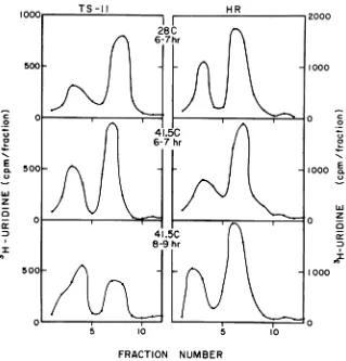
I therefore examined the distribution of
3H-uridinelabelbetween the26sand 42s species of
ts-11-specific, single-stranded RNA under several conditions; the results are shown on
the left-hand side of Fig. 3. Thepattern of ts-11-specific RNA synthesized between 6 and 7 hr after infection at 28C(toppanel) was similar topublished profilesofthe wild-typeRNA
syn-thesis(4, 7, 19). Thecurve duringthe first hour after the shift to 41.5C (middle panel) showed
that incorporation had increased slightly at 41.5 C. The profile was similar to that of the
control, suggesting the stability of the RNA-synthesizingenzymes for atleast1 hrat 41.5C. Close inspection of the profiles, however,
re-vealed that the ratio of 26s RNA to 42s RNA was altered from approximately 2.3 at 28 C to 1.8 at41.5C. Between 2 and 3 hr after temper-ature elevation (bottom panel), this trend had become pronounced. Although the amount of label in the 42s peakactually increased
some-what, the synthesis of 26s RNA was markedly
inhibited, giving a 26s/42s RNA ratio of 0.6. A similar effect was observed by Scheele and Pfefferkorn (19) in cells infected with ts-24
duringthe first hour after temperature shift. Before such a result can be interpreted as
demonstratinga defect ina viral RNA-synthe-sizing enzyme, it is necessary to show that the decline did not result from nonproduction of a
required protein, a situation which might have occurred if the protein was trapped within an
202 J. VIROL.
on November 10, 2019 by guest
http://jvi.asm.org/
TS-II HR
l l
28C
6-7hF 5001
c
0
U
'Z 0
C4
0
w
z
a
I
5C
S
OOF
5 10
hr
I0
1000
0 0o
0
%$..
1000 E0.
u
w
0 z
a
In
1000
0o
FRACTION NUMBER
FIG. 3. Distribution ofvirus-specific RNA between the 26s and 42s single-stranded species under various conditions of incubation. After infection with ts-11orwild-typevirus, replicate cultures were incubated at 28 C in the presence of actinomycin D (1
Ag/ml).
At 6hrafterinfection,somecultures wereshifted to 41.5 C and cycloheximide (25pg/ml)
wasaddedto the cultures infected with thetype virus only. Ts-11 and wild-type-infected culturesat 28C andat 41.5Cwerelabeled for1hrby exposure to medium containing3H-uridine(5 uCi/mI)anddissolved in1 mlof buffered1%SDS solution.Between 8 and 9 hrafter infection(2 and3hr after temperature elevation), an additional mutant-infected culture and one infected with wild-type virus werelabeledat 41.5 C.(Thecultureinfectedwith thewild-typeviruswasmaintained in the continuous
pres-ence ofcycloheximide.) Samples of0.2mlofthedissolved cultureswereanalyzed by ultracentrifugation. Mi-grationwasfromrighttoleft;42sRNA isattheleft,and26sison theright.
uncleaved precursor. The right-hand column showsan experiment using cells, infected with the wild-type virus, which were incubated and shifted like the ts-11-infected cells, but with
cycloheximide (25 gg/ml) added atthe time of shift to inhibit the subsequent production of
new proteins. The ratio of 26s to 42s
virus-specificRNA remained normal in cells infected with the wild-type virus, and the amount of viral RNA made did not decline after several hours at 41.5 C in the absence ofprotein
syn-thesis. Thus, itseems logical toassume thata
temperature-sensitive defect in ts-11 is in a
polypeptide necessaryfor viralRNAsynthesis.
Determination of proteinsizes.Atthe
non-permissive temperature, cells infected with those ts-mutantsofSindbisviruswhichare
de-fective in nucleocapsid formation (ts-2, ts-5
and ts-13), and with the RNA- mutant ts-11,
contained abnormally large proteins (20). To compare the size of the ts-11 precursors with
those of the capsid- mutants, ts-13-specific
protein, made at the nonpermissive
tempera-ture and labeled with
"4C-protein
hydrolysate,was coelectrophoresed with a sample of ts-11
protein labeled at 41.5 C as described in Fig.
2E. The results (Fig. 4) indicate that the
large
precursor protein specified by ts-13 is almost
r
on November 10, 2019 by guest
http://jvi.asm.org/
[image:6.493.88.407.69.402.2]WAITE
FRACTION NUMBER
FIG. 4. Coelectrophoresis of the large proteins made during infection by a capsid- mutarnt and
byts-11.Aculture infected withts-13wasincubated
for3.5 hrat40 C in low amino acid medium. Itwas
then exposed foranadditional 3.5 hrtosimilar
me-dium containing 14Cproteinhydrolysate (5
gCi/ml)
and prepared for electrophoresis. A 0.1-mIsample of
this culture (0) was mixed witha 0.1-mIsample of
3H-leucine-labeled ts-11-specific protein (0) pre-paredasthatinFig. 2E,andtheyweresubjectedto acrylamide gel electrophoresis. No virus protein
markers could be included with this experiment; however, thelocationofthemembrane(M)and cap-sid (C) proteins was known from previous electro-phoresis ofthe ts-11 preparation in thepresence of
markers.
exactly the same size as the largest protein
made during ts-11 infection. Similar results
wereobtained on5% gels.
Since Strauss et al. (23) reported that the
ts-13 precursor had a molecular weight of
130,000 and Scheele and Pfefferkorn (20)
re-ported that the same protein had a molecular
weight of approximately 90,000, it seemed ad-visable to redetermine the molecular weight. Samples of radioactively labeled preparations of ts-11-specific and ts-13-specific proteins
were coelectrophoresed with unboiled BSA
(molecular weight of the monomer 67,000 and
that of the dimer 134,000) and thyroglobulin (molecular weight 160,000; reference 26). After electrophoresis, the gels were stained with
Coomassie brilliant blue toreveal the location ofthemarkers (26) and then sliced witha trans-versegel slicer. The slices were countedto de-termine the location of the radioactive peaks. The molecular weight of the large precursor,
determined asdescribedby Weber and Osborn
(26), is 133,000. The molecular weights of the smaller, abnormal, ts-11-specific precursors are approximately 112,000 and 95,000.
DISCUSSION
Ts-11 is a temperature-sensitive mutant of
Sindbis virus which is phenotypically RNA-;
atthenonpermissivetemperature, itcausesthe formation of
abnormally large proteins
whichare presumably nonfunctional precursors or
large portions of improperly cleaved
precur-sors. There are three
possible
explanations forsuch behavior. (i) An alteration in the amino
acid sequence of a protein required for RNA synthesis causes misfolding of the precursor so that it cannot be properly cleaved. (ii) An
alteration in some other amino acid sequence
changes the conformation of the
polypeptide
and prevents the release of several proteins, includingonerequiredforviral RNAsynthesis,
which would be functional ifreleased;
the mu-tant would then bephenotypically,
but notgenotypically, RNA-. (iii) Ts-11 is a double
mutant with a lesion in a protein required for
RNAsynthesis andasecond defect whichleads
to the production of an uncleaved precursor.
Thislargepolypeptide mightor mightnot con-tain the amino acid sequence for the altered
RNA-synthesizing
enzyme.Evidence presented above indicates that the RNA-synthesizing system produced by ts-11
during incubation at the permissive tempera-ture is less stable after the shifttothe
nonper-missive temperature than thatofthe wild-type
virus. This suggests that at least one protein requiredforviral RNAsynthesis hasanaltered
amino acid sequence and that the mutant is
genotypically as well asphenotypically RNA-. The possibility that ts-11 is a double mutant cannotbe eliminated.
The secondmutation,if itexists, is not inthe membrane or capsid proteins, nor in the
un-known function in whichts-20 isdefective, nor in the RNA-synthesizing protein in which ts-6 is defective (17), because the mutant
comple-ments efficiently with members of all these
groups. If the virus codes for theenzyme
caus-ing precursor cleavage, this enzyme could be the site of the second lesion. If so, the
postu-lated abnormal cleavageenzyme isprobablynot
the one responsible for the cleavageofthe pro-tein L (Fig. 1). Tosylphenylalanylchloromethyl ketone, which apparently prevents the
cleav-age of the Lprotein, does not causethe appear-ance of the larger proteins seen in ts-11-in-fected cells(16).
Atleast one ts mutation of ts-1 1 may be in the A cistron, since thelate-appearing defect in ts-11 RNAsynthesis is similarto that of many of
the accepted members of complementation group A (19; Scheele and Pfefferkorn,
unpub-lished data). However, ts-11 behaves in anomalous fashion
compared
with othermem-bers of group A. It was
originally
assigned
toan
independent complementation
groupbe-204 J. VIROL.
on November 10, 2019 by guest
http://jvi.asm.org/
[image:7.493.61.255.57.198.2]cause it complemented somewhat with other
RNA- mutants which were placed in group A
(3). The discovery of mutant ts-6 (group B), which complements with very high efficiency with all other RNA- mutants, led to a reas-signment of ts-11 to group A (17). It was
sub-sequently reclassified in a new complemen-tation group A', becauseof its unique behavior
in the intrinsic interference assay of Marcus
and Zuckerbraun(15). They foundthat, unlike
members ofgroupsAorB, ts-11couldinterfere with Newcastle diseasevirus replicationat the
nonpermissive temperature, butonlyincertain unusual lots ofprimary African green monkey kidney cells. This led them to believe that the mutanthada tsmutation in athirdprotein (A')
required for RNA synthesis and that a viral contaminant of the unusual monkey kidney cells
provided
thisactivity,although
noSindbis RNA synthesis was detectable.Ts-il
mayin-deed have a mutation in a thirdgene
required
for viral RNA synthesis; however, it could still be amemberofgroup A. Both the
complemen-tation pattern and theunique behavior in the
interference assaycould result fromthe failure
of precursor cleavage in cells infected by this
mutant. The unknownfactorwhich allowed ts-11 to interfere with Newcastle disease virus replication at the nonpermissive temperature
could have been a different protease which could cleave the precursor, thus
releasing
amarginally functional A protein. Alternatively, thepresence of aprotein similartoAand
sup-plied by the
postulated
contaminating virusmightpromotecleavage of theprecursor.
Leak-iness or intracistronic complementation could then explain the interference.
The largest proteins in ts-11- and
ts-13-in-fected CEF have molecular weights of about
133,000. Since both RNA+ and RNA- mutants cause the productionofpolypeptides ofsimilar size, it is tempting to postulate that they are
identical and might represent the entire poly-peptide product of a major messenger RNA species. In astudyofthe
single-stranded
RNA species in Sindbis-infectedcells,
Levin and Friedman (13)found,
inadditionto42s(4x10⁶f
daltons, reference 7) and 26s (1.8 x 10⁶
dal-tons, reference 7) RNA, three minor species:
38s (3.1 x 10⁶ daltons), 33s (2.4 x 10⁶ daltons)
and onesmaller than the 26sRNA, forwhich I
calculated a molecular weight of0.6 x 106 to
0.8 x 10⁶. This should have a sedimentation coefficient of about 16s (14). Recent
investiga-tions of the RNA species associated with the
polysomes ofcells infected with group A arbo-viruses have revealed 33s and 26s RNA(11) or 26sand 16s RNA(8; Rosemond andSreevalsan,
Abstr. Annu. Meeting A.S.M., p. 242, 1972).
These RNAspeciesshouldbecapableof coding
for proteins with molecular weightsof approxi-mately 240,000, 180,000, and 70,000. All large ts-11 polypeptides are too large to be trans-lated from the 16s RNA. However, the
133,000-dalton proteinseems toosmallto representthe entire polypeptide product of the 26s RNA, unless there is alarge segment which remains
untranslated. In addition, even though both
Inembrane
proteins are glycosylated (21), the 133,000-dalton protein seems toosmall to con-taintheaminoacidsequence for twomembraneproteins with molecular weights of about
53,000, a capsid protein with a molecular weight of 30,000, and at least two enzymes
in-volved inRNA synthesis. Thus, the possibility
remains thata largerprecursor willyetbe dis-covered. Analysis oftryptic peptides could
re-veal whether the large precursors of ts-11 and
the capsid- mutants are identical, whether they are overlapping portions of a still larger precursor, orwhether theyarederivedfrom
dif-ferent precursors. Similar studies could
indi-cate whether the smaller proteins in
ts-11-in-fected cells ariseby cleavage ofthe largest or
have unrelated aminoacid sequences.
ACKNOWLEDGMENTS
I thank Elmer R. Pfefferkorn foradviceand encourage-mentand forcritically readinganearlier version of this
manu-script.
Thisinvestigationwassupported byPublic Health Service research grant AI 08238fromthe NationalInstituteofAllergy and Infectious Diseasesand apostdoctoral fellowship from The Anna Fuller Fund.
LITERATURE CITED
1. Baltimore, D. 1971. Polio is notdead. Perspect. Virol. 7:1-12.
2. Burge, B. W., and E. R. Pfefferkorn. 1966.Isolation and characterization of conditional-lethalmutantsof Sind-bis virus.Virology30:204-213.
3. Burge, B. W., and E. R.Pfefferkorn. 1966. Complemen-tation between temperature-sensitive mutants of Sindbisvirus.Virology30:214-233.
4. Burge, B. W., and E. R. Pfefferkorn. 1968. Functional defects oftemperature-sensitive mutants of Sindbis virus.J. Mol. Biol. 35:193-205.
5. Burrel, C. J., E. M. Martin, and P. D. Cooper. 1970. Post-translational cleavage of virus polypeptides in arbovirus infectedcells. J. Gen. Virol. 6:319-323.
6. Chance, R. E., R. M. Ellis, and W. W. Bromer. 1968. Porcine proinsulin: characterization and amino acid
sequence.Science 157:697-700.
7. Dubos, P., and P. Faulkner. 1970. Molecularweightsof
Sindbisvirusribonucleic acidasdeterminedby poly-acrylamide gelelectrophoresis.J. Virol. 6:145-147.
8. Eaton, B. T.,T. P. Donaghue, and P. Faulkner. 1972. Presence of poly(A) in the polyribosome-associated RNAofSindbis-infected BHK cells.Nature N. Biol. 238:109-111.
9. Friedman, R. M.1969.Primarygeneproductsofan
arbo-virus. Biochem. Biophys. Res. Commun. 37:369-373. 10.Jacobson, M. F., J. Asso, and D. Baltimore. 1970.
on November 10, 2019 by guest
http://jvi.asm.org/
206
WAITE Furtherevidence on the formation of poliovirus pro-teins. Proc. Nat. Acad.Sci. U.S.A. 61:77-84. 11. Kennedy, S. I. T. 1972. Isolation and identification ofthe virus-specified RNA species found on the mem-brane-boundpolysomes of chick embryo cells infected with Semliki Forest virus. Biochem. Biophys. Res. Commun. 48:1254-1258.
12. Kiehn, E. D., and J. J. Holland. 1970. Synthesis and cleavage ofenterovirus polypeptides in mammalian cells. J. Virol. 5:358-369.
13. Levin, J., and R. M. Friedman. 1971. Analysis of arbo-virus ribonucleic acid forms by polyacrylamide gel electrophoresis. J.Virol. 7:504-514.
14. Loening, U. E. 1968. Molecular weights of ribosomal RNA inrelation to evolution. J. Mol. Biol. 38:355-365.
15. Marcus, P. I., and H. L. Zuckerbraun. 1970. Viral polymerase proteinsasantiviralagents(intrinsic inter-ference). Ann. N.Y. Acad. Sci. 173:185-198. 16. Pfefferkorn, E. R., and M. K.Boyle. 1972.Selective
in-hibition of thesynthesis ofSindbis virion proteinsby aninhibitor of chymotrypsin. J. Virol. 9:187-188. 17. Pfefferkorn, E. R., and B. W.Burge. 1967. Genetics and
biochemistry ofarbovirus temperature-sensitive
mu-tants, p. 403-426. In J.S. Colter and W. Parenchych (ed.), The molecular biology of viruses. Academic PressInc., New York.
18. Scheele, C. M., and E. R. Pfefferkorn. 1969. Kineticsof
incorporation ofstructural proteins into Sindbis
vir-ions.J. Virol. 3:369-375.
J. VIROL.
19. Scheele, C. M., and E. R. Pfefferkorn. 1969. Inhibition ofinterjacentribonucleic acid (26s) synthesis in cells infected by Sindbis virus. J. Virol. 4:117-122. 20. Scheele, C. M., and E. R. Pfefferkorn. 1970.
Virus-specific proteins synthesized in cells infected with RNA+ temperature-sensitive mutants of Sindbis virus. J.Virol. 5:329-337.
21. Schlesinger, M. J., S. Schlesinger, and B. W. Burge. 1972.Identification of a secondglycoprotein in Sindbis virus.Virology 47:539-541.
22. Shapiro, A. L., E. Vinuela, and J. V. Maizel, Jr. 1967. Molecular weightestimation of polypeptide chains by electrophoresis in SDS-polyacrylamide gels. Biochem. Biophys. Res. Commun.28:815-820.
23. Strauss,J. N., Jr., B. W. Burge, and J. E. Darnell. 1969. Sindbis virus infection of chick and hamster cells: syn-thesis ofvirus-specific proteins. Virology 37:367-376. 24. Strauss, J. N., Jr., B. W. Burge, E. R. Pfefferkorn, and J. E. Darnell. 1968. Identification of the membrane protein and "core" protein of Sindbis virus. Proc. Nat. Acad.Sci.U.S.A. 59:533-537.
25. Waite, M. R. F., and E. R.Pfefferkorn. 1970. Phospho-lipidsynthesis in Sindbis virus-infected cells. J. Virol. 6:637-643.
26. Weber, K., and M. Osborn. 1969. Thereliability of mo-lecularweightdeterminations by dodecylsulfate-poly-acrylamide gel electrophoresis. J. Biol. Chem. 244:
4406-4412.
on November 10, 2019 by guest
http://jvi.asm.org/
Temperature-Sensitive Mutant
of
Sindbis
Virus
MARILYNN R. F. WAITE
Department ofMicrobiology,DartmouthMedicalSchool, Hanover, NewHampshire03755
Volume 11,no. 2,