University of South Carolina
Scholar Commons
Theses and Dissertations
2016
Virus Particles Provide Nanotopographical Cues
For Osteogenic Differentiation Of Mesenchymal
Stem Cells
Kamolrat Metavarayuth University of South Carolina
Follow this and additional works at:https://scholarcommons.sc.edu/etd
Part of theChemistry Commons
This Open Access Dissertation is brought to you by Scholar Commons. It has been accepted for inclusion in Theses and Dissertations by an authorized administrator of Scholar Commons. For more information, please [email protected].
Recommended Citation
Metavarayuth, K.(2016).Virus Particles Provide Nanotopographical Cues For Osteogenic Differentiation Of Mesenchymal Stem Cells.
VIRUS PARTICLES PROVIDE NANOTOPOGRAPHICAL CUES FOR OSTEOGENIC
DIFFERENTIATION OF MESENCHYMAL STEM CELLSby
Kamolrat Metavarayuth
Bachelor of Science in Pharmacy Chulalongkorn University, 2008
Master of Science University of Florida, 2012
Submitted in Partial Fulfillment of the Requirements
For the Degree of Doctor of Philosophy in
Chemistry
College of Arts and Sciences
University of South Carolina
2016
Accepted by:
Qian Wang, Major Professor
Chuanbing Tang, Chair, Examining Committee
Thomas M. Makris, Committee Member
ACKNOWLEDGEMENTS
I first would like to express my sincere gratitude to my advisor Prof. Qian Wang.
As a mentor, he motivated and supported me through my graduate study. Dr.Wang
provided me sincere guidance and has always been confidence in my abilities, allowing
me to pursue my research goals and successful results. He has given me the opportunity
to work in an interdisciplinary field, which expose me to various new techniques. Not
only he is my mentor, but also he is my friend who supported me and guided me through
my hard time. He gave me invaluable suggestions which really helped me overcame
many obstacles. When I first started here, two senior graduate students taught me new
laboratory techniques, Pongkwan Sitasuwan, Ph.D. and Jittima Luckanagul, Ph.D. I
would like to also extend my gratitude to them. Through their experienced lessons, they
always give me helpful suggestions which facilitated me in problem solving and effective
troubleshooting.
I would like to thank former and current fellow laboratory members for creating a
supportive and fun working environment, especially Xiaolei Chang, Ph.D., Hong Guan,
Ph.D., and Lin Lv. I have enjoyed my four years working and spending time with all of
you.
Thank you my beloved family members, my parents, my little sister and brother,
for their unconditional love and support. Their encouragement and understanding has
ABSTRACT
One key aspect of tissue engineering is to develop biomimetic scaffolding
materials that can modulate the proliferation, self-renewal and differentiation of
multipotent stem cells into different lineages. Bone marrow derived mesenchymal stem
cells (BMSCs) can differentiate into several target cells such as osteoblasts,
chondrocytes, adipocytes, and smooth muscle cells. BMSCs are commonly used for in
vitro osteogenesis studies in bone tissue engineering field. However the mechanisms and
signaling pathways that these cells use to recognize and response to biomaterial surface
are still unclear. This dissertation focuses on investigating the effect of chemical and
physical cues introduced by virus nanoparticles on the promotion of osteogenic
differentiation of BMSCs by virus coated two dimensional substrates.
Introduction to surface nanotopography influences on cell behaviors is
highlighted in chapter 1. In this chapter, background and reports on the impact of
different nanotopographies on stem cell behaviors are described.
Then we investigated effects of particle shapes, nanoscale features, and surface
chemistry on osteogenesis of BMSCs by utilizing substrates fabricated from five different
plant viruses nanoparticles in chapter 2. Three shapes of virus nanoparticles (rod, fiber,
On the other hand, the ordered arrangement of coat proteins on virus
nanoparticles has been well documented to exhibit astonishing effect on immune system
stimulation. Likewise, we sought to examine this effect by comparing arrange and
random organization of coat proteins on nanoparticles in chapter 3. For this study, the
randomly coated TMV coat proteins on gold nanorods (TMV-GNRs) was assembled and
used to represent nanoparticles with random TMV coat protein organization.
Chapter 4 focuses on mechanical pathway of virus substrates mediated
osteogenesis of BMSCs through a centralized modulator, bone morphogenetic protein 2
(BMP2) which is believed to be responsible for accelerated osteogenesis. The possible
pathways associated with virus substrates induced BMP2 upregulation is further explored
in this chapter. It was discovered that expression level of BMP2 and many genes
involved in cell motility had significant alteration early after osteoinduction on TMV
substrate. These results suggest stress-induced osteogenesis as the underlying
mechanisms of virus substrates stimulated osteoblastic differentiation.
Collectively, the research presented in this dissertation investigates the underlying
mechanism of virus substrates mediate osteogenic differentiation of BMSCs in order to
gain insights into the design of functional biomaterials for tissue engineering and
TABLE OF CONTENTS
ACKNOWLEDGEMENTS ... iii
ABSTRACT ...v
LIST OF FIGURES ... ix
LIST OF TABLES ... xi
CHAPTER 1INTRODUCTION: THE INFLUENCE OF SURFACE TOPOGRAPHICAL CUES ON THE DIFFERENTIATION OF MESENCHYMAL STEM CELLS...1
1.1 Stem cell fate and microenvironment ...1
1.2 Topological cues from the substrates ...2
1.3 Plant virus provides topographical cues for cell culturing ...10
1.4 Possible mechanism of topographical cues induced stem cell differentiation ...16
1.5 Summary ...19
1.6 References ...21
CHAPTER 2VIRUSNANOPARTICLESMEDIATEDOSTEOGENIC DIFFERENTIATIONOFBONEDERIVEDMESENCHYMALSTEMCELLS ...34
2.1 Introduction ...34
2.2 Results and discussion ...36
2.3 Conclusions ...53
3.1 Introduction ...71
3.2 Results and discussion ...74
3.3 Conclusions ...83
3.4 Experimental section ...83
3.5 References ...90
CHAPTER 4POSSIBLESIGNALINGPATHWAYINVOLVEDINVIRUS SUBSTRATE-MEDIATEDBONEDIFFERENTIATIONOFMESENCHYMAL STEMCELLS ...95
4.1 Introduction ...95
4.2 Results and discussion ...98
4.3 Conclusions ...105
4.4 Experimental section ...106
4.5 References ...111
APPENDIX A–REPRINTPERMISSIONFORCHAPTER1 ...122
LIST OF FIGURES
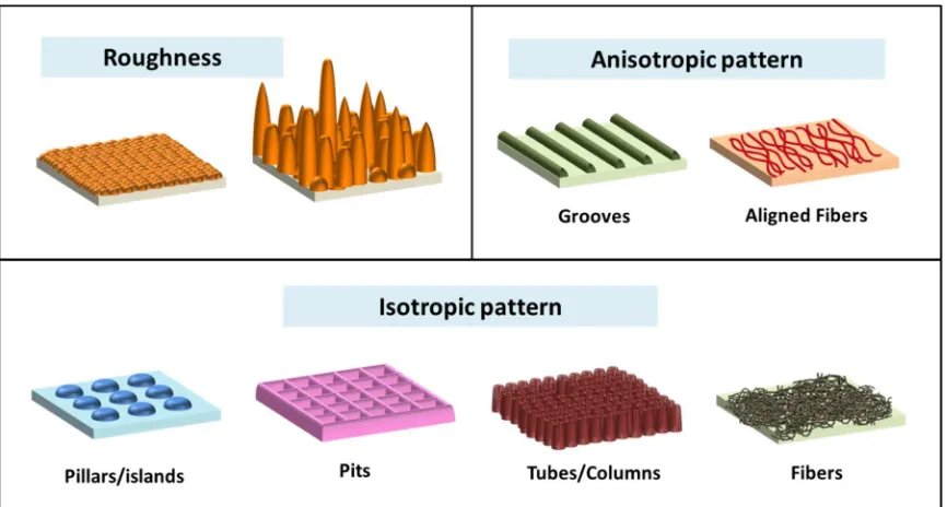
Figure 1.1Schematic illustration of different factors affecting surface topography:
Roughness, Anisotropic pattern, and Isotropic pattern ...3
Figure 1.2 Examples of surface roughness influences cell behaviors ...5
Figure 1.3 Examples of isotropic pattern influences cellular responses ...9
Figure 1.4 Electrospun nanofibres induce stem cell differentiation ... 11
Figure 1.5Genetically engineered TMV-RGD enhances cell adhesion on fibrous substrates ...13
Figure 1.6TMV-induced osteogenic differentiation in BMSCs in vitro ...15
Figure 1.7 Schematic diagram depicts interrelation of common intracellular signaling events triggered by changes in substrate topography and predicting sequential events for TMV-induced BMP-2 upregulation, leading to accelerated osteogenesis...18
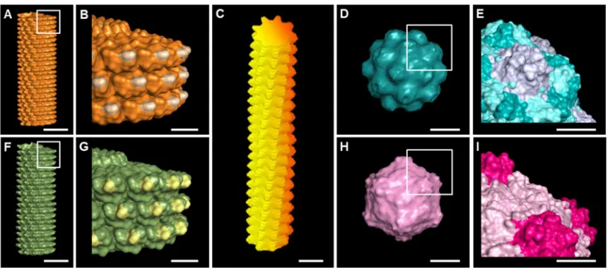
Figure 2.1 Molecular models shows surface topography of plant viruses used in this study ...37
Figure 2.2 Representative AFM micrographs showing the coverage of PDL coated substrate with different virus nanoparticles indicate the viral particles ...41
Figure 2.3 RT-qPCR analysis showed significant BMP2 upregulation in cells grown on TMV, TVCV, PVX, and TYMV coated substrates ...43
Figure 2.4 The expression of osteogenic marker in BMSCs cultured on PDL and different virus nanoparticles coated substrates under osteogenic conditions ...44
Figure 2.5 BMP2 immunohistochemical staining suggests the protein expressions are localized to the cell aggregates; most are found on TMV, TVCV, PVX, and TYMV substrates ...46
Figure 2.8 Immunochemical staining showing the difference in vinculin size of cells on PDL or virus coated substrates for 24 hours ...52
Figure 2.9 Representative actin (top panel) and vinculin (bottom panel)
immunofluorescent heat maps of cells culture on PDL and virus coated substrates ...54
Figure 3.1 Schematic shows TMV coat protein coated gold nanorod (TMV-GNRs) preparation and structural comparison of align and random TMV coat protein (TMV-CP) coated nanorod structure ...75
Figure 3.2 CTAB-GNRs characterization. (a-c) TEM image of (a) wild type TMV (b-c)
CTAB-GNRs (d) height profile AFM image of CTAB-GNRs shows diameter
measurement of the gold nanorod ...76
Figure 3.3Transmission electron microscopy image shows layer by layer coating of CTAB-GNRs with PAA and PAH ...78
Figure 3.4 Characterization of nanoparticles coated substrates for stem cell cultures ...79
Figure 3.5 TEM image shows intact nanorod structure of TMV-GNRs after assembly process...80
Figure 3.6 Osteogenesis of mesenchymal stem cells ...82
Figure 4.1 BMP2 inhibitor (Noggin) inhibits TMV induced osteogenesis of stem cells ..99
Figure 4.2 RhoA/ROCK pathway involves in TMV substrates mediated osteogenesis of stem cells ...101
LIST OF TABLES
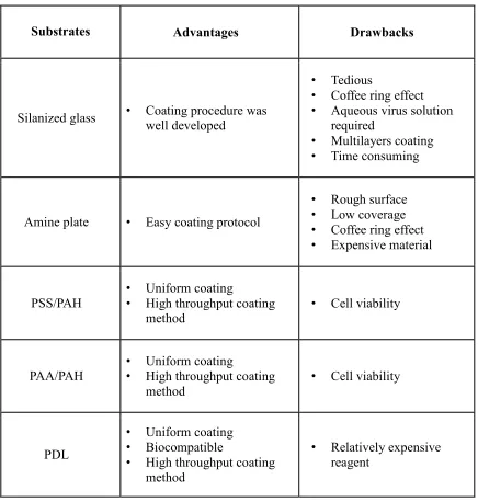
Table 2.1 Summary of advantages and drawbacks of each virus coating procedure ...39
Table 2.2 Primers used for RT-qPCR to measure gene expression levels. BGLAP: bone-gamma-carboxyglutamate protein; BMP2: bone morphogenetic protein 2; SPP1: secreted phosphoprotein 1 ...59
Table 3.1 Primers used for RT-qPCR to measure gene expression levels. BGLAP: bone-gamma-carboxyglutamate protein; BMP2: bone morphogenetic protein 2 ...89
1Metavarayuth K, Sitasuwan P, Zhao X, Lin Y, Wang Q. 2015. ACS. Biomater. Sci. Eng. 2: 142-51. Reprinted here with permission of publisher (Appendix A).
CHAPTER 1
INTRODUCTION: THE INFLUENCE OF SURFACE TOPOGRAPHICAL
CUES ON THE DIFFERENTIATION OF MESENCHYMAL STEM
CELLS
11.1 STEM CELL FATE AND MICROENVIRONMENT
One key aspect of tissue engineering is to develop biomimetic scaffolding
materials that can modulate the proliferation, self-renewal and differentiation of
multipotent stem cells into different lineages. In vivo, stem cells exist in a complex and
active environment, a key component of which is the extracellular matrix (ECM).[1] The
ECM provides physical and chemical supports for the cell and contains supramolecular
assemblies of proteins and glycosaminoglycans, which play a vital role in the cell
behavior. In order to undergo fundamental biological processes, the cells must adhere to
the underlying ECM. As a result, many novel biomaterials, which are purposely created
to improve or replace biological functions, have been designed to resemble the ECM. To
engineer a biomimetic scaffold resembling native ECM, and ultimately, to enable the
tissue regeneration, an extensive study on the interactions between stem cells and
implanted materials is necessary.
During recent years, there is extensive research emphasizing on the chemical (i.e.
functional groups, surface charge, surface energy, hydrophobicity, and protein
composition) and physical (i.e. overall architecture, porosity, surface topography, and
of these properties is crucial to develop biomaterials that guide stem cells for proper
tissue regeneration. In this chapter, we focus our discussion on the surface topography of
2D scaffold, especially, with an emphasis on the virus-based materials.
1.2 TOPOLOGICAL CUES FROM THE SUBSTRATES
It was first demonstrated in 1911 that cell behaviors can be controlled by
topological cues from the underlying substrates.[3] Later, the term contact guidance was
coined by Paul Weiss in 1945.[4] Contact guidance refers to the phenomenon that cells
adjust their orientation and align along the patterns that they are cultured on. Cells can
respond to topographical features as small as 5 nm [5] so it is important to achieve
surface patterns at a nanometer-scale resolution. Current nano- and micro-fabrication
methods include electron beam- or photo-lithography, self-assembling systems,
microcontact printing, particle synthesis, replica casting or molding, chemical etching,
sandblasting and electrospinning.[5, 6] These techniques enable the recapitulation of
topographical cues in the cell niche in a controllable and reproducible fashion. In general,
factors affecting substrate surface topography include (1) roughness of the underlying
surface, and (2) patterns on the surface (Figure 1.1).[7, 8]
The most studied aspect of topography is surface roughness which relates to the
texture of the uppermost layer of a material and is quantified by measuring the
protrusions or depressions at the surface. Numerous experiments have reported that
Figure 1.1Schematic illustration of different factors affecting surface topography:
roughness (Ra) closed to 1 µm.[9-14] For example, Yang et al. investigated the
enhancement of osteogenic differentiation by surface roughness introduced to
hydroxyapatite (HA) discs. The discs have Ra of surface topography ranging from 0.2 to
1.65 µm, and human bone-marrow mesenchymal stem cells (hBMSCs) were cultured in
osteogenic medium (α-MEM supplemented with 10% fetal bovine serum, 50 mg/mL
ascorbic acid, 10 mM glycerophosphate, 100 nM dexamethasone and 100 U/mL
penicillin and 100 mg/L streptomycin) on these discs. The optimal osteogenic
differentiation was observed on discs with surface topography characterized by Ra
ranging from 0.7 to 1.0 µm (Figure 1.2a,b).[9] Recent study by Faia-Torres et al. applied
polycaprolactone (PCL) gradient substrate to study effect of surface roughness on
osteogenesis of MSCs in basal growth media without soluble osteogenic inducers. They
demonstrated that the expression of osteogenic markers (alkaline phosphatase (ALP) and
collagen type I proteins) and mineralization are related to the surface roughness.[10]
More specifically, their results show that peak expression of normalized ALP was found
in the area that has substrate gradient at position 5 mm that corresponded to Ra~0.93 µm.
This trend is also consistent with a systemic review by Wennerberg and Albrektsson
which concluded that moderately rough surface (Ra~1-2 µm) showed strongest bone
responses.[8] In addition, various experiments have shown that nanoscale
surface-roughness can also influence cell behavior. Although the optimal nano-surface-roughness scale
for osteogenic differentiation cannot be specified, introduction of nanoscale roughness to
Figure 1.2 Examples of surface roughness influences cell behaviors (a) Scanning electron microscopic (SEM) images of the surface morphology of hydroxyapatite (HA) discs. Scale bar is 200 µm. (b) Alizarin Red staining of differentiating hBMSCs in osteogenic medium. Note darkest red staining at Ra 0.7 µm. Reproduced with permission from ref 8a. Copyright 2015 Elsevier B.V. (c) Surface morphology of titanium discs for
osteoblastic cell culture. SEM images of machined titanium (A) and acid-etched titanium (B). Bar = 20 µm. AFM images of the machined titanium (C) and the acid-etched
that when cultured on dual acid-etched titanium surface with a Ra of 110 nm rat
bone-marrow derived osteoblast differentiation increased compared to Ra = 49 nm.[15] Similar
observations have been noted by de Oliveira et al.[16, 17] Contrary to this, rat periosteal
cell-differentiation into osteoblasts, which could be seen on machined titanium disk
surfaces (Ra = 49 nm), was inhibited on acid etched surfaces (Ra = 183 nm), while
chondrocyte specific genes were activated when cultured in an osteochondral-defined
culture medium containing both osteogenic and chondrogenic differentiation factors.[18]
In explaining cell responses to surface pattern, we will use a classification based
on the orientation of topography (isotropic or anisotropic). An anisotropic surface has a
clear orientation such as ridges and grooves surfaces. On the other hand, an isotropic
surface is a surface with no orientation, such as evenly or randomly distributed pits,
protrusions, pillars, channels, or etc. Techniques developed to engineer these surface
orientations are not the focus of this mini review and they have been reviewed in detailed
elsewhere.[19, 20]
Cell orientation and migration along the anisotropic direction of ridges and
grooves have long been observed in microscale.[21-25] Multiple studies revealed that
MSCs committed to adipogenic [26] and myogenic [27] phenotypes when microscale
grooves are introduced to substrate surface, especially with the groove scale less than 500
nm. Conversely, osteogenic differentiation is negatively affected by this particular
anisotropic pattern.[28, 29] Periodicity can also modulate differential function of cells. In
techniques, recent studies have focused on whether cells align on nanoscale ridges and
grooves and can still be induced by contact guidance.[31-38] In a study by Zhu et al.,
stem cell derived osteoblasts were cultured on polystyrene (PS) nanogrooves (300-nm
pitch, 60- to 70-nm depth) substrates in dexamethasone, ascorbic acid and
β-glycerophosphate supplemented media, which were found to exhibit anisotropic
orientation in both cellular actin and mineralized matrix.[38] In addition, elongation of
stem cells plays an important role in neuronal differentiation of stem cells. In fact,
nanogroove topography is widely studied for neuron tissue engineering, [39-42] For
example, Yim et al. have shown that hMSCs could differentiate and proliferate on the
nanogratings of 350 nm width. In addition, alignment of cytoskeleton and nuclei of
elongated hMSCs were observed along the nanogratings, and gene profiling and
immunostaining showed significant up-regulation of neuronal markers such as
microtubule-associated protein 2 (MAP2) compared to unpatterned and micropatterned
controls.[39]
In summary, anisotropic topographies induce dramatic morphological changes
(via contact guidance) in cellular, cytoskeletal, and focal adhesions regardless of micro-
or nanoscale, which subsequently could lead to changes in gene expression and modulate
stem cell differentiation into specific lineages.
As comparison, isotropic patterns cannot influence the cell alignment, Instead, it
has been shown to enable the control of more-collective cell functions. Cell response to
isotropic pattern is often inconsistent and difficult for in-depth analysis due to the
isotropic topographies that will be reviewed here are distributed pillars, pit, nanotubes,
and random nanofibers.
Random distributed pillars or islands on a supporting surface has been
demonstrated to influence osteogenesis of stem cells. There are many factors that can be
varied in a pillared surface substrate, for example, height, shape and diameter of island,
and distance between two islands. The complexity of pillared surface makes it
inappropriate to compare the results from each study. Nevertheless, there is a general
trend of cell behaviors attributed to surface isotropic topographies. The introduction of
pillars or islands to substrate surface usually enhances osteogenic differentiation [43-46]
but particular dimension of the island that induces the highest differentiation cannot be
nailed down. Besides dimension of topographical cues, the distribution of topographical
feature may also have significant influences on stem cell behaviors. Dalby et al. reported
that surfaces composed of nanopits with controlled disordered resulted in increased
expression of osteogenic markers relative to surfaces consisting of either highly ordered
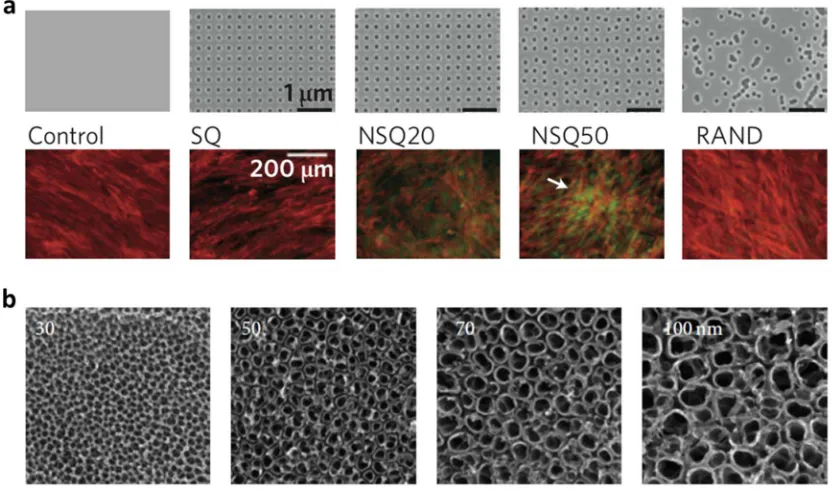
or randomly displaced nanopits (Figure 1.3a).[47] In addition to distribution of nanopits,
depth of nanopits has also been shown to affect cell responses. In general, the deeper pit
tends to enhance higher osteogenesis of stem cells.[48, 49] Another feature of isotropic
surface that is currently explored particularly for bone tissue engineering is nanotubes.
There is an increasing number of data elucidating the benefits of using TiO2 nanotubes,
one of the lateral spacing topographical cues, for enhanced orthopedic implant surfaces,
Figure 1.3 Examples of isotropic patterns that influence cellular responses. (a) Top row shows images of nanotopographies fabricated by electron beam lithography (EBL) on poly(methyl methacrylate) (PMMA). All present 120 nm diameter pits (100 nm deep, absolute or average 300 nm center–center spacing) with square (SQ), control disordered 20 (NSQ 20), control disordered 50 (NSQ 50, ± 20 or 50 nm from true center) and random placements (RAND). Bottom row shows osteoprogenitors cultured on the
control, note the lack of positive osteopontin (OPN) stain; SQ, note reduced cell numbers compared with the control; NSQ20, note some OPN positive cells; NSQ50, note
nanotubes scaffolds for osteogenic differentiation have been obtained from three groups
of pioneers in bone tissue engineering.[50-52]
Electrospun nanofiber webs may also be evaluated within the context of
topographic effect because randomly deposited nanofibers with variable nanoscale
thicknesses provide nanotextures coupled with micropores.[53, 54] Electrospun fibers
have been investigated as promising tissue engineering scaffolds since they mimic the
nanoscale properties of native ECM. It can be aligned on substrate surface to create both
anisotropic and isotropic topography which control commitment of stem cells to a
specific lineage. Recent study by Yin et al. demonstrate that the aligned anisotropic
fibrous scaffold displays promising results in tendon-like tissue regeneration at the early
repair stage, while in the random fibrous scaffold group, they observed the development
of bone formation at the injury site. The two topographically-different scaffolds not only
support MSC adhesion and spreading, but also induce tenogenesis and osteogenesis,
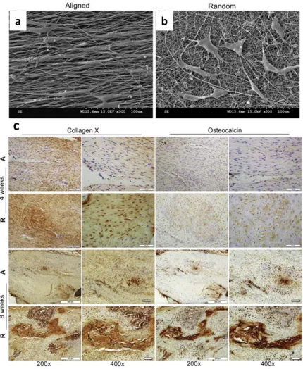
respectively, both in vitro and in vivo (Figure 1.4).[55]
1.3 PLANT VIRUS PROVIDES TOPOGRAPHICAL CUES FOR CELL CULTURING
There are two main categories of biomaterials used to study the influence of
nanotopography on cellular behaviors. The first type is polymeric materials, where
nanostructures could be generated by nanoimprint lithography,[56] capillary force
lithography,[57] ultraviolet assisted lithography,[58] embossing, photolithography, and
Further modifications to the two main types of biomaterials described above
could be achieved by nanoparticle surface coating. In our laboratory, we are creating
materials from plant virus nanoparticles, which can be produced in gram quantities at low
cost, and the resulting particles are highly monodispersed.[60, 61] Other advantages of
these bionanoparticles include the well-defined structural features, unique shapes and
sizes, genetic programmability and robust chemistries.[61] For example, the cell adhesion
motifs, like arginine-glycine-aspartic acid (RGD), have been incorporated into Tobacco
mosaic virus (TMV) coat proteins through genetic engineering to give mutant viruses
(e.g. TMV-RGD).[62] The RGD motif, predominantly found in an extracellular adhesive
glycoprotein, fibronectin, and other extracellular matrix proteins, was reported to mediate
cell adhesion via transmembrane integrin binding.[63] The polyvinyl alcohol (PVA)
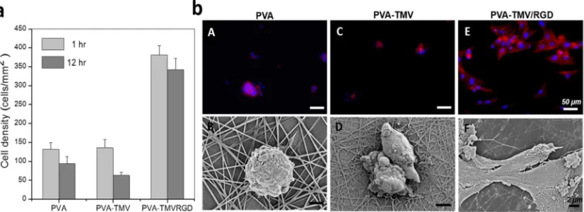
fibers incorporated with genetically engineered TMV-RGD was observed to facilitate cell
adhesion and spreading with prominent actin fibers, even in the absence of serum
supplement (Figure 1.5a). On the contrary, cells remained in a round shape with
randomized actin structure on both PVA and PVA-TMV substrates (Figure 1.5b).[62]
Besides TMV, there are other viruses that have also been used for tissue
engineering application.[64-68] One example of virus that has been widely studied is
M13 bacteriophage (phage), a nanofiber-like virus that has ability of self-assembly into
highly controlled periodic nanostructures when prepared in concentrated solution.[69-71]
Merzlyak et al. have genetically engineered phage to display cell signaling motif on their
Figure 1.5Genetically engineered TMV-RGD enhances cell adhesion on fibrous
growth in three dimensions.[64]
We hypothesized that the unique surface topography and polyvalent nature
provided by the plant virus coat protein assembly can be harnessed to modulate stem cell
responses. In particular, high-order hierarchical structure of plant virus compliments
investigation of the effect of ligand displayed polyvalency on cellular response as they
can be genetically and/or chemically modified to display particular functional groups in a
controlled spatial orientation at nanometer scale. Therefore, we have first utilized
substrates randomly coated with rod-shaped TMV and spherical Turnip yellow mosaic
virus (TYMV) to test osteogenic potential of bone marrow derived mesenchymal stem
cells (BMSCs).[67, 72] Surprisingly, the osteogenic differentiation process was
accelerated by 7 days in both cases. The underlying reasons how topography and
nanopattern of virus-based materials can affect differentiation process is still not well
understood. In an attempt to gain better understanding, early cellular responses to TMV
coated substrate were observed within 24 hours of osteogenic induction.[73] We
discovered that bone morphogenetic protein 2 (BMP-2) was upregulated endogenously
during the first 24 hours with a peak expression at 8 hours (Figure 1.6a, b).
BMP-2 is one of the most potent inducers of bone differentiation in mesenchymal
stem cells [74] and is highly involved in the beginning of bone repair in an animal
study.[75] Recombinant human BMP-2 (rhBMP-2) is commercially available and used as
therapeutic supplement for bone repair in spine fusion surgeries and tibial fracture
Figure 1.6TMV-induced osteogenic differentiation in BMSCs in vitro.[73] (a) Gene expression profile showing an upregulation of BMP2 mRNA level in BMSCs on TMV substrate at 8 hours after osteoinduction. (b) Immunohistochemical staining illustrating an increase in BMP2 expression at the protein level in BMSCs grown on TMV surface. Reproduced with permission from ref 37. Copyright 2012 The Royal Society of
breathing difficulty.[77-80] It was observed that in vitro osteogenic differentiation
induced by supplementing BMP-2 to the cell culture was more effective when cells were
grown on titanium surface with nanometer size of roughness.[81] Therefore,
material-induced BMP-2 endogenous expression may provide an alternative approach to
orthopedic surgeries where the morphogen is localized and the expression level is
self-regulated, leading to reduce adverse effects.
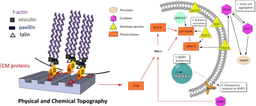
1.4 POSSIBLE MECHANISM OF TOPOGRAPHICAL CUES INDUCED STEM CELL
DIFFERENTIATION
Initial clues of molecular mechanisms by which cells sense different topography
are the differences in focal adhesion (FA) structures of cells on different substrates.
Variations of FA size, strength, and composition often reflect changes in actin
contractility and point to RhoA, a small GTPase whose activation enhances non-muscle
myosin IIa-dependent actin contractility by stimulating the formation of stress fibers and
FAs.[82] Many studies have emphasized the critical role of RhoA, Rho-associated kinase
(ROCK), and its downstream effects on actomyosin contractility on the control of cell
fate by cell spreading.[83-87] General concept acquired from these studies describes that
MSCs differentiate along an osteogenic lineage when RhoA/ROCK pathway is activated
which leads to cell spreading; whereas adipogenesis is dominated when RhoA/ROCK
pathway is inhibited and cell spreading is restricted. Another key regulator of
nanogratings [88] also increased FAK activity. Differential activation of FAK in turn
triggers downstream signaling to the mitogen-activated protein kinase (MAPK) cascade,
which is an intracellular signaling cascade that delivers information about the
extracellular environment to the cell nucleus.[30, 90] Ultimately, this pathway in turn
influenced the transcription factor RUNX2 to control osteoblast differentiation and
matrix mineralization (Figure. 1.7). Collectively, these findings suggest the participation
of FAK/RhoA/ROCK/MAPK signaling pathway in substrate topography influence cell
fate decisions.
Our plant virus based material provided the in situ endogenous BMP-2
stimulation and at the same time nanoscale surface features.[68, 73] One example of an
attempt to combine BMP-2 induction with nanopatterned surface is the immobilization of
BMP-2 peptides on nanoscale grooved and dot-shaped polymer surfaces, resulting in an
improved osteogenesis without any other soluble inducers.[91] In this study, they
discovered cytoskeleton and cell membrane stress induce RhoA/ROCK-mediated
cytoskeletal tension and subsequently osteogenesis. The restrict cell morphology and
stress in cell cytoskeleton observed were similar to what we observed in our plant virus
scaffolds. Therefore, it is possible that nanotopographical features supplied by the virus
nanoparticles influence cell spreading and introduce mechanical stress on the cell
membrane leads to an early osteogenesis via the similar activation of RhoA/ROCK
pathway.[92] To identify the upstream side of RhoA/ROCK signaling pathway, we
started by focusing on identifying cell membrane receptors responsible for sensing the
external stimuli. Our experiment showed that the size of focal adhesion complexes as
standard tissue culture plastics.[68, 73] Furthermore, there were a number of chemokines
and small chemotactic cytokines, in addition to macrophage chemotactic protein 1
(MCP-1), produced in response to cell adhesion on TMV substrate.[93] These chemokines are
not only important in the migration of immune cells during injury and infection,[94] but
also the migration of stem cells during body development and maturation.[95] The
production of these cytokines could be triggered as a consequence of cell membrane
receptor signaling. Gene expression changes in motility genes were briefly investigated.
It was discovered that actinin 3, integrin alpha 4, rhodopsin (Rho), Ras-related C3
botulinum toxin substrate 2 (Rac2), and Rho guanine nucleotide exchange factor 7 were
upregulated.[93] Interestingly, Rac2, a hematopoietic-specific Rho GTPase, has been
reported to play an important role in a success in long-term bone engraftment in
mice.[96] These genes have been reported to be upstream of protein kinases, possibly
leading to BMP-2 endogenous production which subsequently activate
RhoA/ROCK-mediate cytoskeletal tension and ultimately accelerate osteogenesis (Figure 1.4c).
1.5 SUMMARY
Topography of implant materials plays an important role in directing stem cell
fate. Microscale surface roughness has long been recognized to alter osteogenesis of stem
cells. By optimizing roughness scale of material surface to Ra~1-2 µm, bone formation
can be highly induced. Anisotropic surface has been studied in term of a tool to direct cell
alignment which often impacts stem cell fate. Isotropic surface is not determined to
influence cell alignment, instead it is proved to be able to control cell function. Cell
dimensions of islands decorated substrate surface for osteogenesis were varied from one
study to another. However, the concept that the introduction of pillars or islands to
substrate surface can benefit osteogenic differentiation has been verified. In addition, the
studies from Dalby et al. highlighted that the distribution of topographical features can
significantly influence cell response. Many current studies have focused on nanotube
featured materials which would also enhance bone formation; however, proper dimension
of the nanotube has not been endorsed. Furthermore, fibrous materials are widely
explored as promising tissue engineering scaffolds since they mimic the nanoscale
properties of native ECM. It can be placed on substrate surface to create either
anisotropic or isotropic surface and modulate stem cell differentiation to target
phenotype.
The complex signaling cascade involved in nanotopographical cues influence cell
responses is still unclear. The interaction between cells and material surface could be
static or dynamic. Further study is needed to understand how a particular nano-patterning
results in certain cellular response and behavior. For plant virus supplied
nanotopographical cues substrates, even though the endogenous upregulation of BMP-2
was observed for BMSCs in vitro, we cannot be concluded that this is a universal
phenomenon. Inhibiting BMP-2 by using siRNA, BMP-2 knock out, or applying BMP-2
inhibitors such as Noggin and Chordin may reaffirm the involvement of BMP-2 in the
phenomenon. In addition, to investigate the participation of RhoA/ROCK pathway,
For example at molecular level, very limited studies have been reported to systematically
elucidate the correlation between different topographical cues and cellular behaviors. It
would require further investigation from many scientists in different fields, such as
computational biology and molecular biology, to solve the puzzles.
1.6 REFERENCES
[1] Gattazzo F, Urciuolo A, Bonaldo P. Extracellular matrix: a dynamic
microenvironment for stem cell niche. Biochim Biophys Acta. 2014;1840:2506-19.
[2] Kaivosoja E, Barreto G, Levon K, Virtanen S, Ainola M, Konttinen YT. Chemical
and physical properties of regenerative medicine materials controlling stem cell fate. Ann
Med. 2012;44:635-50.
[3] Harrison RG. On the Stereotropism of Embryonic Cells. Science. 1911;34:279-81.
[4] Weiss P. Experiments on cell and axon orientation in vitro; the role of colloidal
exudates in tissue organization. J Exp Zool. 1945;100:353-86.
[5] Curtis A, Wilkinson C. Nantotechniques and approaches in biotechnology. Trends
Biotechnol. 2001;19:97-101.
[6] Ross AM, Jiang Z, Bastmeyer M, Lahann J. Physical aspects of cell culture
substrates: topography, roughness, and elasticity. Small. 2012;8:336-55.
[7] Moore DT. Tutorials in optics. Washington, DC: Optical Society of America; 1992.
[8] Wennerberg A, Albrektsson T. Effects of titanium surface topography on bone
integration: a systematic review. Clin Oral Implants Res. 2009;20 Suppl 4:172-84.
[9] Yang W, Han W, He W, Li J, Wang J, Feng H, et al. Surface topography of
hydroxyapatite promotes osteogenic differentiation of human bone marrow mesenchymal
[10] Faia-Torres AB, Charnley M, Goren T, Guimond-Lischer S, Rottmar M,
Maniura-Weber K, et al. Osteogenic differentiation of human mesenchymal stem cells in the
absence of osteogenic supplements: A surface-roughness gradient study. Acta Biomater.
2015;28:64-75.
[11] London RM, Roberts FA, Baker DA, Rohrer MD, O'Neal RB. Histologic
comparison of a thermal dual-etched implant surface to machined, TPS, and HA surfaces:
bone contact in vivo in rabbits. Int J Oral Maxillofac Implants. 2002;17:369-76.
[12] Grassi S, Piattelli A, de Figueiredo LC, Feres M, de Melo L, Iezzi G, et al.
Histologic evaluation of early human bone response to different implant surfaces. J
Periodontol. 2006;77:1736-43.
[13] Park KH, Heo SJ, Koak JY, Kim SK, Lee JB, Kim SH, et al. Osseointegration of
anodized titanium implants under different current voltages: a rabbit study. J Oral
Rehabil. 2007;34:517-27.
[14] Suzuki K, Aoki K, Ohya K. Effects of surface roughness of titanium implants on
bone remodeling activity of femur in rabbits. Bone. 1997;21:507-14.
[15] Takeuchi K, Saruwatari L, Nakamura HK, Yang JM, Ogawa T. Enhanced intrinsic
biomechanical properties of osteoblastic mineralized tissue on roughened titanium
surface. J Biomed Mater Res A. 2005;72:296-305.
[16] de Oliveira PT, Nanci A. Nanotexturing of titanium-based surfaces upregulates
expression of bone sialoprotein and osteopontin by cultured osteogenic cells.
[17] de Oliveira PT, Zalzal SF, Beloti MM, Rosa AL, Nanci A. Enhancement of in vitro
osteogenesis on titanium by chemically produced nanotopography. J Biomed Mater Res
A. 2007;80:554-64.
[18] Kubo K, Att W, Yamada M, Ohmi K, Tsukimura N, Suzuki T, et al.
Microtopography of titanium suppresses osteoblastic differentiation but enhances
chondroblastic differentiation of rat femoral periosteum-derived cells. J Biomed Mater
Res A. 2008;87:380-91.
[19] Klymov A, Prodanov L, Lamers E, Jansen JA, Walboomers XF. Understanding the
role of nano-topography on the surface of a bone-implant. Biomaterials Science.
2013;1:135-51.
[20] Yao X, Peng R, Ding J. Cell-material interactions revealed via material techniques
of surface patterning. Adv Mater. 2013;25:5257-86.
[21] Wojciak-Stothard B, Madeja Z, Korohoda W, Curtis A, Wilkinson C. Activation of
macrophage-like cells by multiple grooved substrata. Topographical control of cell
behaviour. Cell Biol Int. 1995;19:485-90.
[22] Meyle J, Wolburg H, von Recum AF. Surface micromorphology and cellular
interactions. J Biomater Appl. 1993;7:362-74.
[23] Matsuzaka K, Walboomers XF, Yoshinari M, Inoue T, Jansen JA. The attachment
and growth behavior of osteoblast-like cells on microtextured surfaces. Biomaterials.
2003;24:2711-9.
[24] Charest JL, Bryant LE, Garcia AJ, King WP. Hot embossing for micropatterned cell
[25] Andersson AS, Backhed F, von Euler A, Richter-Dahlfors A, Sutherland D, Kasemo
B. Nanoscale features influence epithelial cell morphology and cytokine production.
Biomaterials. 2003;24:3427-36.
[26] Wang PY, Li WT, Yu J, Tsai WB. Modulation of osteogenic, adipogenic and
myogenic differentiation of mesenchymal stem cells by submicron grooved topography. J
Mater Sci Mater Med. 2012;23:3015-28.
[27] Wang ZY, Lim J, Ho YS, Zhang QY, Chong MS, Tang M, et al. Biomimetic
three-dimensional anisotropic geometries by uniaxial stretching of poly(epsilon-caprolactone)
films: degradation and mesenchymal stem cell responses. J Biomed Mater Res A.
2014;102:2197-207.
[28] Biggs MJ, Richards RG, McFarlane S, Wilkinson CD, Oreffo RO, Dalby MJ.
Adhesion formation of primary human osteoblasts and the functional response of
mesenchymal stem cells to 330nm deep microgrooves. J R Soc Interface.
2008;5:1231-42.
[29] Watari S, Hayashi K, Wood JA, Russell P, Nealey PF, Murphy CJ, et al. Modulation
of osteogenic differentiation in hMSCs cells by submicron topographically-patterned
ridges and grooves. Biomaterials. 2012;33:128-36.
[30] Biggs MJP, Richards RG, Gadegaard N, Wilkinson CDW, Oreffo ROC, Dalby MJ.
The use of nanoscale topography to modulate the dynamics of adhesion formation in
primary osteoblasts and ERK/MAPK signalling in STRO-1+ enriched skeletal stem cells.
[31] Teixeira AI, Abrams GA, Bertics PJ, Murphy CJ, Nealey PF. Epithelial contact
guidance on well-defined micro- and nanostructured substrates. J Cell Sci.
2003;116:1881-92.
[32] Karuri NW, Liliensiek S, Teixeira AI, Abrams G, Campbell S, Nealey PF, et al.
Biological length scale topography enhances cell-substratum adhesion of human corneal
epithelial cells. J Cell Sci. 2004;117:3153-64.
[33] Diehl KA, Foley JD, Nealey PF, Murphy CJ. Nanoscale topography modulates
corneal epithelial cell migration. J Biomed Mater Res A. 2005;75:603-11.
[34] Bunk R, Klinth J, Montelius L, Nicholls IA, Omling P, Tagerud S, et al. Actomyosin
motility on nanostructured surfaces. Biochem Biophys Res Commun. 2003;301:783-8.
[35] Johansson F, Carlberg P, Danielsen N, Montelius L, Kanje M. Axonal outgrowth on
nano-imprinted patterns. Biomaterials. 2006;27:1251-8.
[36] Barbucci R, Pasqui D, Wirsen A, Affrossman S, Curtis A, Tetta C. Micro and
nano-structured surfaces. J Mater Sci Mater Med. 2003;14:721-5.
[37] Hamilton DW, Riehle MO, Monaghan W, Curtis AS. Articular chondrocyte passage
number: influence on adhesion, migration, cytoskeletal organisation and phenotype in
response to nano- and micro-metric topography. Cell Biol Int. 2005;29:408-21.
[38] Zhu B, Lu Q, Yin J, Hu J, Wang Z. Alignment of osteoblast-like cells and
cell-produced collagen matrix induced by nanogrooves. Tissue Eng. 2005;11:825-34.
[39] Yim EK, Pang SW, Leong KW. Synthetic nanostructures inducing differentiation of
[40] Lee MR, Kwon KW, Jung H, Kim HN, Suh KY, Kim K, et al. Direct differentiation
of human embryonic stem cells into selective neurons on nanoscale ridge/groove pattern
arrays. Biomaterials. 2010;31:4360-6.
[41] Ankam S, Suryana M, Chan LY, Moe AA, Teo BK, Law JB, et al. Substrate
topography and size determine the fate of human embryonic stem cells to neuronal or
glial lineage. Acta Biomater. 2013;9:4535-45.
[42] Wang G, Ao Q, Gong K, Wang A, Zheng L, Gong Y, et al. The effect of topology of
chitosan biomaterials on the differentiation and proliferation of neural stem cells. Acta
Biomater. 2010;6:3630-9.
[43] McNamara LE, Sjostrom T, Burgess KE, Kim JJ, Liu E, Gordonov S, et al. Skeletal
stem cell physiology on functionally distinct titania nanotopographies. Biomaterials.
2011;32:7403-10.
[44] Fiedler J, Ozdemir B, Bartholoma J, Plettl A, Brenner RE, Ziemann P. The effect of
substrate surface nanotopography on the behavior of multipotnent mesenchymal stromal
cells and osteoblasts. Biomaterials. 2013;34:8851-9.
[45] You MH, Kwak MK, Kim DH, Kim K, Levchenko A, Kim DY, et al.
Synergistically enhanced osteogenic differentiation of human mesenchymal stem cells by
culture on nanostructured surfaces with induction media. Biomacromolecules.
2010;11:1856-62.
[46] Sjostrom T, Dalby MJ, Hart A, Tare R, Oreffo RO, Su B. Fabrication of pillar-like
[47] Dalby MJ, Gadegaard N, Tare R, Andar A, Riehle MO, Herzyk P, et al. The control
of human mesenchymal cell differentiation using nanoscale symmetry and disorder. Nat
Mater. 2007;6:997-1003.
[48] Wilkinson A, Hewitt RN, McNamara LE, McCloy D, Dominic Meek RM, Dalby
MJ. Biomimetic microtopography to enhance osteogenesis in vitro. Acta Biomater.
2011;7:2919-25.
[49] Zouani OF, Chanseau C, Brouillaud B, Bareille R, Deliane F, Foulc MP, et al.
Altered nanofeature size dictates stem cell differentiation. J Cell Sci. 2012;125:1217-24.
[50] Park J, Bauer S, Schlegel KA, Neukam FW, von der Mark K, Schmuki P. TiO2
nanotube surfaces: 15 nm--an optimal length scale of surface topography for cell
adhesion and differentiation. Small. 2009;5:666-71.
[51] Park J, Bauer S, von der Mark K, Schmuki P. Nanosize and vitality: TiO2 nanotube
diameter directs cell fate. Nano Lett. 2007;7:1686-91.
[52] Kang Y-H, Choi B, Ahn C, Oh S, Lee MS, Jin E-J. Titanium Oxide Nanotube
Surface Topography and MicroRNA-488 Contribute to Modulating Osteogenesis. Journal
of Nanomaterials. 2014;2014:8.
[53] Badami AS, Kreke MR, Thompson MS, Riffle JS, Goldstein AS. Effect of fiber
diameter on spreading, proliferation, and differentiation of osteoblastic cells on
electrospun poly(lactic acid) substrates. Biomaterials. 2006;27:596-606.
[54] Stevens MM, George JH. Exploring and engineering the cell surface interface.
[55] Yin Z, Chen X, Song H-x, Hu J-j, Tang Q-m, Zhu T, et al. Electrospun scaffolds for
multiple tissues regeneration in vivo through topography dependent induction of lineage
specific differentiation. Biomaterials. 2015;44:173-85.
[56] Huang XD, Bao L-R, Cheng X, Guo LJ, Pang SW, Yee AF. Reversal imprinting by
transferring polymer from mold to substrate. Journal of Vacuum Science & Technology
B. 2002;20:2872-6.
[57] Suh K-Y, Park MC, Kim P. Capillary Force Lithography: A Versatile Tool for
Structured Biomaterials Interface Towards Cell and Tissue Engineering. Advanced
Functional Materials. 2009;19:2699-712.
[58] Choi SJ, Yoo PJ, Baek SJ, Kim TW, Lee HH. An ultraviolet-curable mold for
sub-100-nm lithography. J Am Chem Soc. 2004;126:7744-5.
[59] Janson IA, Putnam AJ. Extracellular matrix elasticity and topography:
Material-based cues that affect cell function via conserved mechanisms. Journal of Biomedical
Materials Research Part A. 2015;103:1246-58.
[60] Liu Z, Qiao J, Niu Z, Wang Q. Natural supramolecular building blocks: from virus
coat proteins to viral nanoparticles. Chem Soc Rev. 2012;41:6178-94.
[61] Liu Z, Xiao L, Xu B, Zhang Y, Mak AF, Li Y, et al. Covalently immobilized
biomolecule gradient on hydrogel surface using a gradient generating microfluidic device
for a quantitative mesenchymal stem cell study. Biomicrofluidics.
2012;6:24111-2411112.
[63] Pierschbacher M, Hayman EG, Ruoslahti E. Synthetic peptide with cell attachment
activity of fibronectin. Proc Natl Acad Sci U S A. 1983;80:1224-7.
[64] Merzlyak A, Indrakanti S, Lee SW. Genetically engineered nanofiber-like viruses
for tissue regenerating materials. Nano Lett. 2009;9:846-52.
[65] Wang J, Wang L, Li X, Mao C. Virus activated artificial ECM induces the
osteoblastic differentiation of mesenchymal stem cells without osteogenic supplements.
Sci Rep. 2013;3:1242.
[66] Lin Y, Su Z, Niu Z, Li S, Kaur G, Lee LA, et al. Layer-by-layer assembly of viral
capsid for cell adhesion. Acta Biomater. 2008;4:838-43.
[67] Kaur G, Valarmathi MT, Potts JD, Jabbari E, Sabo-Attwood T, Wang Q. Regulation
of osteogenic differentiation of rat bone marrow stromal cells on 2D nanorod substrates.
Biomaterials. 2010;31:1732-41.
[68] Metavarayuth K, Sitasuwan P, Luckanagul JA, Feng S, Wang Q. Virus
Nanoparticles Mediated Osteogenic Differentiation of Bone Derived Mesenchymal Stem
Cells. Advanced Science. 2015:n/a-n/a.
[69] Lee S-W, Mao C, Flynn CE, Belcher AM. Ordering of Quantum Dots Using
Genetically Engineered Viruses. Science. 2002;296:892-5.
[70] Dogic Z, Fraden S. Smectic phase in a colloidal suspension of semiflexible virus
particles. Physical review letters. 1997;78:2417.
[71] Lee S-W, Wood BM, Belcher AM. Chiral Smectic C Structures of Virus-Based
[72] Kaur G, Valarmathi MT, Potts JD, Wang Q. The promotion of osteoblastic
differentiation of rat bone marrow stromal cells by a polyvalent plant mosaic virus.
Biomaterials. 2008;29:4074-81.
[73] Sitasuwan P, Andrew Lee L, Bo P, Davis EN, Lin Y, Wang Q. A plant virus
substrate induces early upregulation of BMP2 for rapid bone formation. Integr Biol
(Camb). 2012;4:651-60.
[74] Cheng H, Jiang W, Phillips FM, Haydon RC, Peng Y, Zhou L, et al. Osteogenic
activity of the fourteen types of human bone morphogenetic proteins (BMPs). J Bone
Joint Surg Am. 2003;85-A:1544-52.
[75] Ai-Aql ZS, Alagl AS, Graves DT, Gerstenfeld LC, Einhorn TA. Molecular
mechanisms controlling bone formation during fracture healing and distraction
osteogenesis. J Dent Res. 2008;87:107-18.
[76] McKay WF, Peckham SM, Badura JM. A comprehensive clinical review of
recombinant human bone morphogenetic protein-2 (INFUSE Bone Graft). Int Orthop.
2007;31:729-34.
[77] Bishop GB, Einhorn TA. Current and future clinical applications of bone
morphogenetic proteins in orthopaedic trauma surgery. Int Orthop. 2007;31:721-7.
[78] Carragee EJ, Hurwitz EL, Weiner BK. A critical review of recombinant human bone
morphogenetic protein-2 trials in spinal surgery: emerging safety concerns and lessons
learned. Spine J. 2011;11:471-91.
[80] Shields LB, Raque GH, Glassman SD, Campbell M, Vitaz T, Harpring J, et al.
Adverse effects associated with high-dose recombinant human bone morphogenetic
protein-2 use in anterior cervical spine fusion. Spine (Phila Pa 1976). 2006;31:542-7.
[81] Kato RB, Roy B, De Oliveira FS, Ferraz EP, De Oliveira PT, Kemper AG, et al.
Nanotopography directs mesenchymal stem cells to osteoblast lineage through regulation
of microRNA-SMAD-BMP-2 circuit. J Cell Physiol. 2014;229:1690-6.
[82] Hall A. Rho GTPases and the actin cytoskeleton. Science. 1998;279:509-14.
[83] Yao X, Peng R, Ding J. Effects of aspect ratios of stem cells on lineage
commitments with and without induction media. Biomaterials. 2013;34:930-9.
[84] Bhadriraju K, Yang M, Alom Ruiz S, Pirone D, Tan J, Chen CS. Activation of
ROCK by RhoA is regulated by cell adhesion, shape, and cytoskeletal tension.
Experimental Cell Research. 2007;313:3616-23.
[85] Wang YK, Yu X, Cohen DM, Wozniak MA, Yang MT, Gao L, et al. Bone
morphogenetic protein-2-induced signaling and osteogenesis is regulated by cell shape,
RhoA/ROCK, and cytoskeletal tension. Stem Cells Dev. 2012;21:1176-86.
[86] McBeath R, Pirone DM, Nelson CM, Bhadriraju K, Chen CS. Cell shape,
cytoskeletal tension, and RhoA regulate stem cell lineage commitment. Dev Cell.
2004;6:483-95.
[87] Xu Y, Wagner DR, Bekerman E, Chiou M, James AW, Carter D, et al. Connective
tissue growth factor in regulation of RhoA mediated cytoskeletal tension associated
[88] Teo BK, Wong ST, Lim CK, Kung TY, Yap CH, Ramagopal Y, et al.
Nanotopography modulates mechanotransduction of stem cells and induces
differentiation through focal adhesion kinase. ACS Nano. 2013;7:4785-98.
[89] Lim JY, Dreiss AD, Zhou Z, Hansen JC, Siedlecki CA, Hengstebeck RW, et al. The
regulation of integrin-mediated osteoblast focal adhesion and focal adhesion kinase
expression by nanoscale topography. Biomaterials. 2007;28:1787-97.
[90] Khatiwala CB, Kim PD, Peyton SR, Putnam AJ. ECM compliance regulates
osteogenesis by influencing MAPK signaling downstream of RhoA and ROCK. J Bone
Miner Res. 2009;24:886-98.
[91] Kim MJ, Lee B, Yang K, Park J, Jeon S, Um SH, et al. BMP-2
peptide-functionalized nanopatterned substrates for enhanced osteogenic differentiation of human
mesenchymal stem cells. Biomaterials. 2013;34:7236-46.
[92] Wang Y-K, Yu X, Cohen DM, Wozniak MA, Yang MT, Gao L, et al. Bone
Morphogenetic Protein-2-Induced Signaling and Osteogenesis Is Regulated by Cell
Shape, RhoA/ROCK, and Cytoskeletal Tension. Stem Cells and Development.
2012;21:1176-86.
[93] Sitasuwan P. Biomaterial-Induced Osteogenesis of Mesenchymal Stem Cells by
Surface Roughness and Functionalization. Columbia, SC: University of South Carolina;
2013.
[94] Niggli V. Signaling to migration in neutrophils: importance of localized pathways.
[96] Jansen M, Yang F-C, Cancelas JA, Bailey JR, Williams DA. Rac2-Deficient
Hematopoietic Stem Cells Show Defective Interaction with the Hematopoietic
CHAPTER 2
VIRUS NANOPARTICLES MEDIATED OSTEOGENIC
DIFFERENTIATION OF BONE DERIVED MESENCHYMAL STEM
CELLS
12.1 INTRODUCTION
It is well established that cell-material interactions regulate numerous cellular
functions.[1-3] Biological processes such as adhesion, growth, differentiation and
apoptosis, are controlled by cell shape and cytoskeletal organization which is directed by
cell-surface interactions.[4-7] Meanwhile, the surface chemistry and topography of
materials play a very crucial role in altering cell behaviors at many stages of cell growth
and development.[3, 8-13] Although the dimensions of mammalian cells are on the order
of a few micrometers, cellular sensing of the external environment and interaction with
biomaterials occurs at the nanometer level.[14, 15] Cell interactions with nanometric
surfaces often result in a specific sequence of gene and protein regulations. These series
of events initiate as early as the cell begins to sense the surrounding environment.
Therefore, the understanding of various topographical cues that are responsible for
35
the contact area for cell spreading, and (4) the geometry of topological features at a
nanometer scale.[16, 17] Reviewing the effect of individual cues is often complicated due
to the difficulty in controlling and altering particular topographical features while
preserving others. Micro/nanofabrication techniques are required to enable the
recapitulation of topographical cues in the cell niche in a controllable and reproducible
fashion. Examples of these technologies are mechanical roughening,[18] nano- and
microindentation, and substrate-templating using a well-defined relief to impart
topography with solvent-casting, electro-deposition, chemical-vapor depositions, or
compression-molding processes.[19-21] These engineered micro/nanoscale topographical
cues mimic the micro/nanoscale features in the physiological environment, which can be
used to demonstrate how individual cues or the combination of topographical cues affect
a particular cellular response. However, all these methods suffer from the laborious
process, the inability of predictably generating chemistry and topography in a
simultaneous fashion, the requirement for high-cost equipment, or the limited class of
material can be used.[6, 22-24]
Virus particles, especially plant viruses, are uniform in size, have well organized
and characterized 3D structure, and can be produced in high yield and purity.[25, 26] The
symmetrical arrangement of the outer coat proteins of the viral particles makes them
attractive scaffolds for polyvalent display of a variety of functional groups for various
applications.[25, 27-29] In the past two decades, new materials with unique structural
features have been developed for a wide range of applications, including electronics, drug
delivery, imaging, gene therapy, and immunotherapy, by taking advantages of distinctive
systematically investigation about how the nanoscale topographical cues of various plant
virus particles coated substrates impact cell behaviors, specifically, osteogenesis of bone
derived mesenchymal stem cells (BMSCs).
From our unexpected, yet significant, observation that rodlike plant virus
nanoparticle, Tobacco mosaic virus (TMV) coated two dimensional substrate
dramatically accelerates osteogenesis of BMSCs. The study suggested that the virus does
not act as soluble inducer becasue supplementing cell culture media with TMV solution
failed to mediate the differentiation.[32, 33] We have hypothesized that the shape of virus
nanoparticle and/or nanoscale topography provided by surface structure of virus particle
may be necessary for the enhanced osteogenesis. Therefore, in this study, we generate a
series of plant virus nanoparticles coated substrates using a variety of viral nanoparticles
with distinct morphology and nanotopography. We applied these virus based scaffolds to
investigate cellular responses to different types of topographical cues (Figure 2.1). Our
results show that some of these virus based scaffolds accelerate and enhance osteogenic
differentiation of bone derived mesenchymal stem cells (BMSCs). This finding presented
here may provide a new route for enhancing the performance of orthopedic implants by
regulating stem cell differentiation with nanotopography.
2.2 RESULTS AND DISCUSSION
2.2.1 Fabrication of virus-coated scaffolds by layer-by-layer deposition method
We fabricated 2D virus based substrates from five plant viruses which can be categorized
37
viruses morphologically different, but also they are nanotopographically dissimilar as
shown in Figure 2.1
Since all these viral particles have isoelectric points (pI) less than 5.5, overall
surface charges on these particles are negative in neutral pH condition. Via an
electrostatic interaction, negatively charged viral particles can be strongly adsorbed onto
various surfaces containing positively charged functional group. Several coating
strategies and substrates were tested to screen for a high throughput method that offers
uniform coating of every type of five virus particles on substrate surface. Summary of
advantages and drawbacks of each coating procedure is shown in Table 2.1. Glass slide
modified with organofunctional alkoxysilane molecules was first used to construct five
virus coated substrates followed protocol developed by Kaur et al. The primary amine
functional group of the organofunctional alkoxysilane can be protonated and presents
positively charges under neutral condition. General coating method is dropping 0.2 mL of
aqueous virus solution on the silanized glass and let the virus solution dry under sterile
tissue culture hood.[34] Although, high coverage of virus nanoparticles on substrate
surfaces can be achieved on this material, the coating protocol is very tedious and
requires skillful personal to prepare the substrates. In addition, coffee ring effect, a
pattern left by a puddle of particle-laden liquid after it evaporates is also commonly
detected on the substrates prepared by this method if the virus solution is not fully
covered the coating area. The higher number of virus layers could be detected around the
39
Table 2.1 Summary of advantages and drawbacks of different virus coating procedures
Substrates Advantages Drawbacks
Silanized glass • Coating procedure was
well developed
• Tedious
• Coffee ring effect
• Aqueous virus solution required
• Multilayers coating
• Time consuming
Amine plate • Easy coating protocol
• Rough surface
• Low coverage
• Coffee ring effect
• Expensive material
PSS/PAH
• Uniform coating
• High throughput coating method
• Cell viability
PAA/PAH
• Uniform coating
• High throughput coating method
• Cell viability
PDL
• Uniform coating
• Biocompatible
• High throughput coating method
than silanized glass but we still could not get a consistent coating. In particular, during
the drying process, more virus particles were drawn to the wall of the well and the center
of each well had much less virus particle.
Since drop dry coating method failed to prepare uniform coating, we changed to a
layer-by-layer (LbL) approach since it was reported by Zan et al. that the LbL method
could create high coverage regular coating of TYMV on 2D substrate.[35] Two pairs of
polyelectrolytes were tested for fabrication of the five virus particles coated substrates,
polystyrene sulfonate(PSS) alternates with polyallylamine hydrochloride(PAH) and
polyacrylic acid (PAA) alternates with PAH. Both of the polyelectrolytes pairs gave
uniform coating with high coverage of virus particles. However, cells could not survive
on the LbL substrates after a few days of culturing. To achieve a high cell viability, we
employed poly-d-lysine (PDL) in our study. The interaction between positively charged
amine functional group of PDL helps to retain the viral particles on the substrates by
electrostatic interaction. By depositing structurally and nanotopographically distinctive
viral particles on PDL coated substrate, we can readily construct an array of virus-coated
scaffolds with various topographies offered by the intrinsic morphology and
micro/nanotopography of each viral particle. The presence of viral particles on PDL
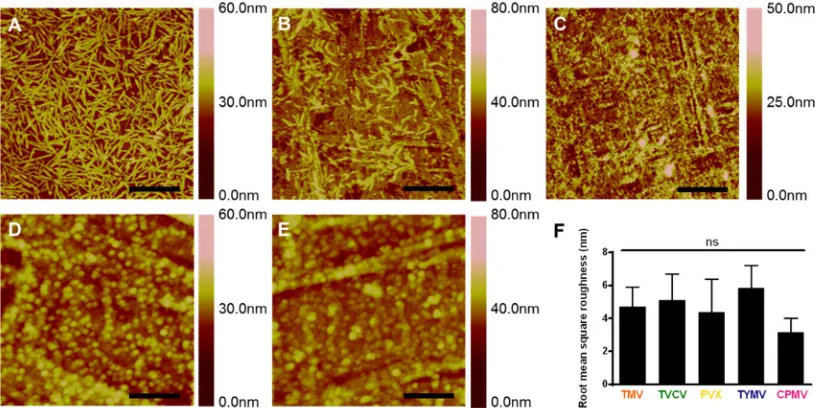
coated surface was confirmed by atomic force microscopy (AFM) (Figure 2.2). The AFM
micrographs also show a nearly complete coverage of substrates by intact viral particles.
The virus particles are randomly adsorbed on 12-well plates coated with PDL, however,
41
well plates was used throughout this study. The virus substrates have been
characterized in term of root mean square roughness from data collected from AFM
micrographs (n = 4). There is no significant difference of microscale roughness across the
virus coated substrates, created from deposition of numerous virus particles on the
substrate surface, across these five virus substrates.
2.2.2 Viral particles coated substrates promote Osteogenesis
To investigate the effect of surface topography on osteogenesis, we cultured BMSCs on
PDL coated substrate and the five virus-based substrates and studied the osteoblastic
differentiation. BMSCs were isolated and cultured as reported in literature.[36, 37]1-3
The purity of the stem cells populations has been previously verified with several stem
cells markers such as CD73 and CD90.[36, 37] The difference in the expression of bone
morphogenetic protein-2 (BMP2) gene, an early osteogenic marker,[33] among BMSCs
cultured on PDL and virus substrates were recorded at 6 hours after osteoinduction
(Figure 2.3). Moreover, after 7 days of induction, osteocalcin (BGLAP) and osteopontin
(SPP1) genes expressions were higher compare to uninduced BMSCs (Figure 2.4). These
two genes are non-collagen genes actively involved during proliferation period.
Osteocalcin is a specific marker for the osteoblast differentiation and mineralization, and
is expressed exclusively during the post-proliferative period and reaches its maximum
expression during mineralization and accumulates in the mineralized bone.[38-40]
Osteopontin is known to serve as a bridge between the cells and the hydroxyapatite
43
Figure 2.3 RT-qPCR analysis showed significant BMP2 upregulation in cells grown on TMV, TVCV, PVX, and TYMV (but not on CPMV) coated substrates at 6 hours after osteogenic induction. This result suggests early osteoblastic differentiation of BMSCs on the four virus coated substrates.
N
o
rm
a
li
z
e
d
f
o
ld
e
x
p
re
s
s
io
n
PDL
TMV TVCV
PVX TYMV CPMV
4
4
of osteoblastic differentiation.[41] We observed significantly changes in the expression
of all three osteospecific genes in cells plated on the virus based substrates, excepted
CPMV coated substrate, compared to cells grew on bare PDL substrate. Interestingly, in
the case of spherical-shaped viral particles, while TYMV coated substrates increased
BMP2 gene expression by 4 folds and dramatically increment of BGLAP and SPP1 were
observed, there was no significant difference in these gene expressions between cells
plated on PDL and CPMV substrates.
Consistent with gene expression data, immunofluorescence imaging of BMP2
(Figure 2.5) and osteocalcin (Figure 2.4C) revealed that the morphogens are localized in
the cell aggregates on the four virus coated substrates. BMSCs cultured on TMV, TVCV,
PVX, and TYMV develop greater cell nodules, a notable feature of BMSCs undergoing
osteogenesis. In order to quantify the differences in the spatial distributions of cells on
each substrates, we acquired the coordination of cells and applied the nearest neighbor
analysis.[42, 43] The spatial distributions of BMSCs on TMV, TVCV, PVX, and TYMV
substrates were similar to the theoretical “cluster” distribution, which indicates cells tend
to cluster to form the cell nodules. (Figure 2.6). On the other hand, the spatial
distribution of BMSCs on PDL and CPMV were similar to the “independent” distribution
and shifted towards a “regular” distribution. The data suggests that TMV, TVCV, PVX,
and TYMV coated substrates are more favorable to the osteogenesis of BMSCs than PDL
and CPMV substrates.
These cell clusters displayed robust positive staining for BMP2 in cell aggregates
(Figure 2.6). No fluorescence signal was detected in cells grew on PDL control and
Figure 2.5 BMP2 immunohistochemical staining suggests the protein expressions are localized to the cell aggregates; most are found on TMV, TVCV, PVX, and TYMV
indicates that the canonical osteogenic marker was exclusively found in cells aggregates
on TMV, TVCV, PVX, and TYMV substrates.
In addition to the analysis of osteo-specific markers, alkaline phosphatase (ALP)
activity and calcium mineralization supported the osteogenic differentiation of cells on
the four virus based scaffolds. ALP is an early marker of osteogenesis and its activity
mediates matrix mineralization.[44] Cytochemical analysis of the osteogenesis process of
BMSCs on PDL and virus coated substrates at day 4 and 7 after osteogenic induction
suggested that cells on TMV, TVCV, PVX, and TYMV substrates had an increase in
ALP activity at day 4, whereas CPMV substrates did not alter the enzyme activity when
compared to PDL control. The enzyme activity drops to baseline at day 7 for cells on
TMV and TVCV substrates (Figure 2.7A). It is possible that cells on these two virus
substrates undergo differentiation and reach mineralization period earlier than cells on
other substrates since alkaline phosphatase activity rises during cell proliferation and
achieves maximum level as the culture progresses into mineralization stage. However,
cellular level of ALP declines as mineralization progresses. [45] Additionally, cells on
the four virus substrates at day 7 were positively stained by Alizarin red S which showed
deep red color for calcium deposition in large cell nodules, whereas negatively stain was
observed on PDL substrates (Figure 2.7B).
Cells on CPMV substrate only formed small nodules that were also stained with
Alizarin red S. Quantification of dissolved Alizarin red S dye from cells nodules by
substrates but not statistically significant (Figure 2.7C). However, as the calcium
mineralization accumulates, longer incubation time of cells on these substrates could
increase the difference in calcium deposition between each substrate and increase the
difference of the mineralization between cells on TVCV and PDL coated substrates. Cells
on CPMV substrate has comparable calcium mineralization to cells on PDL control.
The combined results from RT-qPCR, immunohistochemical staining, nearest
neighbor analysis, enzyme activity and calcium mineralization unambiguously indicate
that TMV, TVCV, PVX, and TYMV substrates can accelerate and enhance osteogenesis
of BMSCs. The accelerated osteogenic differentiation of BMSCs on TMV and TYMV
substrates has been demonstrated before in our previous studies when BMSCs were
cultured on the viruses coated APTES glass coverslips.[32, 33, 46] In this study, we have
confirmed that it is the topography created by deposition of virus nanoparticles on
substrates, not underlying material, which mediates such differentiation.
2.2.3 Nanotopography of viral based scaffolds alters cells morphology and induces
differentiation
The majority of cells cultured on the four virus substrates have noticeably smaller size at
24 hours after seeding compared to those on PDL and CPMV substrates. Previous study
illustrated that cell shape and size are associated with adhesion strength of cells on a
substrate.[47] Additionally, several reports showed that integrin-mediated focal adhesion
is an important regulator of osteogenesis.[48, 49] It is hypothesized that too strong

![Figure 1.6 TMV-induced osteogenic differentiation in BMSCs in vitro.[73] (a) Gene expression profile showing an upregulation of BMP2 mRNA level in BMSCs on TMV substrate at 8 hours after osteoinduction](https://thumb-us.123doks.com/thumbv2/123dok_us/8414040.1386813/27.612.99.524.76.240/figure-osteogenic-differentiation-expression-profile-upregulation-substrate-osteoinduction.webp)