Ovarian Hyperstimulation Syndrome:
Experience of a Reproductive Medicine
Center 2005-2011
Síndrome de Hiperestimulação Ovárica: Experiência de um Centro de
Medicina da Reprodução 2005-2011
1. Departamento de Microscopia, Laboratório de Biologia Celular. Instituto de Ciências Biomédicas de Abel Salazar. Universidade do Porto. Porto. Portugal. 2. Serviço de Ginecologia e Obstetrícia. Centro de Genética da Reprodução Prof. Alberto Barros. Porto. Portugal.
3. Serviço de Embriologia. Centro de Genética da Reprodução Prof. Alberto Barros. Porto. Portugal. 4. Clinical Embryologists. Centro de Genética da Reprodução Prof. Alberto Barros. Porto. Portugal. 5. Departamento de Genética. Faculdade de Medicina do Porto. Porto. Portugal.
Recebido: 13 de Janeiro de 2012 - Aceite: 23 de Janeiro de 2013 | Copyright © Ordem dos Médicos 2013
Mariana LIMA1, Mário SOUSA1, Cristiano OLIVEIRA2, Joaquina SILVA3, José TEIXEIRA DA SILVA2, Mariana CUNHA4, Paulo VIANA4, Alberto BARROS5
Acta Med Port 2013 Jan-Feb;26(1):24-32
RESUMO
Introdução: A Síndrome de Hiperestimulação Ovárica é uma complicação da hiperestimulação controlada do ovário realizada nos ci-clos de reprodução medicamente assistida . O objetivo deste trabalho foi efetuar uma análise desses cici-clos, para melhor compreensão daquela patologia, nomeadamente fatores de risco, formas de prevenção e tratamento da mesma e suas consequências.
Materiais e Métodos: Análise retrospetiva de 4870 ciclos de reprodução medicamente assistida (2005 - 2011) com Síndrome de Hi-perestimulação moderado (27) e grave (24). Foram estudados, os dados das características dos doentes, protocolos de estimulação, resultados embriológicos e clínicos, e tratamento efetuado.
Resultados: No grupo com Síndrome de Hiperestimulação Ovárica a idade média foi inferior, a dose de rFSH + HMG foi mais baixa e os níveis de estradiol foram mais elevados. Nos grupos com Síndrome de Hiperestimulação, as taxas foram significativamente supe-riores para o número médio de ovócitos e blastocistos obtidos, de gravidez bioquímica e clínica, de implantação e de recém-nascidos. O parto muito pré-termo e a proporção de recém-nascidos com peso baixo e muito baixo foram superiores no grupo com Síndrome de Hiperestimulação Ovárica. As doentes com Síndrome de Hiperestimulação Ovárica grave foram hospitalizadas tendo apenas sido necessária medicação de suporte.
Discussão: A Síndrome de Hiperestimulação Ovárica foi associada a condições de risco para o feto, nomeadamente prematuridade e baixo peso ao nascimento, devendo manter-se uma vigilância apertada da gravidez nestes casos.
Conclusão: A idade jovem constitui um fator de risco de Síndrome de Hiperestimulação Ovárica e o nível de estradiol elevado foi preditor do mesmo, devendo levar à adoção de estratégias de prevenção.
Palavras-chave: Síndrome de Hiperestimulação Ovárica; Técnicas de Reprodução Assistida.
ABSTRACT
Introduction: Ovarian Hyperstimulation Syndrome is a complication of controlled ovarian hyperstimulation during cycles of Assisted Medical Reproduction. The objective of this work was to analyze those cycles to achieve a better knowledge of this pathology, namely risk factors and strategies for prevention and treatment of Ovarian Hyperstimulation Syndrome .
Materials and Methods: Retrospective analysis of 4870 ART cycles (2005 - 2011), with moderate (27) and severe (24) Ovarian Hy-perstimulation Syndrome. Data was analyzed for patients’ characteristics, stimulation protocol, embryologic and clinical outcomes, and treatment performed.
Results: In Ovarian Hyperstimulation Syndrome groups the mean ages and the doses of rFSH + HMG were lower, and the serum E2 levels, doses of HCG, number of oocytes retrieved as well as the rates of blastocyst, biochemical and clinical pregnancy, implantation, newborns, very preterm birth and newborns with low and very low weight were significantly higher. Patients with severe Ovarian Hy-perstimulation Syndrome were hospitalized and received only support measures with no complications.
Discussion: Ovarian Hyperstimulation Syndrome is associated with conditions that can bring risk to the fetus, namely prematurity and low birth weight, so the pregnancy should be carefully monitored in these cases.
Conclusions: Young age is a risk factor for Ovarian Hyperstimulation Syndrome and high serum E2 levels may predict a higher risk too and thus should induce the adoption of prevention strategies.
Keywords: Ovarian Hyperstimulation Syndrome; Reproductive Techniques, Assisted.
INTRODUCTION
Ovarian Hyperstimulation Syndrome (OHSS) is based on an excessive answer triggered by therapeutic ovarian controlled hyperstimulation during Assisted Reproductive Techniques (ART) cycles by in vitro fertilisation (IVF) and intracytoplasmic sperm injection (ICSI)1 . It represents one of the most important iatrogenic complications of ART.2 It is characterised by ovarian cystic expansion
associated with ascites, due to an increase of peritoneal capillary patency.3 It may present with a wide clinical spectrum of manifestations ranging from mild to moderate symptomatology. These are characterised by abdominal discomfort, non-tense abdominal distension, nausea, vomiting and diarrhea, requiring only clinical supervision1 and occurr in approximately 5% of hormonal stimulation
ARTIGO ORIGINAL
cycles.4 More serious clinical signs, frequently requiring hospitalisation are described in about 2% of the cycles.5 In turn, these are characterised by severe abdominal pain, incoercible vomiting, tension ascites, hypovolaemia with hypotension, dyspnea, oliguria or anuria, fluid-electrolyte imbalance, haemoconcentration and abnormal liver function tests and may be as severe as to include hepatorenal syndrome or ARDS, with hospitalisation in Intensive Care.1 In the literature, there are well described rare cases of thrombosis mainly, but not exclusively, in patients with thrombotic predisposition: jugular and subclavian veins combined thrombosis,6 intracranial venous
sinus thrombosis7 and left medial cerebral artery thrombosis
causing, in the described cases, irreversible neurological damage.8
Two mortal cases of perforation of duodenal ulcer are also described in this context.9,10 There are even described
mortality rates of approximately three deaths per 100.000 IVF cycles completed11 which, although low, should in fact be non-existent, as they represent a direct consequence of the infertility treatment performed. Therefore, it is most important to recognize the risk factors for development of OHSS, in order to allow adequate strategies and, whenever these fail, the best possible treatment, which should be tailored to each individual case.
Therefore, one of the aims of this work consisted in the analysis of characteristics of patients submitted to ICSI and IVF cycles, including cycles in which OHSS has developed, trying to identify potential risk factors for its development, to be considered when a woman starts a hormone stimulation cycle. Furthermore, we evaluated and compared laboratorial and clinical results obtained in these cycles (namely pregnancy and possible complication rates, as abortion or birth defects) to those cycles in which OHSS did not occurr. We also completed a revision of the hormone stimulation protocols applied in the cycles that were included in the study and their possible relation with OHSS development. Finally, we briefly describe the treatment that has been performed in the severe OHSS cases and its results (namely complications that occurred and time of hospitalisation required for patient’s recovery).
MATERIAL AND METHODS
Patient database have been used after informed and written consent, according to the Portuguese National Assisted Medical Procreation Law (Lei Nacional de Procriação Medicamente Assistida) (Law number 32/2006, from 26th July) and the Portuguese National Assisted Medical
Procreation Council (Conselho Nacional de Procriação Medicamente Assistida) (CNPMA, 2008) criteria.
Participants
The present study consists in a retrospective analysis of IVF and ICSI cycles performed between January 2005 and October 2011 (3.978 ICSI cycles and 893 IVF cycles in total) in a Reproductive Medicine Center, complicated by the occurrence of moderate to severe OHSS. The
remaining ICSI and IVF cycles performed in that period have been included as a control group, including those with mild OHSS. One ICSI cycle has been excluded from the study, after a testicular biopsy revealed the absence of spermatozoa or spermatids.
OHSS classification
OHSS has been considered as (i) mild when clinical signs only include weight increase, thirst, abdominal discomfort and/or mild distension, with an ovarian diameter lower than 5 cm; (ii) moderate in patients which additionally present nausea and vomiting, abdominal distension without tension or pain, dyspnea and/or ascites (identified by ultrasound), with an ovarian diameter between 5 - 12 cm, but when no hospitalisation is required and (iii) severe when a third space fluid collection is identified, a hydrothorax and/or tension ascites is present, when there is evidence of intravascular fluid loss, haemoconcentration, hypovolaemia, oliguria and/ or hepatorenal syndrome, with an ovarian diameter over 12 cm and when hospitalisation is required.12,13
Data
In all the cycles, we analysed data concerning patient characteristics, stimulation protocols, embryological and clinical results. Data was retrieved from the Center computerized database. Information concerning treatment received by OHSS patients and period of hospitalisation was obtained through hospital medical records or, when these were not available or insufficient, by telephone interview with the patients. Data concerning gestation period and newborn weight followed the American Society criteria for ART.14
Stimulation protocol
Controlled ovarian hyperstimulation was more frequently performed using an antagonist (cetrorelix: Merck-Serono, Geneva, Switzerland; ganirelix: Organon, Oss, Holland), the agonist protocol being performed in the remaining patients (buserelin: Sanofi Aventis, Frankfurt, Germany). Recombinant follicle stimulating hormone (rFSH-beta: Organon; rFSH-alfa: Merck-Serono) was used for stimulation, with human menopausal gonadotropin (HMG: Ferring, Kiel, Germany) added infrequently. HMG was used in isolation in only a few patients. Ovulation induction was performed with urinary chorionic gonadotropin (HCG: Organon).
Statistical analysis
RESULTS
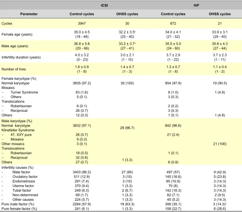
The present study included 3.978 ICSI cycles and 893 IVF cycles performed in our Medical Center over the past seven years. OHSS development occurred in 31 (0.78%) ICSI cycles and in 21 (2.35%) IVF cycles. Among ICSI cycles with OHSS development, 18 (58.1%) were considered moderate, representing a global incidence of 0.45% and the remaining 13 (41.9%) were considered severe, representing 0.33% of the cycles. IVF cycles were complicated by 10 cases of moderate OHSS (47.6%) and 11 cases of severe OHSS (52.4%), representing an incidence of 1.12% and 1.23%, respectively. One of the ICSI cycles with moderate OHSS was cancelled after a testicular biopsy revealed the absence of spermatozoa or spermatid count and therefore the study only included the remaining 30 OHSS cases. In what concerns patient characteristics (Table 1), we observed that both members of the couple were on average younger in the ICSI cycle group with OHSS. There were no differences between both groups in the duration of
infertility and number of treatment attempts, as well as in the frequencies of specific infertility causes. In contrast, in IVF cycles, a higher proportion of a pure masculine infertile factor was observed in the control group (35.1) vs. the OHSS group (14.3). There were no significant differences in what concerned feminine (2.84% in ICSI cycles and 2.06% in IVF cycles) and masculine (2.91% in ICSI cycles and 3.44% in IVF cycles) karyotype abnormalities when compared to the control group. Of note, in the OHSS group, there was a single case of masculine reciprocal translocation in ICSI cycles and isolated cases of Turner syndrome and feminine deletion in IVF cycles were recorded.
There were no significant differences between ICSI and IVF cycles with respect to any of the following spermogram parameters: spermatozoa count, morphology, progressive motility, vitality or hypo-osmolarity test. Spermatozoa were obtained by aspiration or testicular biopsy (each one completed in just one OHSS cycle, representing a total of 6.6% of the cases). There were also no significant Table 1:Patient characteristics.
ICSI IVF
Parameter Control cycles OHSS cycles Control cycles OHSS cycles
Cycles 3947 30 872 21
Female age (years) 35.0 ± 4.5 (18 - 48) 32.2 ± 3.5(25 - 40)a 34.0 ± 4.1 (21 - 52) 33.9 ± 3.1 (29 - 40)
Male age (years) 36.8 ± 5.8 (20 - 66) 33.2 ± 3.7(27 - 41) a 35.5 ± 5.0(24 - 60) 35.8 ± 4.3(27 - 44)
Infertility duration (years) 4.0 ± 3.2 (0 - 23) 3.0 ± 2.1 (1 - 10) 3.7 ± 2.9(1 - 22) 3.7 ± 2.3 (1 - 11)
Number of tries 1.6 ± 0.9(1 - 8) 1.4 ± 0.7 (1 - 3) 1.3 ± 0.7 (1 - 6) 1.1 ± 0.4 (1 - 2)
Female karyotype (%) Normal karyotype Mosaics
- Turner Syndrome
- Others
Translocations
- Robertsonian
- Reciprocal
Others
3835 (97.2)
63 (1.6) 5 (0.1)
6 (0.1) 26 (0.7) 12 (0.3)
30 (100) 854 (97.9)
9 (1.0) 3 (0.3)
2 (0.2) 3 (0.3) 1 (0.1)
19 (90.5)
1 (4.8)
1 (4.8) Male karyotype (%)
Normal karyotype Klinefelter Syndrome
- 47, XXY pure
- Mosaics
Other mosaics Translocations
- Robertsonian
- Reciprocal
Others
3832 (97.1)
26 (0.7) 9 (0.2) 3 (0.1)
18 (0.5) 32 (0.8) 27 (0.7)
29 (96.7)
1 (3.3)
842 (96.6)
21 (2.4)
1 (0.1)
8 (0.9)
21 (100)
Infertility causes (%) - Male factor - Ovulatory factor - Endometriosis - Uterine factor - Tubal factor - Non-determined - Other causes Pure male factor (%) Pure female factor (%)
3403 (86.2) 511 (12.9)
291 (7.4) 370 (9.4) 249 (6.3) 69 (1.7) 224 (5.7) 2284 (57.9)
241 (6.1)
27 (90) 3 (10) 3 (10) 1 (3.3) 2 (6.7) 1 (3.3) 1 (3.3) 19 (63.3)
1 (3.3)
497 (57) 145 (16.6)
95 (10.9) 70 (8) 142 (16.3)
62 (7.1) 45 (5.2) 306 (35.1) 108 (22.7)
9 (42.9) 5 (23.8) 3 (14.3) 3 (14.3) 3 (14.3) 2 (9.5) 3 (14.3) 3 (14.3)a
6 (28.6)
Values expressed as average ± standard deviation (variation), except for those parameters otherwise indicated
a Statistically significant difference between OHSS group and control group. * Ovarian Hyperstimulation Syndrome
Table 2: Stimulation data.
ICSI IVF
Parameter Control cycles OHSS cycles Control cycles OHSS cycles
Basal FSH (mIU/mL) (0.1 - 86.7)7.3 ± 4.1 (2.5 - 13.5)6.6 ± 2.2 (0.1 - 55.3)6.6 ± 3.6 (3.5 - 11.3)6.3 ± 1.9
Stimulation protocol (n; %)
- Antagonist
- Agonist 3034 (89.5)356 (10.5) 27 (90.0)3 (10) 692 (92.9)53 (7.1) 21 (100.0)
Total rFSH dose (IU/mL) 1573 ± 608.2(300 - 7200) 1322.5 ± 608.2(800 - 2700) 1493.4 ± 501.4(600 - 3600) 1221.3 ± 319(800 - 1800)a
Total HMG dose (IU/mL) 2220 ± 1018.3(1200 - 5400) 1680 ± 812.6(750 - 3375)
Total rFSH + HMG dose (IU/mL) 2834.6 ± 1057.2(750 - 9000) 1717.5 ± 742.9(925 - 3225)a 2519.4 ± 985.7(875 - 6875) (1275 - 3225)2362.5 ± 843
Stimulation duration (days) 8.5 ± 1.7 (1 - 19) 8.5 ± 1.4 (7 - 11) 8.3 ± 1.5 (5 - 15) 7.9 ± 1.6 (6 - 12)
Estradiol measurement at the time of
HCG administration (pg/mL) 1246.3 ± 891.1(1 - 14630.4) 1707.9 ± 646.5
a
(455.7 - 3142) 1337.3 ± 863.8(7.3 - 8213) 1594.6 ± 504.5(885 - 2706.4) Ovulation triggering (n; %)
- HCG 2756 (98.1) 15 (100) 544 (96.6) 12 (100)
HCG dose (IU/mL) 9612.1 ± 1874.6(5000 - 15000) (10000 - 10000)10000 ± 0a 9364.6 ± 1780.5(5000 - 15000) (10000 - 10000)10000 ± 0a
Values expressed as average ± standard deviation (variation), except for those parameters otherwise indicated
a Statistically significant difference between OHSS and control groups, Ovarian Hyperstimulation Syndrome (OHSS).
Table 3: Embryological results.
ICSI IVF
Parameter Control cycles OHSS cycles Control cycles OHSS cycles
Cycles (n) 3947 30 872 21
Number of oocyte obtained (n) 29716 324 7475 232
Average number of oocyte obtained/
cycle 7.53 ± 4.8 (0 - 36) 10.8 ± 4.1
a
(3 - 19) 8.57 ± 4.6 (0 - 34) 11 ± 4.2
a
(6 - 21)
MII (n) 24251 257 6981 208
2PN (n) 16841 189 4824 155
MII/number of oocyte obtained (n; %) 24251 / 29716 (81.6) 257 / 324 (79.3) 6981 / 7475 (93.4) 208 / 232 (89.7)a
Oocyte immaturity rate (n; %) 5465 / 29716 (18.4) 67 / 324 (20.7) 494 / 7475 (6.6) 24 / 232 (10.3)a
Fertilisation rate (n; %) 16841 / 24251 (69.4) 189 / 257 (73.5) 4824 / 6981 (69.1) 155 / 208 (74.5)
Cleavage rate (n; %) 16483 / 16841 (97.9) 182 / 189 (96.3) 4732 / 4824 (98.1) 153 / 155 (98.7)
Blastocyst rate (%) 4030 / 16841 (23.9) 75 / 189 (39.7)a 1694 / 4824 (35.1) 81 / 155 (52.3)a
Transferred embryo type (n; %)
- 2-4 cells
- 5-17 cells
- Morula
- Compactation
- Blastocyst
824 (11.9) 3436 (49.5)
286 (4.1) 446 (6.4) 1945 (28)
2 (3.8) 14 (26.9)a
1 (1.9) 3 (5.8) 32 (61.5)a
120 (7.5) 670 (42.1)
60 (3.8) 67 (4.2) 673 (42.3)
11 (28.2) 1 (2.6) 1 (2.6) 26 (66.7)a Values expressed as average ± standard deviation (variation), except for those parameters otherwise indicated.
aStatistically significant difference between OHSS group and control group; * Ovarian hyperstimulation syndrome (OHSS);
† Metaphase II (MII);
‡ Two pro-nucleus (2PN);
§ Maturity rate (nº MII/nº oocyte obtained); Immaturity rate [(nº oocyte – nº MII) / nº oocyte]; Fertilisation rate (2PN/MII); Cleavage rate (Obtained embryo number/2PN); Blastocyst rate (blastocyst number/2PN).
differences between the use of these techniques and the ejaculated sperm obtained in both groups.
In what concerns ovarian stimulation (Table 2), in ICSI cycles from the control group, rFSH was administered in 2.088 (61.6%), rFSH + HMG in 1.280 (37.8%) and HMG in 20 (0.6%) of the patients. In OHSS group, rFSH was used in 20 (66.7%) and rFSH + HMG in 10 (33.3%) of the cases, in the absence of HMG. The average total dose of rFSH + HMG used was lower in the OHSS group.
In the control group of IVF cycles, rFSH was used in 540 (72.5%), rFSH + HMG in 195 (26.2%) and HMG in 10 (1.3%) of the patients. In the OHSS group, rFSH was used in 17 (81.0%) and rFSH + HMG in 4 (19.0%) of the cases, all in the absence of HMG. The average total dose of rFSH administered was lower in OHSS group and there were no significant differences in stimulation duration.
The stimulation protocol performed was of the antagonist type in the majority of patients with only a small percentage having received the agonist protocol in ICSI cycles complicated with OHSS. There were no significant differences between the groups.
In all OHSS cases, the drug used for ovulation triggering was HCG. The dose of HCG used in the OHSS group was significantly higher than the one used in the control groups and estradiol measured on the day of HCG administration was higher in the ICSI-OHSS group,.
In what concerns embryological results (Table 3), the
average number of oocytes retrieved per cycle was higher in the OHSS groups. Immaturity rate was higher in IVF-OHSS group. There were no significant differences in fertilisation and cleavage rates between the groups.
The rate of blastocyst was significantly higher in the OHSS groups. In the same way, transferred embryo type was significantly different in both groups, with a higher proportion of transferred blastocyst in the OHSS groups. In both groups, the majority of transferred embryos were of good quality (A and B), both in ICSI and IVF cycles (96% in the control group vs. 100% in the OHSS group, in both cycles).
Clinical results analysis (Table 4) showed no significant differences in the transferred average number of embryos per embryo transfer (ET) cycle between the groups. Biochemical and clinical pregnancy success rates by ET cycle were higher in the OHSS groups. There were no differences between the groups in ectopic, twin and triplet pregnancy rates (nor in the total number of multiple pregnancies). In what concerns the type of birth, there was a predominance of dystocic delivery in all groups (for ICSI, 0.38% / ET cycles in the OHSS cases and 0.17% / ET cycles in control cases; for IVF, 0.48% / ET cycles in OHSS cases and 0.24% / ET cycles in control cases; for all cases, 0.42% / ET cycles in OHSS cases and 0.19% / ET cycles in control cases). Implantation rate was higher in the OHSS groups. There was no difference in the proportion of abortions for Table 4: Clinical results (Pregnancies and Births).
ICSI IVF
Parameter Control cycles OHSS cycles Control cycles OHSS cycles
ET cycles (n) 3606 29 832 21
Total transferred embryos (n) 6937 52 1590 39
Average number of transferred embryos
(n; % of ET cycles) 6937 / 3606 (1.9) 52 / 29 (1.8) 1590 / 832 (1.9) 39 / 21 (1.9)
Biochemical pregnancy
(n; % of ET cycles) 1518 / 3606 (42.1) 27 / 29 (93.1)a 477 / 832 (57.4) 21 / 21 (100)a
Clinical pregnancy
(n; % of ET cycles) 1303 / 3606 (36.1) 26 / 29 (89.7)a 418 / 832 (50.2) 21 / 21 (100)a
Ectopic pregnancy
(n; % of ET cycles and CP) 23 / 3606 (0.6)23 / 1303 (1.8) 1 / 29 (3.4)1 / 26 (3.8) 8 / 418 (1.9)8 / 832 (1) 1 / 21 (4.8)1 / 21 (4.8)
Single pregnancy (n) 961 17 274 12
Twin pregnancy (n; % of CP) 334 / 1303 (25.6) 9 / 26 (34.6) 141 / 418 (33.7) 8 / 21 (38.1)
Triplet pregnancy (n; % of CP) 8 / 1303 (0.6) 3 / 418 (0.7) 1 / 21 (4.8)
Abortion (n; % of CP) 199 / 1303 (15.3) 4 / 26 (15.4) 47 / 418 (11.2) 1 / 21 (4.8)
Eutocic delivery (n) 253 3 78 3
Dystocic delivery
(n; % of total deliveries) 630 / 883 (71.3) 11 / 14 (78.6) 198 / 276 (71.7) 10 / 13 (76.9)
Implantation rate
(n; % of ET cycles) 1636 / 6937 (23.6) 34 / 52 (65.4)a 559 / 1590 (35.2) 31 / 39 (79.5)a
Values expressed as average ± standard deviation (variation), except for those parameters otherwise indicated.
aStatistically significant difference between OHSS and control groups; * Ovarian Hyperstimulation Syndrome (OHSS);
† Embryo Transfer (ET);
‡ Clinical Pregnancy (CP);
§ Implantation Rate (gestational sacs number/transferred embryo number).
clinical pregnancy between the groups.
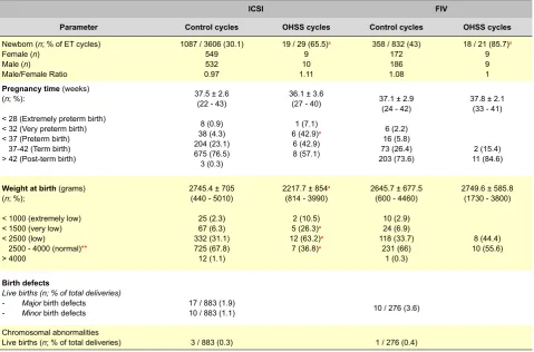
In what concerns newborns (NB) and complications (Table 5), the proportion of NB per ET cycle was significantly higher in OHSS groups, with a male/female ratio of approximately 1 in all groups. There were no significant differences in the pregnancy average time in weeks between both groups. However, in ICSI-OHSS cycles, less than 32 weeks pregnancy proportion was significantly higher. Average weight at birth was lower in the ICSI-OHSS group, with a higher proportion with very low and low weight. As far as birth defects and chromosomal abnormalities, these were only identified in the control groups. Chromosomal abnormalities found in NB in ICSI cycles included one case of trisomy 21, two NB with Robertsonian translocations and one with reciprocal translocation. In IVF cycles, there was only one NB with trisomy 21.
With regard to hospitalised patients, only 24 severe OHSS cases were admitted in the Hospital, with a hospitalisation average of 8.2 days (5 - 15). All patients presented on admission with fatigue, nausea, abdominal distension and discomfort. Of note, there was no vomiting or dyspnea and bowel movements remained unchanged. There was also no anaemia, no fever and the patients remained normotensive. The exams showed haemoconcentration and an increase in ovarian volume, with on average for the right ovary of 7.89
cm3 (5.5 - 10) and on average for the left ovary of 6.9 cm3 (6.3 – 8.2). A diagnosis of late (after a positive βHCG) and severe OHSS was reached. Patients were evaluated by vaginal touch, speculum examination, ultrasound (which showed moderate to voluminous ascites, with no abnormality in bowel loops and normal uterus and adnexa). Haematocrit, serum ions, liver function, renal function and coagulation parameters were evaluated during hospitalisation. Patients were rehydrated (in order to reduce haemoconcentration) and required furosemide (in order to improve diuresis, as fluid therapy may actually increase the risk of ascites and pleural effusion and in view of the association of OHSS with increased vascular permeability, which results in increased intra-abdominal pressure when renal function may be compromised). Enoxaparine was administered in patients who were breastfeeding. Ascites and pleural effusion were evaluated by abdominal ultrasound extension to the lung surface. Most patients reported miction difficulties on admission and required urinary catheterisation with urinary output and fluid balance monitoring. No other complications were observed during hospital admission and therefore neither thoracocentesis nor paracentesis/culdocentesis were required. Admission lasted from 2 to 15 days and upon discharge patients were advised rest, fluid restriction, appropriate nutrition and paracetamol for analgesia. Even Table 5. Clinical Results (Newborn Frequency and Complications).
ICSI FIV
Parameter Control cycles OHSS cycles Control cycles OHSS cycles
Newborn (n; % of ET cycles) Female (n)
Male (n) Male/Female Ratio
1087 / 3606 (30.1)
549 532
0.97
19 / 29 (65.5)a
9
10 1.11
358 / 832 (43) 172 186 1.08
18 / 21 (85.7)a
9 9 1
Pregnancy time (weeks) (n; %):
< 28 (Extremely preterm birth) < 32 (Very preterm birth) < 37 (Preterm birth) 37-42 (Term birth) > 42 (Post-term birth)
37.5 ± 2.6 (22 - 43)
8 (0.9) 38 (4.3) 204 (23.1) 675 (76.5) 3 (0.3)
36.1 ± 3.6 (27 - 40)
1 (7.1) 6 (42.9)a
6 (42.9) 8 (57.1)
37.1 ± 2.9 (24 - 42)
6 (2.2) 16 (5.8) 73 (26.4) 203 (73.6)
37.8 ± 2.1 (33 - 41)
2 (15.4) 11 (84.6)
Weight at birth (grams) (n; %);
< 1000 (extremely low) < 1500 (very low) < 2500 (low)
2500 - 4000 (normal)** > 4000
2745.4 ± 705 (440 - 5010)
25 (2.3) 67 (6.3) 332 (31.1) 725 (67.8) 12 (1.1)
2217.7 ± 854a
(814 - 3990)
2 (10.5) 5 (26.3)a
12 (63.2)a
7 (36.8)a
2645.7 ± 677.5 (600 - 4460)
10 (2.9) 24 (6.9) 118 (33.7)
231 (66) 1 (0.3)
2749.6 ± 585.8 (1730 - 3800)
8 (44.4) 10 (55.6)
Birth defects
Live births (n; % of total deliveries) - Major birth defects
- Minor birth defects 17 / 883 (1.9)10 / 883 (1.1) 10 / 276 (3.6)
Chromosomal abnormalities
Live births (n; % of total deliveries) 3 / 883 (0.3) 1 / 276 (0.4)
Values expressed as average ± standard deviation (variation), except for those parameters otherwise indicated.
aStatistically significant difference between OHSS group and control group; * Ovarian Hyperstimulation Syndrome (OHSS);
† Embryo transfer (ET);
** Normal: weight within the normal range for newborns.
though it did not prove necessary, a specific recommendation was made for immediate return to the Hospital should any sign of deterioration arise. There was also no need for re-hospitalisation in the literature review performed.
DISCUSSION
Patients who develop OHSS were globally younger in ICSI than in IVF cycles, a finding already described in previous studies.15,16 This relation is probably due to the
fact that in both groups younger patients have a higher number of recrutable follicles and a higher gonadotropin receptor density, developing a more intense response to gonadotropin.17 Male age was also lower, a fact that might
be related to a tendency for couples to be within the same age group.
In the present study, there were no significant differences between the groups in what concerns infertility causes. This finding is not in agreement with a revision in 214.219 ICSI/IVF cycles, 1.523 with moderate OHSS and 655 with severe OHSS, where a relation was found between OHSS development and tubal and ovulatory factors, including Polycystic Ovary Syndrome (PCOS) 15.
PCOS was already related with OHSS in previous studies18 in which only severe OHSS cases were included
in the OHSS group (165) and another study19 where 24 moderate OHSS and 29 severe OHSS were described. This association is probably due to the presence of an increased ovarian sensitivity to gonadotropin in PCOS. In a systematic review which included 10 studies, there was a relation between PCOS and OHSS.20 In addition, there was also a
lower proportion of patients with reduced ovarian reserve in the group that developed OHSS.15 In the present study (51 cases, 24 severe), it is likely that a lower number of OHSS cycles may explain the lack of association with PCOS. On the other hand, the discrepancy between our results and those that have been described may be explained by the fact that our classification of infertility due to ovulatory disorders included not only PCOS patients, but also other ovulatory problems. The latter may be associated to a lower ovarian sensitivity to gonadotropin action and therefore inversely related with OHSS development. Furthermore, the association between female pathology as cause of infertility and OHSS development found in the referred studies may also be explained by a lower pure masculine factor cycle proportion in the IVF-OHSS group.
In the present work, there were no significant differences between groups in what concerns stimulation protocols but this study only included three ICSI-OHSS cases using an agonist protocol. A Cochrane systematic review made in 2006, including 29 trials,21 revealed a 2% lower OHSS incidence when using an antagonist vs. an agonist protocol. A more recent revision22 involving 45 randomised clinical trials showed an incidence difference of 2.7%. This was due to an OHSS incidence reduction by half when the antagonist was compared to the agonist protocol and a lower probability of cycle cancelling due to high risk of OHSS development. In another randomised clinical trial23 it was also shown that
the antagonist protocol was associated to a lower OHSS incidence. One possible explanation for higher safety of the antagonist protocol may be the immediate action of the drug, its administration being indicated specifically for the suppression of premature and spontaneous LH release, thereby reducing the stimulation duration and secondary effects due to hypoestrogenemia.24
In the present study (4.871 cycles), we found no significant differences between the groups in the use of rFSH, HMG or both, in agreement with a meta-analysis of seven randomised clinical trials25 involving 2.159 patients. The average total dose of rFSH + HMG used was lower in the ICSI-OHSS group, as well as the average total dose of rFSH administered in the IVF-OHSS group. A meta-analysis26 demonstrated that the OHSS development risk
increases significantly with daily FSH doses higher than 200 IU and with average stimulation duration of 8.5 days, corresponding to a total dose of 1.700 IU. This apparent inversion of the expected results may be related to the fact that in this study, as a preventative measure, we performed a reduction of the administered dose in patients that were judged to present a OHSS development risk.
Estradiol quantification on the day of HCG administration revealed a higher value in the ICSI-OHSS group, what is in agreement with a prospective study27 and with
the observation that the OHSS development is usually associated with an increase in estradiol levels.28
All OHSS cases underwent ovulation induction with HCG. Several studies showed a lower OHSS incidence using a GnRH agonist.29-31 The higher incidence in our
study may thus be explained by the longer half-life of HCG, remaining high until six days after administration. In a revision study,33 as well as in a prospective study of 102 oocyte donors with high OHSS development risk,34 the authors concluded that using a GnRH agonist as ovulation inductor results in a total absence of OHSS. In addition, a retrospective study demonstrates that HCG dose reduction may result in a reduction of OHSS incidence.35 In the present study, the administered HCG dose in the OHSS group was significantly higher.
In the present study, the average number of obtained oocytes was higher in the OHSS group, both in ICSI (10.8
vs. 7.53) and in IVF (11 vs. 8.57) cycles, in agreement with retrospective studies (13 vs. 10)36 and (13 vs. 9)37 which
is in keeping with the fact that a high number of follicles obtained by aspiration is a good predictive factor for early OHSS development.
Blastocyst rate and transferred blastocyst proportion was significantly higher in the OHSS groups. In ICSI cycles, this may have been attributed to embryo transfer being predominantly performed on the fifth day. However, in IVF cycles, embryos were mainly transferred on the fifth day in both OHSS positive and negative groups. Therefore, the higher blastocyst proportion should not be exclusively related to the transfer day, but rather to a relationship between embryo maturation frequency to blastocyst and OHSS development.
In the present work, both biochemical and clinical pregnancy were higher in OHSS groups, as in several previous studies,15 as pregnancy increases frequency, duration and severity of OHSS symptoms.1 In fact, in a retrospective study, the comparison between the clinical characteristics of patients who develop early and late OHSS, revealed that the conception cycle was the main cause of late OHSS, as opposed to OHSS which occurs earlier and is mainly related to gonadotropin stimulation.38 These
authors remark that the sustained action of HCG increased due to pregnancy might be related with longer duration and higher severity of late vs. early OHSS, implying a higher magnitude and duration of the granulosa cell stimulation.37
Implantation rate was higher in OHSS groups, related with a similar transferred embryo average number with a higher number of gestational sacs. The same result has been obtained in a prospective study that suggested that OHSS severity increases the number of gestational sacs, independently of the moment in which it starts, confirming the hypothesis that a higher severity of late OHSS might be due to a pregnancy effect.37
Contrary to one study,15 we did not find any significant differences in multiple pregnancy rate between the groups. However, a faster and more sustained increase in HCG levels occurs in multiple pregnancy.39 In our work, the number of transferred embryos, a risk factor for multiple pregnancy development, was not significantly associated with OHSS development.
In the present study, higher prematurity rates and low weight at birth have been found in the OHSS group, in agreement with other results.40 In another study, a relation
between OHSS development and low weight at birth, but not with prematurity was found.15 Higher prematurity rates have however been observed.36 There are still no data that
support a pathogenic association between prematurity and OHSS, except when there is a high prevalence of multiple pregnancies and in fact, in the present work, there are no significant differences between both groups. The higher rate of low weight at birth in the OHSS group might be explained by OHSS induction of placental developmental abnormalities and gestational hypertension and these have been observed at a higher prevalence in patients that develop OHSS.36
We observed no significant differences in abortion rate between the groups, as in other previous studies.19,36 A
multicentric study that found a higher abortion rate reported a higher number of multiple pregnancies.36
In our work, birth defects only occurred in a small percentage of newborns in the control group, corresponding approximately, in ICSI cycles (1.9% major and 1.1%
minor), to those found by another study.42 The latter was
observational and analysed congenital birth defects after ICSI cycles between 1994 - 1997, in 13 reproductive medicine centers (five public and eight private) which found 2.2% major and 1.2% minor abnormalities. Although the birth defect rate that we obtained in IVF cycles was slightly higher (3.6%), this difference was not statistically significant. In another study,43 there was also no significant differences in birth defect rates between ICSI and IVF cycles, with a
major birth defect rate of 3.8% in IVF cycles, in agreement with the present work. The chromosomal abnormality rate found in live births from the control group of 0.3% in ICSI cycles and 0.4% in IVF cycles did not exceed the 0.92% rate obtained for general population.44
During hospitalisation, no patient presented any relevant clinical signs or required re-hospitalisation, in agreement with a literature review.
CONCLUSIONS
When starting an ART cycle, young age and estradiol levels seem to be relevant risk factors for OHSS, alerting the physician for this possible complication and allowing for adoption of adequate preventative measures. OHSS is also positively associated with laboratory and clinical results obtained during ART cycles, with a higher average oocyte number, biochemical and clinical pregnancy, implantation, as well as a higher proportion of newborn. However, beyond maternal risk, OHSS may also cause foetal abnormalities, namely prematurity and low weight at birth, for which we recommend close pregnancy supervision.
ACKNOWLEDGEMENTS
We are grateful and acknowledge the fundamental role of both Jorge Beires, Ginaecology Department, São João Hospital, Porto and José Manuel Teixeira da Silva, responsible for ovarian punction and follicular aspiration; Maria José Correia, Anaesthesiology Department, São João Hospital, Porto; Luís Ferraz, Urology Department, Vila Nova de Gaia Hospital Center, Portugal; Ana Gonçalves (Biochemistry), Cláudia Osório (Biology) and Nuno Barros (Microbiology), responsible for work performed at the laboratory of Andrology and Armando Teixeira-Pinto, Biostatistics and Medical Informatic Department, Medicine Faculty, Porto University, Porto, for his help in statistics.
CONFLICT OF INTERESTS
All authors declare that there are no conflicts of interest concerning the present manuscript.
SOURCES OF FUNDING
There have been no external sources of funding for writing this manuscript.
REFERENCES
1. The Practice Committee of the American Society for Reproduc-tive Medicine. Ovarian hyperstimulation syndrome. Fertil Steril. 2008;90(5Suppl):S188-93.
2. Elchalal U, Schenker JG. The pathophysiology of ovarian hyperstimula-tion syndrome-views and ideas. Hum Reprod. 1997;12:1129-37.
3. Rizk B, Aboulghar M, Smitz J, Ron-El R. The role of vascular endothelial growth factor and interleukins in the pathogenesis of severe ovarian hy-perstimulation syndrome. Hum Reprod Update. 1997;3:255-66. 4. Delvigne A. Symposium: Update on prediction and management of
OHSS. Epidemiology of OHSS. Reprod Biomed Online. 2009;19:8-13.
5. Papanikolaou EG, Tournaye H, Verpoest W, Camus M, Vernaeve V, Van Steirteghem A, et al. Early and late ovarian hyperstimulation syndrome: early pregnancy outcome and profile. Hum Reprod. 2005;20:636-41. 6. Salomon O, Schiby G, Heiman Z, Avivi K, Sigal C, Levran D, et al.
Combined jugular and subclavian vein thrombosis following assisted reproductive technology-new observation. Fertil Steril. 2009;92:620-5. 7. Edris F, Kerner CM, Feyles V, Leung A, Power S. Successful
manage-ment of an extensive intracranial sinus thrombosis in a patient undergo-ing IVF: case report and review of literature. Fertil Steril. 2007;88:705 e9-14.
8. Jing Z, Yanping L. Middle cerebral artery thrombosis after IVF and ovar-ian hyperstimulation: a case report. Fertil Steril. 2011;95:2435 e13-5. 9. Uhler ML, Budinger GR, Gabram SG, Zinaman MJ. Perforated duodenal
ulcer associated with ovarian hyperstimulation syndrome: Case Report. Hum Reprod. 2001;16:174-6.
10. Memarzadeh MT. A fatal case of ovarian hyperstimulation syndrome with perforated duodenal ulcer. Hum Reprod. 2010;25:808-9.
11. Braat DD, Schutte JM, Bernardus RE, Mooij TM, van Leeuwen FE. Ma-ternal death related to IVF in the Netherlands 1984-2008. Hum Reprod. 2010;25:1782-6.
12. Schenker JG, Weinstein D. Ovarian hyperstimulation syndrome: a cur-rent survey. Fertil Steril. 1978;30:255-68.
13. Rizk B AM. Classification, pathophysiology and management of ovarian
hyperstimulation syndrome. 2nd ed. New York: The Parthenon Publishing
Group; 1999.
14. Zegers-Hochschild F, Adamson GD, de Mouzon J, Ishihara O, Mansour R, Nygren K, et al. International Committee for Monitoring Assisted Reproductive Technology (ICMART) and the World Health Organiza-tion (WHO) revised glossary of ART terminology, 2009. Fertil Steril. 2009;92:1520-4.
15. Luke B, Brown MB, Morbeck DE, Hudson SB, Coddington CC, 3rd, Stern JE. Factors associated with ovarian hyperstimulation syndrome (OHSS) and its effect on assisted reproductive technology (ART) treat-ment and outcome. Fertil Steril. 2010;94:1399-1404.
16. Delvigne A, Dubois M, Battheu B, Bassil S, Meuleman C, De Sutter P, et al. The ovarian hyperstimulation syndrome in in-vitro fertilization: a Belgian multicentric study. II. Multiple discriminant analysis for risk pre-diction. Hum Reprod. 1993;8:1361-6.
17. Whelan JG, 3rd, Vlahos NF. The ovarian hyperstimulation syndrome. Fertil Steril. 2000;73:883-96.
18. Wiser A, Levron J, Kreizer D, Achiron R, Shrim A, Schiff E, et al. Out-come of pregnancies complicated by severe ovarian hyperstimulation syndrome (OHSS): a follow-up beyond the second trimester. Hum Re-prod. 2005;20:910-4.
19. Papanikolaou EG, Pozzobon C, Kolibianakis EM, Camus M, Tournaye H, Fatemi HM, et al. Incidence and prediction of ovarian hyperstimula-tion syndrome in women undergoing gonadotropin-releasing hormone antagonist in vitro fertilization cycles. Fertil Steril. 2006;85:112-20. 20. Tummon I, Gavrilova-Jordan L, Allemand MC, Session D. Polycystic
ovaries and ovarian hyperstimulation syndrome: a systematic review. Acta Obstetr Gynecol Scand. 2005;84:611-6.
21. Al-Inany HG, Abou-Setta AM, Aboulghar M. Gonadotrophin-releasing hormone antagonists for assisted conception. Cochrane Database Syst Rev. 2006;CD001750.
22. Al-Inany HG, Youssef MA, Aboulghar M, Broekmans F, Sterrenburg M, Smit J, et al. GnRH antagonists are safer than agonists: an update of a Cochrane review. Hum Reprod Update. 2011;17:435.
23. Lainas TG, Sfontouris IA, Zorzovilis IZ, Petsas GK, Lainas GT, Alexo-poulou E, et al. Flexible GnRH antagonist protocol versus GnRH ag-onist long protocol in patients with polycystic ovary syndrome treated for IVF: a prospective randomised controlled trial (RCT). Hum Reprod. 2010;25:683-9.
24. Borm G, Mannaerts B. Treatment with the gonadotrophin-releasing hor-mone antagonist ganirelix in women undergoing ovarian stimulation with recombinant follicle stimulating hormone is effective, safe and conve-nient: results of a controlled, randomized, multicentre trial. The Euro-pean Orgalutran Study Group. Hum Reprod. 2000;15:1490-8. 25. Coomarasamy A, Afnan M, Cheema D, van der Veen F, Bossuyt PM,
van Wely M. Urinary hMG versus recombinant FSH for controlled ovar-ian hyperstimulation following an agonist long down-regulation protocol
in IVF or ICSI treatment: a systematic review and meta-analysis. Hum Reprod. 2008;23:310-5.
26. Sterrenburg MD, Veltman-Verhulst SM, Eijkemans MJ, Hughes EG, Macklon NS, Broekmans FJ, et al. Clinical outcomes in relation to the daily dose of recombinant follicle-stimulating hormone for ovarian stimu-lation in in vitro fertilization in presumed normal responders younger than 39 years: a meta-analysis. Hum Reprod Update. 2011;17:184-96. 27. Lee TH, Liu CH, Huang CC, Wu YL, Shih YT, Ho HN, et al. Serum anti-Mullerian hormone and estradiol levels as predictors of ovarian hyper-stimulation syndrome in assisted reproduction technology cycles. Hum Reprod. 2008;23:160-7.
28. Fauser BC, Diedrich K, Devroey P. Predictors of ovarian response: prog-ress towards individualized treatment in ovulation induction and ovarian stimulation. Hum Reprod Update. 2008;14:1-14.
29. Acevedo B, Gomez-Palomares JL, Ricciarelli E, Hernandez ER. Trigger-ing ovulation with gonadotropin-releasTrigger-ing hormone agonists does not compromise embryo implantation rates. Fertil Steril. 2006;86:1682-7. 30. Radesic B, Tremellen K. Oocyte maturation employing a GnRH agonist
in combination with low-dose hCG luteal rescue minimizes the severity of ovarian hyperstimulation syndrome while maintaining excellent preg-nancy rates. Hum Reprod. 2011;26:3437-42.
31. Humaidan P, Papanikolaou EG, Tarlatzis BC. GnRHa to trigger final oo-cyte maturation: a time to reconsider. Hum Reprod. 2009;24:2389-94. 32. Gonen Y, Balakier H, Powell W, Casper RF. Use of
gonadotropin-releas-ing hormone agonist to trigger follicular maturation for in vitro fertiliza-tion. J Clin Endocrinol Metabol. 1990;71:918-22.
33. Humaidan P, Kol S, Papanikolaou EG. GnRH agonist for triggering of final oocyte maturation: time for a change of practice? Hum Reprod Up-date. 2011;17:510-24.
34. Bodri D, Guillen JJ, Trullenque M, Schwenn K, Esteve C, Coll O. Early ovarian hyperstimulation syndrome is completely prevented by go-nadotropin releasing-hormone agonist triggering in high-risk oocyte donor cycles: a prospective, luteal-phase follow-up study. Fertil Steril. 2010;93:2418-20.
35. Shapiro BS, Daneshmand ST, Garner FC, Aguirre M, Ross R, Morris S. Effects of the ovulatory serum concentration of human chorionic go-nadotropin on the incidence of ovarian hyperstimulation syndrome and success rates for in vitro fertilization. Fertil Steril. 2005;84:93-8. 36. Courbiere B, Oborski V, Braunstein D, Desparoir A, Noizet A, Gamerre
M. Obstetric outcome of women with in vitro fertilization pregnancies hospitalized for ovarian hyperstimulation syndrome: a case-control study. Fertil Steril. 2011;95:1629-32.
37. Mathur RS, Akande AV, Keay SD, Hunt LP, Jenkins JM. Distinction be-tween early and late ovarian hyperstimulation syndrome. Fertil Steril. 2000;73:901-7.
38. Lee KH, Kim SH, Jee BC, Kim YJ, Suh CS, Kim KC, et al. Compari-son of clinical characteristics between early and late patterns in hos-pitalized patients with ovarian hyperstimulation syndrome. Fertil Steril. 2010;93:2274-80.
39. Beerendonk CC, van Dop PA, Braat DD, Merkus JM. Ovarian hy-perstimulation syndrome: facts and fallacies. Obstetr Gynecol Surv. 1998;53:439-49.
40. Chung K, Coutifaris C, Chalian R, Lin K, Ratcliffe SJ, Castelbaum AJ, et al. Factors influencing adverse perinatal outcomes in pregnancies achieved through use of in vitro fertilization. Fertil Steril. 2006;86:1634-41.
41. Abramov Y, Elchalal U, Schenker JG. Obstetric outcome of in vitro fertil-ized pregnancies complicated by severe ovarian hyperstimulation syn-drome: a multicenter study. Fertil Steril. 1998;70:1070-6.
42. Loft A, Petersen K, Erb K, Mikkelsen AL, Grinsted J, Hald F, et al. A Danish national cohort of 730 infants born after intracytoplasmic sperm injection (ICSI) 1994-1997. Hum Reprod. 1999;14:2143-8.
43. Bonduelle M, Liebaers I, Deketelaere V, Derde MP, Camus M, Devroey P, et al. Neonatal data on a cohort of 2889 infants born after ICSI (1991-1999) and of 2995 infants born after IVF (1983-(1991-1999). Hum Reprod. 2002;17:671-94.
44. Jacobs PA, Browne C, Gregson N, Joyce C, White H. Estimates of the frequency of chromosome abnormalities detectable in unselected new-borns using moderate levels of banding. J Med Genet. 1992;29:103-8.