THE EFFECTS OF LIMB LOAD ON MUSCLE ACTIVITY DURING ISOLATED REFLEX TESTING AND WALKING POST-STROKE
Amy West Hardee
A thesis submitted to the faculty of the University of North Carolina at Chapel Hill in partial fulfillment of the requirements of the degree of Masters of Science in the
Department of Biomedical Engineering.
Chapel Hill 2010
ii Abstract
Amy West Hardee: The Effects of Limb Load on Muscle Activity During Isolated Reflex Testing and Walking Post-Stroke
(Under the direction of Michael Lewek and Richard Goldberg)
Gait impairments are common after stroke, and are most often characterized by difficulty flexing the leg during swing phase. Limb load, or the weight born through the leg, can influence extensor muscle excitability and may be responsible for this ‘stiff-legged gait pattern’. We hypothesized that there would be a direct relationship between limb load mediated knee extensor excitability during an isolated setting and during late stance/early swing of gait. Nine subjects had electromyography and kinematics recorded during walking with 0, 20, and 40% body weight support. I also collected quadriceps reflex excitability with and without an imposed limb load in an isolated setting. No significant relationship was observed between limb load and quadriceps muscle activity during reflex testing or walking. Limb load did not influence quadriceps activity suggesting that limb load sensors, which are believed to reside in the
iii
This thesis is dedicated to my husband, Thomas, whose love and positivity kept me motivated these past two years, to my family for being my constant cheerleaders, and to Lucy for always being there as my sounding board and
iv
Acknowledgements
I would like to thank Richard Goldberg, my biomedical engineering advisor, for taking me on as a student as I entered graduate school and for going above and beyond to find me a biomechanics based rehabilitation project that he knew I would enjoy.
I would like to thank Mike Lewek, my research advisor, for encouraging me to formulate my own ideas and to be confident in them. Your patience, support, and sense of humor taught me to never be afraid to ask questions.
I would like to thank Bing Yu, my committee member, for always being available for questions or discussion regarding my research.
I would like my entire committee to know how much I appreciated your understanding and support through issues that were not only academic, but personal as well.
v
Table of Contents
List of Tables ... vii
List of Figures ... viii
List of Abbreviations and Symbols ... x
1.0 Introduction and Background ... 1
1.1 Stroke... 1
1.2 Gait ... 2
1.2.1 The ‘Normal Gait’ Cycle ... 2
1.2.2 The Effect of Stroke on Gait ... 4
1.3 Treatment of Stiff-Legged Gait ... 6
1.3.1 Muscle Studies ... 6
1.3.2 Body Weight Support ... 7
1.4 The Effects of Limb Load on Gait ... 9
1.4.1 The Proprioceptive System ... 9
1.4.2 Movements Produced by the Nervous System ... 11
1.4.3 Central Pattern Generators ... 12
1.4.4 Previous Studies Examining the Neurophysiological Effect of Loading in Cats ... 13
1.4.5 Previous Studies Examining the Neurophysiological Effect of Loading in Humans ... 17
1.4.6 Spasticity in the Human Adult ... 18
1.5 Specific Aims and Hypotheses ... 20
2.0 Methods ... 23
2.1 Subjects ... 23
2.2 Testing Equipment ... 24
2.2.1 Test Apparatus – Isolated reflex testing ... 24
2.2.2 Test Apparatus – Gait Analysis Unloading ... 28
vi
2.2.2.2 Theraband Properties ... 31
2.3 Methods – isolated reflex testing ... 32
2.4 Methods – Gait Analysis ... 34
2.5 Statistical Analysis ... 36
3.0 Results ... 38
3.1 Isolated Reflex Testing ... 38
3.2 Gait Data ... 41
3.3 Relationship Between Gait and Reflex Testing ... 44
4.0 Discussion ... 47
4.1 Interpretation of Results ... 48
4.1.1 Relationship Between Isolated Reflex Testing and Gait Analysis ... 48
4.1.2 The Relationship Between Speed and Load ... 48
4.1.3 Dynamic Unweighting System ... 49
4.2 Positive Aspects of the Study ... 49
4.3 Limitations of the Study ... 50
Appendix A: Subject Information Sheet ... 52
Appendix B: Subject Consent Form... 53
Appendix C: Device Figures ... 58
Appendix D: Isolated Reflex Testing Raw Data ... 60
vii
List of Tables
Table 1. Subject Data………..…………....23
Table 2. Theraband Properties………...………32
Table 3. Knee Flexion Angle………..………....42
Table 4. All Subjects – Regression Analysis Comparison……….…..…..44
viii
List of Figures
Figure 1. The Gait Cycle………..………..………3
Figure 2. Quadriceps Anatomy………..4
Figure 3. Knee Flexion Angle……….……….5
Figure 4. Paretic vs. Non-Paretic Knee Flexion Angle………..………….………...6
Figure 5. Extensor Excitability vs. Ankle Load (Hypothesized) ………..……..………….…………21
Figure 6. Extensor Excitability vs. Body Weight Support (Hypothesized) ……..…………...………...21
Figure 7. Relationship Between Gait Extensor Excitability and Isolated Reflex Extensor Excitability (Hypothesized)……….………..………….………...22
Figure 8. Isolated Reflex Testing Station ………...………...24
Figure 9. Ankle Loader Dimensions– Side View ………..25
Figure 10. Ankle Loader Dimensions – View of Foot Plate………..………..25
Figure 11. Ankle Loader Device ………..26
Figure 12. Ankle Loader with Knee in 30° of Flexion ………....26
Figure 13. Ankle Loader with Knee in 90° of Flexion ………..……….….27
Figure 14. Isolated Reflex Testing Full Setup Diagram ………..……….….28
Figure 15. Unweighting Structure Dimensions……….29
Figure 16. Unweighting System Diagram……….……….29
Figure 17. Constructed Unweighting Structure………..………..……..…..……30
Figure 18. Thera-Band Tubing………..………..………..……..…..……31
Figure 19. Treadmill and Harness Setup……….………..………...…..35
Figure 20. Camera Marker Placement……….………..36
Figure 21. Variables Obtained During Isolated Reflex Testing………..………….…………..…..38
Figure 22. Peak Muscle Response During Isolated Reflex Testing……….40
ix
Figure 24. Muscle Activity During Gait……….………..……43
Figure 25. Unweighting Device: Top View……….………..……58
Figure 26. Unweighting Device: Front View……….……….………..……59
Figure 27. VL Peak Response……….………….……….………..……60
Figure 28. VM Peak Response……….………….……….………..……61
Figure 29. RF Peak Response……….………….………..……….………..……62
Figure 30. VL Prolonged Response……….………….……..……….………..……63
Figure 31. VM Prolonged Response……….………….……..……….………..……64
x
List of Abbreviations and Symbols
ANOVA Analysis of Variance BWS Body Weight Support
BWSTT Body Weight Support Treadmill Training CNS Central Nervous System
COM Center of Mass
CPG Central Pattern Generator EMG Electromyography
IRB Institutional Review Board MG Medial Gastrocnemius
MVIC Maximum Voluntary Isometric Contraction Nm Newton meter
1.0 Introduction and Background
The purpose of this study is to examine the effects of limb load on knee extensor excitability during gait in patients who have experienced a stroke. We are also interested in observing muscle excitability of the knee extensors during an isolated reflex testing situation.
1.1 Stroke
Stroke is the third leading cause of death in America following diseases of the heart and cancer. Out of the 795,000 Americans that have experienced a new or recurring stroke each year, about 137,000 die from complications. For those that survive, however, stroke is one of the leading causes of long term disability in adults. Dealing with the initial care and subsequent long-term disability required an estimated direct and indirect cost of $68.9 billion in 2009 (AHA 2009).
2
stroke occurs on the right side of the brain, the patient may experience paralysis of the left side of the body and right side of the face, memory loss, and vision problems.
The length of time to recover from a stroke depends on its severity. 15%-30% of those experiencing a stroke are permanently disabled (AHA 2009). According to the National Heart, Blood, and Lung Institute’s Framingham Heart Study, among ischemic stroke survivors who were older than 65 years of age, 50% had hemiparesis, 30% were unable to walk without some assistance, 26% were dependent in ADL (activities of daily living), and 19% had aphasia (AHA 2009).
1.2 Gait
1.2.1 The ‘Normal Gait’ Cycle
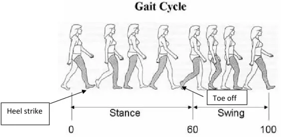
3
Figure 1: The Gait Cycle
http://me.queensu.ca/people/deluzio/GaitAnalysis.php
During normal gait, the knee must flex and extend to allow for shock
absorption during stance and foot clearance during swing. Specifically, the knee flexes in preswing to approximately 35° and then to 6 0° in mid-swing (Wheeless 2007). The vasti become active in the terminal swing phase and remain active until the early midstance phase (Figure 2). Normally, the absence of quadriceps activity in the preswing phase is important to allow the knee to flex 35° before toe-off (Wheeless 2007). Rapid flexion of the knee prior to toe-off has been correlated with the amount of knee flexion achieved during swing phase
(Goldberg, Ounpuu et al. 2003). The rectus femoris (RF), a hip flexor and knee extensor, has a burst of activity during the preswing and initial swing phases. As a hip flexor, the RF increases knee flexion during swing, which is largely a
passive movement, but as a knee extensor, can impair the ability to adequately flex the knee. Activity of the hamstrings begins in the midswing phase and ends
Heel strike
4
in the loading response phase and therefore has little influence on knee flexion during swing.
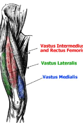
Figure 2: Quadriceps Anatomy
http://msjensen.cehd.umn.edu/webanatomy/muscular/musc_thigh_1_m.htm
1.2.2 The Effect of Stroke on Gait
After stroke, muscle activity patterns and thus kinematics during walking are often altered. This can result in decreased gait speed, postural instability,
reduced energy efficiency, and kinematic abnormalities (Lewek, Hornby et al. 2007).
5
associated with stroke and other diagnoses that affect the brain or spinal cord. Reduced knee flexion in swing can contribute to toe drag and creates a very large moment of inertia that significantly increases the amount of mechanical energy required to walk (Kerrigan, Gronley et al. 1991). Patients with stiff-legged gait typically try to compensate for a lack of knee flexion with hip hiking,
circumduction, or toe-drag, which require the patient to exert more energy, and may lead to additional biomechanical injuries.
Figure 3: Knee Flexion Angle
6
Figure 4: Paretic vs. Non-Paretic Knee Flexion Angle (Lewek, Hornby et al. 2007)
1.3 Treatment of Stiff-Legged Gait
1.3.1 Muscle Studies
Although hip and ankle impairments can contribute to the presence of stiff-legged gait, research on this condition has focused on the impact of knee
7
that “spastic paretic stiff-legged gait can be attributed to multiple impairments besides spastic quadriceps activity” (Kerrigan, Gronley et al. 1991). In 2001, Kerrigan et al hypothesized that subjects with spastic paretic stiff-legged gait have altered kinetics not only about the knee, but also about the hip and ankle (Kerrigan, Karvosky et al. 2001). After collecting joint kinetic data of 20 subjects with spastic paretic stiff-legged gait and comparing the data with that of 20 control subjects the authors observed increases in peak external hip flexion torque in stance, hip-power generation in loading response, knee-extension torque in midstance, ankle-dorsiflexion torque, and ankle-power absorption in stance (Kerrigan, Karvosky et al. 2001). These findings supported the likelihood of multiple mechanisms being responsible for reduced knee flexion in swing. Also in 2001, Kerrigan et al found that when subjects were asked to walk on their toes compared with normal heel-toe walking, there was a 17 degree reduction in peak knee flexion. This suggested that ankle function is related to knee flexion in swing. These previous studies have suggested that there are kinetic
mechanisms taking place throughout the lower limb that can lead to stiff-legged gait that are not only confined to the knee, but could be related to the ankle and hip flexors as well.
1.3.2 Body Weight Support
Treadmill training in conjunction with body weight support (BWSTT) has become a common rehabilitation strategy to restore locomotor function in
8
Visintin 2003). From a motor learning perspective, BWSTT may be successful because complete gait cycles can be practiced with many repetitions (massed practice) instead of focusing on single elements. While there is some suggestion that BWSTT is better than training without body weight support, the underlying mechanism is not clear (Finch, Barbeau et al. 1991; Barbeau and Visintin 2003). The clinical rationale of this study is to elucidate the mechanism underlying the benefit of BWSTT.
Previous studies have suggested that after training with a body weight support system, subjects displayed greater increases in terminal stance hip extension angle and hip flexion power (Visintin, Barbeau et al. 1998; Hesse, Konrad et al. 1999; Barbeau and Visintin 2003; Moseley, Stark et al. 2005). One study recruited 100 subjects following stroke and assigned 50 to receive
locomotor treadmill training with body weight support and 50 subjects performed this training without body weight support. The researchers took measures
9
the affected limb increased, and the activity of the gluteus increased. Their findings suggested that body weight support did not lead to an inefficient gait cycle (Hesse, Konrad et al. 1999). In 1998, Visintin et al demonstrated that participants who trained with body weight support scored significantly higher than a control group (without body weight support) for functional balance, motor
recovery, overground walking speed, and overground walking endurance. They also found that 3 months after training, the body weight support group continued to have significantly higher scores for overground walking speed and motor recovery (Visintin, Barbeau et al. 1998). This suggested that body weight support treadmill training could have promising long term effects for stroke patients. While these studies demonstrated that subjects benefitted from body weight support during training, the mechanism underlying the benefit is still unclear. A Cochrane review published in 2005 to assess the effectiveness of treadmill training and body weight support in the treatment of walking after stroke, however, showed inconclusive data to support the use of BWSTT
(Moseley, Stark et al. 2005). So while some studies have shown positive effects after BWSTT, some have not. Understanding the mechanism underlying muscle activity responses to BWSTT may help elucidate who would respond well to such training and who would not.
1.4 The Effects of Limb Load on Gait
1.4.1 The Proprioceptive System
10
limb in space, the length and rate of contraction of each muscle, and the tension which each muscle is applying at its point of insertion (Lin 1990). Muscle
spindles, golgi tendon organs, and sensory receptors in joint, subcutaneous tissue, and skin are the primary sensory inputs to the system (Kandel 2000).
Muscles are innervated by alpha and gamma motor neurons. The alpha motor neurons are large lower motor neurons of the brainstem and spinal cord. These neurons innervate extrafusal muscle fibers of skeletal muscle (Hammer 2007). Their cell bodies are found in the central nervous system but are considered to be part of the peripheral nervous system because their axons extend into the body’s periphery to innervate skeletal muscles. Alpha motor neurons and the muscle fibers they innervate are called the motor unit. The gamma motor neurons innervate the intrafusal fibers within the muscle spindles (Hammer 2007). These motor neurons set the tension of the spindle cell and control the sensitivity of the spindle receptors.
11
The Ib afferents arise from the golgi tendon organs and simultaneously provide information about the tension in the muscle and the rate of change of tension. During many circumstances, these afferents selectively inhibit the alpha motor neurons of the agonist muscles while facilitating those of the antagonist muscles (Hammer 2007). Golgi tendon organs can be important signals for regulating the step cycle. Electrical stimulation of the afferents from these
receptors prolongs the stance phase and often delays the onset of swing until the stimulus has terminated (Kandel 2000). With both the la and lb afferents active during stance, the golgi tendon organs provide a measure of the load carried by the leg. The excitatory action of the golgi tendon organs on extensor motor neurons during walking is opposite to their inhibitory action when locomotor activity is not being generated (Kandel 2000). The consequence of this is that the swing phase will not be initiated until the leg is unloaded and the forces exerted by extensor muscles are low. These golgi tendon organs also contribute greatly to the generation of burst activity in extensor motor neurons (Kandel 2000).
1.4.2 Movements Produced by the Nervous System
12
signal into the spinal cord, synapsing monosynaptically with a quadriceps alpha motor neuron, which produces a quick unsustained contraction of the quadriceps (Kandel 2000). Through reciprocal inhibition, the antagonist muscle, in this case the hamstrings, would be inhibited.
Rhythmic patterns, such as walking, however, are much more complex. Various work and studies have shown that rhythmic motor patterns are produced by neural networks called central pattern generators.
1.4.3 Central Pattern Generators
Central pattern generators (CPGs) are neural networks in the spinal cord that can produce rhythmic patterned outputs such as walking, breathing, flying, or swimming without rhythmic sensory or central input. The basic pattern produced by a CPG is usually modified by sensory information from peripheral receptors or signals from other regions of the central nervous system (Kuo 2002). The
generation of rhythmic motor activity by CPGs depends on the cellular properties of individual nerve cells in the network, the properties of the synaptic junctions between neurons, and the pattern of interconnections between neurons (Kuo 2002).
13
of a group of neurons. These are important when it is necessary to rapidly generate a high intensity burst within a group of neurons.
Marder and Calabrese showed that the locust nervous system, when isolated from the animal, could produce rhythmic output resembling that observed during flight, which was some initial evidence that rhythmic motor patterns are centrally generated (Calabrese 1996). Later work showed that in a wide variety of animals, nervous systems isolated from sensory feedback could produce rhythmic outputs resembling those observed during rhythmic motor pattern production.
1.4.4 Previous Studies Examining the Neurophysiological Effect of Loading in Cats
Somatosensory input from the receptors of muscle and skin, input from the vestibular apparatus, and visual input are important types of sensory information that are used to regulate stepping. Many studies have demonstrated that
somatosensory input from the limbs regulate the step cycle in spinal and
decerebrate cats. When these cats are placed on a treadmill, they can use the somatosensory input to the CPG to attain a rate of stepping that matches the speed of the treadmill. Specifically, afferent input regulates the duration of the stance phase. It has been shown that when the stepping rate increases, the stance duration decreases. This suggested that some form of sensory input signals the end of stance and the initiation of swing.
14
neuronal activity. In 1978, Grillner and Rossignol had cats walk on a treadmill with their hind legs. They held one limb and allowed the other to continue to walk. They found that if the held limb was slowly brought backwards to a
particular point, the limb flexes and continues walking. The hip position at which the leg lifts off during this reaction was very close to the hip angle at the initiation of the swing phase during walking (Grillner and Rossignol 1978). They
concluded that the fixed limb tended to initiate lift-off during the midstance or the midswing of the contralateral limb cycle and that hip position and the
contralateral step cycle phase are two important factors in determining the initiation of swing in one leg. In 1980, Duysens and Pearson performed an experiment in which they rigidly fixed the left hindlimb in one position after denervating almost all the leg muscles except the ankle extensor and ankle flexor. Rhythmic alternating contractions of the isolated ankle flexor and
extensor occurred in the fixed hindleg during periods of walking in the other three intact limbs (Duysens and Pearson 1980). They then stretched the triceps surae so to increase the force of the active contractions to beyond 4 kg and found that these rhythmic contractions disappeared. The contractions in the ankle flexor and extensor only returned after the force in the stretched triceps fell below 4 kg. They also found that inhibition of the locomotor rhythm could be produced by clamping the ankle joint in a flexed position so as to stretch the ankle extensor. This suggested that the triceps surae proprioceptors signaling the presence of loading in the hindlimb extensor muscles inhibit the generation of hindlimb
15
importance during stance to prevent the initiation of the swing phase when hindlimb extension is needed to fully support the animal. Therefore, the
unloading of leg extensor muscles may be necessary in the initiation of swing. In 1995, Pearson found that during locomotor activity, the influence of
feedback from the golgi tendon organs in extensor muscles onto extensor motor neurons is reversed from inhibition to excitation and that the excitatory action of tendon organs during stance ensures that stance is maintained while extensor muscles are loaded. It was concluded that extensor activity may be regulated according to the load carried by the leg (Pearson 1995).
16
gait, their spindle afferents acted to inhibit the spinal center generating extensor activity and facilitating the initiation of swing (Hiebert, Whelan et al. 1996).
Finally, in 2001, Lam and Pierson examined the influence of proprioceptive input from hip flexor muscles on hip flexor activity during the swing phase of walking in the decerebrate cat (Lam and Pearson 2001). When the hip was manually assisted into flexion, there was a reduction in hip flexor burst activity. When hip flexion was manually resisted or blocked during swing, the duration and amplitude of hip flexor activity increased. Because of these results, they concluded that during swing, feedback from hip flexor muscle afferents, particular those from the sartorius muscles enhanced flexor activity (Lam and Pearson 2001). They also found that if they delayed the onset of flexor activity in the contralateral hindlimb, blocking hip flexion often resulted in the prolongation of ipsilateral flexor activity for long periods of time.
17
1.4.5 Previous Studies Examining the Neurophysiological Effect of Loading in Humans
In 2000 Pang and Yang chose to test whether the same sensory information that controlled the phases of stepping in the spinal cat also controlled stance and swing phase in a human infant. They studied the forces exerted and the hip angle of the lower limbs in 22 infants while walking supported on a treadmill. They manipulated the hip position and the amount of load experienced by the right limb during stepping. It was found that when the hip was flexed and the load on the limb was high, the stance phase was prolonged and the swing phase was delayed (Pang and Yang 2000). When the hip was extended and the load was low, the stance phase was shortened and swing phase occurred
18
These studies also showed compelling evidence for the existence of spinal rhythm generating networks in humans. The fact that human infants at a time prior to the completion of all cortico-spinal connections could produce rhythmic stepping movements, shortly after birth, when held erect over a horizontal surface suggests the some of the basic neuronal circuits of locomotion are developed genetically and these circuits may possibly be entirely within the spinal cord.
1.4.6 Spasticity in the Human Adult
19
authors felt that the difference between the responses to hip flexion and hip extension suggested organized pathways for coordinating leg movements.
Wu et al have also done studies examining extensor spasms. In 2005, they did a study examining the contributions of knee proprioceptors to the origination of extensor spasms (Wu, Hornby et al. 2005). They conducted tests in both the flexed and extended positions and found that larger responses were observed in the EMGs with the hip in the extended position which emphasized the role of the knee and hip proprioceptors in the initiation of extensor spasms. In 2006, Wu and Schmit performed ramp and hold movements on 12 patients with SCI (Wu and Schmit 2006). They imposed an ankle plantarflexion load and released it after the hip reached a targeted position. Reflexes characterized by hip flexion torque, knee extension, and coactivation of ankle flexors and extensors were trigged by ankle load released when the hip was in an extended position. The authors found that the ankle load release enhanced the reflexes triggered by hip extension itself which suggested that ankle load afferents play an important role in spastic reflexes and that reflex pathways associated with ankle load afferents have important implications in the spinal reflex regulation of human movement (Wu and Schmit 2006).
20
effect that limb load has on gait. This may also help us discover why some subjects benefit from body weight support training. During body weight support training, our subjects will experience decreased limb load. It is expected that this decreased limb load will cause a decrease in knee extensor excitability.
Because it is believed that prolonged knee extensor excitability is a main cause of stiff-legged gait, this decrease in knee extensor excitability should facilitate knee flexion during swing. Increased knee flexion during swing should lead to a more energy efficient gait cycle. If our study proves supports this contention, then the use of BWSTT may be a rehabilitation strategy that should be continued in the future to train subjects to walk more “normally.”
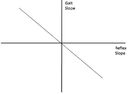
1.5 Specific Aims and Hypotheses
Aim 1: To determine the effects of isolated limb loading on reflex excitability of
the knee extensor muscles post-stroke.
Hypothesis 1: We hypothesize that increased load at the ankle in individuals
Figure 5: Extensor Excitability vs. Ankle Load (Hypothesized)
Aim 2: To determine the contribution of limb load on muscle activity during
walking in individuals
post-Hypothesis 2.1: We hypothesize that there will be a direct relationship between
the limb load and knee extensor excitability (Figure 6).
Figure 6: Extensor Excitability vs. Body Weight Support (Hypothesized)
21
Extensor Excitability vs. Ankle Load (Hypothesized)
the contribution of limb load on muscle activity during -stroke.
We hypothesize that there will be a direct relationship between the limb load and knee extensor excitability during late stance/early swing of gait
Figure 6: Extensor Excitability vs. Body Weight Support (Hypothesized) Extensor Excitability vs. Ankle Load (Hypothesized)
the contribution of limb load on muscle activity during
We hypothesize that there will be a direct relationship between during late stance/early swing of gait
Hypothesis 2.2: We hypothesize
excitability with limb load in an isolated situation will exhibit greater knee extensor activity during gait (Figure 7)
Figure 7: Relationship Between Gait Extensor Excitability & Isolated Refl Extensor Excitability (Hypothesized)
22
We hypothesize that individuals who show greater extensor excitability with limb load in an isolated situation will exhibit greater knee extensor
(Figure 7).
Figure 7: Relationship Between Gait Extensor Excitability & Isolated Refl Extensor Excitability (Hypothesized)
who show greater extensor excitability with limb load in an isolated situation will exhibit greater knee extensor
2.0 Methods
2.1 Subjects
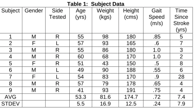
Nine subjects who exhibited clinically identifiable stiff-legged gait participated in one testing session that consisted of two parts. The first part consisted of isolated reflex testing while the second was a gait analysis. These nine subjects had an average age of 53±5.5 years (Table 1). Six subjects were male while 3 subjects were female. Six subjects were right side paretic while 3 subjects were left side paretic. The subjects had a mean weight of 81.6±16.9 kg. The mean height was 68.8±4.9 inches (174.7±12.5 cm).Individuals were included if they showed clinical symptoms consistent with ischemic or hemorrhagic unilateral brain lesion resulting in sensory motor dysfunction more than 6 months prior to testing. Individuals had to be able to step independently without assistance while walking on the treadmill.
Table 1: Subject Data Subject Gender Side
Tested Age (yrs) Weight (kgs) Height (cms) Gait Speed (m/s) Time Since Stroke (yrs)
1 M R 55 98 180 .85 5
2 F L 57 93 165 .6 7
3 M R 55 86 180 1.0 3
4 M R 60 68 170 1.0 2
5 F R 51 43 150 .5 8
6 M L 49 90 188 .55 6
7 F L 54 83 170 .9 28
8 M R 57 79 178 .65 4
9 M R 41 93 191 .75 4
AVG 53.3 81.6 174.7 .72 7.4
24 2.2 Testing Equipment
2.2.1 Test Apparatus – Isolated reflex testing
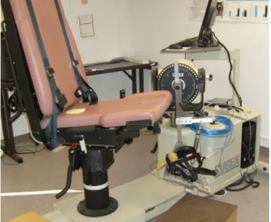
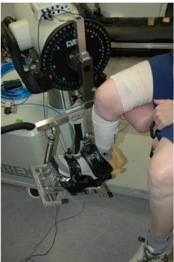
For the isolated reflex testing, the experimental apparatus was an instrumented ankle/knee actuator. Each subject was seated on a Cybex isokinetic dynamometer (Humac NORM; CSMI, Massachusetts) (Figure 8). A rigid frame was constructed for imposing and measuring ankle joint torque (Figures 9 and 10). The frame was affixed to the motor axis of the Cybex. The sagittal plane knee axis of rotation was aligned with the axis of the Cybex motor and the ankle axis of rotation was aligned with the center of rotation of the ankle loader by adjusting the subject’s position in the chair and the length of the ankle actuator device. The foot was secured onto a footplate with two dorsal straps (Figures 11, 12, 13). This isolated reflex testing component is able to determine the relative contribution of ankle load to the knee extensor reflex excitability.
25
Figure 9: Ankle Loader Dimensions – Side View
26
Figure 11: Ankle Loader Device
27
Figure 13: Ankle Loader with Knee in 90° of flexion
Figure 14: Isolated Reflex Testing Full Setup Diagram
2.2.2 Test Apparatus 2.2.2.1
A custom unweighting structure was constructed using 8020 aluminum. The structure was rigidly mounted to the floor to improve stability and safety. The structure is 5 feet tall, 3 feet wide, and 2 feet deep (Figure
connected to the top and bottom bars that allow for the dynamic counter weight theraband system. The 42’’ bar across the front
sliding up and down along the structure to allow for dynamic adjustment as the subject completed their walking trial. T
moving bar at the bottom, while the top of the bar attached to the harness through the use of a winch on the 36’’ front bar (Figure
28
Isolated Reflex Testing Full Setup Diagram
Test Apparatus – Gait Analysis Unloading 2.2.2.1 Unweighting Device for Gait
A custom unweighting structure was constructed using 8020 aluminum. The mounted to the floor to improve stability and safety. The 5 feet tall, 3 feet wide, and 2 feet deep (Figure 15). Pulleys
op and bottom bars that allow for the dynamic counter weight theraband system. The 42’’ bar across the front was mobile and capable of sliding up and down along the structure to allow for dynamic adjustment as the subject completed their walking trial. The theraband load was attached to this moving bar at the bottom, while the top of the bar attached to the harness through the use of a winch on the 36’’ front bar (Figures 16 and 17).
Isolated Reflex Testing Full Setup Diagram
A custom unweighting structure was constructed using 8020 aluminum. The mounted to the floor to improve stability and safety. The
Pulleys were op and bottom bars that allow for the dynamic counter weight
was mobile and capable of sliding up and down along the structure to allow for dynamic adjustment as the
Figure 15: Unweighting Structure Dimensions
Figure
29
: Unweighting Structure Dimensions
30
Figure 17: Constructed Unweighting Structure
The harness was attached to a rope that crosses over a pulley system mounted to a track above the test subject on the ceiling so that it could move freely with the subject. This allowed the subject to drift forward or back as they were walking on the treadmill. The rope then continued from the other side of the pulley to the moving bar on the unweighting structure. The load from the
31
amount. This allowed the unloading to be dynamic and to account for the change in movement of the center of mass when taking a step.
2.2.2.2 Theraband Properties
Theraband latex resistance bands were used to unload the subject instead of static weights (Figure 18). Due to the elastic properties of theraband, a long piece of Theraband will change its load only minimally with the relatively smaller superior/inferior changes in COM throughout the gait cycle.
Figure 18: Thera-Band Tubing
http://www.thera-band.com/store/products.php?ProductID=27
32
Table 2: Theraband Properties (Patterson, Stegink Jansen et al. 2001)
Force Elongation (lbs) after stretching by percentage of its original length (%) 25 50 75 100 125 150 175 200 225 250 Yellow 1.1 1.8 2.4 2.9 3.4 3.9 4.3 4.8 5.3 5.8 Red 1.5 2.6 3.3 3.9 4.4 4.9 5.4 5.9 6.4 7.0 Green 2.0 3.2 4.2 5 5.7 6.5 7.2 7.9 8.8 9.6 Blue 2.8 4.6 5.9 7.1 8.1 9.1 10.1 11.1 12.1 13.3 Black 3.6 6.3 8.1 9.7 11 12.3 13.5 14.8 16.2 17.6 Silver 5.0 8.5 11.1 13.2 15.2 17.1 18.9 21.0 23.0 25.3 Gold 7.9 13.9 18.1 21.6 24.6 27.5 30.3 33.4 36.6 40.1
There were many possible combinations of weights that could be used in the unweighting device. It was set up with 5 different strengths available, 5 lbs, 20 lbs, 20 lbs, 40 lbs, and 10 lbs. They could be attached in any different
combination to meet the necessary weight. For example, when trying to unload a 185 pound man by 40% of his body weight we would attach the 5 lb, 20 lb, 40 lb, and 10 lb therabands to reach a total of 75 lbs.
2.3 Methods – isolated reflex testing
Knee extensor reflex responses were elicited by rapidly rotating the knee into flexion using the Cybex isokinetic dynamometer. This provided a rapid passive stretch to the knee extensor muscles. The knee was rotated from 30 degrees to 90 degrees of knee flexion at both speeds of 90°/sec an d 180°/sec (Figures 12 and 13). This was done with muscles relaxed and with the hip flexors
33
Before knee flexion stretches were performed, the hemiparetic foot was fit into the previously described (see Section 2.2.1) custom brace which had the
capability of providing a sustained load to the ankle extensors. This was done to simulate the load through the ankle (limb) during the stance phase of gait. The magnitude of the quadriceps muscle’s response to the imposed knee movement was compared under different limb load conditions (no load, sustained load of 20Nm, and sustained load of 30 Nm). Five trials of each combination were performed of Cybex velocity (90°/sec and 180°/sec), appl ied load (no load,
sustained load at the ankle plantarflexor of 20nm, and sustained load at the ankle plantarflexor of 30 nm.), and quadriceps activation level (relaxed and 10% pre-activated). The order of the conditions was randomized among the different subjects to account for an order effect. A 16-channel electromyographic (EMG) system (Motion Lab Systems, Baton Rouge, LA) was used to collect muscle responses from the vastus lateralis, vastus medialis, rectus femoris, medial hamstrings, medial gastrocnemius, tibialis anterior, and soleus using active surface electrodes.
34
muscle was combined into an ensemble average of the 5 trials for all conditions. The individual muscle responses were compared for each condition.
2.4 Methods – Gait Analysis
All subjects also underwent gait analysis to determine lower extremity kinematics and EMG immediately following the reflex testing. The EMG electrodes stayed on the subject from the isolated reflex testing. Subjects walked on a Bertec, dual belt, instrumented treadmill (Bertec Corp, Columbus, OH) with different body weight support conditions which were controlled by the dynamic unweighting device (Figure 19). The motions of the lower extremity segments were tracked with an eight-camera motion capture system
35
thigh muscle activity during late stance/early swing under the different unweighting conditions.
36
Figure 20: Camera Marker Placement
2.5 Statistical Analysis
37
difference in EMG magnitude between the baseline average (250 ms before the movement started) and the final position average in muscle activity (250 ms after termination of the knee movement). Group means and standard deviations were compared using a two-way repeated measures ANOVA (repeated for both speed and load). If differences were found to be significant, post hoc testing was
performed.
SPSS software was also used to analyze the gait data. An ensemble
average of VL, RF, and VM activity was taken for each condition (0% BWS, 20% BWS, and 40%BWS). This was normalized to one gait cycle (heel strike to heel strike). An ANOVA was used to compare peak knee flexion angles during swing between conditions. An ANOVA was also used to analyze the VM, VL, and RF muscle activity between the three different load conditions (0%, 20%, and 40%).
Regression analyses were performed on both the isolated reflex data and the gait data. For the reflex data, slopes were calculated across the 3 different loads (no load, 20 Nm, and 30 Nm) for each muscle. This was done for both
3.0 Results
3.1 Isolated Reflex Testing
As the knee began to flex rapidly, there was a brief burst of muscle activity from the quadriceps. In most cases, this muscle activity was sustained and tended to be greater than the Baseline period (Figure 21). We had unusable data from 2 subjects leaving N=7 during reflex testing.
Figure 21: Variables Obtained During Isolated Reflex Testing
Baseline muscle activity was 4±6 %MVIC for the VL, 6±7 %MVIC for the RF, and 7±5 %MVIC for the VM prior to the knee flexion movement during the
39
load (VL: p=0.798; VM: p=0.217; RF: p=0.522) or speed (VL: p=0.565; VM: p=0.794; RF: p=0.902), and no interaction effect was present (VL: p=0.394; VM: p=0.588; RF: p=0.691) for Baseline muscle activity. Confirming this lack of difference between conditions provides confidence that the level of pre-activation did not influence the reflex results between conditions.
The initial peak muscle activity of the RF (p=0.027) and VM (p=0.035)
Figure 22: Peak Muscle Response During Isolated Reflex Testing
Although muscle activity appeared slightly greater immediately following the knee flexion movement compared to Baseline, this increased muscle activity was not influenced by load (VL: p=0.354; VM: p=0.208; RF: p=0.630) or speed (VL: p=0.646; VM: p=0.367; RF: p=0.123) with no significant interaction effect (VL: p=0.285; VM: p=0.217; RF: p=0.095) (Figure
0 10 20 30 40 50 60 VL % M V IC 40
Peak Muscle Response During Isolated Reflex Testing
Although muscle activity appeared slightly greater immediately following the knee flexion movement compared to Baseline, this increased muscle activity was not influenced by load (VL: p=0.354; VM: p=0.208; RF: p=0.630) or speed (VL:
F: p=0.123) with no significant interaction effect (VL: p=0.285; VM: p=0.217; RF: p=0.095) (Figure 23).
RF VM
Muscle
Peak Response
90°/sec 0 Nm load
180°/sec 0 Nm load
90°/sec 20 Nm load
180°/sec 20 Nm load
90°/sec 30 Nm load
180°/sec 30 Nm load
Peak Muscle Response During Isolated Reflex Testing
Although muscle activity appeared slightly greater immediately following the knee flexion movement compared to Baseline, this increased muscle activity was not influenced by load (VL: p=0.354; VM: p=0.208; RF: p=0.630) or speed (VL:
F: p=0.123) with no significant interaction effect (VL:
/sec 0 Nm load
/sec 0 Nm load
/sec 20 Nm load
/sec 20 Nm load
/sec 30 Nm load
Figure 23: Prolonged
3.2 Gait Data
Subjects walked on the treadmill at their comfortable One subject had quadriceps data
may have been due to bad
walking. This left N=8 during gait. With no body weight support,
had an average knee flexion angle of 34±14° on their pa retic side and an average knee flexion angle of 65±4° on their non
significant difference of knee flexion at different BWS was a range of paretic kne
to nearly normal (i.e., 55.4°) (Table
-5 0 5 10 15 20 25 30 35 40 VL % M V IC 41
Muscle Response During Isolated Reflex Testing
Subjects walked on the treadmill at their comfortable pace (mean quadriceps data that was too noisy to use in the analysis may have been due to bad EMG electrode placement or movement during
N=8 during gait. With no body weight support, the subjects an average knee flexion angle of 34±14° on their pa retic side and an average knee flexion angle of 65±4° on their non -paretic side. There was no significant difference of knee flexion at different BWS (p=0.569). However, there was a range of paretic knee flexion values, ranging from very stiff knee (i.e., 8.5°) to nearly normal (i.e., 55.4°) (Table 3).
RF VM
Muscle
Prolonged Response
90°/sec 0 Nm load
180°/sec 0 Nm load
90°/sec 20 Nm load
180°/sec 20 Nm load
90°/sec 30 Nm load
Muscle Response During Isolated Reflex Testing
pace (mean: 0.72 m/s.) too noisy to use in the analysis. This electrode placement or movement during
subjects an average knee flexion angle of 34±14° on their pa retic side and an
paretic side. There was no (p=0.569). However, there e flexion values, ranging from very stiff knee (i.e., 8.5°)
/sec 0 Nm load
/sec 0 Nm load
/sec 20 Nm load
/sec 20 Nm
42
Table 3: Knee Flexion Angle Results
Paretic Knee Flexion Angle Non-Paretic Knee Flexion Angle 0% BWS 20% BWS 40%
BWS 0% BWS
20% BWS
40% BWS All
Subjects (±14.4°) 34.1° (±14.7°) 33.3° (±14.8°) 33.7° (±4.6°) 65.1° (±4.4°) 64.4° (±5.7°) 62.9° Stiff-Knee Gait (n=4) 20.9° (±8.5°) 19.8° (±7.9°) 21.1° (±8.2°) 65.1° (±4.2°) 63.5° (±3.9°) 62.1° (±6.7°) “Normal’ knee flexion (n=5) 44.7° (±6.6°) 44.0° (±7.6°) 43.7° (±10.2°) 65.0° (±5.4°) 65.1° (±5.1°) 63.6° (±5.4°)
The muscle activity of the VL, RF, and VM were compared between the three different body weight support conditions (0% BWS, 20% BWS, and 40% BWS) for all subjects. For all subjects, the integral of muscle activity during late stance/early swing was not significantly different for VL (p = 0.687), VM
43
Figure 24: Muscle Activity During Gait
0.0 5.0 10.0 15.0 20.0 25.0 30.0 35.0
VL RF VM
%
M
V
IC
Muscle
Gait Muscle Activity
"Normal" - 0% BWS
Stiff-Knee - 0% BWS
"Normal" - 20% BWS
Stiff-Knee - 20% BWS
"Normal" - 30% BWS
44
3.3 Relationship Between Gait and Reflex Testing
The regression analysis between the slopes of gait testing and the slopes for reflex testing showed no significant relationships (N=7) (Table 4).
Table 4: All Subjects – Regression Analysis Comparison
Muscle Isolated Reflex condition
Gait condition P value R2 value
VL
Final 90°/sec
VL Gait
.814 .012
Final 180°/sec .969 .000
Peak 90°/sec .584 .064
Peak 180°/sec .903 .003
RF
Final 90°/sec
RF Gait
.616 .054 Final 180°/sec .466 .110
Peak 90°/sec .192 .312
Peak 180°/sec .536 .081
VM
Final 90°/sec
VM Gait
.476 .106
Final 180°/sec .971 .000
Peak 90°/sec .419 .134
Peak 180°/sec .351 .175
Total Muscle Activity
Final 90°/sec
Total Muscle Activity Gait
.711 .030
Final 180°/sec .663 .041
Peak 90°/sec .084 .481
45
When doing regression analysis on those subjects who exhibited very strong stiff-legged gait (knee flexion less than 30° during ga it, N = 4) significant
46
Table 5: Stiff-Legged Gait Subjects – Regression Analysis Comparison
Muscle Isolated Reflex condition
Gait condition P value R2 value
VL
Final 90°/sec
VL Gait
.792 .043
Final 180°/sec .867 .018
Peak 90°/sec .896 .011
Peak 180°/sec .601 .159
RF
Final 90°/sec
RF Gait
.011 .977
Final 180°/sec .040 .922
Peak 90°/sec .597 .162
Peak 180°/sec .563 .191
VM
Final 90°/sec
VM Gait
.576 .180
Final 180°/sec .868 .017
Peak 90°/sec .625 .140
Peak 180°/sec .596 .163
Total Muscle Activity
Final 90°/sec
Total Muscle Activity Gait
.823 .031
Final 180°/sec .874 .016
Peak 90°/sec .283 .514
4.0 Discussion
My hypotheses were not supported by these data. Overall, these data indicated that in both an isolated setting and a functional gait setting, the modulation of limb load did not have a significant effect on quadriceps muscle activity. There did, however, seem to be an effect of speed in the isolated reflex setting for the rectus femoris and vastus medialis. Despite the lack of significant findings in regards to my hypotheses, I believe that there are important
implications for gait retraining post-stroke.
Most studies have focused heavily on the role of limb load on the ankle muscles (gastrocnemius, soleus, tibialis anterior). My study, however, focused on the knee extensors (vastus lateralis, rectus femoris, vastus medialis). Klarner et al, performed a gait analysis study with a range of body weight support from 0% to 100% monitoring the knee extensors and the ankle plantarflexors. They found the largest modulation of muscle activity in the medial gastrocnemius, lateral gastrocnemius, and soleus (Klarner, Chan et al.). Another study that confirmed our findings was performed by van Hedel, et al. This study also examined muscle activity with varying amounts of body weight support. They found that the VM, RF, and VL were much less influenced by unloading
48
primarily within the ankle plantarflexor muscles act more strongly at the local level and have less of an excitatory input to the other musculature throughout the limb.
4.1 Interpretation of Results
4.1.1 Relationship Between Isolated Reflex Testing and Gait Analysis
We hypothesized that the individuals who showed greater extensor excitability with limb load in an isolated situation would exhibit greater knee extensor activity during gait. However, when looking at the subjects as a whole group, no significant relationship was found between the slopes of the regression lines of gait for each muscle and that of isolated reflex testing. When examining the regression data of only those subjects who exhibited strong stiff-knee gait (knee flexion<40°), there was a significant relationshi p for the RF prolonged response at both speeds of 90°/sec and 180°/sec. Howeve r, upon further
analysis, we observed that one subject was an extreme outlier because only 2 of the 3 conditions (0 Nm and 20Nm) were accounted for due to not being able to get a 30 Nm stretch. This altered the slope of the isolated reflex data. Once this subject’s data was removed, no significant relationship existed among the
remaining data points.
4.1.2 The Relationship Between Speed and Load
The vastus medialis showed a significant interaction between load and speed during the isolated reflex testing. Further testing revealed that the
49
modulates the effect of speed. This could imply that as locomotor training is performed at increasing speeds, there is a potential to change the way that the load is sensed. However, we are unable to make a full conclusion because we did not do the gait analysis at different speeds. Therefore, we do not know if the same effect can be observed during walking.
4.1.3 Dynamic Unweighting System
As described in the methods, our unweighting system allows for dynamic unweighting. This differs from many other unweighting systems because it keeps the load constant. This could possibly explain why we are getting different
results than some other published research (Mulroy, Klassen et al.; Visintin and Barbeau 1989; Finch, Barbeau et al. 1991; Hesse, Konrad et al. 1999; Barbeau and Visintin 2003). Others may find differences because their load is variable during walking rather than nearly constant (within 7%), as our subjects are experiencing. This implies that in other studies, the subject is generating additional muscle activity by the constant weighting and unweighting that they experience in their counterweight unloading devices. Our constant load system may be eliminating the change in muscle activity that is caused by the rapid loading and unloading in the counterweight systems.
4.2 Positive Aspects of the Study
50
Another positive aspect of this study was the dynamic unweighting system. Our body weight support system allowed for there to be a constant load on the subject at all times unlike many of the static counterweight systems often used.
Each subject completed their test session in a short period of time, about an hour for the isolated reflex testing and only a few minutes on the treadmill for gait analysis. This reduced the potential effects of muscle fatigue.
Lastly, both test setups in the study could accommodate a large size range of subjects. The ankle loader could be adjusted to accommodate from a
woman’s size 5 shoe to a men’s size 15 shoe. The ankle loader and Cybex both had adjustments for different leg lengths as well. The unweighting device also allowed room for a height of 6’8’’. This was positive as we were not required to limit our subject pool.
4.3 Limitations of the Study
One limitation of this study was the small sample size (n=9). While there were several values that demonstrated significance, it is unknown whether a larger sample size would have lead to more observations of statistical or clinical significance.
51
52
Appendix A: Subject Information Sheet
Subject: _____________Today’s Date: _____________Birthdate: ___________ Height: _______Weight:________Stroke Onset: ______Paretic Side: L R Foot Width: Left _________Right_________ Pelvic Depth: ___________ Fugl-Meyer: _________/(34) Berg: _______/(56)
Minute Gait
Speed
Pounds Unweighted
FP0 Left FP0 Right
0% 20% 40%
Channels
0 Cybex Position
1 Cybex Velocity
2 Cybex Torque
3 Loader Torque (Force really)
4 Vastus Lateralis
5 Rectus Femoris
6 Vastus Medialis
7 Medial Hamstring
8 Medial Gastroc
9 Soleus
10 Tib Anterior
11 Load release (5V for loaded, 0V when release)
53
Appendix B: Subject Consent Form
University of North Carolina-Chapel Hill Consent to Participate in a Research Study Adult Subjects
Biomedical Form
___________________________________________________________ IRB Study #____07-0845_________________
Consent Form Version Date: __6/18/07________
Title of Study: Limb Load Affects Reflexes During Walking Post-Stroke
Principal Investigator: Michael Lewek, PT, PhD
UNC-Chapel Hill Department: Department of Allied Health Sciences, Div of PT UNC-Chapel Hill Phone number: 919-966-9732
Email Address: [email protected]
Co-Investigator: Amy West; Richard Goldberg, PhD; Vicki Mercer, PT, PhD Funding Source: University Research Council
Study Contact telephone number: 919-966-9732 Study Contact email: [email protected]
________________________________________________________________ _
What are some general things you should know about research studies? You are being asked to take part in a research study. To join the study is voluntary.
You may refuse to join, or you may withdraw your consent to be in the study, for any reason.
Research studies are designed to obtain new knowledge that may help other people in the future. You may not receive any direct benefit from being in the research study. There also may be risks to being in research studies.
Deciding not to be in the study or leaving the study before it is done will not affect your relationship with the researcher, your health care provider, or the University of North Carolina-Chapel Hill. If you are a patient with an illness, you do not have to be in the research study in order to receive health care.
54 What is the purpose of this study?
Unloading patients with stroke using a harness support for walking training has become routine in the clinical setting. The purpose of this research study is to learn if weight bearing through your legs increases the amount of muscle activity while walking in people who have had a stroke. Walking difficulties are common in individuals who have had a stroke, and may be due to abnormal muscle control. Muscles are controlled, in part, by sensing the weight through the legs while walking. The aim of this study is to see if the amount of weight bearing during walking alters muscle activity in individuals who have had a stroke. You are being asked to be in the study because you have either had a stroke or because you have not experienced a stroke and wish to volunteer as a control subject.
Are there any reasons you should not be in this study?
You should not be in this study if you have heart, muscle, or bone problems that would limit your ability to move your legs or walk.
How many people will take part in this study?
If you decide to be in this study, you will be one of approximately 40 people in this research study (20 individuals following stroke, and 20 individuals with no history of neurological injury). All individuals will perform identical testing. How long will your part in this study last?
If you decide to participate, you will be asked to attend 2 sessions of testing. Each session will last approximately 3 hours, for a total of 6 hours. There will be no follow-up required to participate in this study.
What will happen if you take part in the study?
You will need to come to UNC-CH for 2 sessions of testing to participate in this study. For one session you will be asked to walk on a treadmill with a portion of your body weight supported by a harness, and for the other session, you will be asked to sit in a chair to have your reflexes tested. You will need to wear loose fitting shorts and comfortable shoes for both testing sessions. If you do not have a pair of shorts, we can provide them for you. The study will begin with a series of tests in which you will have your strength, balance, and walking ability
assessed by a physical therapist. Once these tests are completed, you will have sensors taped to your thighs, calves, and shins to monitor the activity of your leg muscles. Before walking on the treadmill, we will also tape small reflective balls (about the size of a dime) to your legs to monitor how your hips, knees, and ankles bend. Once this equipment is taped to your legs, we will ask you to do various leg movements to ensure that the sensors are working properly. You will then perform one of the following two protocols, with the other protocol being performed on a separate day:
55
• You will be seated in a chair to begin the data collection.
• You will be asked to kick out a small amount against a machine that will bend your knee.
• The machine will bend your knee, while another machine either provides or does not provide a stretch to your calf muscles. When the stretch is applied, there will be some trials in which the stretch is released at the end of the knee movement, and some trials in which the stretch will persist for several seconds after the knee movement.
• The stretch and the knee movement should not hurt, and are just intended to produce a reflexive response in your thigh muscles (like a “tendon tap” or “knee-jerk response”).
Walking Analysis:
• A harness will be placed around your waist that will be connected
overhead to an unweighting system. The unweighting system will support a portion of your body weight while you walk. Support is provided at 75%, 50%, 25%, and 0% of your body weight.
• You will walk on a treadmill at a comfortable speed for 5 minutes at each body weight support level.
• While you are walking, we will be collecting data about how your legs are moving and how active your muscles are. Approximately 20 minutes of walking on the treadmill will be required for this session with rest breaks provided as needed.
Both of these procedures are required for participation in the study. What are the possible benefits from being in this study?
Research is designed to benefit society by gaining new knowledge that will
improve rehabilitation programs. You will not benefit personally from being in this research study.
What are the possible risks or discomforts involved with being in this study?
Participation in this study may involve the following risks:
There is a risk of muscle fatigue from walking on the treadmill, although this risk will be minimized by providing you with long rest breaks as needed. In addition, the skin may need to be shaved for the placement of sensors, and although there is a risk that the skin may receive a small cut from the electric shaver, our
shavers are designed to minimize this risk. Aloe vera shaving cream can be used to minimize the risk of skin irritation that may be caused by shaving. There is also the possibility that the knee movement could elicit short lasting spasms in individuals with stroke. These spasms would be similar to what you may
experiences on a day-to-day basis during rapid leg movements.
56 How will your privacy be protected?
No subjects will be identified in any report or publication about this study. Although every effort will be made to keep research records private, there may be times when federal or state law requires the disclosure of such records, including personal information. This is very unlikely, but if disclosure is ever required, UNC-Chapel Hill will take steps allowable by law to protect the privacy of personal information. In some cases, your information in this research study could be reviewed by representatives of the University, research sponsors, or government agencies for purposes such as quality control or safety.
Any personal information collected will be stored in a locked office. This information will be linked to the collected data by a code that does not identify you. Only the investigator and study team will have access to this information. What will happen if you are injured by this research?
All research involves a chance that something bad might happen to you. This may include the risk of personal injury. In spite of all safety measures, you might develop a reaction or injury from being in this study. If such problems occur, the researchers will help you get medical care, but any costs for the medical care will be billed to you and/or your insurance company. The University of North Carolina at Chapel Hill has not set aside funds to pay you for any such reactions or
injuries, or for the related medical care. However, by signing this form, you do not give up any of your legal rights.
What if you want to stop before your part in the study is complete?
You can withdraw from this study at any time, without penalty. The investigators also have the right to stop your participation at any time. This could be because you have had an unexpected reaction, or have failed to follow instructions, or because the entire study has been stopped.
Will you receive anything for being in this study?
You will receive $40 for each testing session that you participate in. This means that you can receive up to $80 for completing both sessions involved in this study.
Will it cost you anything to be in this study?
If you enroll in this study, you will have some tests and procedures that are only part of the research study. You will not be billed for these tests and procedures. You will be responsible for the costs of transportation to and from the laboratory and for parking fees during the test session.
What if you are a UNC employee?
Taking part in this research is not a part of your University duties, and refusing will not affect your job. You will not be offered or receive any special job-related consideration if you take part in this research.
57
This research is funded by the University Research Council of the University of North Carolina at Chapel Hill.
What if you have questions about this study?
You have the right to ask, and have answered, any questions you may have about this research. If you have questions, or if a research-related injury occurs, you should contact the researchers listed on the first page of this form.
What if you have questions about your rights as a research subject? All research on human volunteers is reviewed by a committee that works to protect your rights and welfare. If you have questions or concerns about your rights as a research subject you may contact, anonymously if you wish, the Institutional Review Board at 919-966-3113 or by email to
- - - - - -
Title of Study: Limb Load Affects Reflexes During Walking Post-Stroke Principal Investigator: Michael Lewek, PT, PhD
Subject’s Agreement:
I have read the information provided above. I have asked all the questions I have at this time. I voluntarily agree to participate in this research study. _________________________________________
_________________
Signature of Research Subject Date _________________________________________
Printed Name of Research Subject
_________________________________________ _________________
Signature of Person Obtaining Consent Date _________________________________________
58
Appendix C: Device Figures
59
60
Appendix D: Isolated Reflex Testing Raw Data
Figure 27: VL Peak Response
Conditions: 0- 0 load, 90 speed; 1- 0 load, 180 speed; 2 - 20 load, 90 speed; 3 - 20 load, 180 speed; 4 - 30 load, 90 speed; 5 - 30 load, 180 speed
0 10 20 30 40 50 60
0 1 2 3 4 5 6
%
M
V
IC
Load and Speed
VL Muscle Activity Peak Response
s1
s2
s3
s4
s5
s6
s7
61
Figure 28: VM Peak Response
Conditions: 0- 0 load, 90 speed; 1- 0 load, 180 speed; 2 - 20 load, 90 speed; 3 - 20 load, 180 speed; 4 - 30 load, 90 speed; 5 - 30 load, 180 speed
0 5 10 15 20 25 30 35 40 45
0 1 2 3 4 5 6
%
M
V
IC
Load and Speed
VM Muscle Activity Peak Response
s1
s2
s3
s4
s5
s6
s7
62
Figure 29: RF Peak Response
Conditions: 0- 0 load, 90 speed; 1- 0 load, 180 speed; 2 - 20 load, 90 speed; 3 - 20 load, 180 speed; 4 - 30 load, 90 speed; 5 - 30 load, 180 speed
0 20 40 60 80 100 120 140 160 180 200
0 1 2 3 4 5 6
%
M
V
IC
Load and Speed
RF Muscle Activity Peak Response
s1
s2
s3
s4
s5
s6
s7
63
Figure 30: VL Prolonged Response
Conditions: 0- 0 load, 90 speed; 1- 0 load, 180 speed; 2 - 20 load, 90 speed; 3 - 20 load, 180 speed; 4 - 30 load, 90 speed; 5 - 30 load, 180 speed
-15 -10 -5 0 5 10 15
0 1 2 3 4 5 6
%
M
V
IC
Load and Speed
VL Muscle Activity Prolonged Response
s1
s2
s3
s4
s5
s6
s7
64
Figure 31: VM Prolonged Response
Conditions: 0- 0 load, 90 speed; 1- 0 load, 180 speed; 2 - 20 load, 90 speed; 3 - 20 load, 180 speed; 4 - 30 load, 90 speed; 5 - 30 load, 180 speed
-10 -5 0 5 10 15 20
0 1 2 3 4 5 6
%
M
V
IC
Load and Speed
VM Muscle Activity Prolonged Response
s1
s2
s3
s4
s5
s6
s7
65
Figure 32: RF Prolonged Response
Conditions: 0- 0 load, 90 speed; 1- 0 load, 180 speed; 2 - 20 load, 90 speed; 3 - 20 load, 180 speed; 4 - 30 load, 90 speed; 5 - 30 load, 180 speed
-20 -10 0 10 20 30 40 50 60 70
0 1 2 3 4 5 6
%
M
V
IC
Load and Speed
RF Muscle Activity Prolonged Response
s1
s2
s3
s4
s5
s6
s7