Prospective One Year Follow Up of HIV
Infected Women Screened for Cervical
Cancer Using Visual Inspection with Acetic
Acid, Cytology and Human Papillomavirus
Testing in Johannesburg South Africa
Cynthia Firnhaber1,2*, Bridgette Goeieman2, Mark Faesen2, Simon Levin2,3,
Sophie Williams2, Sibongile Rameotshela2, Avril Swarts1, Pam Michelow4,5, Tanvier Omar5, Anna-Lise Williamson6,7, Bruce Allan7, Kate Schnippel2, Jennifer
S. Smith8,9
1Clinical HIV Research Unit, Faculty of Health Sciences, Department of Internal Medicine, University Witwatersrand, Johannesburg, South Africa,2Right to Care, Johannesburg, South Africa,3Department of OB/GYN, Coronation Hospital, University of Witwatersrand, Johannesburg, South Africa,4Cytology Unit, Department of Anatomical Pathology, Faculty of Health Science, University of Witwatersrand, Johannesburg, South Africa,5National Health Laboratory Service, Johannesburg, South Africa,6Institute of Infectious Disease and Division of Medical Virology, Department of Clinical Laboratory Sciences, University of Cape Town, Cape Town, South Africa,7National Health Laboratory Service, Groote Schuur Hospital, Cape Town, South Africa,8Department of Epidemiology, Gillings School of Global Public Health, University of North Carolina, Chapel Hill, North Carolina, United States of America,9Lineberger Comprehensive Cancer Center, University of North Carolina, Chapel Hill, North Carolina, United States of America
Abstract
Background
Cervical cancer is the most common cancer in Sub-Saharan Africa. There are little of HIV-infected women one-year after screening using visual inspection with acetic acid (VIA), HPV or cytology in sub-Saharan Africa.
Methods
HIV-infected women in Johannesburg South Africa were screened one year later by Pap smear, VIA and human papillomavirus (HPV) testing. Women qualified for the 12 month fol-low-up visit if they had a negative or cervical intra-epithelial neoplasia (CIN) 1 results at the baseline visit. Modified Poisson regression was used to analyse associations between patient baseline characteristics and progression.
Results
A total of 688 of 1,202 enrolled at baseline study who were CIN-2+ negative and qualified for a 12 month follow-up visit. Progression to CIN-2+ was higher in women with positive VIA results (12.6%; 24/191) than those VIA-negative (4.4%; 19/432). HPV-positive women at
a11111
OPEN ACCESS
Citation:Firnhaber C, Goeieman B, Faesen M, Levin S, Williams S, Rameotshela S, et al. (2016) Prospective One Year Follow Up of HIV Infected Women Screened for Cervical Cancer Using Visual Inspection with Acetic Acid, Cytology and Human Papillomavirus Testing in Johannesburg South Africa. PLoS ONE 11(1): e0144905. doi:10.1371/journal. pone.0144905
Editor:Maria Lina Tornesello, Istituto Nazionale Tumori, ITALY
Received:August 31, 2015
Accepted:November 26, 2015
Published:January 5, 2016
Copyright:© 2016 Firnhaber et al. This is an open access article distributed under the terms of the
Creative Commons Attribution License, which permits unrestricted use, distribution, and reproduction in any medium, provided the original author and source are credited.
baseline were more likely to progress to CIN-2+ (12.3%; 36/293) than those HPV-negative (2.1%; 7/329). Cytology-positive women at baseline were more likely to progress to CIN-2+ (9.6%; 37/384) than cytology-negative women (2.5%; 6/237). Approximately 10% (10.4%; 39/376) of women with CIN 1 at baseline progressed to CIN 2+. Women who were VIA or HPV positive at baseline were more likely to progress aIRR 1.85, CI 95% (1.46 to 2.36), aIRR 1.41 CI 95% (1.14 to 1.75) respectively.
Conclusion
Progression to CIN-2+ in HIV-infected women is significant when measured by baseline positive VIA, HPV or Pap and yearly screening by any method should be considered in this population if possible.
Introduction
Cervical Cancer, in a vast majority of cases, is preventable with adequate screening and early treatment of dysplasia. Effective cytology-based screening programs in North American, Europe and Australia/New Zealand have reduced the incidence of this cancer to<10/100,000. In contrast, in Africa where there are inadequate screening programmes, the incidence is above 30/100,000 women [1]. Cervical cancer is the most common cancer and the most common cause of cancer death in women in Sub-Saharan Africa [1]. The reason for this high prevalence is multifactorial. Inadequate access to cervical cancer screening and treatment program due to lack of skilled specialists (i.e. pathologists, gynacologists), clinic infrastructure, and transport for women to visit the clinic are a few of the reasons why appropriate screening programs have not been successfully implement in sub-Saharan Africa.
Another important reason for the high prevalence of cervical cancer is the HIV epidemic in sub-Saharan Africa. HIV-infected women have more persistent HPV infections, are more likely to be infected with multiple HPV types and are more likely to have high-grade cervical disease as compared with HIV-negative women [2]. HIV-infected women in this region are 3 to 5 times more likely to develop cervical cancer [3,4]. Beginning in 1993, cervical cancer became one of three AIDS defining cancers per the Centers for Disease Control’s AIDS defin-ing illnesses [5]. The World Health Organization classified cervical cancer as stage 4 AIDS defining illness in 2005 [6].
As women live longer due to widespread roll-out of antiretroviral therapy (ART), prevent-ing other opportunistic infections, the risk of developprevent-ing cervical cancer may actually increase in these countries. At present conventional Pap smears is the standard of care cervical cancer screening in South Africa but coverage is limited in many areas. Adequate implementation and access to cervical screening and treatment programs is therefore essential to maintain the health improvements achieved with ART.
Visual inspection of the cervix with acetic acid (VIA) in HIV-infected women has been shown to have a similar sensitivity for CIN-2+ to cervical cytology, albeit with lower specificity [7–9]. HPV testing has been found to have the higher sensitivity for CIN-2+, with relatively lower specificity. The World Health Organization (WHO) in the recent (December 2014) cer-vical cancer guidelines states there is a research gap examining the screening and follow up in HIV-infected women in resource-limited countries. It is acknowledged that, for HIV-infected populations, guidelines are based largely on expert opinion as there is insufficient scientific Requests for the data from The South African
Department of Health may be done at the following site:http://nhrd.hst.org.za/Home/Index.
Funding:This work was funded under United States Aid for International Development Public Health Evaluation ZA.09.0265 grant awarded to CF. United States Aid for International Development Presidents Emergency Plan For AIDs Relief (PEPFAR) 674-A-00-08-00007-00 helped fund CF, BG, MF, SL, SW, SR and KS. University of North Carolina Center for AIDS Research P30-AI50410 was awarded to JSS. African Research Chairs Initiative of the Department of Science and Technology was awarded to ALW and BA. A small portion of this work is based on the research supported by the South African Research Chairs Initiative of the Department of Science and Technology and Nation Research Foundation (NRF), South Africa awarded to ALW. The funders had no role in study design, data collection and analysis, decision to publish, or preparation of the manuscript.
Competing Interests:JS Smith has received
evidence regarding how often to screen in HIV-infected women, or comparing different screening modalities [10].
We present 1 year findings for a cervical screening study conducted among HIV-infected women in Johannesburg, South Africa. The parent study is described in detail elsewhere [7]. The objective of the current sub-study is to present the one year follow up in HIV–infected women who had negative or CIN 1+ on either Pap smear or colposcopic biopsy at the baseline visit. Three cervical screening tests were compared: VIA, cytology, and HPV DNA testing.
Methods
Ethics approvals
The study protocol and consent were reviewed and approved by the Human Ethics Committee (Medical) of the University of the Witwatersrand, by the University of Cape Town for HPV testing, and by the University of North Carolina for secondary data analyses.
Study design
Women were educated in regards to the study and signed a written consent according to South African Good Clinical Practice and University of Witwatersrand ethics committee. Participants were enrolled in a prospective screening observational cohort study [7] from a Johannesburg HIV treatment clinic located in a tertiary teaching hospital. Each woman at the baseline visit was screened with a Pap smear, HPV and VIA. All women with ASCUS+ or a positive VIA had a colposcopic biopsy. In addition, every fourth participant who had both a negative Pap smear and VIA had a colposcopic biopsy at the baseline visit. Women with CIN 2+ on biopsy were referred for treatment by Loop electrical excision procedure. The other women were followed-up one year later in a sub study, if they were willing to participate and met the following crite-ria: qualified and participated in the baseline study visit, not pregnant, had a negative or CIN 1 histology on colposcopic biopsy or had a negative Pap smear and VIA and did not received a verification biopsy. Women who presented with sexual transmitted infections (STI) or menses were asked to return after completion of STI treatment or upon resolution of menses. A follow-up questionnaire at the one year follow-follow-up visit was obtained through participant interviews to update status on socio-demographic characteristics, changes in medical history, ART regimen, reproductive and menstrual characteristics, and other lifestyle factors, including smoking. For further recruitment, enrolment criteria and study methodology see the parent study [7].
Study-related procedures
Each woman at the one year follow up visit was screened using the same three screening meth-ods as in the baseline visit: HPV Hybrid Capture 2 DNA test (QIAGEN GmbH, Hilden, Ger-many), conventional Pap smear cytology, and VIA, all performed using the techniques previously described[7]. Similar to the baseline visit, HPV DNA specimens were collected by the clinician and test results were not used for clinical management. The Pap smear, HPV test and VIA were all performed by study nurses. All laboratory personnel were blinded to VIA results; the HPV laboratory team was also blinded to the cytology results. Conventional Pap smears results were analyzed at the National Health Laboratory Services (NHLS) cytology unit according to Bethesda 2001 guidelines [11]; liquid based cytology is not available in the South African public sector.
Agency for Research on Cancer guidelines [13]. Digital images were taken using a commer-cially available digital camera for physician review. The final‘VIA‘reading used in the analysis was the reading done after review by the doctors using the digital camera images at a weekly quality assurance meeting. A colposcopic-directed biopsy was taken by study doctors for histo-logical confirmation by an anatomical pathologist for all women with any abnormal Pap smear (ASCUS+) or a positive VIA. Lesions were biopsied and often greater than one biopsy was taken. If there were no lesions on colposcopy, biopsies were taken at the location of clock posi-tion 6 and 12 on the cervix. The study cytopathologist and anatomical pathologist were blinded to the VIA, HPV and other study results.
Quality Assurance
For quality assurance (QA) of the VIA technique, the study gynecologist and a medical officer trained in colposcopy reviewed each digital picture and the initial VIA diagnosis of the nurse within two weeks of the VIA procedure. The medical staff reviewing the pictures were blinded to the Pap and HPV results at the time of the image review.
The cytology unit undergoes several accreditation processes from the South African National Accreditation System (SANAS) and undergoes regular proficiency testing by the Royal College of Pathologist of Australasia Quality Assurance Programme (RCPA). The cytol-ogy unit also undergoes several internal quality assurance procedures. Discrepant results between cytology and histology resulted in a review of the Pap smear slide. If discrepancy was confirmed, then a repeat colposcopic biopsy was conducted, if clinically indicated.
HPV testing QA was done per recommendation on the manufacture’s guidelines.
Statistical methods
Subjects who were followed at 12 months were compared with those who were not in terms of demographic and clinical characteristics, {i.e. age (<30, 30–49, 50+ years), parity, CD4 count (<250,250 cells/mm3), HIV viral load (<40,40 copies/ml) cytology] in order to ensure that there was no selection bias in the follow-up cohort. Statistical differences were assessed using the (nonparametric) Wilcoxon rank-sum test for the continuous variables and the chi-square test for categorical ones.
At 12-month follow-up, VIA, HPV, cytology and histological biopsy results (for ASCUS + Pap smear results) were analyzed, stratified by baseline histology or cytology status (negative or CIN 1). A modified Poisson regression approach [14] was used to calculate relative risk of progression according to baseline characteristics (i.e. age, CD4 count, VIA result, and HPV DNA infection status), with the progression of pathology at study follow-up. Progression was defined as CIN 1 progressing to CIN 2+ on colposcopic biopsy or a negative cytology/histology result to CIN 1 +. A model for progression was built and then adjusted for the baseline status (negative or CIN1) as a model covariate. 95% confidence intervals for the incidence rate ratios were computed using robust standard errors. All analysis was done within Stata v13.1 (College Station, TX).
Results
CD4 count and viral load), or reported sexual history between the women who were re-screened at 12 months and those who were not (data not shown).
Table 1shows the results of the 12-month follow-up cervical screening tests, stratified by the baseline results from the same screening method. Of the 478 women who were VIA nega-tive at baseline, 92 (19.2%) progressed to a posinega-tive VIA at the 12-month follow-up visit. Of the 210 women who were VIA positive at baseline who qualified for a follow-up visit (negative or CIN-1 histology), 78 (37.1%) were VIA positive (persistent) and 132 (62.9%) were VIA nega-tive at 12 months. Of the 358 women who were neganega-tive for HPV DNA infection at baseline, 58 (16.2%) had acquired HPV infection by the time of the follow-up visit. Of the 322 women who were HPV DNA positive at baseline and eligible for the 12-month follow-up, 155 (48.1%) were HPV DNA negative at follow-up.
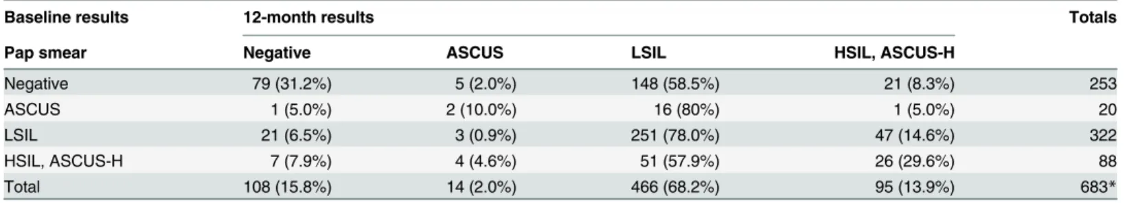
Pap smear results at follow-up compared to Pap smear results at baseline are shown in
Table 2. Of the 253 women who had a negative Pap smear at baseline, 21 (8.3%) had progressed to HSIL or ASC-H, 148 (58.5%) had progressed to LSIL and 5 (2.0%) to ASCUS. From baseline ASCUS and LSIL, 48 (19.6%) had progressed to HSIL or ASC-H. In total, compared to baseline Pap smear, 34.9% (238/683) of follow-up Pap smears indicated progression at the 12 months follow-up visit. Of the women who had a HSIL or ASC-H Pap smear at baseline (with either Fig 1. Consort diagram of participant follow-up one year later.CIN = Cervical Intraepithelial Neoplasia HPV = Human papillomavirus.
doi:10.1371/journal.pone.0144905.g001
Table 1. 12-month follow-up results against baseline results from same screening methodology, VIA and HPV.
Screening method Baseline results 12-month results Totals
Negative Positive
VIA Baseline negative 386 (80.8%) 92 (19.2%) 478
Baseline positive 132 (62.9%) 78 (37.1%) 210
Total 518 (75.3%) 170 (24.7%) 688
HPV Baseline negative 300 (83.8%) 58 (16.2%) 358
Baseline positive 155 (48.1%) 167 (51.9%) 322
Total 455 (66.9%) 225 (33.1%) 680^
^Baseline HPV missing (n = 1), month 12 HPV missing (n = 7)
CIN 1 or negative histology), 26 (29.6%) persisted at HSIL or ASC-H cytology and 62 (70.4%) regressed LSIL, ASCUS, or normal Pap smear result.
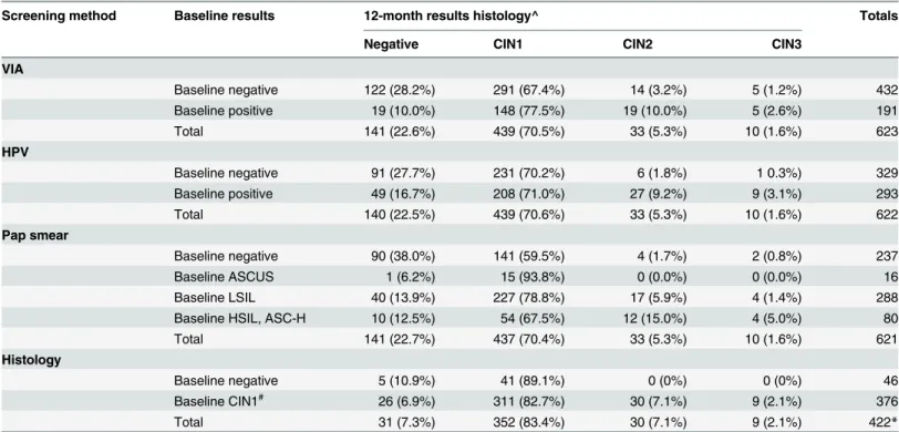
Table 3shows the colposcopic biopsy histology results (the gold standard) at 12-months compared to the baseline results from the different screening methods. Close to 90% (41/46) of the women who were histology negative at baseline had progressed at the 12-month follow-up screening: CIN 1. Among women that were CIN 1 at the baseline visit approximately 10% (39/ 376) progressed to CIN 2+.
Most of the 376 women who were CIN 1 at baseline persisted at CIN 1 (n = 311, 82.7%); 39 (10.4) progressed to CIN 2 or CIN 3 and 26 (6.9%) regressed to a negative histology result. In total, approximately 19% % (80/422) women with histology results at baseline and at 12 months, the cervical histology had progressed by the 12-month follow-up visit.
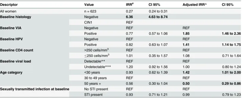
The relative risks of progression from baseline negative or CIN 1 and an adjusted risk of progression to CIN-2+ holding the baseline histology status constant are reported inTable 4. Most progression was from baseline histology negative (RR: 6.36, 95% CI: 4.63 to 8.74). Pro-gression to CIN-2+ was more likely in women with positive VIA results (12.6%, 24/191) than those VIA-negative (4.4%, 19/432) aIRR: 1.85 (95% CI: 1.46 to 2.36). HPV-positive women at baseline were more likely to progress to CIN-2+ (12.3%, 36/293) than those HPV-negative (2.1%, 7/329) aIRR: 1.41 (95% CI: 1.14 to 1.75). Baseline CD4 count less than or equal to 250 cells/ mm3or baseline HIV viral load undetectable (<40 copies) were not associated with pro-gression in either the unadjusted or adjusted models. Having an STI at baseline screening was also not associated with progression. Compared to women aged 30–49 years old, women youn-ger than 30 were at higher risk of progression (aIRR: 1.42, 95% CI: 1.01 to 2.00) and women who were 50 years or older were less likely to experience progression (aIRR: 0.50, 95% CI: 0.29 to 0.86), once the model was adjusted for baseline histology status.
Discussion/Conclusion
Previous cross-sectional and prospective studies have shown both high rates of cervical dyspla-sia (LSIL+) at baseline and high rates of incident disease in HIV-infected women in South Africa ranging from 34% to 75% [7,15,16]. Progression rates measured by cervical Pap smear results in HIV-infected women in South Africa have been shown to be high in two cohorts of HIV-infected women from Johannesburg. Omar et al data showed the rate of progression from negative to LSIL was 9.6/100 person years (95% CI: 8.3 to 11.1) and from LSIL to HSIL 4.6/100 py (95% CI: 3.9 to 5.5) after approximately 1-year follow-up [15]. Firnhaber et al showed pro-gression rates of 14.6/100 py (95% CI: 11.5 to 18.5) from negative to LSIL and LSIL to HSIL of 10.8/100 py (95% CI: 8.1 to 14.4) [17]. Our current study demonstrated similar disease Table 2. 12 month follow-up results against baseline results from same screening methodology, Pap smear.
Baseline results 12-month results Totals
Pap smear Negative ASCUS LSIL HSIL, ASCUS-H
Negative 79 (31.2%) 5 (2.0%) 148 (58.5%) 21 (8.3%) 253
ASCUS 1 (5.0%) 2 (10.0%) 16 (80%) 1 (5.0%) 20
LSIL 21 (6.5%) 3 (0.9%) 251 (78.0%) 47 (14.6%) 322
HSIL, ASCUS-H 7 (7.9%) 4 (4.6%) 51 (57.9%) 26 (29.6%) 88
Total 108 (15.8%) 14 (2.0%) 466 (68.2%) 95 (13.9%) 683*
*Baseline Pap smear missing (n = 2), month 12 Pap smear missing (n = 3)
progression from a negative histology baseline result to CIN 1 in approximately 60% of women. Histological progression from CIN 1 to CIN 2+ was approximately 10% in the one year of follow-up.
Comparing these results to HIV-negative women seen within the same government public system in Johannesburg showed a baseline abnormal cytology of 11.4% (1.8% HSIL) with a progression rates from a negative Pap smear to cervical abnormalities of 6.5% within a year. Progression to HSIL during this time period was rare, less than 0.5% which would allow for longer intervals for screening in HIV-negative women [18].
Our data shows that if a woman is VIA positive but has normal or CIN 1 histology, her risk of progression to CIN 1+ histology within one year is nearly two times higher than the risk of progression in VIA negative women. Similarly, if a women is HPV positive but has normal or CIN1 histology, her risk of progression within one year is 40% higher than the risk for HPV negative women. Importantly, 19% (80/422) women progressed from negative or CIN 1 histol-ogy to CIN1+ histolhistol-ogy, suggesting a treatment by cryotherapy or other method at the same visit (see and treat) may be warranted. Kuhn et al showed that HIV-infected women compared to HIV-negative women who were screened by either HPV or VIA but not treated had a high rate of CIN 2+.Of the untreated women 14.9% progressed to CIN 2+ compared to 4.6% who were treated. HIV-infected women screened with VIA and treated with cryotherapy had a drop in CIN 2+ by 7.4% and women screened by HPV and treated with cryotherapy had a 12% reduction of CIN 2+ [8].
Table 3. 12-month follow-up histology results against baseline results from different screening methods among women without CIN-2+ at baseline.
Screening method Baseline results 12-month results histology^ Totals
Negative CIN1 CIN2 CIN3
VIA
Baseline negative 122 (28.2%) 291 (67.4%) 14 (3.2%) 5 (1.2%) 432
Baseline positive 19 (10.0%) 148 (77.5%) 19 (10.0%) 5 (2.6%) 191
Total 141 (22.6%) 439 (70.5%) 33 (5.3%) 10 (1.6%) 623
HPV
Baseline negative 91 (27.7%) 231 (70.2%) 6 (1.8%) 1 0.3%) 329
Baseline positive 49 (16.7%) 208 (71.0%) 27 (9.2%) 9 (3.1%) 293
Total 140 (22.5%) 439 (70.6%) 33 (5.3%) 10 (1.6%) 622
Pap smear
Baseline negative 90 (38.0%) 141 (59.5%) 4 (1.7%) 2 (0.8%) 237
Baseline ASCUS 1 (6.2%) 15 (93.8%) 0 (0.0%) 0 (0.0%) 16
Baseline LSIL 40 (13.9%) 227 (78.8%) 17 (5.9%) 4 (1.4%) 288
Baseline HSIL, ASC-H 10 (12.5%) 54 (67.5%) 12 (15.0%) 4 (5.0%) 80
Total 141 (22.7%) 437 (70.4%) 33 (5.3%) 10 (1.6%) 621
Histology
Baseline negative 5 (10.9%) 41 (89.1%) 0 (0%) 0 (0%) 46
Baseline CIN1# 26 (6.9%) 311 (82.7%) 30 (7.1%) 9 (2.1%) 376
Total 31 (7.3%) 352 (83.4%) 30 (7.1%) 9 (2.1%) 422*
^ Negative cytology results presented if patient did not have colposcopy because Pap smear and VIA negative
#Women with baseline CIN2+ were referred for further management at baseline and were not eligible for the 12-month follow-up study *Baseline histology not available for n = 10; 12-month histology (or negative Pap smear) not available for n = 65 persons
Two studies from Johannesburg have shown that HAART in this population reduces the cervical dysplasia progression rate [17,19], but in should be noted this gain in slowing the rate of cervical disease progression may be overcome by women living longer. In this current study, the baseline cervical dysplasia prevalence were high. One year later there was significant pro-gression, despite over 90% of women taking ARVs the majority of the women on ARVs had suppressed HIV viral loads (<1000 copies/ml) at the month 12 study visit.
Interestingly, our study showed that age over 50 years was protective of progression. This could be due to a selection bias as the women who had CIN 2+ disease were sent for treatment and not eligible for a follow-up visit at month 12.
HIV infection has been shown to increase persistence and reduce clearance of HPV infec-tion [20,21]. At the baseline visit of this study, 731 women were HPV positive. Of the women who qualified for a month 12 visit 155 women no longer had HPV infection for a rate of HPV infection clearance of 21.2% (155/731). This is higher than the Denny et al cohort where only 6% cleared there HPV in 36 months [16]. This difference maybe as result of the women with high-grade lesions were referred for treatment and did not qualify for the 12-month screening follow up. Additionally, the Denny et al study was done in the early 2000’s when ARVs were not readily available in South Africa. In contrast, approximately 90% of the women in our study were on ARVs and virally suppressed. Good adherence to ARV medication has been shown to decrease the persistence of HPV infection in HIV-infected women [22,23].
This study was not designed to evaluate regression as women with CIN 2+ histologically were referred for treatment and did not qualify for the month 12 visit. However, the likelihood of regression from VIA positive to negative results or HPV infection clearance over the one fol-low up might be inferred or estimated from these study results. Women who were VIA positive at the baseline visit (n = 529) and qualified for month 12 visit, 24.8% (132/529) of these women Table 4. Relative risk of progression (from baseline negative cytology or histology to CIN 1 or from baseline CIN 1 to CIN 2+) at 12 months.
Descriptor Value IRR# CI 95% Adjusted IRR^ CI 95%
All women n = 623 0.27 0.24 to 0.31
Baseline histology Negative 6.36 4.63 to 8.74
CIN1 REF
Baseline VIA Negative REF REF
Positive 0.77 0.57 to 1.06 1.85 1.46 to 2.36
Baseline HPV Negative REF REF
Positive 0.82 0.63 to 1.07 1.41 1.14 to 1.75
Baseline CD4 count >250 cells/mm3 REF REF
250 cells/mm3 1.01 0.35 to 1.57 1.08 0.71 to 1.64
Baseline viral load Detectable** REF REF
Undetectable*** 1.20 0.92 to 1.56 1.00 0.80 to 1.24
Age category <30 years 0.93 0.62 to 1.39 1.42 1.01 to 2.00
30 to 49 years REF REF
50 years + 0.56 0.30 to 1.04 0.50 0.29 to 0.86
Sexually transmitted infection at baseline No STI present REF REF
STI present 0.93 0.71 to 1.21 0.99 0.79 to 1.23
#Relative risk calculated using incidence rate ratios, Poisson distribution with robust standard errors. ^ Adjusted for baseline histology results.
**detectable is40 copies/ml ***undetectable is<40 copies/ml
regressed to a negative VIA result. This information and the increased risk of women who are VIA positive progressing to high-grade disease in areas with limited access to pathology, treat-ment for dysplasia or cervical cancer, may negate the concerns of overtreattreat-ment of HIV-infected women when using the VIA see and treat method.
Another limitation of this study is the progression may not have reflected incident disease or change of disease but prevalent disease that was missed by initial cytology or histology. This possibility was minimized by the continuing internal and external quality assurance programs by the NHLS anatomical pathology services, secondary review of discrepant results and if nec-essary a second biopsy.
These results provide important information required for resource allocation for cervical cancer screening, both in terms of guidelines of when to screen and also improving access to screening in the country with the largest HIV epidemic. Progression rates are high in this pop-ulation and a positive VIA and HPV test demonstrated a higher risk of progression within one year. VIA is at present is being implemented in several sub-Saharan Africa countries through a variety of governmental and donor funded programs. Of the 432 women with a baseline nega-tive VIA, 19 (4.4%) had CIN 2+ a year later. Although comparing different modalities, this is about 8 times the progression seen in the HIV-negative population (<0.5%) in Johannesburg using Pap smears indicating that a screen and treat program in HIV-infected women with neg-ative baseline results should consider reduced screening intervals.
HIV-infected women are at significant risk for progression of their cervical dysplasia. Whilst there is some evidence that ARVs might slow progression, the disease is not eliminated in many of these women. In this cohort, progression still occurred with a relative high median CD4 count and with the majority of women with suppressed viral load. Countries need to aggressively ramp-up the scale of cervical cancer screening and frequency of screening (maybe yearly) using one of the appropriate screening methods per the capacity and resources of the country for HIV infected women. VIA is a possible option in countries with limited resources and poor access to screening. Longer term studies with information on the relative costs of screening methods are needed to better inform screening policies for the individual countries. Improved access to screening is imperative to maintain the gains of health achieved in our HIV-infected women.
Acknowledgments
Dr Carla Chibwesha for her review and suggestions for the article.
"This work was funded under United States Aid for International Development Public Health Evaluation ZA.09.0265 grant awarded to CF. United States Aid for International Devel-opment Presidents Emergency Plan For AIDs Relief (PEPFAR) 674-A-00-08-00007-00 helped fund CF, BG, MF, SL. SW, SR and KS. University of North Carolina Center for AIDS Research P30-AI50410 awarded to JSS. African Research Chairs Initiative of the Department of Science and Technology awarded to ALW and BA. A small portion of this work is based on the research supported by the South African Research Chairs Initiative of the Department of Sci-ence and Technology and Nation ResearchFoundation (NRF), South Africa awarded to ALW. The funders had no role in study design, data collection and analysis, decision to publish, or preparation of the manuscript"
Author Contributions
References
1. International Agency for Research on Cancer. GLOBOCAN 2012: Estimated Cancer Incidence, Mortal-ity and Prevalence Worldwide in 2012 [Internet]. 2012. Available from:http://globocan.iarc.fr/Pages/ fact_sheets_population.aspx
2. Williamson A-L. The Interaction between Human Immunodeficiency Virus and Human Papillomavi-ruses in Heterosexuals in Africa. J Clin Med [Internet]. 2015; 4(4):579–92. Available from:http://www. mdpi.com/2077-0383/4/4/579/
3. Belhadj H, Rasanathan JJK, Denny LE, Broutet N. Sexual and reproductive health and HIV services: integrating HIV/AIDS and cervical cancer prevention and control. Int J Gynaecol Obstet [Internet]. Inter-national Federation of Gynecology and Obstetrics; 2013 May [cited 2014 Oct 9]; 121 Suppl: S29–34. Available from:http://www.ncbi.nlm.nih.gov/pubmed/23477703
4. Frisch M, Biggar RJ, Goedert JJ. Human papillomavirus-associated cancers in patients with human immunodeficiency virus infection and acquired immunodeficiency syndrome. J Natl Cancer Inst. 2000; 92(18):1500–10. PMID:10995805
5. Centers for Disease Control and Prevention. 1993 Revised Classification System for HIV Infection and Expanded Surveillance Case Definition for AIDS Among Adolescents and Adults. MMWR Recomm Rep [Internet]. 1992 [cited 2015 May 18]; 18(41):1–19. Available from:http://www.cdc.gov/mmwr/ preview/mmwrhtml/00018871.htm
6. World Health Organization. Interim WHO Clinical Staging of HIV/AIDS and HIV/AIDS Case Definitions for Surveillance: African Region [Internet]. Geneva; 2005. Available from:http://www.who.int/hiv/pub/ guidelines/clinicalstaging.pdf
7. Firnhaber C, Mayisela N, Mao L, Williams S, Swarts A, Faesen M, et al. Validation of cervical cancer screening methods in HIV positive women from Johannesburg South Africa. PLoS One [Internet]. 2013; 8(1):2–9. Available from:http://journals.plos.org/plosone/article?id=10.1371/journal.pone. 0053494#pone-0053494-g001
8. Kuhn L, Wang C, Tsai W-Y, Wright TC, Denny LE. Efficacy of human papillomavirus-based screen-and-treat for cervical cancer prevention among HIV-infected women. AIDS [Internet]. 2010 Oct 23 [cited 2014 Oct 9]; 24(16):2553–61. Available from:http://www.ncbi.nlm.nih.gov/pubmed/20706107
9. Mabeya H, Khozaim K, Liu T, Orango O, Chumba D, Pisharodi L, et al. Comparison of Conventional Cervical Cytology Versus Visual Inspection With Acetic Acid Among Human Immunodeficiency Virus– Infected Women in Western Kenya. J Low Genit Tract Dis. 2012; 16(2):92–7. doi:10.1097/LGT. 0b013e3182320f0cPMID:22126834
10. World Health Organization (WHO). Comprehensive Cervical Cancer Control [Internet]. 2014. Available from:http://apps.who.int/iris/bitstream/10665/144785/1/9789241548953_eng.pdf
11. Solomon D, Nayar R, editors. The Bethesda System for Reporting Cervical Cytology: Definitions, Crite-ria, and Explanatory Notes. 2nd ed. Springer; 2001. 191 p.
12. Mwanahamuntu MH, Sahasrabuddhe V V, Pfaendler KS, Mudenda V, Hicks MLM, Vermund SH, et al. Implementation of see and treat cervical cancer prevention services linked to HIV care in Zambia. AIDS. 2009; 23(6):1–9.
13. Sankaranarayanan R, Wesley R. A Practical Manual on Visual Screening for Cervical Neoplasia [Inter-net]. 2003. Available from:http://screening.iarc.fr/doc/viavilimanual.pdf
14. Zou G. A Modified Poisson Regression Approach to Prospective Studies with Binary Data. Am J Epide-miol. 2004; 159(7):702–6. PMID:15033648
15. Omar T, Schwartz S, Hanrahan CF, Modisenyane T, Tshabangu N, Golub JE, et al. Progression and regression of premalignant cervical lesions in HIV-infected women from Soweto : a prospective cohort. Aids [Internet]. 2011 Jan 2 [cited 2014 May 4]; 25(September 2010):87–94. Available from:http://www. pubmedcentral.nih.gov/articlerender.fcgi?artid=3166782&tool = pmcentrez&rendertype = abstract
16. Denny LE, Boa R, Williamson A-L, Allan B, Hardie D, Stan R, et al. Human papillomavirus infection and cervical disease in human immunodeficiency virus-1-infected women. Obstet Gynecol [Internet]. 2008 Jun; 111(6):1380–7. Available from:http://www.ncbi.nlm.nih.gov/pubmed/18515522
17. Firnhaber C, Westreich D, Schulze D, Williams S, Siminya M, Michelow P, et al. Highly active antiretro-viral therapy and cervical dysplasia in HIV-positive women in South Africa. J Int AIDS Soc. 2012; 15 (2):2–7.
18. Adam Y, Mcintyre JA, De Bruyn G. Incidence of cytological abnormalities within 24 months of a normal cervical smear in Soweto, South Africa. S Afr Med J [Internet]. 2013 Jan [cited 2014 Oct 9]; 103(1):34– 9. Available from:http://www.ncbi.nlm.nih.gov/pubmed/23237122
http://www.pubmedcentral.nih.gov/articlerender.fcgi?artid=3709565&tool = pmcentrez&rendertype = abstract
20. Chaturvedi AK, Madeleine MM, Biggar RJ, Engels EA. Risk of human papillomavirus-associated can-cers among persons with AIDS. J Natl Cancer Inst. 2009; 101(16):1120–30. doi:10.1093/jnci/djp205
PMID:19648510
21. Didelot-Rousseau M-N, Nagot N, Costes-Martineau V, Vallès X, Ouedraogo a, Konate I, et al. Human papillomavirus genotype distribution and cervical squamous intraepithelial lesions among high-risk women with and without HIV-1 infection in Burkina Faso. Br J Cancer. 2006; 95(3):355–62. PMID:
16832413
22. Minkoff H, Zhong Y, Burk RD, Palefsky JM, Xue X, Watts DH, et al. Influence of adherent and effective antiretroviral therapy use on human papillomavirus infection and squamous intraepithelial lesions in HIV-positive women. J Infect Dis. 2010; 201(5):681. doi:10.1086/650467PMID:20105077