University of Warwick institutional repository: http://go.warwick.ac.uk/wrap
A Thesis Submitted for the Degree of PhD at the University of Warwick
http://go.warwick.ac.uk/wrap/77814
This thesis is made available online and is protected by original copyright. Please scroll down to view the document itself.
Pathogen Detection Based on
Carbohydrate Adhesion
by
Lucienne Carlien Otten
Thesis
Submitted to The University of Warwick
for the degree of
Doctor of Philosophy
Systems Biology DTC
i
Contents
List of Schemes and Figures iv
List of Tables vii
Abbreviations viii
Acknowledgments xiv
Declaration xvi
Abstract xviii
Chapter 1 1
1.2 Introduction 1
1.2.1 Carbohydrates 2
1.2.2 Glycan complexity 2
1.2.3 Mammalian Glycocalyx 5
1.2.4 Glycosylation in disease susceptibility 7
1.2.5 Altered glycosylation in disease states 8
1.2.6 Protein-Carbohydrate Interactions 11
1.2.7 Pathogen exploitation 13
1.2.8 Disease prevention 23
1.2.9 Pathogen detection 27
1.3 Summary 32
1.4 Aims and Thesis summary 34
1.5 References 36
Chapter 2: The power and challenges of glycomics databases and the use of statistical tools in the extraction of crucial binding information; opportunities for chemists and biologists. 46
2.1 Abstract 46
2.2 Introduction 47
2.3 Results and discussion 52
2.3.1 Toxin modes of action 52
2.3.2 Inhibitor design 54
2.3.3 Identification of lectins from a carbohydrate binding ‘bar code’ 58
2.3.4 Challenges when using database profiles 65
2.4 Conclusion 68
2.5 Methods 70
2.6 References 73
Chapter 3: Optimisation of glycan-surface conjugation 76
3.1 Abstract 76
3.2 Introduction 77
3.2.1 Carbohydrate Microarrays 77
3.2.2 Surface functionalisation 78
3.2.3 Surface functionalisation considerations 82
3.3 Results and Discussion 85
3.3.1 Solvent Testing 85
3.3.2 Hydroxylation of Polypropylene Surface with GM1 86
3.3.4 Hydroxylation of Polypropylene Surface with Octanol 89
3.3.5 Hydroxylation of Polypropylene Surface with Dodecan-1-ol 90
3.3.6 Hydrazide Functionalised 96-Well Plates 96
3.3.7 Hydrazide surface blocking 99
3.4 Conclusion 101
3.5 Materials and methods 103
3.6 References 109
Chapter 4: Discrimination between lectins with similar specificities by ratiometric profiling of binding to glycans 111
4.1 Abstract 111
4.2 Introduction 112
4.3 Results and Discussion 117
4.3.1 Lectin binding profiles 117
4.3.2 Linear Discriminant analysis 119
4.3.3 Identification of mixed samples 121
4.3.4 Solution based assay 122
4.3.5 Mixed surfaces 125
4.6 References 133
Chapter 5: Carbohydrate functionalised surfaces for the rapid identification of bacterial species to improve point-of-care diagnostics 136
5.1 Abstract 136
5.2 Introduction 137
5.2.1 Bacterial infections 137
5.2.2 Antibiotic resistance 137
5.2.3 Point-of-care diagnostics 140
5.2.4 Bacterial Biofilm formation 141
5.2.5 Role of protein-carbohydrate interactions in bacterial infection 143
5.2.6 Carbohydrate interactions for bacterial detection 146
5.3. Results and discussion 149
5.3.1 Bacterial cell membrane labelling 149
5.3.2 Bacterial differential binding 153
5.3.3 Specificity of bacterial binding 154
5.3.4 Bacterial species discrimination 156
5.3.4 Blind sample identification 158
5.4 Conclusions 160
5.5 Materials and methods 162
5.6 References 165
Chapter 6: Differential glycan binding for the detection of Plasmodium falciparum infected erythrocytes and identification of drug
resistance. 169
6.1 Abstract 169
6.2 Introduction 170
6.3 Results and discussion 179
6.3.1 Level of infection in infected red blood cell samples 179
6.3.2 Labelling techniques 180
6.3.3 Differential binding on carbohydrate surfaces 185
6.3.4 Differential binding for diagnostics 188
6.3.5 Detection of drug resistance 191
6.4 Conclusion 200
6.5 Materials and methods 203
6.6 References 208
iii
Appendices 218
Appendix 1: Gold nanoparticle-linked analysis of carbohydrate-protein interactions and polymeric inhibitors, using unlabelled proteins; easy measurements using a
‘simple’ digital camera 219
Appendix 2: Glycopolymers with secondary binding motifs mimic glycan branching
and display bacterial lectin selectivity in addition to affinity. 227
Appendix 3: Discrimination between lectins with similar specificities by ratiometric
profiling of binding to glycosylated surfaces; a chemical ‘tongue’ approach. 233
Appendix 4: Permission for use of figures 237
List of Schemes and Figures
Chapter 1 Introduction
Figure 1.1 Schematic showing different glucose anomers and optical
isomers. 3
Figure 1.2 Schematic showing different stereochemistry in the glycosidic
linkage.
4
Figure 1.3 Schematic showing different regiochemistry in the glycosidic
linkage
4
Figure 1.4 Schematic of epimerisation. 5
Figure 1.5 Electron microscopy image of the mammalian glycocalyx. 5
Figure 1.6 The ten mammalian monosaccharides displayed using
standard CFG nomenclature. 6
Figure 1.7 Schematic of serological blood groups. 8
Figure 1.8 Tumour altered glycosylation. 10
Figure 1.9 Mechanisms for multivalent binding. 12
Figure 1.10 Pathogen exploitation of protein-carbohydrate interactions. 13
Figure 1.11 Crystal structure of cholera toxin. 16
Figure 1.12 Crystal structure of Ricin. 17
Figure 1.13 Species differences in terminal sialic acid linkages. 19
Figure 1.14 Bacterial biofilm formation. 20
Figure 1.15 Schematic depicting anti-adhesion therapy. 26
Chapter 2 The power and challenges of glycomics databases and the
use of statistical tools in the extraction of crucial binding information; opportunities for chemists and biologists
Figure 2.1 Tissue differences in glycosylation. 48
Figure 2.2 Variation in database nomenclature. 49
Figure 2.3 Enzymatic and targeting domain of cholera toxin and ricin. 52
Figure 2.4 Binding profiles of cholera toxin and ricin. 53
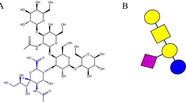
Figure 2.5 Structure of GM1. 55
Figure 2.6 Crystal structure of GM1 bound to cholera toxin and a
schematic representation of a polymer inhibitor.
57
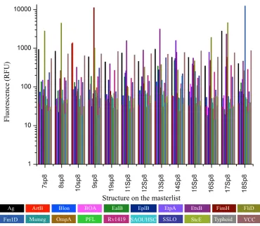
Figure 2.7 Relative binding profiles of 20 bacterial lectins from the CFG. 59
Figure 2.8 Example of LDA 60
Figure 2.9 Model fitting example 61
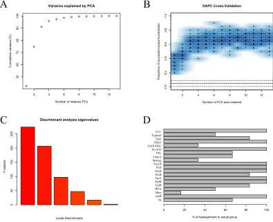
Figure 2.10 LDA output 62
Figure 2.11 Random Forest model evaluation 64
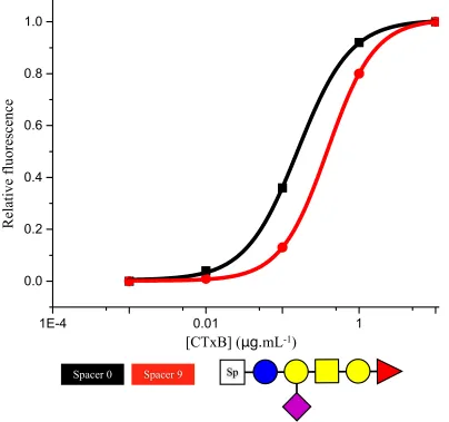
Figure 2.12 Spacer effect on binding curves 66
v
Chapter 3 Optimisation of glycan-surface conjugation
Figure 3.1 Schematic highlighting the importance of carbohydrate
presentation distance. 84
Figure 3.2 Structure of Cyanuric chloride. 85
Figure 3.3 Drop Shape Analysis of GM1 based functionalisation of
polypropylene slides. 87
Figure 3.4 Affect of blocking agents on non-specific binding. 88
Figure 3.5 Schematic of surface functionalisation technique. 91
Figure 3.6 Drop Shape Analysis after dodecan-1-ol based
functionalisation.
92
Figure 3.7 Variable angle XPS. 93
Figure 3.8 Serial dilution of PNA on a galactose surface. 94
Figure 3.9 Differential PNA binding on different glycan surfaces. 94
Figure 3.10 Comparison of lectin and bacterial multivalent binding to a surface.
95
Figure 3.11 Hydrazide surface functionalisation. 97
Figure 3.12 IC50 of Con A on a galactose and mannose surface. 98
Figure 3.13 IC50 of PNA on a galactose and mannose surface. 99
Figure 3.14 BSA blocking of a hydrazide surface. 100
Figure 3.15 Modified DSA methodology. 107
Chapter 4 Discrimination between lectins with similar specificities by
ratiometric profiling of binding to glycans
Figure 4.1 Comparison of oligosaccharide and mono-/d-saccharide
binding.
112
Figure 4.2 Schematic of cholera toxin action. 113
Figure 4.3 Schematic of ricin action. 115
Figure 4.4 Binding of five fluorescently labelled lectins to a galactose surface.
118
Figure 4.5 Binding profiles of five fluorescently labelled lectins. 119
Figure 4.6 LDA model of lectin binding. 120
Figure 4.7 Binding profile for mixed ricin and cholera samples. 122
Scheme 4.1 Synthetic route to carbohydrate functionalised AuNPs. 123
Figure 4.8 Aggregation of glycosylated gold nanoparticles. 123
Figure 4.9 Binding profiles of lectins with GlycoAuNPs and the
subsequent LDA model.
124
Figure 4.10 Mixed GlycoAuNPs schematic. 126
Figure 4.11 Concentration independent LDA model. 127
Figure 4.12 Concentration dependent LDA model. 128
Chapter 5 Carbohydrate functionalised surfaces for the rapid identification of bacterial species to improve point-of-care diagnostics
Figure 5.1 Antibiotic resistance time line. 138
Figure 5.2 Predicted number of deaths from antibiotic resistance by
2050. 139
Figure 5.3 Bacterial biofilm formation. 142
Figure 5.4 Global emergence of drug resistant tuberculosis. 145
Scheme 5.1 Fluorescent labelling of bacterial cells. 149
Figure 5.5 Confirmation of biotinylation. 150
Figure 5.6 Fluorescent labelling images. 151
Figure 5.7 Fluorescence measurements after labelling. 152
Figure 5.8 Bacterial binding profiles. 154
Figure 5.9 Specificity of E. coli binding. 155
Figure 5.10 Binding inhibition curve. 156
Figure 5.11 Bacterial LDA model. 157
Figure 5.12 Bacterial random forest analysis. 158
Figure 5.13 Blind sample identification using LDA model. 159
Chapter 6 Differential glycan binding for the detection of
Plasmodium falciparum infected erythrocytes and identification of drug resistance
Figure 6.1 P. falciparum erythrocyte life cycle. 171
Figure 6.2 Schematic showing cytoadhesion of infected RBCs. 172
Figure 6.3 Giemsa stained RBCs. 179
Figure 6.4 Schematic of RBC labelling methodologies. 180
Figure 6.5 RBC autofluorescence. 181
Figure 6.6 Colourimetric analysis. 182
Figure 6.7 Serial dilution of infected and uninfected RBCs. 184
Figure 6.8 Fluorescence microscopy images of labelled RBCs. 184
Figure 6.9 Binding profiles of different life stages of infected RBCs. 186
Figure 6.10 Life stages LDA model. 189
Figure 6.11 Life stages LDA model with blind samples. 190
Figure 6.12 Ring stage drug resistance profiles. 193
Figure 6.13 Trophozoite stage drug resistance profiles. 194
Figure 6.14 Life stage separated LDA model of resistance strains. 198
vii
List of Tables
Chapter 2 The power and challenges of glycomics databases and the
use of statistical tools in the extraction of crucial binding information; opportunities for chemists and biologists
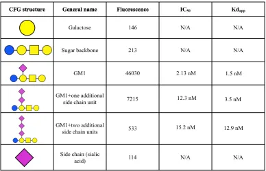
Table 2.1 Binding of cholera toxin to a variety of GM1 based structures. 56
Table 2.2 CFG data used for analysis. 70
Chapter 3 Optimisation of glycan-surface conjugation
Table 3.1 Methods of surface functionalisation for carbohydrate
microarray production.
78
Chapter 6 Differential glycan binding for the detection of
Plasmodium falciparum infected erythrocytes and identification of drug resistance
Abbreviations
3D7 Plasmodium falciparum 3D7
Ara Arabinose
Biotin-NHS (+)- Biotin N-hydroxysuccinimide ester
BSA Bovine Serum Albumin
C. difficile Clostridium difficile
cAMP Cyclic adenosine monophosphate
CDC Centre for Disease Control
Cel Cellobiose
CFG Consortium for Functional Glycomics
CFTR Cystic fibrosis transmembrane conductance regulator
Con A Concanavalin A
CTx Cholera toxin
CTxB Cholera toxin B subunit
DBA Dolichos biflorus Agglutinin
ix
DMSO Dimethylsulfoxide
DNA Deoxyribonucleic acid
DSA Drop shape analysis
E. coli Escherichia coli
FDA Food and Drug Administration
FITC Fluorescein isothiocyanate
Gal Galactose
GalNAc N-Acetyl-D-Galactosamine
GI Gastrointestinal
Glc Glucose
GlcNAc N-Acetyl-D-Glucosamine
Gly Glyceraldehyde
GlycoAuNPs Sugar functionalised gold nanoparticles
GM1 Monosialyltetrahexosylganglioside
GPI Glycophosphatidylinositol
HEPES 2-[4-(2-hydroxyethyl)piperizan-1-yl]ethanesulfonic acid
HIV Human immunodeficiency virus
HSB Hue, Saturation, Brightness
IC50 Half maximal binding concentration
Kd Dissociation constant
Kdapp Apparent dissociation constant
L-PHA Plant lectin leukoagglutinin
L. casei Lactobacillus casei
Lac Lactose
M. marinum Mycobacterium marinum
M. smegmatis Mycobacterium smegmatis
M. tuberculosis Mycobacterium tuberculosis
Man Mannose
ManNAc N-Acetyl-D-Mannosamine
MHC1 Major histocompatibility complex 1
xi
N. americanus Necator americanus
N. brasiliensis Nippostrongylus brasiliensis
NHS N-hydroxysuccinimide
P. aeruiginosa Pseudemonas aeruginosa
P. falciparum Plasmodium falciparum
P. knowlsei Plasmodium knowlsei
P. malariae Plasmodium malariae
P. ovale Plasmodium ovale
P. putida Pseudemonas putida
P. vivax Plasmodium vivax
PBS Phosphate buffered saline
PCR Polymerase chain reaction
PCs Principle components
PfEMP1 Plasmodium falciparum erythrocyte membrane protein 1
Plasmodium spp. Plasmodium species
pRBC Trophozoite infected red blood cells
RBCs Red blood cells
RCA Ricin
RCA120 Non-toxic derivative of the ricin B subunit
RNA Ribonucleic acid
S. aureus Staphylococcus aureus
S. pneumoniae Streptococcus pneumoniae
S. suis Streptococcus suis
SBA Soybean Agglutinin
SiNW Silicon nanowires
Sp Spacer
Strain 4 Plasmodium falciparum TH004-004
Strain 23 Plasmodium falciparum TH004-023
Strain 36 Plasmodium falciparum TH004-036
xiii
UV Ultraviolet
V. cholerae Vibrio cholerae
VBNC Viable but not culturable
Acknowledgments
First of all I would like to thank Matt, for his endless patience, enthusiasm and
optimism. For always putting a positive spin on even the worst results and for
providing the positive outlook necessary to help me complete my thesis. I would also
like to thank Stephen Edmondson for his assistance with the variable angle XPS, Liz
Fullam and Alasdair Hubbard for their assistance with the microbiology work and
Eva Caamaño-Gutiérrez, Matthew Phanchana and Ahmed Saif for their help with the
Malaria work.
I would also like to thank all current and past members of the Gibson group. It has
been a pleasure to work with all of you. Special thanks goes to Daniel Phillips,
Caroline Biggs, Sarah-Jane Richards and Tom Congdon for all their support during
my Masters project and throughout my PhD and for generally putting up with me in
the office! Thanks also to Richard Lowery for all your support and friendship, for
providing general entertainment in the office and for science balloons.
Without the funding and support from the BBSRC and the Systems Biology DTC this
work would not have been possible. Being part of the Systems Biology DTC provided
me with the skills necessary to help make this project my own and allowed me to
make some amazing friends. Specifically Eva and Nikita, without whom my Masters
year would not have been the enjoyable experience it was. Thank you both for your
endless support, friendship and for all the days we just span on the chairs in the office.
I would also like to thank Nick Barker for his inspirational dedication to Outreach and
xv
allowing me to make slime!). Thanks also to all those at the Brilliant Club for giving
me the opportunity to teach elements of my thesis to students around the West
Midlands. A special mention to the student who hand drew their presentation images,
as they did not have access to PowerPoint and the person who rapped their
presentation to music- you are both inspirational. Having students be enthusiastic
about your research really helps after a bad day!
To all those at St John Ambulance, especially those of you that have waded through
fields, sat in first aid posts, frozen at the pitch side and crewed ambulances with me, I
thank you for; all the experiences you have given me, providing an endless source of
procrastination (when needed), being a source of endless entertainment and for
generally feigning interest in my science. You were literally ‘the difference’ when it
came keeping me sane at times and I could not have done this without your support!
I would like to thank my friends and family for always being there to support me not
just through my PhD but throughout my life. Thanks to Fiona for always being there
and for providing emergency supplies of ‘happy hippos’ when times got tough, you
truly are an inspiration to me and I will forever be thankful you chose me to live in
your house all those years ago! I would also like to thank Aoife for her many
motivational messages, Akshay for his general enthusiasm about all of my work and
especially Chris for listening to my endless rants about science, work and life in
general and for constantly reminding me why I do the things that I do.
Finally, heartfelt thanks to my mum, dad and my sisters Lysandra and Lesley for
providing constant love and support without which this thesis would never have
Declaration
The work presented in this thesis is entirely original and my own work, except where acknowledged below. I confirm that this thesis has not been submitted for another degree at another University.
The inhibitor design element of Chapter 2 was published as:
Jones, M.W., Otten, L., Richards, S-J., Lowery, R., Phillips, D.J., Haddleton, D.M.,
Gibson, M.I. (2014) Glycopolymers with secondary binding motifs mimic glycan
branching and display bacterial lectin selectivity in addition to affinity. Chemical
science, 5, 1611-1616.
Chapter 3:
All variable angle XPS was carried out by Stephen Edmondson, School of Materials Science at the University of Manchester.
Chapter 4 contains elements published as:
Otten, L., Gibson, M.I. (2015) Discrimination between lectins with similar
specificities by ratiometric profiling of binding to glycosylated surfaces; a chemical
‘tongue’ approach. RSC Advances, 5, 53911-53914.
Richards, S-J., Otten, L., Gibson, M.I. (2016) Glycosylated gold nanoparticle
libraries for label-free multiplexed lectin biosensing. Journal of Materials Chemistry
xvii
And is in preparation for:
Otten, L., Vlachou, D., Richards, S-J., Gibson, M.I. Glycan heterogeneity on gold
nanoparticles increases lectin discrimination capacity in label-free multiplexed
bioassays.
Chapter 5 has been published as:
Otten, L., Fullam, E., Gibson, M.I. (2016) Discrimination between bacterial species
by ratiometric analysis of their carbohydrate binding profile. Molecular Biosystems,
DOI: 10.1039/C5MB00720H.
Chapter 6 is in preparation for:
Caamaño-Gutiérrez, E., Otten, L., Phanchana, M., Saif, A., Biagini, G., Gibson, M.I.
The ‘sweet tooth’ of Plasmodium falciparum: a key for Artemisinin resistance
classification.
Within this chapter all parasite work was completed at the Liverpool School of
Tropical medicine with the assistance of Eva Caamaño-Gutiérrez, Matthew
Phanchana and Ahmed Saif from the Department of Parasitology. I completed all data
Abstract
The rapid detection of pathogenic organisms to ensure appropriate
administration of treatment remains a global healthcare challenge. This is becoming
increasingly difficult, as identification of the organism alone is no longer enough,
with the rise of drug resistance amongst many pathogens it is becoming increasingly
important that both the pathogen and drug resistance are identified.
Currently, rapid identification can be achieved through a variety of techniques.
However, many of these techniques are expensive, require extensive sample
preparation, or highly trained personnel to run with results often not rapidly available.
This leaves health care professionals to make point-of-care treatment decisions based
on symptoms without any indication of drug resistance. The use of carbohydrate
microarrays for pathogen detection has been identified as both a method for detection
but also as a basis for identifying new drug targets. This exploits the initial
protein-carbohydrate interaction that many pathogens utilise in the initial stages of infection.
However, the use of microarrays is also challenging, as highly sensitive identification
of pathogens often requires expensive or synthetically challenging oligosaccharides or
coupling with a highly sensitive detection method thus limiting its point of care
application.
Herein we describe the coupling of a facile surface chemistry for glycan
addition with a powerful statistical algorithm to improve the sensitivity of a cheap
monosaccharide functionalised surface without using expensive detection
methodologies. This technique was then applied to the detection and identification of
toxic lectins, bacterial samples and finally the life-stage specific detection of
Plasmodium falciparum (one of the parasites responsible for human malaria). In this
1
Chapter 1
1.2 Introduction
Carbohydrates in the glycocalyx, which surrounds almost all cells, mediate a
multitude of biological processes through protein-carbohydrate interactions. The
glycocalyx is also important for many diseases and can reflect disease states, such as
cancer, through aberrant glycosylation patterns. Many pathogens also utilise proteins
to interact with carbohydrates as an important step in pathogenicity.
Protein-carbohydrate interactions are exploited by Vibrio cholerae in order to mediate
toxin binding thus causing cholera infection, which results in the death of around
100,000 people a year.1 Malaria is estimated to kill one child in Africa every 8
minutes. It is caused by infection with parasites of the Plasmodium spp. and initial
binding and internalisation of Plasmodium falciparum is mediated by a lectin-like
protein. Protein-carbohydrate interactions are also crucial for infection with Influenza
A and for the direct binding of pathogenic strains of Escherichia coli to give but a few
examples.2
Many of the current detection methodologies for these pathogens are complex or too
expensive for robust point-of-care diagnostics and as such, increasing the resolution
of current cheap and facile methods without the use of expensive detection systems is
1.2.1 Carbohydrates
Carbohydrates were once described as a molecule “in search of a function”3 and
thought to be less information rich than proteins.4 The importance of carbohydrate
signalling has also been underestimated previously.5 However, in the last 20 years
their role in complex biological processes; such as cell-cell communication,
immunity, cell differentiation and fertilisation, has come to light and it is now known
that carbohydrates play a crucial role in many processes.4 This has contributed to the
delay in generating techniques for their study when compared to DNA and protein
techniques and many challenges remain in the study of carbohydrates.
Unlike proteins and DNA, carbohydrate synthesis is not trivial, it is possible to
remove carbohydrate chains from cells but most procedures require multiple
chromatographic or purification steps. In nature, sugar chain synthesis requires
multiple enzymes and highly complex synthesis pathways and whilst there has been
some success in the production of synthetic oligosaccharides the process is often
laborious involving many protection and deprotection steps.6-10 Generation of the
carbohydrate structures found in bacteria is further complicated by the presence of
different glycosylation enzymes and their ability to utilise a larger library of
monosaccharides than mammalian cells resulting in the production of even more
complex structures.5
1.2.2 Glycan complexity
Carbohydrates are now regarded as being unsurpassed in their structural diversity,
3
mammalian cells but that is enough to potentially generate ten million different
tetrasaccharides.3
The monosaccharides are themselves able to exist as different enantiomers known as
D- and L- depending on the spatial configuration around the chiral carbon (Figure
1.1A) and as different anomers depending on the configuration on the anomeric
carbon (Figure 1.1B). The complexity of glycans goes beyond just the number of
available monosaccharide building blocks, with chain lengths ranging from single
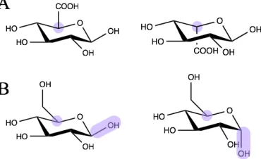
[image:23.595.205.390.327.440.2]monosaccharide units through to polysaccharides containing 500,000 sugar units.
Figure 1.1 D- and L- optical isomers (left and right respectively) are determined by
the arrangement around the chiral carbon (A). Monosaccharides can also exist as
different anomers such as α and β glucose (left and right respectively). This is defined
as the direction of the hydroxyl group on the anomeric carbon relative to the
configuration around the chiral carbon (B).
Glycosidic linkages between monomers allow the formation of large glycan chains
and these linkages can also show differences in both stereo- and regiochemistry
adding another layer of complexity to glycan chains. Differences in stereochemistry
shown by maltose and cellobiose, both of which are disaccharides of D-glucose
(Figure 1.2).
Figure 1.2 The glycosidic linkage between monosaccharides can show differences in
stereochemistry with either an α linkage, such as that maltose, or a β linkage as in
cellobiose (left and right respectively).
Differences in regiochemistry can also occur as the glycosidic bond can form between
any of the hydroxyl groups as demonstrated by maltose and trehalose (Figure 1.3).
These chains can also be highly branched utilising both α and β linkages. Often 3D
structure of the carbohydrate, density, linker to the cell and topological arrangement
all have to be maintained for recognition to occur.
Figure 1.3 The glycosidic linkage can also show differences in regiochemistry as the
linkage can form between any of the hydroxyl groups in the monosaccharide
5
After assembly by glycosyltransferases the glycan complexity is further increased by
site-specific modifications of the monosaccharide units within a glycan chain. This
modification can include sulfation, acylation or epimerisation of glucuronic acid to
iduronic acid (Figure 1.4).
Figure 1.4 Epimerisation of glucuronic acid (left) to iduronic acid (right) is a
common mammalian monosaccharide modification.
1.2.3 Mammalian Glycocalyx
Carbohydrates are covalently linked to membrane proteins and lipids to form a dense
glycocalyx (Figure 1.5). Cell surface glycans are positioned optimally to mediate
cell-cell interactions and protect the cell-cell from the extracell-cellular environment.11, 12 They also
act as recognition elements and binding sites for a multitude of other components and
are crucial for preventing non-specific protein-protein interactions.13, 14
Figure 1.5 Electron microscopy image of a cross section of a rat myocardial capillary
showing the glycocalyx of the endothelial cells (reprinted with permission from
The diversity of the glycans present in the glycocalyx is determined by the
mammalian glycome. The glycome is comprised of ten key mammalian
monosaccharides (Figure 1.6), which are added to other molecules in a non-template
driven process mediated by the expression of several different glycosyltransferases
(several of which have tissue specific isoforms).16-18
Figure 1.6 The ten mammalian monosaccharides displayed using standard
consortium for functional glycomics (CFG) nomenclature.19
Whilst the theoretical glycospace (which is the number of all the possible
combinations of monosaccharides) is vast only a small subspace is actually used in
mammals20 and it is thought that there are only around 2000 different mammalian
glycans with many highly conserved core structures.19 Prokaryotes utilise many more
than ten monosaccharides and use a much larger subspace of their theoretical glycome
as their outer membrane has to protect the cell from many harsh and changing
environments.20
Galactose Glucose Mannose N-acetyl galactosamine
N-acetyl glucosamine Sialic acid
(N-acetyl neuraminic acid)
Glucuronic acid
Fucose
7
1.2.4 Glycosylation in disease susceptibility
As the glycocalyx is crucial in the interactions of a cell with its environment it plays a
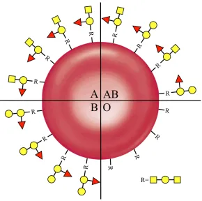
crucial role in disease susceptibility and pathogen interaction. For example, the ABO
blood system is determined by differences in antigenic oligosaccharides on the
surface of red blood cells (RBCs, Figure 1.7). Serological subgroups are further
defined by the number of oligosaccharides per RBC highlighting the importance of
both the specific antigen and the distribution of those antigens.21, 22 Whilst the
biological role of the system remains a mystery, anthropological studies on the
population and geographic distributions of blood groups indicate that they may be
related to group susceptibility to certain disease.21 It is known that blood group
determines susceptibility to or severity of diseases such as small pox, cholera and
malaria.23-25 The susceptibility of people with different blood groups to malaria is
thought to be due to the invasion of the pathogen into uninfected RBCs.25 It has been
hypothesised that the individual variation of glycans on the surface of human tissues
Figure 1.7 Serological blood groups are determined by oligosaccharides on the
surface of red blood cells. Oligosaccharides shown are depicted using the standard
CFG nomenclature.19
1.2.5 Altered glycosylation in disease states
Differences in glycosylation have been detected in diseased tissue and many different
cancer cells. It is thought that changes in the expression of glycosyltransferases and
glycosidases result in the changes in the glycans found on the surfaces of tumour
cells, it has also been suggested that these differences affect interactions between
lectins at the tumour surface which in turn determines a tumour’s metastatic
[image:28.595.156.446.74.362.2]9
Cancer cells show a number of differential glycosylation patterns but these changes
tend to be a specific set of permeations that result in malignant transformation and
tumour progression. These changes have been selected for as a form of
‘microevolution’. Most notable are the differences in glycosylation of mucins.27
Mucins are large glycoproteins found on the surfaces of endothelial cells. They are
also secreted by healthy cells into the lumens of organs in order to limit adhesion of
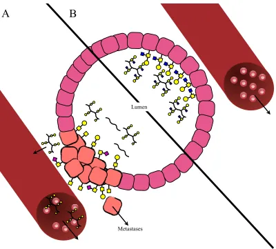
pathogens (Figure 1.7B).21 However, in tumours they have been found to be
over-expressed28 and a number of changes occur; firstly incomplete glycosylation of the
mucins occurs resulting in truncated glycan patterns29 or increased sialic acid
moieties.30 These truncated mucins can generate an immune response. The secreted
mucins are also truncated and in some instances there is a total loss of glycosylation
and the protein backbones are just secreted (Figure 1.8A).
The altered topology of the tumour itself means that the mucins are added to all
surfaces of the cells whereas normally they are only expressed on the surface facing
the lumen. It also results in mucins being secreted into places other than the lumen
and can be secreted into the blood stream (Figure 1.8A). As in healthy cells, the
mucins can still act in an anti-adhesion capacity but in cancerous tissue they can
prevent the adhesion of cancer cells to neighbouring cancer cells resulting in cell
Figure 1.8 Glycosylation and secretion of mucins is altered in tumours (A) compared
to normal tissue (B).
Other cancer-associated glycosylation changes include the increased β1-6 branching
of N-linked glycans,31 increased addition of sialic acid to surface glycans and
polysialylation,32 increased presentation of ligands specific to selectins33 and
increased presentation of sialylated blood-group associated glycans on cell surfaces
(e.g. Lewisx glycans).34 Incomplete synthesis and altered expression of blood-group
associated glycans coupled with the loss of normal AB blood group glycan expression
is associated with poorer clinical outcome.35 Metastases
A
B
11
Other diseases are also characterised by glycosylation differences. Mutations in
certain glycosyltransferases will result in differences in glycosylation associated with
many congenital diseases including galactosemia, muscle-eye brain disease and
congenital muscular dystrophy.18 Glycoforms (isoforms of a protein that differ only in
glycosylation) of immunoglobulin-γ antibodies have been associated with rheumatoid
arthritis.36 Overexpression of mucins has also been associated with many chronic lung
conditions including asthma, chronic-obstructive pulmonary disease, bronchitis and
cystic fibrosis.37
1.2.6 Protein-Carbohydrate Interactions
Protein-carbohydrate interactions mediate many crucial biological processes including
(but not limited to); cell-cell communication, fertilisation, pathogen recognition and
response, protein folding and both passive and innate immunity.6, 26 All known
organisms are covered by either free or bound glycan structures and proteins known
as lectins bind to these carbohydrates during signal transduction.21 These interactions
are typically weak with a dissociation constant (Kd) typically between 10-3 and 10-6 M.
This is overcome in nature by presentation of multiple copies of the carbohydrate
structures on the cells. The increase in affinity in binding is greater than the sum of
the individual binding events that take place and this is known as the cluster glycoside
effect.38
This multivalent interaction means that many lectins have multiple binding subunits,
which can interact with the surface in one of two manners; either the bind-and-slide39
method or face-to-face binding40 (Figure 1.9). In the bind-and-slide model, a
lectin will slide along until it binds to another region and in the face-to-face model the
subunits of the multivalent lectin face in opposite directions to each other and thus
can engage in crosslinking.41 Graded affinity binding can also increase the binding of
a lectin to a surface where one subunit interacts with the native version of the ligand
and other subunits interact more weakly with truncated versions of the ligand in order
to increase the avidity of the binding event.42
Figure 1.9 Mechanisms for multivalent binding can be achieved by either binding of
a multivalent protein which then dissociates and binds to the next ligand effectively
sliding along the chain of saccharides with each binding event essentially increasing
the residence time of the protein within the area of the ligand (A) or through face to
face binding of multiple dimers binding to multiple ligands on a surface (B).
Bind-and-slide
Face-to-face
13
1.2.7 Pathogen exploitation
The carbohydrates on a cell surface determine everything from blood type to disease
susceptibility and they are often exploited by pathogens (Figure 1.10).21 This
exploitation falls in to three broad categories; mimicry of the host glycome to avoid
detection and eradication by the host immune system, adhesion of the pathogen as a
crucial step in infection or for internalisation, and production of lectins similar to
those produced by the host’s immune system in order to produce an
immune-compromised niche for the pathogen.
Figure 1.10 Protein-carbohydrate interactions are crucial for many biological
processes including pathogen-associated adhesion.
Bacterial adhesion
Toxin adhesion
Cell-cell adhesion
Viral adhesion
Fungal adhesion Phage adhesion
1.2.7.1 Host glycome mimicry
Pathogenic bacteria often find themselves in a “dual glycan speedway” where they
evolve to avoid phage recognition but also detection by the host.43, 44 One mechanism
by which they avoid host recognition is through mimicry of the host glycome and
many species have evolved a variety of mechanisms for doing this. In general, many
human pathogens have a glycome that is very similar to the mammalian glycome.20
This avoids triggering the immune system by introducing antigenic glycans such as
the non-mammalian monosaccharide D-galactofuranose, which is found in
Mycobacterium tuberculosis in arabinogalactan chains and has been shown to be
highly antigenic.45
Many mammalian glycans are either fucosylated or contain a sialic acid cap. This cap
forms a crucial part of protecting host cells from the immune system. They mediate
the binding of Factor H (which protects the host cells from attack by the complement
immune system)46 and also engage siglecs, which are sialic acid binding
immunoglobulin-like lectins. Siglecs are known to have an inhibitory affect on
immune cells thus protecting the cell they are bound to from the immune response.47
As sialic acid mediates both of these processes it would be extremely beneficial for
pathogenic organisms to adopt the sialic acid cap into their glycans to offer protection
from the host immune system. Indeed, strains of Haemophilus influenza that have
sialic acid incorporated in their glycome were shown to be more resistant to
complement-mediated killing by human serum. Mutation of the gene controlling
sialylation resulted in loss of sialic acid residues and a marked reduction in the
tolerance to human serum.48 A similar affect has been shown in pathogenic Neisseria
15
killing by human serum.49 Whilst sialic acid is used in both of these examples not all
bacteria have evolved to use it but many of those that have not will utilise bacterial
mimics of sialic acid.50-52
L-fucose is another important terminal monosaccharide in mammalian glycans but
this is only found in low abundance amongst bacterial species.20 The exception to this
is one class of bacterial species, which contain L-fucose in high abundance; and
includes the pathogenic bacteria Helicobacter pylori, which contains large
proportions of fucosylated Lewis A glycans in order to mimic the host glycome and
avoid an immune response.53
Not all bacterial classes mimic the mammalian glycome, for example actinobacteria
contain a large number of actinobacter specific monosaccharide residues.20 This class
contains a number of specialised soil bacteria crucial for nitrogen fixation or other
elements of the soil cycle but also includes a number of human pathogens from the
class of Mycobacterium including M. tuberculosis.54 As already mentioned, this
species utilises D-galactofuranose to form antigenic arabinogalactan. M. tuberculosis
also contains very few sialic acid mimics and thus its glycome is a poor mimic of the
host glycome but, in this instance, mimicry is less important for pathogen survival as
M. tuberculosis is an intracellular pathogen.20
Many pathogenic bacterial species mimic the host glycome, at least in part, to avoid
detection by the immune system or to aid in the binding of immunosuppressant lectins
often intracellular based pathogens and thus do not experience the same selection
pressures to evolve a glycome more similar to that of the host.
1.2.7.2 Toxin adhesion
As all cells are glycosylated, many pathogens exploit this glycosylation to mediate
binding and internalisation of toxins. Cholera is caused by cell internalisation of a
toxin produced by the bacteria Vibrio cholerae.55, 56 The toxin is an AB5 toxin, and is
made up of one subunit that causes cell damage by constitutively activating a protein
resulting in the excretion of ions from the cell into the lumen of the small intestine
(Figure 1.11A) and five lectin subunits (Figure 1.11B) that bind the
monosialyltetrahexosylganglioside (GM1) found on endothelial cells in the small
intestine.57
Figure 1.11 Crystal structures of cholera toxin which contains one enzymatic A
subunit shown in grey (A) and five glycan binding B subunits (B).58
17
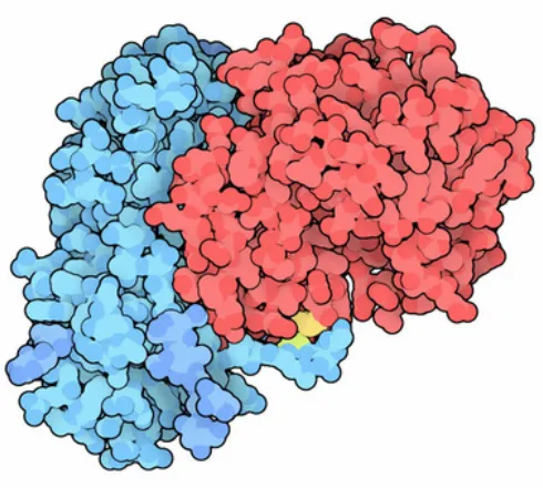
Ricin is a highly toxic lectin extracted from the seeds of the Ricinus communis plant.
This toxin is an AB toxin and thus contains one glycan binding targeting domain (the
B subunit) and an enzymatic A subunit. The targeting domain mediates the binding to
galactose-rich glycans on the cell surface and upon binding the toxin is internalised by
the cell. Once inside the cell the A subunit and the B subunit are cleaved at the
disulphide bond (shown in yellow Figure 1.12) and the A subunit cleaves an adenine
residue from the 28S ribosome effectively halting protein synthesis and triggering cell
death.59 Whilst somewhere between 106 and 108 molecules of ricin will bind to the
cell surface, few will make it into the cytosol but entry of just a single A subunit of
[image:37.595.176.421.359.579.2]ricin is sufficient to deactivate 1500 ribosome molecules per second.60
Figure 1.12 Crystal structure of Ricin, which is comprised of one A subunit (red) and
a carbohydrate binding B subunit (blue). A disulphide bond connects the A subunit
and the B subunit (yellow).61
Lectins are also found in many vegetables and consumption of improperly cooked
that are not degraded during digestion can bind to the endothelial cells in the
intestines and are often toxic.63 Whilst in humans the affects tend to be nausea,
vomiting and diarrhoea, long term consumption (in animal models) has been
associated with endothelial cell necrosis and increased cell turn over.64 Upon
incubation of healthy cultured endothelial cells with plant based lectins, mitogenesis65
and inhibition of exocytosis is observed.66 Many of the endothelial cells in the GI tract
experience mechanical stress resulting in damage to the plasma membrane.67 Healthy
cells can rapidly repair this damage.68 However, lectin binding to these damaged cells
inhibits this healing mechanism and results in cell necrosis and it has been proposed
that this is responsible for plant-based food poisoning.69
1.2.7.3 Virus attachment
Many viruses exploit protein-carbohydrate interactions for the initial adhesion phase
but by far the most studied example is that of hemagglutinin found in influenza A and
B. This lectin is co-localised as trimmers onto the viral surface and mediates binding
to the underlying surfaces through sialic acid terminated glycans. After binding, the
viral envelope fuses to the cell membrane, the virus is internalised and replication
occurs.
Viral specificity is determined by the linkage of the sialic acid to the terminal glycan
and this differs between species; avian species have α(2-3) linked residues (Figure
1.13A), humans have predominantly α(2-6) linkages (Figure 1.13B) and porcine
species have both α(2-3) and α(2-6) (Figure 1.13C). As such avian flu binds strongest
to α(2-3) linked sialic acid residues and human strains of influenza A binds
19
virus to infect a human it must first infect a porcine species at the same time as either
a porcine or human strain in order for the avian species to recombine with either the
human or porcine viral DNA that allows binding to α(2-6) linked sialic acids.
Figure 1.13 Avian tissue glycans are terminated by α(2-3) linked sialic acids (A),
human tissue glycans are predominantly terminated by α(2-6) linked sialic acids (B)
and porcine tissue glycans are terminated by a mixture of α(2-3) and α(2-6) linked
sialic acids (C).
Interestingly, the carbohydrate binding properties of viruses are not limited to
mammalian viruses but are also found amongst bacteriophage (which are viruses that
target bacteria). The bacteriophage PL-1, which infects Lactobacillus casei was found
to interact specifically with L-rhamonosyl residues in the lipopolysaccharide chains
which cover the surface of this bacteria. Addition of a lectin capable of binding L
-rhamanose resulted in a dose-dependent inhibition in phage absorption by this
bacterial species.70
1.2.7.4 Bacterial adhesion and biofilm formation
Lectins play a crucial role in bacterial adhesion and colonisation both by pathogenic
bacteria to host cells and subsequent biofilm formation, but also in symbiotic
relationships where bacteria bind to plants and confer nitrogen-fixing properties.
During bacterial infection, a single bacterial cell will bind to the underlying host cell
surface. The initial adhesion phase is often a protein-carbohydrate interaction either
through bacterial adhesins binding to host glycans or vice versa.
Figure 1.14 Schematic depicting bacterial biofilm formation. The example shown is
the bacterial adhesin FimH interacting with a mannose rich tetrasaccharide.
For example the adhesin FimH is a crucial virulence factor found in uropathogenic
Escherichia coli strains.71, 72 FimH is found on the end of fimbrae and mediates the
binding to mannose rich glycans on the cell surface (Figure 1.14). A combination of
the adhesins present on the bacterial surface will define the range of susceptible
tissues, which will differ between species and strains within the same species. For
example, E. coli strains containing type 1 pili are responsible for initial colonisation
and infection of the bladder whereas those that possess P-pili result in infection of the
kidney through binding to glycans.2 Loss or inhibition of these adhesins can prevent
21
In the opposite case, binding of the symbiotic Rhizobium spp. to the root hairs of
plants is mediated by polysaccharides on the bacterial cell (either the
exopolysaccharide, the lipopolysaccharide or the capsular polysaccharide depending
on the species). For example, on the roots of the Dutch clover plant (Trifolium repens)
the protein trifoliin is expressed.73 Trifoliin is a lectin that binds the
exopolysaccharide of Rhizobium trifolii.74 Colonisation of the plant’s roots by the
bacteria is the formation of a symbiotic relationship that gives the plant
nitrogen-fixing properties.
1.2.7.5 Parasite exploitation
Many parasites are known to exploit protein-carbohydrate interactions during
infection. The production of parasite specific lectins are thought to play a role in
down-regulating the immune response upon parasite infection to create an
immune-compromised niche for the parasite to live in. A lectin-like protein is also thought to
play a key role in Plasmodium falciparum (one of the species that results in human
malaria) infiltration of red blood cells.
Generation of an immune-compromised niche involve interactions with host c-type
lectins. C-type lectins are calcium dependent lectins that play a crucial role in the
mammalian immune system; encompassing molecules such as collectins (crucial for
innate bacterial and viral recognition),75 selectins (crucial for mediating adhesion of
leukocytes at the site of inflammation)33 and Natural Killer cell receptors (which bind
lysis and provide triggers for cell death upon detection of altered MHC 1 molecules)76
to name but a few. Many parasites are known to produce c-type lectins upon infection
or have had putative c-type lectin domains identified in their genome. Necator
americanus77 and Nippostrongylus brasiliensis78 are both gastrointestinal worms with
N. americanus a human pathogen and N. brasiliensis a rodent-specific parasite. Both
of these parasites have been shown to produce c-type lectins, which show binding
properties similar to mammalian selectins. It is thought that these play a role in
suppressing the immune system during parasite infection.78 The parasitic nematode
Toxocara canis also secretes a glycoprotein, which has been shown to bind to
mammalian cell surface glycans in a Ca2+ dependent manner and is thought to play a
role in immune suppression.79, 80
Lectins have also been shown to have a more direct role in parasite infection. During
infection with Plasmodium falciparum it has been shown that a lectin-like protein is
involved in binding of the parasite to the surface of the red blood cells prior to
internalisation of the parasite. This process was prevented upon the addition of high
concentrations of N-Acetyl-D-Glucosamine to the media.81 Many of the
parasite-derived proteins that are expressed on the surface of an infected red blood cell (during
a late stage in the infection process) have also been shown to contain putative
carbohydrate binding domains.82, 83
1.2.7.6 Exploitation by Fungi
Lectins play an important role in the adhesion of pathogenic fungi and has been
shown to occur in many species including Chryosporium keratinophilum,84
23
Aspergillus fumigatus, which is the most prevalent airborne fungal pathogen of the
developed world and causes life-threatening infections in immune-compromised
people.87 Infection involves the inhalation of conidia followed by adherence to the
lung surface and then germination. This adhesion is known to be sialic acid dependent
but many other lectins are also present on the surface of the fungi and play an
important role in adhesion including a fucose binding lectin.88
1.2.8 Disease prevention
The prevalence of protein-carbohydrate interactions in pathogen mechanisms makes
them an important target for disease prevention in the form of carbohydrate vaccine
targets, new bacterial species-specific antibiotic targets and anti-adhesion therapy.
These will now be addressed.
1.2.8.1 Carbohydrate vaccine targets
Despite many pathogens mimicking the host glycome, many still have pathogen
specific oligosaccharide presentations, which could provide targets suitable for
carbohydrate-based vaccines. For example, the antigenic response to arabinogalatan
from M. tuberculosis is a potential vaccine candidate as the component
galactofuranose is not present in mammalian cells; this minimising the chance of
generating an autoimmune response.20 Monoclonal antibodies have been generated
that are directed at the cluster presented mannose-rich oligosaccharides present on
human immunodeficiency virus (HIV) glycoprotein 120 and new broadly neutralizing
carbohydrate based antibodies have also been extracted from patients.89, 90 A
carbohydrate specific immune response to the Streptococcus pneumonia bacteria has
peptide and the tetrasaccharide repeating unit of the S. pneumoniae capsular
polysaccharide.91
Protein conjugated carbohydrate vaccines have also been developed for H. influenza
type b, where meningitis caused by this bacteria has been eradicated in areas with a
vaccine program.92 A species specific tetrasaccharide has also been identified for
Bacillus anthracis93 and administration of this allowed the generation of antibodies
specific to this bacteria although more work needs to be done to develop this into a
usable vaccine against anthrax.94 Proteins conjugated to the P. falciparum specific
glycophosphatidylinositol (GPI) hexasaccharide have also been developed as a
potential conjugate vaccine. This vaccine conferred reduced mortality to malaria in
mice without any cross-reactivity with human GPI although more work to develop
this is still on going.95
Carbohydrate vaccines are also being pursued as an anti-cancer treatment by utilising
antibodies produced by the aberrant glycosylation of mucins on tumour cells. This
involved the coupling of the sialylated Tn antigen (where the Tn Antigen is just N
-Acetyl-D-Galactosamine coupled to a serine or a threonine residue) that is produced
on aberrant mucins. This vaccine was able to generate a humoral and a cellular
immune response to the cancer cells.96
1.2.8.2 Pathogen specific glycosyltransferase targets
As glycosylation is a process involving the expression of many glycosyltransferases
and the existence of non-mammalian monosaccharides there will be pathogen specific
25
susceptible to the host’s immune system by removing its ability to mimic host glycan
presentation on its surface or by reducing its pathogenicity through other mechanisms.
Neuraminidases play a crucial role in influenza infection. Influenza particles adhere to
the cell surface through adhesion to sialic acid residues. Upon creation of a new virus
particle, the viral neuraminidase clips the terminal sialic acid residue from the cellular
glycan thus allowing release of the viral particle from the cell surface where it can
then go on and infect other cells.97 This process is the target of neuraminidase
inhibitors such as oseltamivir and zanamivir although resistance to such drugs is
becoming more widespread.
V. cholerae can also produce a bacterial neuraminidase and this capability is found
amongst all strains that cause the most severe cholera infection.98 The role of this
neuraminidase is to clip sialic acid residues from glycans on the cell surface in order
to truncate the glycans to form GM1 (the natural binding ligand of the cholera
toxin).99, 100 This increases the cellular presentation of the cholera toxin binding ligand
thereby increasing the amount of toxin binding and causing increased disease
severity.101 Inhibition of this neuraminidase could reduce disease severity in severe
cholera infections.
As previously mentioned, the presence of sialic acid terminated glycans play a crucial
role in the protection of H. influenza and Neisseria spp. from the complement immune
system. As such, inhibition of the enzymes responsible for addition of the sialic acid
cap to the glycans on the surface of these bacteria will make them more susceptible to
1.2.8.3 Anti-adhesion therapy
For those pathogens that have an essential carbohydrate mediated adhesion phase
(Figure 1.15A), anti-adhesion therapy has potential for prevention of infection and
treatment of disease. Many examples of carbohydrate based anti-adhesion compounds
exist, but some examples include the use of glycopolymers for the inhibition of
cholera toxin binding102, 103 and for the specific inhibition of fimbriated (and thus
pathogenic) strains of E. coli.104105 This mechanism is also used in nature to protect
cells from bacterial infection, mucins which are found on the surface of epithelial
cells are also secreted thus trapping pathogens before they can invade the underlying
tissue (Figure 1.15B).21, 106
Figure 1.15 Schematic depicting anti-adhesion therapy. (A) Normally a bacteria will
bind to the underlying cell glycans resulting in infection (B) whereas a drug or a
mucin that mimcs these glycans can prevent binding and thus prevent infection.
A
B
Bacterial adhesion
Infection
Prevention of adhesion
27
A form of anti-adhesion therapy has also been proposed for the prevention of cancer
metastases. Many cancer cells contain regions of aberrant glycosylation, one common
modification is the presence of many selectin ligands. This would mask the cancer
cells from the immune system as selectins are used to suppress the immune system
and prevent it from attacking host cells. Several compounds have been designed to
mimic the glycan-binding domain of selectins in order to prevent binding of selectins
to tumour cells.107-109
1.2.8.4 Carbohydrate based drugs
Carbohydrates also form the basis of a number of other drug compounds including a
number of antibiotics such as kanamycin, vancomycin and teicoplannin.110 Heparin
and related compounds such as dermatan sulphate form the basis of many
anti-thrombotic agents.111, 112 Glycoproteins have been used as enzyme replacement
therapy in patients with Hurler and Hurler-Scheie forms of mucopolysaccharidosis
I.113
1.2.9 Pathogen detection
As differential carbohydrate binding and glycosylation are often present in disease
states or determine tissue specificity of pathogens they can be used as a detection
mechanism too. Detection of disease specific glycosylation can be used in the
detection of cancers or the presence of pathogens mimicking the host glycome and
detection of pathogen specific lectins (either as secreted toxins or surface localised)
1.2.9.1 Host detection of pathogen mimicry
Whilst many pathogens mimic the host glycome in the presentation of the glycans on
their cell surface, the host immune system contains mechanisms for identifying
mimicry. For example, sulfation is a site-specific modification that is common in
mammals but very rare amongst bacterial species. Sulfation tends to occur on
glycosaminoglycans and these sulfation events are recognised by immune lectins,
which identify cells containing these compounds as belonging to the host. The
sulfation of these glycans coupled with the epimerisation of glucuronic acid to
iduronic acid allow host immune lectins to distinguish between host proteins and
pathogen mimics.20
1.2.9.2 Detection of disease specific glycosylation
Disease specific changes in glycosylation can be detected through the use of labelled
lectins or carbohydrate specific antibodies and many applications of this has been
shown.
Lectins have been used for the staining of tissue in the detection of cancer for many
years and hundreds of examples exist in the literature, to take but one example, the
staining of tissue samples with the plant lectin leukoagglutinin (L-PHA) was able to
detect cancerous cells through binding to the aberrant β(1-6) linkages present in
oligosaccharides on their surfaces. Not only did L-PHA binding increase with
cancerous tissues when compared to normal, but this increase was found to correlate
29
Recently lectin microarrays have been used in the differentiation of gastric cancer
tissue sections from gastric ulcers,115 quantum dot conjugated lectins have been used
for the evaluation of the glycophenotype of breast cancer116 and a recombinant fungal
lectin has been used in the detection of aberrant cancer associated glycosylation in
tissue samples where the lectins were able to specifically bind cancer cells (reducing
the risk of false positives).117
Not only has lectin binding been used to determine the cancerous state of tissues but
this information has been used to inform treatment by identifying crucial cancer
antigens. Lectin coated nanoparticles have also been shown to enhance drug delivery
to the brain after intranasal administration of a drug with the aim of combatting
Alzheimer’s disease.118
Cancerous secretion of mucins into the bloodstream has been used as a marker for
cancer through detection with monoclonal antibodies and many glycan-specific
cancer-associated antibodies can be found in the blood of patients with cancer and
these can be used for detection. Whilst many of these antibodies have become
potential therapeutic targets their use may be limited by the high level of mucins
coated in their glycan targets, which are secreted into the blood by cancerous tissues
and thus would compete with tissue bound glycans for the drug.31
1.2.9.3 Detection of pathogen specific lectins
Many methods for detection of pathogen specific lectins have been explored in the
literature. Glycan functionalised magnetic particles have been used for the detection
magnetic particles for the detection of Streptococcus suis (a zoonotic pathogen),2, 120
mannose conjugated gold nanoparticles for the detection of FimH positive (and thus
pathogenic) strains of E. coli104 and label-free real-time detection of lectins down to
femtogram levels has also been achieved through the conjugation of unmodified
carbohydrates to silicon nanowires.121
Whilst those examples were all particle based, carbohydrate microarrays were first
used for mapping epitopes of HIV-related antibodies which has now been used to
inform vaccine design.122-125 They have also been used for the detection of food-based
bacterial pathogens for which standard culturing methods are challenging. This
methodology coupled a mono- and di-saccharide based microarray with surface
plasmon resonance for a real-time label-free output.126 Detection and typing of
bacteria in the blood has also been achieved127 along with; detection of
oligosaccharide binding of avian and human influenza strains128, identification of
antibodies that protect against severe malaria129 and epitope mapping of
tumour-associated antigens.130
However, many of the techniques that are currently available for pathogen detection
either have poor sensitivity as they utilise monosaccharides or those that are based on
monosaccharides that have good sensitivity are often coupled to expensive detection
methods such as surface plasmon resonance, mass spectrometry or quartz crystal
microbalance. Long-chain oligosaccharides are often used as sensors in order to
increase the sensitivity of detection systems but these are expensive or difficult to
synthesise. Many other techniques require either expensive or synthetically
31
chemistry. Whilst non-carbohydrate based-detection systems exist, many utilise
proteomic or antibody based detection methods that require preparation, storage,
expensive detection equipment or highly trained staff to implement and they are not