Direct Detection of
Shigella
in Stool Specimens by Use of a
Metagenomic Approach
Jie Liu,
aMathieu Almeida,
b,dFurqan Kabir,
cSadia Shakoor,
cShahida Qureshi,
cAnita Zaidi,
cShan Li,
dBoubou Tamboura,
eSamba O. Sow,
eInacio Mandomando,
fPedro L. Alonso,
fThandavarayan Ramamurthy,
gDipika Sur,
gKaren Kotloff,
dJames Nataro,
hMyron M. Levine,
dO. Colin Stine,
dEric Houpt
aaDivision of Infectious Diseases and International Health, University of Virginia, Charlottesville, Virginia, USA
bCenter for Bioinformatics and Computational Biology, University of Maryland, College Park, Maryland, USA
cAga Khan University, Karachi, Pakistan
dSchool of Medicine, University of Maryland, Baltimore, Maryland, USA
eCentre pour le Développement des Vaccins, Bamako, Mali
fCentro de Investigação em Saúde da Manhiça, Maputo, Mozambique
gNational Institute of Cholera and Enteric Diseases, Kolkata, India
hDepartment of Pediatrics, University of Virginia, Charlottesville, Virginia, USA
ABSTRACT
The underestimation of
Shigella
species as a cause of childhood
diar-rhea disease has become increasingly apparent with quantitative PCR (qPCR)-based
diagnostic methods versus culture. We sought to confirm qPCR-based detection of
Shigella
via a metagenomics approach. Three groups of samples were selected from
diarrheal cases from the Global Enteric Multicenter Study: nine
Shigella
culture-positive and qPCR-culture-positive (culture
⫹qPCR
⫹) samples, nine culture-negative but
qPCR-positive (culture
⫺qPCR
⫹) samples, and nine culture-negative and qPCR-negative
(culture
⫺qPCR
⫺) samples. Fecal DNA was sequenced using paired-end Illumina
HiSeq, whereby 3.26
⫻
10
8⫾
5.6
⫻
10
7high-quality reads were generated for each
sample. We used Kraken software to compare the read counts specific to “
Shigella
”
among the three groups. The proportions of
Shigella
-specific nonhuman sequence
reads between culture
⫹qPCR
⫹(0.65
⫾
0.42%) and culture
⫺qPCR
⫹(0.55
⫾
0.31%)
samples were similar (Mann-Whitney U test,
P
⫽
0.627) and distinct from the
cul-ture
⫺qPCR
⫺group (0.17
⫾
0.15%,
P
⬍
0.05). The read counts of sequences
previ-ously targeted by
Shigella/
enteroinvasive
Escherichia coli
(EIEC) qPCR assays, namely,
ipaH
,
virA
,
virG
,
ial
,
ShET2
, and
ipaH3
, were also similar between the culture
⫹qPCR
⫹and culture
⫺qPCR
⫹groups and distinct from the culture
⫺qPCR
⫺groups (
P
⬍
0.001). Kraken performed well versus other methods: its precision and recall of
Shi-gella
were excellent at the genus level but variable at the species level. In summary,
metagenomic sequencing indicates that
Shigella
/EIEC qPCR-positive samples are
sim-ilar to those of
Shigella
culture-positive samples in
Shigella
sequence composition,
thus supporting qPCR as an accurate method for detecting
Shigella
.
KEYWORDS
PCR, shigella, diarrhea, metagenomics
S
higella
was first recognized as the etiologic agent of bacillary dysentery or
shigel-losis in the 1890s and is grouped into four species:
Shigella dysenteriae
,
S. flexneri
,
S.
boydii
, and
S. sonnei
. There are still more than 50,000 deaths annually from
Shigella
,
primarily in children in developing countries (1, 2). It was identified as one of the top
four pathogens causing moderate-to-severe diarrhea in the recent Global Enteric
Multicenter Study (GEMS) (3). However, stool culture, as the traditional diagnostic
method for
Shigella
, has limited sensitivity and is dependent on the quality of the
specimen, its bacterial load, the type of culture media, the time of collection after the
Received28 August 2017Returned for modification18 September 2017 Accepted2 November 2017
Accepted manuscript posted online8 November 2017
CitationLiu J, Almeida M, Kabir F, Shakoor S, Qureshi S, Zaidi A, Li S, Tamboura B, Sow SO, Mandomando I, Alonso PL, Ramamurthy T, Sur D, Kotloff K, Nataro J, Levine MM, Stine OC,
Houpt E. 2018. Direct detection ofShigellain
stool specimens by use of a metagenomic approach. J Clin Microbiol 56:e01374-17.
https://doi.org/10.1128/JCM.01374-17.
EditorAndrew B. Onderdonk, Brigham and Women's Hospital
Copyright© 2018 Liu et al. This is an open-access article distributed under the terms of theCreative Commons Attribution 4.0 International license.
Address correspondence to O. Colin Stine, [email protected], or Eric Houpt, [email protected].
J.L., M.A., and F.K. contributed equally to this article.
crossm
on May 17, 2020 by guest
http://jcm.asm.org/
onset of diarrhea, the time of culturing after the sample collection, and utilization of
antibiotics. Molecular tests, including commercially available multiplex panels, are now
in wide use and have demonstrated that culture underestimates
Shigella
burden (4–13),
for example, 2-fold in the GEMS (11). Most molecular diagnostics amplify the
ipaH
gene
of
Shigella
, a gene that is also shared by enteroinvasive
Escherichia coli
(EIEC).
Shigella
likely constitutes the vast majority of this burden, since most specimens are positive for
gene regions specific to
Shigella
species and enteroinvasive
E. coli
has not been found
to be as prevalent in similar settings (4–13). We sought to utilize a non-PCR
metag-enomic approach to more definitively characterize
Shigella
-positive specimens. Our
hypothesis was that culture-positive and quantitative-PCR-positive (culture
⫹qPCR
⫹)
samples should be similar to culture
⫺qPCR
⫹samples in terms of “
Shigella
” sequence.
MATERIALS AND METHODS
Specimens.Twenty-seven samples were selected from diarrheal cases from three countries of the Global Enteric Multicenter Study (GEMS) (3), Mali, Mozambique, and India (nine samples each). Selection was based on budget availability and prior microbiological and molecular testing results (11, 14). All specimens from these countries that were tested by the quantitative PCR (qPCR) GEMS reanalysis were eligible for testing (11). We restricted theShigellaqPCR-positive samples to those with a cycle number of 17 to 20 in order to avoid any bias toward or againstShigella. Thereafter, nineShigellaculture⫹qPCR⫹ samples, nine culture⫺qPCR⫹samples, and nine culture⫺qPCR⫺samples were selected randomly. The exact same aliquot of nucleic acid extract that was tested by qPCR was subjected to metagenomics sequencing. Ethics approval was obtained from the University of Maryland, all field sites, and the University of Virginia.
To evaluate the performance of metagenomic taxonomical read assignment methods, we evaluated the performance and limits of four popular tools: Kraken (15), Clark (16), MetaPhlan2 (17), and Kaiju (18). As a benchmark, we used 12Shigellaisolates selected from 11Shigella-positive diarrheal stool specimens from a different study of childhood diarrhea conducted in Naushero Feroze, Pakistan: the Etiology, Risk Factors, and Interactions of Enteric Infections and Malnutrition and the Consequences for Child Health and Development Project (MAL-ED). Colonies were picked from xylose lysine deoxycholate agar plates inoculated directly from the swab or inoculated after overnight growth in brain heart infusion, Gram-negative, or Selenite F broth. Each isolate was identified to species by serotyping (19). One stool specimen contained isolates from two distinct species (S. flexneriandS. sonnei), and an isolate of each species was selected.
Construction of Illumina HiSeq library.DNA was prepared for Illumina sequencing with a KAPA high-throughput library preparation kit (Kapa Biosystems, Wilmington, MA). DNA was fragmented with the Covaris E210. Libraries were prepared using a modified version of manufacturer’s with-bead protocol (Kapa Biosystems). The libraries were enriched and barcoded by 10 cycles of PCR amplification with primers containing an index sequence seven nucleotides in length.
Metagenomic andShigellaisolate sample sequencing and processing methods.The libraries were sequenced using paired-end Illumina HiSeq sequencing (Illumina, San Diego, CA), with a read length of 151 nucleotides and an insert size of⬃350 nucleotides. Each sample contained an average of 300 million high-quality reads prior to host removal filtering as described in Table S1 in the supplemental material. The 12 PakistaniShigellaisolates were sequenced using paired-end MiSeq Illumina sequencing with a read length of 101 nucleotides and an insert size of 350 nucleotides. In both cases, the same read quality trimming method was applied using Sickle v1.33 (https://github.com/najoshi/sickle), with a PHRED quality threshold of 30 and a minimum read length after trimming of 75 nucleotides. The sequences were deposited under BioProjectPRJNA394687.
The reads from the 12 Shigellaisolate libraries were assembled using Spades v3.10.0, using the “careful” mode and with “⫺cov-cutoff auto” to reduce the number of misassemblies. The genomes were then compared to selected representative strains of the phyletic lineages previously defined (20) and withShigellaandEscherichiaNCBI reference genomes detected in high abundance by Kraken in the metagenomic samples. For that, the genomes were compared and aligned using Parsnp (21) with the “-c” parameter to constrain the use of all input genomes. The Parsnp tree generated was finally visualized using iTOL (22).
Kraken.Since Kraken demonstrated the best balance between precision and recall forShigella detection, we used the Kraken software v0.10.6 with the April 2016 NCBI database version built with the “kraken-build standard” method (15). The database includes 2,787 bacteria and archaea and 4,339 viruses. We used the Kraken with the default setting with no output filtering, which provided an expected precision of 95.43% and a recall of 77.32% per the Kraken authors (15). In all cases, the reads were aligned using the “paired” mode, which uses the paired-end read information to theoretically increase the precision of Kraken by 3%.
Metagenomic read alignment toShigella/EIEC-specific virulence genes.To improve the preci-sion ofShigella detection, we further examined the counts of sixShigella/EIEC-specific virulence genes, includingipaH,ShET2,ial, virA,virG, andipaH3, as well as the markers in the Mxi-Spa-ipa region (23), in these metagenomic samples. To avoid any impact of fragment length, a fragment that was exactly 1,000 nucleotides was analyzed for each gene, except foripaH3(where 531 nucleotides was analyzed to maintain uniqueness [see Table S2 in the supplemental material]). The
on May 17, 2020 by guest
http://jcm.asm.org/
enomic reads were then aligned to these 1-kb virulence genes using bowtie2 using the paired-end end-to-end read mode assignment; i.e., a read is assigned to an amplicon only if both pairs align entirely on the same amplicon.
Statistics.We used Mann-Whitney U test to examine whether the read counts ofShigellaspecies, human sequences, or virulence factors in the culture⫺qPCR⫹samples were different from the culture⫹ qPCR⫹or culture⫺qPCR⫺samples. Correlation of read counts between various virulence factors was tested by regression analysis using analysis of variance (ANOVA). Two-tailedPvalues were calculated, and values of⬍0.05 were considered statistically significant. All analyses were performed using IBM SPSS version 24.
Accession number(s).Sequences were deposited under BioProjectPRJNA394687, accession
num-bersSRX3008896toSRX3008922.
RESULTS
Direct detection of
Shigella
in stool samples by metagenomics sequencing.
The
27 selected diarrheal samples were previously tested with a broad range of
pathogen-specific qPCRs utilizing TaqMan Array Cards as described previously (11).
Shigella
/EIEC
was the primary diarrhea-associated pathogen (i.e., with the highest odds ratio for
diarrhea) in both the culture
⫹qPCR
⫹and the culture
⫺qPCR
⫹groups (average
quantification cycles of 18.7
⫾
1.03 versus 18.7
⫾
1.05;
P
⫽
0.594). Additional
diarrhea-associated pathogens were identified in specimens, as indicated in Table S3 in the
supplemental material, and included rotavirus,
Cryptosporidium
spp., astrovirus,
Helico-bacter pylori
, and adenovirus 40/41.
The 27 metagenomic libraries had an average of 300M high-quality paired-end
reads. There was no difference in total reads among the three sample groups (295.4
⫾
42.8
⫻
10
6for
Shigella
culture
⫹qPCR
⫹samples, 295.4
⫾
47.7
⫻
10
6for
Shigella
culture
⫺qPCR
⫹samples, and 329.5
⫾
59.2
⫻
10
6for
Shigella
culture
⫺qPCR
⫺samples;
P
⬎
0.05). However, nearly half of the reads (41.8
⫾
39.5%) were human sequences and
varied from 0.3 to 97.8% of the reads in individual libraries.
Shigella
culture
⫺qPCR
⫺samples had a much lower composition of human sequences (13.2
⫾
20.0%,
P
⬍
0.05)
than the other two groups of samples (61.7
⫾
43.1% for
Shigella
culture
⫹qPCR
⫹samples and 50.3
⫾
37.0% for
Shigella
culture
⫺qPCR
⫹samples;
P
⫽
not significant).
Shigella
qPCR
⫹samples had high read counts of human sequences (82.4% for the 4
dysenteric samples and 58.5% for the 14 nondysenteric samples;
P
⫽
0.142). To adjust
for potential sampling bias introduced by the variable human sequence composition,
the read counts were compared as the proportion of nonhuman sequence reads (Fig.
1). Overall, 0.65
⫾
0.42% of nonhuman sequence reads were assigned by Kraken as
Shigella
species for culture
⫹qPCR
⫹samples and 0.55
⫾
0.31% for culture
⫺qPCR
⫹FIG 1Proportion of read counts assigned toShigellaspecies by Kraken. The percentage was calculated by dividing theShigellareads with the nonhuman sequence reads in a given sample.
on May 17, 2020 by guest
http://jcm.asm.org/
[image:3.585.84.327.72.281.2]samples (
P
⬎
0.05). Both were significantly higher than those of culture
⫺qPCR
⫺samples (0.17
⫾
0.15%,
P
⬍
0.05).
The read counts of
Shigella
/EIEC-specific virulence genes
ipaH
,
ipaH3
,
ial
,
ShET2
,
virA
,
and
virG
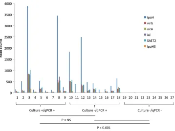
are shown in Fig. 2. For all six gene targets, culture
⫺qPCR
⫹samples yielded
read counts similar to those of the
Shigella
culture
⫹qPCR
⫹samples, whereas few reads
were generated among
Shigella
culture
⫺qPCR
⫺samples. A high correlation was
observed between these genes (e.g.,
R
2⫽
0.889 to 0.932 between
ipaH
and any of the
other 5 genes). The read counts of 18
ipaH
-positive samples showed low correlation
(e.g.,
R
2⫽
0.0288, 0.006 for
ipaH
and
virA
, respectively) with the corresponding
ipaH
qPCR quantification cycles; however, this improved when normalized to nonhuman
sequence reads (e.g.,
R
2⫽
0.259 and 0.361). Unfortunately, the genes responsible for
biosynthesis of
Shigella
O antigen are common to many other bacteria and thus cannot
be used to detect or distinguish
Shigella
from EIEC (24). There is, however, another
region, Mxi-Spa-ipa, that has been reported to have some ability to discriminate
Shigella
from EIEC (23), and these genes showed a similar pattern of positivity among
ipaH
PCR
⫹specimens and negativity among
ipaH
PCR
⫺specimens, supporting the idea
that these
ipaH
PCR
⫹specimens were from
Shigella
(see Table S4 in the supplemental
material).
Testing Kraken with whole-genome sequences of
Shigella
isolates.
In order to
validate the precision and recall of Kraken for
Shigella
at the genus and species level,
we tested the 12 DNA sequences from well-characterized isolates of
Shigella
from
Pakistan. At the
Shigella
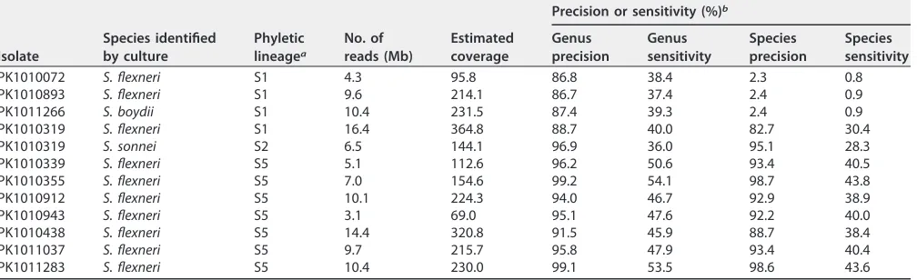
genus level, the Kraken precision ranged from 86 to 99.2%
(corresponding to the proportion of reads assigned to
Shigella
out of the reads
classified to a genus [15]). At the species level, the precision was either close to 2% or
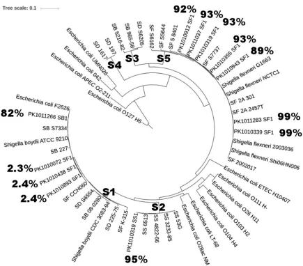
above 80% (Table 1). As shown in Fig. 3, all of the low precision occurred among
S.
flexneri
isolates that were in the S1 phyletic lineage (the S1 lineage includes
S. flexneri
,
S. dysenteriae
, and
S. boydii
as described previously [20]). In contrast, species precision
for
S. sonnei
and
S. flexneri
of the S2 and S5 lineages, respectively (that contain one
nominal species), was
⬎
90%. Accordingly, among the nine culture
⫹qPCR
⫹stool
FIG 2Read counts of sixShigellavirulence genes in 27 stool samples. A 1-kb region was interrogated for each target, except for 531 bp foripaH3. The sample order remained the same as in Table S1 in the supplemental material. NS, not significant.
on May 17, 2020 by guest
http://jcm.asm.org/
[image:4.585.43.401.68.337.2]samples, four
S. sonnei
(S2 lineage) and three
S. flexneri
(all of S5 lineage)
culture-positive specimens were correctly identified by metagenomics sequencing as those
with the highest read count, whereas two
S. flexneri
samples (1 of S1 lineage) were
misclassified as other
Shigella
species (see Table S5 in the supplemental material).
We further compared Kraken to other tools, including Clark, MetaPhlan2, and Kaiju
(see Table S6 in the supplemental material). Kraken and Clark (93.2
⫾
4.7% versus
91.7
⫾
5.6%; Wilcoxon signed-rank test,
P
⬍
0.05) outperformed MetaPhlan2 (66.7
⫾
33.1%,
P
⬍
0.05) and Kaiju (67.5
⫾
9.6%,
P
⬍
0.05) for precision. For sensitivity, although
MetaPhlan2 showed good performance for the S2 and S5 lineages (74.2
⫾
13.4%), its
recall rate was poor for the S1 lineage (2.1
⫾
0.3%). In contrast, Kraken showed
consistent recall rates across lineages (47.8
⫾
5.6% for S2 and S5 and 38.8
⫾
1.2 for S1),
higher than both Clark (28.3
⫾
4.9%,
P
⬍
0.05) and Kaiju (10.7
⫾
2.3%,
P
⬍
0.05).
Detection of other pathogens through metagenomics sequencing.
In addition,
we examined whether the detection of DNA viruses and certain other bacterial
patho-gens by qPCR (see Table S3 in the supplemental material) could also be supported by
the metagenomic results. For many pathogens, such as
Campylobacter jejuni
and
C. coli
,
adenovirus 40/41, enteroaggregative
E. coli
(EAEC), and typical enteropathogenic
E. coli
(EPEC), the pathogen-specific read counts identified by Kraken demonstrated a
quan-titative relationship with qPCR quantification cycle (
C
q) values of the relevant targetgenes (see Fig. S1 in the supplemental material). However, there was also substantial
metagenomic background in qPCR-negative specimens.
DISCUSSION
The main finding of this work is that the metagenomic composition of
Shigella
qPCR-positive samples is similar to that of culture-positive samples. In other words,
the
Shigella
qPCR results based on
ipaH
appear to be robust even if cultures are
negative. Our result is consistent with those of Lindsay et al. (10) that showed that
the culture
⫺qPCR
⫹samples were indistinguishable from culture
⫹qPCR
⫹samples
by clinical criteria.
[image:5.585.43.547.84.238.2]In the metagenomic analysis, we examined the read counts of known
Shigella
/EIEC-specific virulence genes. We surveyed
ipaH
,
ial
,
virA
,
virG
,
ShET2
, and
ipaH3. ipaH
is the
most commonly used target gene for diagnostic PCR assays and is present in multiple
copies on both chromosome and plasmid (13).
ial
,
virA
,
virG
, and
ShET2
are found in
single copy on the plasmid (25–27), whereas
ipaH3
, which is a conserved region
previously identified to distinguish
Shigella
from the majority of
E. coli
, is present in
single copy on the chromosome (20). The metagenomic read counts of these genes
were highly correlated with each other within a sample, which is consistent with our
previous results (9, 11). The read counts distinguished the
Shigella
qPCR positives from
TABLE 1Benchmark of Kraken’s precision and sensitivity for isolates ofShigellaspp.
Isolate
Species identified by culture
Phyletic lineagea
No. of reads (Mb)
Estimated coverage
Precision or sensitivity (%)b
Genus precision
Genus sensitivity
Species precision
Species sensitivity
PK1010072 S. flexneri S1 4.3 95.8 86.8 38.4 2.3 0.8
PK1010893 S. flexneri S1 9.6 214.1 86.7 37.4 2.4 0.9
PK1011266 S. boydii S1 10.4 231.5 87.4 39.3 2.4 0.9
PK1010319 S. flexneri S1 16.4 364.8 88.7 40.0 82.7 30.4
PK1010319 S. sonnei S2 6.5 144.1 96.9 36.0 95.1 28.3
PK1010339 S. flexneri S5 5.1 112.6 96.2 50.6 93.4 40.5
PK1010355 S. flexneri S5 7.0 154.6 99.2 54.1 98.7 43.8
PK1010912 S. flexneri S5 10.1 224.3 94.0 46.7 92.9 38.9
PK1010943 S. flexneri S5 3.1 69.0 95.1 47.6 92.2 40.0
PK1010438 S. flexneri S5 14.4 320.8 91.5 45.9 88.7 38.4
PK1011037 S. flexneri S5 9.7 215.7 95.8 47.9 93.4 40.4
PK1011283 S. flexneri S5 10.4 230.0 99.1 53.5 98.6 43.6
aPhyletic lineage was assigned according to the system of Sahl et al. (20).
bSensitivity refers to the proportion of sequences assigned to the correctShigellagenus or species. Precision, i.e., the positive predictive value, refers to the proportion
of correct classifications, out of the total number of classifications attempted.
on May 17, 2020 by guest
http://jcm.asm.org/
negative and were similar between culture
⫹qPCR
⫹and culture
⫺qPCR
⫹samples. As
expected,
ipaH
had the highest read counts for all the
Shigella
qPCR-positive samples,
usually 5- to 7-fold higher than
ial
,
virA
,
virG
, and
ShET2
. In contrast,
ipaH3
had the
lowest read counts, usually 20- to 30-fold lower than that of
ipaH
. This confirmed that
ipaH
is likely the most efficient available target for PCR detection of
Shigella
species.
As a benchmark comparison, we tested 12 genome sequences from
Shigella
isolates
carefully identified to the species level to determine the precision and recall for Kraken,
Clark, MetaPhlan2, and Kaiju. Kraken outperformed for both precision and recall at the
genus level. We also tested the assignment of reads to the species level. The
Shigella
genus includes four nominal species—
S. sonnei
,
S. flexneri
,
S. dysenteriae
, and
S.
boydii
—that can be accurately distinguished by serotyping. The precision for the genus
was very good (
>
86%) and, although the precision for the species could also be very
good (
>
88%), sometimes it was extremely poor (
⬃
2.4%). The precision of the
se-quences was dependent upon the position of the sequence in the phylogenetic tree
(Fig. 3). The previously defined phyletic lineage S1 (20) was the source of all of the
assignments of sequences that had low precision. This lineage, S1, is characterized by
the presence of three nominal species:
S. flexneri
,
S. dysenteriae
, and
S. boydii
. Thus, in
this case (and in the S3 lineage, where
S. dysenteriae
and
S. boydii
co-occur) (20, 28), all
programs, including Kraken, that assume a monophyletic lineage for its assignment (29)
FIG 3Phylogenetic tree of selected isolates ofShigellaspp. TheShigellalineages were determined previously (20). The Pakistani isolates are denoted by PK*. SF,S. flexneri; SD,S. dysenteriae; SS,S. sonnei; SB,S. boydii. The percentage displays the Kraken annotation precision at the species level forShigella.
on May 17, 2020 by guest
http://jcm.asm.org/
[image:6.585.40.475.66.448.2]produce low precision. However, for species such as
S. sonnei
that are monophyletic,
the assignment to species is very good.
Another observation was that
Shigella
qPCR-positive samples yielded a much higher
proportion of human sequence reads. We suspect this is because clinical shigellosis is
invasive and characterized by the presence of blood, mucus, and epithelial disruption.
Shigella
was most likely the causative agent in these samples because of its strong
association with diarrhea at high quantity, even if in the presence of other pathogens
(7, 10, 11).
This study had limitations, such as the small sample size (limited by cost), the depth
of metagenomic sequencing, the challenges posed by variable levels (up to 97.9%) of
human DNA in fecal samples, low
Shigella
abundance (0.27
⫾
0.32%), the high level of
sequence similarity of
Shigella
genome with
E. coli
genome in available databases, and
imperfect
S. flexneri
species assignment by Kraken. Nonetheless, our study clearly
demonstrates
Shigella ipaH
qPCR-positive specimens contain genes from
Shigella
,
whereas qPCR-negative specimens do not. Thus, culture misses true cases of
Shigella
and molecular diagnosis with appropriate quantitative cutoffs provides a more accurate
method for detecting
Shigella
.
SUPPLEMENTAL MATERIAL
Supplemental material for this article may be found at
https://doi.org/10.1128/JCM
.01374-17
.
SUPPLEMENTAL FILE 1,
PDF file, 0.5 MB.
ACKNOWLEDGMENTS
This study was supported by the Bill and Melinda Gates Foundation (OPP1019093).
We thank the families who participated, the project field staff, and administration
staff at all GEMS sites for assistance. The Etiology, Risk Factors and Interactions of
Enteric Infections and Malnutrition and the Consequences for Child Health and
Devel-opment Project (MAL-ED) is a collaborative project supported by the Bill and Melinda
Gates Foundation, the Foundation for the NIH, and the National Institutes of Health,
Fogarty International Center. We thank the staff and participants of the MAL-ED
Network Project for their important contributions. We also thank Mihai Pop for his
careful review of the manuscript.
REFERENCES
1. Kotloff KL, Winickoff JP, Ivanoff B, Clemens JD, Swerdlow DL, Sansonetti PJ, Adak GK, Levine MM. 1999. Global burden of Shigella infections: implications for vaccine development and implementation of control strategies. Bull World Health Organ 77:651– 666.
2. GBD 2015 Mortality and Causes of Death Collaborators. 2016. Global, regional, and national life expectancy, all-cause mortality, and cause-specific mortality for 249 causes of death, 1980-2015: a systematic analysis for the Global Burden of Disease Study 2015. Lancet 388: 1459 –1544.https://doi.org/10.1016/S0140-6736(16)31012-1.
3. Kotloff KL, Nataro JP, Blackwelder WC, Nasrin D, Farag TH, Panchalingam S, Wu Y, Sow SO, Sur D, Breiman RF, Faruque AS, Zaidi AK, Saha D, Alonso PL, Tamboura B, Sanogo D, Onwuchekwa U, Manna B, Ramamurthy T, Kanungo S, Ochieng JB, Omore R, Oundo JO, Hossain A, Das SK, Ahmed S, Qureshi S, Quadri F, Adegbola RA, Antonio M, Hossain MJ, Akinsola A, Mandomando I, Nhampossa T, Acacio S, Biswas K, O’Reilly CE, Mintz ED, Berkeley LY, Muhsen K, Sommerfelt H, Robins-Browne RM, Levine MM. 2013. Burden and aetiology of diarrhoeal disease in infants and young children in developing countries (the Global Enteric Multicenter Study, GEMS): a prospective, case-control study. Lancet 382:209 –222.https://
doi.org/10.1016/S0140-6736(13)60844-2.
4. Buss SN, Leber A, Chapin K, Fey PD, Bankowski MJ, Jones MK, Ro-gatcheva M, Kanack KJ, Bourzac KM. 2015. Multicenter evaluation of the BioFire FilmArray gastrointestinal panel for etiologic diagnosis of infec-tious gastroenteritis. J Clin Microbiol 53:915–925. https://doi.org/10
.1128/JCM.02674-14.
5. Duong VT, Phat VV, Tuyen HT, Dung TT, Trung PD, Minh PV, Tu le TP,
Campbell JI, Le Phuc H, Ha TT, Ngoc NM, Huong NT, Tam PT, Huong DT, Xang NV, Dong N, Phuong le T, Hung NV, Phu BD, Phuc TM, Thwaites GE, Vi LL, Rabaa MA, Thompson CN, Baker S. 2016. Evaluation of Luminex xTAG gastrointestinal pathogen panel assay for detection of multiple diarrheal pathogens in fecal samples in Vietnam. J Clin Microbiol 54: 1094 –1100.https://doi.org/10.1128/JCM.03321-15.
6. Harrington SM, Buchan BW, Doern C, Fader R, Ferraro MJ, Pillai DR, Rychert J, Doyle L, Lainesse A, Karchmer T, Mortensen JE. 2015. Multi-center evaluation of the BD max enteric bacterial panel PCR assay for rapid detection ofSalmonellaspp.,Shigellaspp.,Campylobacterspp. (C. jejuniand C. coli), and Shiga toxin 1 and 2 genes. J Clin Microbiol 53:1639 –1647.https://doi.org/10.1128/JCM.03480-14.
7. Lindsay B, Ochieng JB, Ikumapayi UN, Toure A, Ahmed D, Li S, Panchalingam S, Levine MM, Kotloff K, Rasko DA, Morris CR, Juma J, Fields BS, Dione M, Malle D, Becker SM, Houpt ER, Nataro JP, Sommerfelt H, Pop M, Oundo J, Antonio M, Hossain A, Tamboura B, Stine OC. 2013. Quantitative PCR for detection ofShigellaimproves ascertainment ofShigellaburden in children with moderate-to-severe diarrhea in low-income countries. J Clin Microbiol 51:1740 –1746.https://doi.org/10.1128/JCM.02713-12.
8. Lindsay B, Oundo J, Hossain MA, Antonio M, Tamboura B, Walker AW, Paulson JN, Parkhill J, Omore R, Faruque AS, Das SK, Ikumapayi UN, Adeyemi M, Sanogo D, Saha D, Sow S, Farag TH, Nasrin D, Li S, Pan-chalingam S, Levine MM, Kotloff K, Magder LS, Hungerford L, Sommerfelt H, Pop M, Nataro JP, Stine OC. 2015. Microbiota that affect risk for shigellosis in children in low-income countries. Emerg Infect Dis 21: 242–250.https://doi.org/10.3201/eid2101.140795.
on May 17, 2020 by guest
http://jcm.asm.org/
9. Lindsay B, Pop M, Antonio M, Walker AW, Mai V, Ahmed D, Oundo J, Tamboura B, Panchalingam S, Levine MM, Kotloff K, Li S, Magder LS, Paulson JN, Liu B, Ikumapayi U, Ebruke C, Dione M, Adeyemi M, Rance R, Stares MD, Ukhanova M, Barnes B, Lewis I, Ahmed F, Alam MT, Amin R, Siddiqui S, Ochieng JB, Ouma E, Juma J, Mailu E, Omore R, O’Reilly CE, Hannis J, Manalili S, Deleon J, Yasuda I, Blyn L, Ranken R, Li F, Housley R, Ecker DJ, Hossain MA, Breiman RF, Morris JG, McDaniel TK, Parkhill J, Saha D, Sampath R, Stine OC, Nataro JP. 2013. Survey of culture, gold-engate assay, universal biosensor assay, and 16S rRNA gene sequencing as alternative methods of bacterial pathogen detection. J Clin Microbiol 51:3263–3269.https://doi.org/10.1128/JCM.01342-13.
10. Lindsay B, Saha D, Sanogo D, Das SK, Omore R, Farag TH, Nasrin D, Li S, Panchalingam S, Levine MM, Kotloff K, Nataro JP, Magder L, Hungerford L, Faruque AS, Oundo J, Hossain MA, Adeyemi M, Stine OC. 2015. Association betweenShigellainfection and diarrhea varies based on location and age of children. Am J Trop Med Hyg 93:918 –924.https://
doi.org/10.4269/ajtmh.14-0319.
11. Liu J, Platts-Mills JA, Juma J, Kabir F, Nkeze J, Okoi C, Operario DJ, Uddin J, Ahmed S, Alonso PL, Antonio M, Becker SM, Blackwelder WC, Breiman RF, Faruque AS, Fields B, Gratz J, Haque R, Hossain A, Hossain MJ, Jarju S, Qamar F, Iqbal NT, Kwambana B, Mandomando I, McMurry TL, Ochieng C, Ochieng JB, Ochieng M, Onyango C, Panchalingam S, Kalam A, Aziz F, Qureshi S, Ramamurthy T, Roberts JH, Saha D, Sow SO, Stroup SE, Sur D, Tamboura B, Taniuchi M, Tennant SM, Toema D, Wu Y, Zaidi A, Nataro JP, Kotloff KL, Levine MM, Houpt ER. 2016. Use of quantitative molecular diagnostic methods to identify causes of diarrhoea in children: a reanalysis of the GEMS case-control study. Lancet 388: 1291–1301.https://doi.org/10.1016/S0140-6736(16)31529-X.
12. von Seidlein L, Kim DR, Ali M, Lee H, Wang X, Thiem VD, Canh DG, Chaicumpa W, Agtini MD, Hossain A, Bhutta ZA, Mason C, Sethabutr O, Talukder K, Nair GB, Deen JL, Kotloff K, Clemens J. 2006. A multicentre study ofShigelladiarrhoea in six Asian countries: disease burden, clinical manifestations, and microbiology. PLoS Med 3:e353.https://doi.org/10
.1371/journal.pmed.0030353.
13. Vu DT, Sethabutr O, Von Seidlein L, Tran VT, Do GC, Bui TC, Le HT, Lee H, Houng HS, Hale TL, Clemens JD, Mason C, Dang DT. 2004. Detection ofShigellaby a PCR assay targeting theipaHgene suggests increased prevalence of shigellosis in Nha Trang, Vietnam. J Clin Microbiol 42: 2031–2035.https://doi.org/10.1128/JCM.42.5.2031-2035.2004. 14. Panchalingam S, Antonio M, Hossain A, Mandomando I, Ochieng B,
Oundo J, Ramamurthy T, Tamboura B, Zaidi AK, Petri W, Houpt E, Murray P, Prado V, Vidal R, Steele D, Strockbine N, Sansonetti P, Glass RI, Robins-Browne RM, Tauschek M, Svennerholm AM, Berkeley LY, Kotloff K, Levine MM, Nataro JP. 2012. Diagnostic microbiologic methods in the GEMS-1 case/control study. Clin Infect Dis 55(Suppl 4):S294 –S302.
https://doi.org/10.1093/cid/cis754.
15. Wood DE, Salzberg SL. 2014. Kraken: ultrafast metagenomic sequence classification using exact alignments. Genome Biol 15:R46.https://doi
.org/10.1186/gb-2014-15-3-r46.
16. Ounit R, Wanamaker S, Close TJ, Lonardi S. 2015. CLARK: fast and accurate classification of metagenomic and genomic sequences using discriminative k-mers. BMC Genomics 16:236.https://doi.org/10.1186/
s12864-015-1419-2.
17. Truong DT, Franzosa EA, Tickle TL, Scholz M, Weingart G, Pasolli E, Tett A, Huttenhower C, Segata N. 2015. MetaPhlAn2 for enhanced meta-genomic taxonomic profiling. Nat Methods 12:902–903.https://doi.org/
10.1038/nmeth.3589.
18. Menzel P, Ng KL, Krogh A. 2016. Fast and sensitive taxonomic classifi-cation for metagenomics with Kaiju. Nat Commun 7:11257.https://doi
.org/10.1038/ncomms11257.
19. Houpt E, Gratz J, Kosek M, Zaidi AK, Qureshi S, Kang G, Babji S, Mason C, Bodhidatta L, Samie A, Bessong P, Barrett L, Lima A, Havt A, Haque R, Mondal D, Taniuchi M, Stroup S, McGrath M, Lang D, et al. 2014. Microbiologic methods utilized in the MAL-ED cohort study. Clin Infect Dis 59(Suppl 4):S225–S232.https://doi.org/10.1093/cid/ciu413. 20. Sahl JW, Morris CR, Emberger J, Fraser CM, Ochieng JB, Juma J, Fields B,
Breiman RF, Gilmour M, Nataro JP, Rasko DA. 2015. Defining the phy-logenomics ofShigellaspecies: a pathway to diagnostics. J Clin Microbiol 53:951–960.https://doi.org/10.1128/JCM.03527-14.
21. Treangen TJ, Ondov BD, Koren S, Phillippy AM. 2014. The Harvest suite for rapid core-genome alignment and visualization of thousands of intraspecific microbial genomes. Genome Biol 15:524.https://doi.org/10
.1186/s13059-014-0524-x.
22. Letunic I, Bork P. 2016. Interactive tree of life (iTOL) v3: an online tool for the display and annotation of phylogenetic and other trees. Nucleic Acids Res 44:W242–W245.https://doi.org/10.1093/nar/gkw290. 23. Hazen TH, Leonard SR, Lampel KA, Lacher DW, Maurelli AT, Rasko DA.
2016. Investigating the relatedness of enteroinvasiveEscherichia colito otherE. coliandShigellaisolates by using comparative genomics. Infect Immun 84:2362–2371.https://doi.org/10.1128/IAI.00350-16.
24. Lan R, Alles MC, Donohoe K, Martinez MB, Reeves PR. 2004. Molecular evolutionary relationships of enteroinvasiveEscherichia coliandShigella spp. Infect Immun 72:5080 –5088.https://doi.org/10.1128/IAI.72.9.5080
-5088.2004.
25. Villalobo E, Torres A. 1998. PCR for detection ofShigellaspp. in mayon-naise. Appl Environ Microbiol 64:1242–1245.
26. Alipour M, Talebjannat M, Nabiuni M. 2012. Polymerase chain reaction method for the rapid detection of virulentShigellaspp. Int J Mol Clin Microbiol 2:134 –137.
27. Vargas M, Gascon J, Jimenez De Anta MT, Vila J. 1999. Prevalence of Shigella enterotoxins 1 and 2 among Shigella strains isolated from patients with traveler’s diarrhea. J Clin Microbiol 37:3608 –3611. 28. The HC, Thanh DP, Holt KE, Thomson NR, Baker S. 2016. The genomic
signatures ofShigellaevolution, adaptation, and geographical spread. Nat Rev Microbiol 14:235–250.https://doi.org/10.1038/nrmicro.2016.10. 29. Lindgreen S, Adair KL, Gardner PP. 2016. An evaluation of the accuracy and speed of metagenome analysis tools. Sci Rep 6:19233.https://doi
.org/10.1038/srep19233.