BIROn - Birkbeck Institutional Research Online
Addyman, Caspar and Rocha, S. and Mareschal, Denis (2014) Mapping
the origins of time: Scalar errors in infant time estimation. Developmental
Psychology 50 (8), pp. 2030-2035. ISSN 0012-1649.
Downloaded from:
Usage Guidelines:
Please refer to usage guidelines at
or alternatively
Mapping the Origins of Time: Scalar Errors in Infant Time Estimation
Caspar Addyman, Sinead Rocha, and Denis Mareschal
Birkbeck, University of London
Time is central to any understanding of the world. In adults, estimation errors grow linearly with the length of the interval, much faster than would be expected of a clock-like mechanism. Here we present the first direct demonstration that this is also true in human infants. Using an eye-tracking paradigm, we examined 4-, 6-, 10-, and 14-month-olds’ responses to the omission of a recurring target, on either a 3-or 5-s cycle. At all ages (a) both fixation and pupil dilation measures were time locked to the periodicity of the test interval, and (b) estimation errors grew linearly with the length of the interval, suggesting that trademark interval timing is in place from 4 months.
Keywords: interval timing, time perception, infancy
Supplemental materials:http://dx.doi.org/10.1037/a0037108.supp
Interval timing concerns our ability to judge, compare, and reproduce time intervals for durations from about half a second to a few minutes (Grondin, 2008; Zakay & Block, 1997). It is fundamental to the way we structure our social and physical interactions with the world. One surprising thing about our interval timing abilities (which contrast sharply with precision timing involved in motor control) is how bad they are. Errors in our time estimates grow linearly with the length of the interval, much faster than would be expected of a repetitive clock-like mechanism (Hass & Hermann, 2012). This so-called scalar property of interval timing is shared by humans, rats, and pigeons (Gibbon, 1977) and can even be found in rate dependent habituation in C. elegans (Staddon & Higa, 1996). Interval timing is typically investigated in human adults and rats by requiring them to reproduce a target interval. However, it has never been directly demonstrated in infants because of the difficulty of getting them to generate re-peated estimates of fixed intervals.
Timing abilities develop slowly from birth.Clifton (1974) es-tablished that even neonates could show a response (heart rate
deceleration) to an omitted timed event. This pattern was also found at 2 months old (Boswell, Garner, & Berg, 1994) and 4 months old (Colombo & Richman, 2002). Heart rate measures sometimes also show anticipation developing from 4 months on-ward, but the data quickly become difficult to interpret (Boswell et al., 1994;Donohue & Berg, 1991). Studies using visual habitua-tion paradigms have shown that infants’ ability to discriminate temporal intervals increases from a ratio 1:2 at 6 months ( van-Marle & Wynn, 2006) to a ratio of 2:3 at 10 months (Brannon, Suanda, & Libertus, 2007), mirroring the development of numer-ical discrimination (Lipton & Spelke, 2003). Measurement of event-related potentials (ERPs) reveals that 10-month-olds show brain responses to omitted stimuli similar to those of adults ( Bran-non, Libertus, Meck, & Woldorff, 2008;Brannon, Roussel, Meck, & Woldorff, 2004). Although a sensitivity to regular timings seems to be present from early infancy, as evidenced above, timing accuracy continues to develop well into childhood (e.g., Droit-Volet, Tourret, & Wearden, 2004; Friedman, 2008; Goldberg, 1995).
The existing research on infant timing has some methodological limitations. Many previous studies of infant time estimation have relied on violation of expectation paradigms with binary measures of novelty and familiarity or habituation and dishabituation, which provide limited information (Aslin, 2007). In particular, they do not allow the detection of time-locked behaviors. Heart rate vari-ability (e.g., Donohue & Berg, 1991) does provide a temporal signal, but this is of low resolution (0.5 Hz) and is subject to rapid habitation, which makes data difficult to interpret. ERPs (e.g.,
Brannon et al., 2008) have high temporal resolution but require extensive repetition with averaging over large numbers of trials, and can produce noisy data that are difficult to analyze and interpret. There is also a theoretical difficulty in that ERPs need a clear external “event” marker to line up multiple trials. This makes it less suitable for studying subjective interval timing where the events of interest are internally generated and have substantial temporal variability. Finally, none of these studies directly address infants’ ability to anticipate and act on temporally predictable events.
This article was published Online First June 30, 2014.
Caspar Addyman, Sinead Rocha, and Denis Mareschal, Centre for Brain and Cognitive Development, Birkbeck, University of London.
This work was funded by Economic and Social Research Council Grant RES-062-23-0819. Denis Mareschal is partly supported by a Royal Society Wolfson research merit award. We are grateful to all the parents and infants who took part in this study and to Hannah Smolar-Fourniol and Jennifer Taylor for dual coding the infant data.
This article has been published under the terms of the Creative Com-mons Attribution License (http://creativecommons.org/licenses/by/3.0/), which permits unrestricted use, distribution, and reproduction in any me-dium, provided the original author and source are credited. Copyright for this article is retained by the author(s). Author(s) grant(s) the American Psychological Association the exclusive right to publish the article and identify itself as the original publisher.
Correspondence concerning this article should be addressed to Caspar Addyman, Centre for Brain and Cognitive Development, Birkbeck, Uni-versity of London, Malet Street, London, WC1E 7HX United Kingdom. E-mail:[email protected]
In the present study we use an eye-tracking system to provide online measures of infant voluntary and involuntary responses in an interval timing task. We use a “peek-a-boo” animation in which a cartoon character pops up on the screen at regular intervals (every 3 s in one condition, 5 s in another) accompanied by a socially engaging sound effect (adult female voice addressing the infant). The infants saw seven repetitions of this event followed by a blank screen for a further 6 or 10 s, respectively. Modern eye trackers provide an accurate measure of fixation location and pupil diameter with high temporal resolution. Fixation data can show where infants look, when they saccade, and for how long they fixate. This provides a measure of voluntary response to expecta-tions about the timing of events. In contrast, changes in pupil diameter are involuntary and accompany violations of expectation in infants (Jackson & Sirois, 2009). They can also provide a proxy for attention or cognitive load (Laeng, Sirois, & Gredebäck, 2012). Responses in both modalities provide estimates of the infant’s repeated subjective judgments of the duration of the interval be-tween predictable stimuli.
Method
Participants
Nineteen four-month-olds (11 female; mean age⫽ 133 days; range: 118 –144 days), 20 six-month-olds (5 female; mean age⫽ 181 days; range: 166 –197 days), 20 ten-month-olds (14 female; mean age⫽ 308 days; range: 293–320 days), and 20 fourteen-month-olds (10 female; mean age⫽ 433 days; range: 415– 447 days) took part in the 5-s condition.
Seventeen four-month-olds (9 female; mean age⫽ 133 days; range: 118 –144 days), 12 six-month-olds (5 female; mean age⫽ 186 days; range: 166 –215 days), 13 ten-month-olds (6 female; mean age⫽ 308 days; range: 291–323 days), and 13 fourteen-month-olds (6 female; mean age ⫽ 428 days; range: 419 – 447 days) took part in the 3-s condition.
Of these, 35 infants (13 four-month-olds, 9 six-month-olds, 10 ten-month-olds, and 3 fourteen-month-olds) took part in both conditions. These infants took part in the 5-s condition first, had a 10-min break, then completed the 3-s condition. Finally, across both conditions 29 infants were excluded: seven infants because of fussiness, four because of experimenter error or computer failure, and 18 due to insufficient eye-tracking data.
Apparatus
Stimuli were presented on a Tobii T120 eye-tracking monitor with a 17-in. (43.18-cm) screen, presentation was controlled by an Apple PowerMac OS X Intel 2.8 Ghz, running MATLAB R2009b (MathWorks Ltd.) with Psychtoolbox (Brainard, 1997; Pelli, 1997).
Stimuli and Design
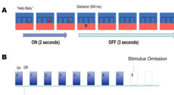
Each condition (5- or 3-s intervals) consisted of three blocks of eight learning trials in which a cartoon character moved up from behind an occluder into a target area directly above the occluder and then back down again, with a periodicity of either 5 s or 3 s (seeFigure 1). This was followed by a “missed beat” in which the animated character did not appear and the screen remained un-changed. The missed beat lasted twice as long as a regular beat, thereby enabling us to assess responses occurring after the ex-pected appearance of a character.
[image:3.603.147.447.502.666.2]Each block featured one of six different cartoon characters (either one of two teddy bears, one of two dinosaurs, a fish, or a tiger), and each character was paired with a single verbal prompt (“Coo-ee!,” “Hey, Baby,” then “What’s that?”). The three target areas (defined by black borders) were located to the left, to the right, or at the center of the screen. The target area for the character remained the same for all the three blocks. For those infants participating in both conditions, both the target area and set of characters were changed between conditions.
Figure 1. Schematic representation of stimulus presentation in the 5-s condition. (A) In one on– off cycle the target rose into the target area accompanied by a vocal prompt. It remained stationary for 1 s before disappearing again. A rapidly shrinking distractor then briefly appeared, and the frame remained empty for the remainder of the period. (B) Each block consisted of seven cycles with the character reappearing at fixed intervals followed by a longer test period in which nothing happened. See the online article for the color version of this figure.
In the 5-s trials, a complete beat cycle consisted of the character moving from behind the occluder to the target area in 500 ms accompanied by a verbal prompt. It then remained stationary for 1,000 ms before moving back behind the occluder in 500 ms. The screen then remained unchanged for 3 further seconds. This blank interval was cut to 1 s in the 3-s cycle condition. In both conditions a distractor (an orange star or a blue circle rapidly decreasing in size) was shown on the lower part of the screen after the character had disappeared. This was to stop infants remaining fixated on the target area.
All animated characters were of comparable size (6° of visual angle). The luminosity of the screen did not significantly differ when the stimulus was present or absent, nor did it differ between characters (see Figure S1 in supplemental materials).
Procedure
Each infant was seated on his or her carer’s lap, approximately 60 cm from the eye tracker screen. A 5-point calibration sequence was run until at least 4 points were properly calibrated for each eye. In addition, 61 of the infants took part in a simple habituation-to-a-checker-board task before and after testing as part of a sepa-rate study.
Results
For each infant the eye tracker recorded fixation and pupil diameter information at a sampling rate of 60 Hz. Because eye-tracking data, especially with infants, can be intermittent, we analyzed the data by fitting smoothed curves to both the fixation and pupil data as described below (see also Jackson & Sirois, 2009;Ramsay & Silverman, 2002).
To normalize the raw data from the eye tracker and allow for comparison between participants, we first divided the data into fixed 50-ms bins. Fixation distance was calculated as the linear distance (in pixels) from the center of the target area to the mean fixation point found by averaging fixation data for left and right eyes (if available), or for a single eye, if only one eye was available. Pupil diameter values were similarly combined. We excluded blocks of trials in which there was tracking data for less than 50% of the total presentation time. A total of 92/284 (.32) where excluded in the 5-s condition and 39/171 (.23) in the 3-s condition. The remaining data still had an average of .26 missing samples. Missing values were linearly interpolated, and a low-pass filter was applied with the “filter” function built into MATLAB (R2009b) with a tau parameter of 0.15.
Next, these data were fit with a piecewise smooth B-spline function of order 4 with 1 base per second to give either 30 bases (3-s condition) or 50 bases (5-s condition). This value smoothed the data while preserving the major event related peaks (Ramsay & Silverman, 2002). It gives our B-spline functions a comparable temporal resolution toJackson and Sirois (2009), who fitted a 26-s presentation with 24 bases (1.08 bases per second). The resulting continuous time series data are shown in Figure S1 in the supple-mental materials.
To investigate infants’ responses to the missed beat, we ana-lyzed the peak-to-peak measurements (for both fixation distance and pupil dilation) for the value associated with the last occurrence of a visible target and the value associated with the missed beat in
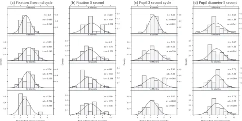
which the screen remains blank (see Figure S2 in the supplemental materials). For fixation distance this corresponds to the distance between minima, whereas for pupil size this corresponds to dis-tance between maxima. The second peak was assumed to be associated with infants’ expectation of the target reappearing at a predictable time, and the interval between the peaks was taken as a measure of the infants’ subjective time estimate of when the target should reappear. In other words, it is a direct measure of the infants’ temporal interval judgments.Figure 2plots histograms for peak-to-peak fixation and pupil diameter grouped by age and time interval.
Approximately 55% of fixation data and 66% of pupil data yielded interval estimates via this method. For each data type we ran an analysis of variance with age and time interval as between-subject factors.1
For fixation, we found that estimates clearly varied according to the time interval, F(1, 192)⫽82.4, p⬍.0001,
2⫽
.30, with no effect of age, F(3, 192)⬍1, and no interaction,
F(3, 192) ⫽ 1.48, p⬍ .22. For pupil diameter, we found that estimates clearly varied according to the time interval, F(1, 233)⫽ 140.5, p⬍.0001,2⫽
.38, with a trend toward a main effect of age, F(3, 233)⫽2.3, p⬍.08, and no interaction, F(3, 233)⬍1. The lack of an age effect shows that interval timing ability is present as young as 4 months of age. In both cases, data failed Levene’s test for homogeneity of variance: fixation, F(7, 192)⫽ 5.62, p⬍.0001; pupil data, F(7, 232)⫽4.19, p⬍.0002. This suggests, in line with the scalar property of interval timing, that variance increases with length of the target interval. However, this breaks the homogeneity of variance assumptions of analysis of variance. Consequently, even though t and F tests with near equal
ns are generally robust to variations in variance across samples
(Boneau, 1960), we also looked at relative time data. All infant time estimates for the 3- and 5-s conditions were divided by 3 and 5, respectively, and the above analyses were rerun. Levene’s tests were no longer significant: for fixation, F(7, 192)⫽2.18, p⬍.09; and for pupil, F(7, 232)⫽ 1.52, p⬍.16. For fixation, time no longer had a significant difference between groups, F(1, 192)⫽ .01, p⬍.94,2⬍
.0001, with no effect of age, F(3, 192)⬍1, and no interaction, F(3, 192)⫽2.18, p⬍.09. For pupil diameter, time was no longer significant, F(1, 232)⫽0.24, p⬍.62,2⫽
.001, and there were no other effects (Fs⬍ 1). Taken together, these analyses support the idea that infant time estimates approximate the target interval with linear growth in error.
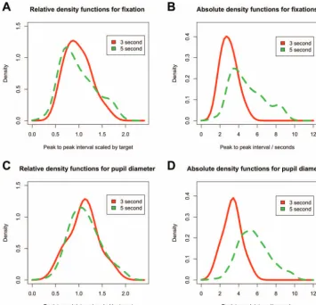
Finally, we combined data across all ages in order to plot density distributions of estimates (see Figure 3). Peak-to-peak distances for both the fixation distance and the pupil dilation were modu-lated by the time interval. In both cases, the value closely approx-imated the interval (3 s or 5 s) of the test condition, showing that infants’ interval estimates were tuned to the particular intervals that they experienced. In addition, their errors (the deviation of the their estimates from the actual interval values) grew in proportion to the length of the target interval (Figures 3Band3D). When the individual peak-to-peak estimates are divided by the actual target interval length (3 or 5, respectively), the curves have the same
1Although some participants took part in both conditions, time interval
shape (Figures 3Aand3C). The curves are centered around target interval, and the width of the distributions is proportional, as is characteristic of the scalar property of interval timing. This is the signature of the interval timing in adults (Gibbon, 1977;Rakitin et al., 1998).
Discussion
This study provides the first direct measurement of infants’ interval timing abilities. Using an eye tracker, we examined the reaction of infants aged 4 –14 months to the omission of a recur-ring target, on either a 3- or 5-s cycle. We found that infants at all ages showed a broadly similar pattern of responses. By analyzing both fixation data and pupil diameter changes, we attempted to dissociate voluntary from involuntary timing responses in infancy, based on the assumption that pupil dilation is involuntary and anticipatory eye gaze is under voluntary control. When examining the peak-to-peak variations on individual trials, we found evidence of interval timing ability in both measures. Specifically, we found that responses were approximately normally distributed about the target interval and the widths of the distributions were proportional to the length of the interval. These are the features of the scalar property in interval timing (Gibbon, 1977). To our knowledge, this has never been directly demonstrated in infants.
The scalar property is the key characteristic of interval timing. Timing is noisy, and errors grow linearly with the length of the interval. Previous studies have shown some interval timing abili-ties in infants, but their conclusions have been limited by meth-odological constraints. Habituation studies (e.g., Brannon et al., 2007;vanMarle & Wynn, 2006) only measure a binary response to
a change in stimulus timing, whereas ERP measures (Brannon et al., 2008,2007) only test for a miss-match negativity reaction to oddballs with very short intervals (⬍1,500 ms). Heart rate vari-ability studies (e.g.,Boswell et al., 1994;Colombo & Richman, 2002) sometimes show an anticipatory change to an omitted stim-ulus, but this response adapts rapidly. Our paradigm overcomes all of these limitations. Infants’ responses are measured during an omission period when nothing happens. Infants may generate timing predictions in the form of timed saccades to the target area. Or they may show involuntary responses to the task, as indexed by changing pupil diameter.
Developing a sense of timing is likely to involve the coordina-tion of these explicit (gaze) control mechanisms with some means of internal time keeping. There is no direct means of assessing internal time keeping, but changes in pupil diameter might provide the best available signal. We found that pupil diameter changes were stronger predictors of the timing of the event than fixation; peak-to-peak changes in pupil diameter showed a much closer fit to Weber’s law (seeFigure 3). But why might pupil diameter index timing ability? There is a well-established literature on using measures of pupil diameter to track preconscious or automatic processing in adults (seeLaeng et al., 2012). Recent advances in analysis techniques suggest that changes in pupil diameter can show attentional effects (Wierda, van Rijn, Taatgen, & Martens, 2012), and attention is a key component of interval timing mech-anisms (Block, Hancock, & Zakay, 2010). Research has shown that pupil diameter changes can indicate violation of certain phys-ical expectations in 8-month-olds (Jackson & Sirois, 2009) and social expectations in 6- and 12-month-olds (Gredebäck & Me-(a) Fixation 3 second cycle (b) Fixation 5 second (c) Pupil 3 second cycle (d) Pupil diameter 5 second
Peak to Peak interval (seconds)
De nsit y 0.2 0.4 0.6
1 2 3 4 5 6
m = 2.64
sd = 0.764
cv = 0.289 14 Months
0.2 0.4 0.6 m = 3.04
sd = 0.776
cv = 0.256 10 Months
0.2 0.4
0.6 m = 3.29
sd = 0.931
cv = 0.283 6 Months
0.2 0.4 0.6 m = 2.8
sd = 0.689
cv = 0.246 4 Months
Peak to Peak interval (seconds)
De nsit y 0.1 0.2 0.3 0.4
2 4 6 8 10
m = 5.09
sd = 1.78
cv = 0.35 14 Months
0.1 0.2 0.3 0.4 m = 4.62
sd = 1.84
cv = 0.399 10 Months 0.1
0.2 0.3 0.4
m = 4.8
sd = 1.78
cv = 0.372 6 Months
0.1 0.2 0.3 0.4 m = 5.29
sd = 1.89
cv = 0.358 4 Months
Peak to Peak interval (seconds)
De nsit y 0.2 0.4 0.6
1 2 3 4 5 6
m = 3.07
sd = 0.863
cv = 0.281 14 Months
0.2 0.4 0.6 m = 3.36
sd = 1.23
cv = 0.365 10 Months
0.2 0.4
0.6 m = 3.21
sd = 1.09
cv = 0.339 6 Months
0.2 0.4 0.6 m = 3.44
sd = 0.689
cv = 0.201 4 Months
Peak to Peak interval (seconds)
De nsit y 0.1 0.2 0.3 0.4
2 4 6 8 10
m = 5.73
sd = 1.68
cv = 0.293 14 Months
0.1 0.2 0.3 0.4 m = 5.71
sd = 1.53
cv = 0.269 10 Months 0.1
0.2 0.3 0.4
m = 5.67
sd = 1.66
cv = 0.292 6 Months
0.1 0.2 0.3 0.4 m = 5.52
sd = 1.88
[image:5.603.51.551.78.328.2]cv = 0.341 4 Months
Figure 2. Histograms of the peak-to-peak value for fixation distance and pupil diameter as a function of timing condition and age. In general, median distribution values are close to 5 in the 5-s conditions and 3 in the 3-s conditions. cv⫽coefficient of variation.
linder, 2010). In both of these cases, pupil diameter changes happen in response to an event occurring on the screen rather than in anticipation coupled with an unchanging screen, as we find here. Research into infants’ volitional interval timing may also help address theoretical issues in the time perception literature. Addy-man, French, Mareschal, and Thomas (2011)recently proposed a developmental model of interval timing in which there is an important role for embodied experience during the acquisition and calibration of time. Research with young infants may help decide between this type of developmental, embodied model and more traditional cognitive models of timing (e.g., Gibbon, Church, & Meck, 1984;Matell & Meck, 2004;Staddon & Higa, 1999). A growing body of research links time and space in adults (Dehaene & Brannon, 2010; Núñez & Cooperrider, 2013; Walsh, 2003); embodiment may provide a framework to account for this (Kranjec & Chatterjee, 2010). Accurate measures of individual variability in the subjective time estimation enables an exploration of the rela-tionship between motor activity in infants and their subjective experience of time.
In summary, this study introduces a new method for investigat-ing the interval timinvestigat-ing abilities requirinvestigat-ing the intrinsic generation of responses by young infants. Our results show that both voluntary
and involuntary components of interval timing are present from 4 months of age. In particular, the errors generated by infants when anticipating the end of an interval follow the same scalar law as those observed in adults.
References
Addyman, C., French, R. M., Mareschal, D., & Thomas, E. (2011). Learning to perceive time: A connectionist, memory-decay model of the development of interval timing in infants. In L. Carlson, C. Hölscher, & T. Shipley (Eds.), Proceedings of the 33rd Annual Conference of the Cognitive Science Society (pp. 354 –359). Austin, TX: Cognitive Science Society.
Aslin, R. N. (2007). What’s in a look? Developmental Science, 10, 48 –53.
doi:10.1111/j.1467-7687.2007.00563.x
Block, R. A., Hancock, P. A., & Zakay, D. (2010). How cognitive load affects duration judgments: A meta-analytic review. Acta Psychologica, 134, 330 –343.doi:10.1016/j.actpsy.2010.03.006
Boneau, A. C. (1960). The effect of violations of assumptions underlying the t test. Psychological Bulletin, 57, 49 – 64.doi:10.1037/h0041412
[image:6.603.117.473.77.419.2]Boswell, A. E., Garner, E. E., & Berg, W. K. (1994). Changes in cardiac components of anticipation in 2-, 4-, and 8-month-old infants [Abstract]. Psychophysiology, 31, S28.doi:10.1111/j.1469-8986.1994.tb02378.x
Brainard, D. H. (1997). The Psychophysics Toolbox. Spatial Vision, 10, 433– 436.doi:10.1163/156856897X00357
Brannon, E. M., Libertus, M. E., Meck, W. H., & Woldorff, M. G. (2008). Electrophysiological measures of time processing in infant and adult brains: Weber’s law holds. Journal of Cognitive Neuroscience, 20, 193–203.doi:10.1162/jocn.2008.20016
Brannon, E. M., Roussel, L. W., Meck, W. H., & Woldorff, M. G. (2004). Timing in the baby brain. Cognitive Brain Research, 21, 227–233.
doi:10.1016/j.cogbrainres.2004.04.007
Brannon, E. M., Suanda, S., & Libertus, K. (2007). Temporal discrimina-tion increases in precision over development and parallels the develop-ment of numerosity discrimination. Developdevelop-mental Science, 10, 770 – 777.doi:10.1111/j.1467-7687.2007.00635.x
Clifton, R. K. (1974). Cardiac conditioning and orienting in the infant. In P. A. Obrist, A. H. Black, J. Brener, & L. V. DiCara (Eds.), Cardiovascular psychophysiology: Current issues in response mecha-nisms, biofeedback and methodology (pp. 479 –504). Chicago, IL: Al-dine.
Colombo, J., & Richman, W. A. (2002). Infant timekeeping: Attention and temporal estimation in 4-month-olds. Psychological Science, 13, 475– 479.doi:10.1111/1467-9280.00484
Dehaene, S., & Brannon, E. (2010). Space, time, and number: A Kantian research program. Trends in Cognitive Sciences, 14, 517–519. doi: 10.1016/j.tics.2010.09.009
Donohue, R. L., & Berg, W. K. (1991). Infant heart-rate responses to temporally predictable and unpredictable events. Developmental Psy-chology, 27, 59 – 66.doi:10.1037/0012-1649.27.1.59
Droit-Volet, S., Tourret, S., & Wearden, J. (2004). Perception of the duration of auditory and visual stimuli in children and adults. Quarterly Journal of Experimental Psychology: Section A. Human Experimental Psychology, 57, 797– 818.doi:10.1080/02724980343000495
Friedman, W. J. (2008). Developmental perspectives on the psychology of time. In S. Grodin (Ed.), Psychology of time (pp. 345–366). Bingley, England: Emerald.
Gibbon, J. (1977). Scalar expectancy theory and Weber’s law in animal timing. Psychological Review, 84, 279 –325.doi:10.1037/0033-295X.84 .3.279
Gibbon, J., Church, R. M., & Meck, W. H. (1984). Scalar timing in memory. Annals of the New York Academy of Sciences, 423, 52–77.
doi:10.1111/j.1749-6632.1984.tb23417.x
Goldberg, E. C. (1995). Emergent forms: Origins and early development of human action and perception. Oxford, England: Oxford University Press Gredebäck, G., & Melinder, A. (2010). Infants’ understanding of everyday social interactions: A dual process account. Cognition, 114, 197–206.
doi:10.1016/j.cognition.2009.09.004
Grondin, S. (2008). Methods for studying psychological time. In S. Grodin (Ed.), Psychology of time (pp. 51–74). Bingley, England: Emerald. Hass, J., & Hermann, M. J. (2012). The neural representation of time: An
information-theoretic perspective. Neural Computation, 24, 1519 –1552.
doi:10.1162/NECO_a_00280
Jackson, I., & Sirois, S. (2009). Infant cognition: Going full factorial with pupil dilation. Developmental Science, 12, 670 – 679. doi:10.1111/j .1467-7687.2008.00805.x
Kranjec, A., & Chatterjee, A. (2010). Are temporal concepts embodied? A challenge for cognitive neuroscience. Frontiers in Psychology, 1, 240.
doi:10.3389/fpsyg.2010.00240
Laeng, B., Sirois, S., & Gredebäck, G. (2012). Pupillometry. Perspectives on Psychological Science, 7, 18 –27.doi:10.1177/1745691611427305
Lipton, J. S., & Spelke, E. S. (2003). Origins of number sense: Large-number discrimination in human infants. Psychological Science, 14, 396 – 401.doi:10.1111/1467-9280.01453
Matell, M. S., & Meck, W. H. (2004 &). Cortico-striatal circuits and interval timing: Coincidence detection of oscillatory processes. Cogni-tive Brain Research, 21, 139 –170. doi:10.1016/j.cogbrainres.2004.06 .012
Núñez, R., & Cooperrider, K. (2013). The tangle of space and time in human cognition. Trends in Cognitive Sciences, 17, 220 –229. doi: 10.1016/j.tics.2013.03.008
Pelli, D. G. (1997). The VideoToolbox software for visual psychophysics: Transforming numbers into movies. Spatial Vision, 10, 437– 442.doi: 10.1163/156856897X00366
Rakitin, B. C., Gibbon, J., Penney, T. B., Malapani, C., Hinton, S. C., & Meck, W. H. (1998). Scalar expectancy theory and peak-interval timing in humans. Journal of Experimental Psychology: Animal Behavior Pro-cesses, 24, 15–33.doi:10.1037/0097-7403.24.1.15
Ramsay, J. O., & Silverman, B. W. (Eds.). (2002). Applied functional data analysis: Methods and case studies. New York, NY: Springer-Verlag.
doi:10.1007/b98886
Staddon, J. E. R., & Higa, J. J. (1996). Multiple time scales in simple habituation. Psychological Review, 103, 720 –733. doi:10.1037/0033-295X.103.4.720
Staddon, J. E. R., & Higa, J. J. (1999). Time and memory: Towards a pacemaker-free theory of interval training. Journal of the Experimental Analysis of Behavior, 71, 215–251.doi:10.1901/jeab.1999.71-215
vanMarle, K., & Wynn, K. (2006). Six-month-old infants use analog magnitudes to represent duration. Developmental Science, 9, F41–F49.
doi:10.1111/j.1467-7687.2006.00508.x
Walsh, V. (2003). A theory of magnitude: Common cortical metrics of time, space and quantity. Trends in Cognitive Sciences, 7, 483– 488.
doi:10.1016/j.tics.2003.09.002
Wierda, S. M., van Rijn, H., Taatgen, N. A., & Martens, S. (2012). Pupil dilation deconvolution reveals the dynamics of attention at high tempo-ral resolution. Proceedings of the National Academy of Sciences of the United States of America, 109, 8456 – 8460. doi:10.1073/pnas .1201858109
Zakay, D., & Block, R. A. (1997). Temporal cognition. Current Directions in Psychological Science, 6, 12–16.doi:10.1111/1467-8721.ep11512604
Received August 8, 2013 Revision received January 10, 2014
Accepted April 11, 2014 䡲