ED N
URSES
Authors:Karen Bergman, RN, PhDc, Dean Kindler, MD, and Lindsy Pfau, RN, Kalamazoo, MI
S
troke affects approximately 795,000 persons in the United States annually, with most of those being first-time strokes and about 185,000 being recur-rent strokes.1 Stroke is the third-leading cause of death in the United States for both men and women.2The cost for stroke care in the United States is estimated at $68.9 billion in 2009, which includes health care as well as lost productivity costs.1 Stroke is the leading cause of long-term disability.3ED nurses often are responsible for the immediate triage and initial assessment of patients, including those presenting with stroke-like symptoms. Early recognition of stroke symptoms is critical to enable the provision of stroke treatments that are based on the time the stroke symptoms began. Time is brain is a key concept in the management of persons who have had a stroke, because everything related to the early acute management of stroke is time sensitive, and nurses’ability to recognize signs and symptoms of a stroke is a crucial factor in preparing patients for stroke intervention.
Assessment
Neurologic assessment in the emergency department is essential for identification and appropriate triage of patients suspected of having a stroke, as well as for ongoing evalua-tion of patients with neurologic condievalua-tions. These present-ing symptoms can be key to proper triage and early identification of stroke and are vital to the ability to provide time-sensitive stroke treatment options.
Assessment of the history of the presenting complaint is critical to determining when the patient had the onset of symptoms. The onset of symptoms is sometimes referred to as the “time last known well.” Patients occasionally will report that their symptoms started when they woke up. In this type of situation, it is important to ask when they were last known to be well, or at their baseline, which often will be before the patient went to bed. Because stroke treat-ment decisions are made on the basis of symptom onset, it is vital to thoroughly question the patient to clarify the time. In cases in which symptom onset has been witnessed, speak with the person who saw the symptoms in evolution to clarify that the patient was at baseline before the symp-toms began and to clearly identify the time the patient was last known to be well.
In addition to ascertaining the time the patient was last known to be well, a thorough medical and medica-tion history should be obtained. Assessing for stroke risk factors is important because additional information can further help the nurse recognize that the patient is experi-encing a stroke. Tables 1 and 2 identify nonmodifiable and modifiable risk factors. In addition, the history should include anything that could exclude the patient from receiving thrombolytic therapy. Potential exclusion factors for intravenous thrombolysis include intracranial or spine surgery, serious head trauma or stroke in the previous 3 months, active internal bleeding, myocardial infarction within 21 days, arterial puncture in a noncom-pressible site or lumbar puncture within 7 days, gastrointest-inal or urinary tract hemorrhage in the previous 21 days, intracranial neoplasm, and arteriovenous malformation (AVM) or aneurysm. Emergency departments should have an easily accessible list of all exclusions to intravenous thrombolytic drugs that can be checked quickly when stroke is suspected.
ASSESSMENT TOOLS
Becausetime is brain, it is vital that nurses recognize stroke symptoms quickly. Below are common tools used for asses-sing stroke:
Warning signs of stroke3 that all persons should be aware of include sudden onset of:
• Numbness or weakness of the face, arm, or leg, espe-cially on one side of the body
Karen Bergman is Neuroscience Coordinator, Bronson Hospital, Kalamazoo, MI.
Dean Kindler is Executive Medical Director for Medical Neuroscience, Bronson Hospital, Kalamazoo, MI.
Lindsy Pfau is Neurovascular Nurse, Bronson Hospital, Kalamazoo MI. For correspondence, write: Karen Bergman, RN, PhDc, 601 John St, Box 67, Kalamazoo, MI 49007; E-mail:[email protected].
J Emerg Nurs 2012;38:36-42. Available online 12 November 2011. 0099-1767/$36.00
Copyright © 2012 Emergency Nurses Association. Published by Elsevier Inc. All rights reserved.
TABLE 1
Modifiable stroke risk factors
Risk factor Implications
High blood pressure This is the leading cause of stroke and is a very important controllable risk factor
Smoking The nicotine and carbon monoxide associated with smoking damage the cardiovascular system in many ways
Diabetes Diabetes is an independent risk factor for stroke; blood sugar can be controlled, but the presence of the disease remains a risk for stroke
Carotid or arterial disease The fatty buildup of plaque in the arterial walls leads to blood vessel narrowing with the potential for occlusion
Atrial fibrillation In this condition the atria quiver rather than beat regularly, creating opportunities for blood to clot; these clots could then dislodge and travel to cerebral blood vessels, causing stroke
Other heart disease Coronary heart disease, heart failure, cardiomyopathy, heart valve disease, or congenital heart mal-formations such as patent foreman ovale increase the risk for stroke
Sickle cell disease Sickled cells tend to adhere to arterial walls and can lead to occlusions in cerebral blood vessels; this disease is more common in African Americans and Hispanics
High blood cholesterol Fasting ideal levels: total cholesterol <200; low-density lipoprotein <100 and high-density lipoprotein >40 increase risk for stroke
Poor diet Diets with high levels of saturated fat can increase cholesterol, high-calorie diets can lead to obesity, and high-sodium diets can cause hypertension; diets containing more than 5 servings of fruits or vegetables per day can lower stroke risk
Physical inactivity and obesity
These can increase the risk of high blood pressure, high cholesterol, and diabetes
Excessive alcohol use Alcohol abuse can lead to medical complications, including stroke; moderate alcohol use is considered to be 1 to 2 drinks per day; more than this amount can be linked to stroke risk
Drug abuse Use of cocaine, amphetamines, and heroin is associated with increased risk of stroke From theNational Stroke Association.3
TABLE 2
Nonmodifiable stroke risk factors
Risk factor Implications
Age The chance of having a stroke about doubles for each decade of life after the age of 55 years; however, many strokes occur in persons in younger age groups
Heredity and race Stroke risk is greater if an immediate family member has had a stroke; in addition, African Americans have a higher risk of death from stroke than do white people
Gender Stroke is more common in men than women; however, at all ages, more women than men die of stroke
Prior stroke, transient ischemic attack, or heart attack
A person who has had one or more transient ischemic attack is almost 10 times more likely to have a stroke than someone of the same age and gender that has not had one; persons who have had a heart attack are at increased risk for having a stroke
• Confusion, difficulty speaking, or difficulty under-standing
• Difficulty seeing in one or both eyes
• Difficulty walking, dizziness, or loss of balance or coordination
• Severe headache with no known cause
The Cincinnati Stroke Scale is a tool commonly used by EMS and emergency response teams for rapid assess-ment of stroke symptoms and includes the following signs:
• Face: Does one side of the face droop?
• Arm: When the patient raises both arms, does one drift down?
• Speech: Are words slurred or inappropriate, or is the patient mute?
These items are scored as normal or abnormal and give first responders a quick way to assess for some but not all signs/symptoms of stroke.4
An additional quick way to triage stroke is to use the FAST acronym, which is based on the Cincinnati Stroke Scale and was developed to raise public recognition of stroke. FAST stands for Face, Arm, Speech, and Time. Time is critical in management of stroke, and the public is being encouraged through various media campaigns to use 911 and treat stroke symptoms as an emergency.
The National Institute of Health Stroke Scale (NIHSS) is a valid and reliable tool for assessing stroke symptoms.5,6 This 11-item scale includes the following elements:
• Level of consciousness (LOC)
• LOC questions • LOC commands • Best gaze • Visual • Facial palsy • Motor arm • Motor leg • Limb ataxia • Sensory • Best language • Dysarthria
• Extinction and inattention
The NIHSS is designed to capture deficits that would occur after stroke in a variety of areas of the brain, includ-ing differentiatinclud-ing right and left brain functions. The Scale has been proven reliable among a variety of health care providers trained to use the Scale.6 NIHSS scores range from 0 to 42; the higher the score, the more severe the stroke. The total NIHSS score can be used to quan-tify the severity of the stroke and enable longitudinal
comparison of function over time. It should be noted that although the total NIHSS score gives an indication of the severity of stroke, patients with low scores still should be considered for interventions such as thrombolysis.7,8 The NIHSS provides a more comprehensive assessment of stroke symptoms compared with a more general tool such as the Glasgow Coma Scale that simply assesses level of consciousness.
Several tools are available to help health care providers perform the NIHSS. The National Stroke Association pro-vides free training,9 which includes watching a series of recorded scenarios and then accurately scoring them with use of the NIHSS. Another helpful tool is a pocket book available for purchase through the National Institute of Neurological Disorders and Stroke. The book has instruc-tions for providers regarding what to have the patient do to elicit the response, as well as explanations of how to score each item. The books are available at National Institute of Neurological Disorders Web site.10
Once the patient has been assessed and the nurse and health care team determine that the patient may be having a stroke, a stat noncontrast computed tomography (CT) scan of the head should be ordered and completed. This CT scan is performed to identify a hemorrhage or other intracranial abnormalities that would exclude the patient from receiving thrombolytic therapy. It is not expected that an acute ischemic stroke would be identi-fied on this CT scan, because the acute ischemic changes of stroke may not be evident on this initial CT scan. At times, the radiologist may be able to identify a dense artery on the CT scan, indicating that the vessel is occluded and an ischemic event is taking place. This find-ing should be correlated with the patient’s presenting symptoms and the last time he or she was known to be well to make treatment decisions.
At times, further diagnostic imaging can be helpful in making clinical decisions about treatment of stroke. Latchaw et al.11describe advanced imaging as having 4 purposes in stroke management: (1) identify hemorrhage if present, (2) identify the presence of intravascular thrombus that could be treated with thrombolysis or thrombectomy, (3) evaluate the presence and size of a core of irreversibly infarcted brain tissue, and (4) identify the presence of hypoperfused tissue at risk for infarction. Advanced tech-niques in neuroimaging such as CT angiography and CT perfusion studies are helpful in visualizing occluded or stenosed blood vessels and in identifying brain tissue con-sidered at risk if perfusion is not fully restored. Advanced magnetic resonance (MR) imaging techniques such as MR diffusion weighted imaging and MR perfusion can be used to detect early ischemic changes and can differentiate
viable from nonviable brain tissue.11 Determination of the need for such techniques and which techniques will be most helpful is a clinical team decision based on the patient’s clinical presentation and availability of such resources.
Ischemic Stroke
Ischemic stroke occurs when a blockage is present in a blood vessel that supplies an area of the brain. Ischemic stroke accounts for approximately 85% of all strokes1 and can be classified as thrombotic, embolic, or lacunar. Thrombus-producing ischemic stroke is common in areas where plaque has built up over time because pla-telets adhere and aggregate at that site until eventually the blood vessel is occluded, causing an ischemic event. Embolic strokes occur when an embolus forms in another area of the body and travels to the brain, blocking the flow of blood. Lacunar strokes are throm-boses of small penetrating arteries that often are caused by hypertension and hipohyalinosis.12,13 Approximately 30% of strokes are cryptogenic in origin—that is, a thorough workup does not reveal a known cause of the stroke.1
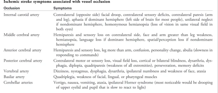
Disruption of blood flow to large cerebral arteries can cause an expected set of symptoms. Knowing the symp-toms, or stroke syndromes, can be helpful in assessing per-sons suspected of having a stroke. Stroke symptoms associated with the arterial occlusion are summarized in
Table 3.12,13Understanding how the disruption of blood flow to a certain cerebral artery creates a certain set of symptoms can be helpful in recognizing why persons pre-sent with different symptoms depending on the area of brain ischemia.
ISCHEMIC STROKE TREATMENT OPTIONS
Although the primary purpose of this article is to review nursing assessment for stroke, the goal of assessment is to be able to provide valuable, lifesaving, and disability-spar-ing treatment options for persons experiencdisability-spar-ing an acute stroke. Therefore a brief review of available treatment options is provided.
As previously discussed, the time of“last known well” is critical for identifying available treatment options for ischemic stroke (Table 4). Patients should be screened for inclusion and exclusion criteria for intravenous throm-bolytic drugs. If the patient meets the criteria, then the physician should discuss administration of thrombolytic agents, including the risks and benefits of the therapy. Emergency departments should have the medication for thrombolysis (Alteplase/recombinant tissue plasminogen activator) readily available because the time window for administration is critical. Thrombolytic therapy is approved by the Food and Drug Administration for treat-ment of ischemic stroke for persons within 0 to 3 hours of last known well for those who meet all inclusion and
no exclusion criteria. TABLE 3
Ischemic stroke symptoms associated with vessel occlusion
Occlusion Symptoms
Internal carotid artery Contralateral (opposite side) facial droop, contralateral sensory deficits, contralateral paresis (arm and leg), aphasia if dominant hemisphere (left side of brain for most people), unilateral neglect if nondominant hemisphere, homonymous hemianopsia (loss of vision in same visual field in both eyes)
Middle cerebral artery Hemiparesis and sensory loss on contralateral side, face and arm greater than leg weakness, hemianopsia, language loss if dominant hemisphere, spatial/perception loss if nondominant hemisphere
Anterior cerebral artery Hemiparesis and sensory loss, leg more than arm, confusion, personality change, abulia (slowness in responding to commands)
Posterior cerebral artery Contralateral motor or sensory loss, visual field loss, cortical or bilateral blindness, dysarthria, dys-phagia, diplopia, quadriparesis (weakness of all extremities), perseveration, memory deficits Vertebral artery Dizziness, nystagmus, dysphagia, dysarthria, ipsilateral numbness and weakness of face, ataxia Basilar artery Quadriplegia, weakness of facial, lingual, or pharyngeal muscles
Cerebellar arteries Vertigo, nausea, vomiting, ataxia, ipsilateral Horner syndrome (most noticeable would be drooping of upper eyelid and pupil that is slow to react to light)
Some patients can be considered for thrombolytic therapy from 3 to 4.5 hours from the time of last known well. Additional inclusion/exclusion criteria to be consid-ered include exclusion for persons older than 80 years, per-sons taking oral anticoagulant drugs, initial NIHSS score >25, and/or a combined history of diabetes and stroke.14
Patients who are not appropriate for thrombolytic therapy may be candidates for endovascular treatment options for stroke. These options include an embolectomy
via mechanical clot retrieval such as with the Merci retrieval device (Figure 1) or the Penumbra device. In this proce-dure a thrombus is identified inside of the artery and a phy-sician trained in endovascular procedures pulls the clot out of the vessel, thus restoring blood flow. These therapies are considered if the patient is within 8 hours of last known well; however, consideration for the amount of time needed to obtain arterial access and get microcatheters to the site of the occlusion must be taken into account.
Intra-arterial thrombolysis is an additional treatment option for centers that offer endovascular procedures.15 In this procedure, much smaller doses (compared with intravenous dosing) of a thrombolytic agent are given at the site of the thrombus. Use of this treatment option can be considered within 8 hours from the time the patient was last known to be well, again with the understanding that it takes time to gain access and get catheters to the site of the clot.
As previously noted, stroke treatments are dependent on the time the patient was last known to be well. It is cru-cial that nursing and medical staff accurately identify when patients presenting with stroke symptoms were last to be known well in order to determine what stroke interven-tions are appropriate to maximize outcomes.
Hemorrhagic Stroke
Hemorrhagic stroke occurs when a blood vessel in the brain ruptures. The problem is twofold; the area of the brain that the blood vessel is supposed to supply is now without blood flow, and the bleeding into the cranial vault causes pressure on the brain. Although hemorrhagic stroke is less common than ischemic stroke, the mortality asso-ciated with hemorrhage is 3 to 5 times higher.16
Hemorrhagic stroke can be caused by spontaneous rupture of a blood vessel, which typically is associated with long-term uncontrolled hypertension, rupture of aneur-ysms (outpouching of blood vessels), or AVMs (abnormal connection of veins and arteries). Symptoms associated FIGURE 1
Merci retrieval device. TABLE 4
Ischemic stroke treatment options
Last known well <3 h Last known well 3-4.5 h Last known well <8 h Consider thrombolytics (intravenous
or intra-arterial); if not thrombolytic candidate, consider endovascular treatment options
Consider thrombolytic drugs with additional exclusion criteria for this time period
Consider endovascular treatment optionsa,b
a
Endovascular treatment options vary among institutions and can include mechanical clot retrieval or clot disruption or intra-arterial thrombolysis.
with hemorrhagic stroke depend on the location and size of the bleed. Often, symptoms begin as a sudden severe head-ache, progressing to a decreased level of consciousness. If the hemorrhage is smaller, focal neurologic signs associated with the area of the brain involved may be present, such as changes in speech or vision or weakness.
Management of hemorrhagic stroke is based on the
cause of the bleeding.17 Options are summarized in
Table 5. Management of a ruptured aneurysm or AVM is different from management of an intracerebral hemor-rhage resulting from spontaneous rupture of a blood ves-sel, such as with hypertensive bleeding. Either surgical or endovascular approaches can be used to treat a ruptured aneurysm or AVM. Treatment of spontaneous bleeding, as in the case of hypertensive bleeding, typically is aimed at medical management. Medical management includes correction of coagulopathy and management of hyperten-sion, as well as monitoring for signs of increased intracra-nial pressure and neurologic decline.
Patients who have had a hemorrhage stroke require ongoing monitoring of airway, breathing, and circulation. Neurologic assessment of patients experiencing a hemor-rhagic stroke is vital for monitoring of the neurologic decline that can occur as the area affected by bleeding expands in size and brain tissues are compressed by the pressure caused by the bleeding. Thorough neurologic assessments should be performed frequently and compared with baseline, as well as with previous neurologic assess-ments. The NIHSS typically is not useful in patients who have experienced a hemorrhagic stroke. More com-monly, the Glasgow Coma Scale is used to monitor level of consciousness and other additional measures of neurolo-gic status are used, such as pupil checks, motor function, and vital signs. Vital signs are a part of the neurologic assessment because of the changes that can occur when intracranial pressure rises, such as in Cushing’s Triad. Cushing’s Triad includes widened pulse pressure (ie, systolic
pressure rises while diastolic pressure remains lower), brady-cardia, and abnormal respirations. These vital sign changes are a response to pressure increasing in the cranial vault, and combined with an abnormal neurologic examination, they should prompt the nurse that intracranial pressure may be elevated.12,13Ongoing assessment of the patient while he or she is in the emergency department is essential for making treatment decisions such as the need for intuba-tion, intracranial monitoring or surgery, blood pressure management, or other medical management.
Conclusion
Emergency nurses are key members of the stroke team and provide a vital role that is imperative to treatment of per-sons who are experiencing an acute stroke. Knowing that
“time is brain,” nurses who can quickly and accurately triage patients with stroke symptoms can help save lives and improve the chances of a meaningful recovery. Assess-ment of patients presenting with stroke-like symptoms, including a thorough medical history, onset and descrip-tion of initial symptoms, assessment of current symptoms, and evaluation for the possibility of acute stroke treat-ments, are critical skills for ED nurses. Knowing that stroke treatment options are highly dependent on the time of the onset of symptoms, nurses and the stroke team must work diligently and have processes in place to quickly triage, assess, and perform diagnostic tests in order to provide treatment for patients who have had a stroke, when provid-ing treatment is possible.
REFERENCES
1. Lloyd-Jones D, Adams R, Carnethon M, et al. Heart disease and stroke statistics—2009 update. A report from the American Heart Association Statistics Committee and Stroke Statistics Subcommittee.Circulation.
2009;119:21-181.
2. Heron MP, Hoyert DL, Murphy SL, Xu JQ, Kochanek KD, Tejada-Vera B. Deaths: final data for 2006.Natl Vital Stat Rep. 2009;57 (14):1-134.
TABLE 5
Hemorrhagic stroke treatment options
Option Details
Consultation considerations Neurosurgery/neurologic interventionalist/neurologic intensivist
Control coagulation abnormalities Consider the need for fresh frozen plasma, vitamin K, prothrombin complex, factor VII Control blood pressure Blood pressure control is determined by the cause of the hemorrhage; hypertension
should be avoided Monitor for increased intracranial
pressure
Assess level of consciousness frequently; a decline in level of consciousness may indicate increasing intracranial pressure and the need for intensive monitoring and possible surgical intervention
site/PageServer?pagename=RISK. Accessed August 25, 2010.
4. Kothari RU, Pancioli A, Liu T, Brott T, Broderick J. Cincinnati Pre-hospital Stroke Scale: reproducibility and validity.Ann Emerg Med.
1999;33(4):373-8.
5. Lyden P, Lu M, Jackson C, et al. Underlying structure of the National Institutes of Health Stroke Scale: results of a factor analysis.Stroke.
1999;30:2347-54.
6. Goldstein L, Samsa G. Reliability of the National Institutes of Health Stroke Scale.Stroke.1997;28:307.
7. Chernyshev OY, Martin-Schild S, Albright KC, et al. Safety of tPA in stroke mimics and neuroimaging-negative cerebral ischemia.Neurology.
2010;74:1340-5.
8. Hassan AE, Hassanzedeh B, Tohidi V, Kirmani J. Very mild stroke pa-tients benefit from intravenous tissue plasminogen activator without in-crease of intracranial hemorrhage.South Med J.2010;103(5):398-402. 9. American Stroke Association. NIHSS training.http://www.stroke.org/
site/PageServer?pagename=NIHSS. Accessed August 23, 2011.
10. National Institute of Neurological Disease and Stroke. www.ninds.nih. gov. Accessed August 23, 2011.
acute ischemic stroke: a scientific statement from the American Heart Association.Stroke.2009;40:3646-78.
12. Hinkle JL, Guanci MM, Bowman L, Hermann L, Mcginty LB. Cere-brovascular events of the nervous system. In:AANN Core Curriculum for Neuroscience Nursing. 4th ed. Washington, DC: American Association of Neuroscience Nurses; 2004.
13. Hickey JV, Todd AQ. Nursing management of patients with cerebro-vascular problems. In:The Clinical Practice of Neurological and Neuro-surgical Nursing. 6th ed. Philadelphia, PA: Wolters Kluwer/Lippincott Williams and Wilkins; 2009.
14. ECASS III. Thrombolysis with Alteplase 3 to 4.5 hours after acute ischemic stroke.N Engl J Med.2008;359:1317-29.
15. Furlan A, Higashida R, Wechsler L, et al. Intra-arterial Prourokinase for acute ischemic stroke: the PROACT II study.JAMA.1999;282:2003-11. 16. Andersen KK, Olsen TS, Dehlendorff C, Kammersgaard LP. Hemorrha-gic and ischemic strokes compared: stroke severity, mortality, and risk factors.Stroke.2009;40:2068-72.
17. Broderick J, Adams H, Barsan W, et al. Guidelines for the management of spontaneous intracerebral hemorrhage.Stroke.1999;30:905-15.