DIETARY EXPOSURES CONTRIBUTE TO DIFFERENT DISEASES THROUGH THE MICROENVIRONMENT
Yuanyuan Qin
A dissertation submitted to the faculty at the University of North Carolina at Chapel Hill in partial fulfillment of the requirements for the degree of Doctor of Philosophy in the Department
of Nutrition (Biochemistry) in the Gillings School of Global Public Health.
Chapel Hill 2015
ABSTRACT
Yuanyuan Qin: Dietary exposures contribute to different diseases through the microenvironment (Under the direction of Liza Makowski)
Genetic predispositions play an important role in our health, but these are not absolute; lifestyle choices help determine how long and how well we live. The global pandemic of obesity and related diseases is a good example. Lifestyle can contribute to diseases through altering the microenvironment of important tissues including adipose in obesity.
In this dissertation, the consequences of lifestyle components on local and systemic inflammation and hyperinsulinemia were investigated, both of which contribute to risk of diseases such as obesity, insulin resistance, and breast cancer. Using preclinical mouse models, we aimed to uncover mechanisms by which alcohol and obesogenic diet exposures alter the microenvironment in the pathogenesis of adipose and mammary gland dysfunction. We found that binge ethanol exposure followed by burn injury exacerbated the adipose inflammatory response induced by burn alone, with significant elevations in macrophage infiltration and secretion of pro-inflammatory mediators when compared to controls not exposed to binge alcohol intake. It is well established that adipose inflammation contributes to insulin resistance, especially associated with obesity. We discovered that fatty acid transporter 1 (FATP1)
transplanted with Fatp1-/- bone marrow and fed an obesogenic diet gained more weight and greater epididymal white adipose accretion, and became hyperglycemic and glucose intolerant. Obesity is a strong risk factor for an aggressive subtype of breast cancer called basal-like breast cancer (BBC) and reducing adiposity is predicted to lower incidence of BBC in human
To my parents, Yueqing Qin and Yuqin Wu, for their unconditional love and support throughout my graduate studies.
To my mentor, Liza Makowski, for serving as an incredible role model of women in science, guiding me the way to a scientist, trusting my ability and potientials, encouraging and pushing
me forward all the time.
To my committee members, for their guidance, support and advice towards my successes as a doctoral student.
PREFACE
Chapter 2 of this work was published in Alcohol Clin Exp Res. in 2014. The following authors contributed to the work in the following manner. Y. Qin designed, conducted and analyzed majority of the experiments, wrote manuscript, J. L. Hamilton conducted and analyzed gene expression data, M. D. Bird conducted the experiments and analyzed protein expression data, M. M. Chen and A. Zahs provided input on protein expression, E. J. Kovacs designed and conducted the experiments, L. Makowski oversaw studies, provided intellectual input and participated in all aspects of analyzing data and preparation of manuscript.
Chapter 3 of this work is in revision. The following authors contributed to this work: Amy R. Johnson, Yuanyuan Qin, Alex J. Freemerman, Megan J. Huang, Alyssa J. Cozzo, Liyang Zhao, Brante P. Sampey, J. Justin Milner, Melinda A. Beck, Blossom A. Damania, Joseph A. Galanko, Matthew L. Edin, Darryl C. Zeldin, Patrick T. Fueger, Brittney Bivins, Andreas Stahl, Liza Makowski. Specific author contributions to this work are as described. A. R. Johnson designed, conducted and analyzed majority of experiments and wrote manuscript. Y. Qin designed, conducted and analyzed majority of experiments and wrote manuscript. A. J. Freemerman and M. J. Huang conducted and analyzed gene expression data, J. A. Galanko performed statistical analysis for the data, B. Bivins conducted immunoblot for FATP1 protein expression. A. Stahl provided FATP1-/- mice and provided intellectual input. L. Makowski oversaw all studies and analyses and contributed to the writing of the manuscript.
Chen, Samantha M. Miller, David B. Darr, Joseph Galanko, Stephanie A. Montgomery, Ben Major, Gary L. Johnson, Melissa A. Troester, Liza Makowski. Specific author contributions to this work are as described. Y. Qin designed, conducted and analyzed majority of experiments and wrote manuscript, X. Chen conducted Multiplexed Inhibitor Bead Affinity Chromatography. J. A. Galanko performed statistical analysis for the data, S. A. Montgomery conducted the pathological analysis. L. Makowski oversaw all studies and analyses and contributed to the writing of the manuscript.
TABLE OF CONTENTS
LIST OF TABLES ... xi
LIST OF FIGURES ...xii
LIST OF ABBREVIATIONS...xiv
CHAPTER I:INTRODUCTION...1
Significance ...1
Alcohol consumption in the US ...4
Alcohol Metabolism...5
Binge Drinking and Trauma...5
The Obesity Pandemic ...6
Macrophage Subtypes ...8
Fatty Acid Transporter Protein 1 ...10
Breast Cancer...13
Risk Factor...14
Intrinsic Subtypes Based on Gene Expression...15
Basal-like Breast Cancer ...16
C3(1)-Tag Mouse Model...18
High Fat Diet, Obesity and Breast Cancer...19
Weight Loss and Breast Cancer...21
The Possible Mechanisms of Obesity-induced Cancer...22
CHAPTER П: ADIPOSE INFLAMMATION AND MACROPHAGE
INFILTRATION AFTER BINGE ETHANOL AND BURN INJURY...27
Overview...27
Introduction ...28
Results ...29
Discussion ...34
Materials and methods ...40
Tables and Figures ...44
CHAPTER III: MACROPHAGE FATTY ACID TRANSPORTER 1 (FATP1) DRIVES ALTERNATIVE MACROPHAGE POLARIZATION AND LIMITS OBESITY-INDUCED INFLAMMATION...50
Overview...50
Introduction ...51
Results ...54
Discussion ...64
Materials and methods ...72
Tables and Figures ...82
CHAPTER IV: REMODELING THE MICROENVIRONMENT IN EARLY ADULTHOOD BY WEIGHT LOSS RESTRAINED DIET-INDUCED BASAL-LIKE BREAST CANCER PROGRESSION...100
Overview...100
Introduction...102
Results ...104
Discussion ...110
Materials and methods ...118
CHAPTER V:SYNTHESIS...141
Summary of research findings ...141
Direction for future research ...142
Public Health Significance...144
LIST OF TABLES
Table 3.1: Hematologic analysis of Fatp1+/+ and Fatp1-/- blood...96 Table 3.2: Flow cytometric analysis of circulating Fatp1+/+
LIST OF FIGURES
Figure 1.1: Macrophage phenotypes...10
Figure 1.2: FATP1 topology model...12
Figure 1.3: Progression of breast cancer...14
Figure 1.4: C3(1)-Tag Mouse Model...19
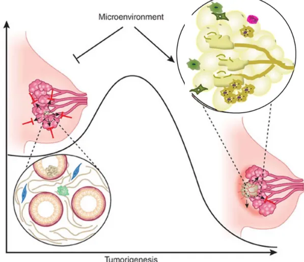
Figure 1.5: Microenvironment regulates tumor induction and progression...23
Figure 2.1: Interleukin-6 (IL-6) levels increase after single and episodic binge ethanol exposure and burn injury...44
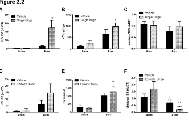
Figure 2.2: Adipose expression of neutrophil chemokine KC after single and episodic binge ethanol and burn injury...46
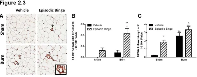
Figure 2.3: Episodic binge ethanol and burn injury drives crown like structure formation...47
Figure 2.4: Adipose chemokine levels of monocyte chemotactic protein-1 (MCP-1) and adiponectin were inversely regulated after single and episodic binge ethanol and burn injury...48
Figure 2.5: Single and episodic binge followed by burn injury failed to upregulate pro-inflammatory mediators...49
Figure 3.1: FATP1 is predominantly expressed in AAMs and transplant of fatp1-/- marrow results in FATP1 deficient macrophages...82
Figure 3.2: Deletion of MΦ Fatp1 increased susceptibility to weight gain, glucose intolerance, and increased white fat mass. Lipids are absorbed by small intestinal enterocytes and repackaged into chylomicrons for distribution to peripheral tissues... ...83
Figure 3.3: Lack of MΦ FATP1 increased adipose inflammation, inflammasome priming, and markers of oxidative stress in HFD-fed mice...85
Figure 3.4: Fatp1 deletion decreased acyl-CoA synthetase (ACSL) activity and resulted in a metabolic shift from lipid oxidation to glycolysis that resulted in exacerbated CAM and less AAM activation...87
Figure 3.6: Over-expression of FATP1 in RAW264.7 MΦs induced a substrate switch with enhanced lipid metabolism and reduced glucose
metabolism resulting in blunted CAM-activation...91
Figure 3.7: Over-expression of FATP1 reduced inflammatory response in RAW264.7 macrophages...93
Figure 3.8: FATP1 expression level had no effect on lean body mass, liver weight or brown adipose (BAT) weight...98
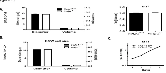
Figure 3.9: Deletion or over-expression of FATP1 had no effect on cell size or viability...99
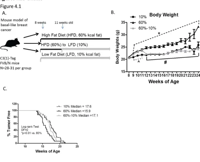
Figure 4.1: Weight loss protected against HFD-mediated early BBC onset...124
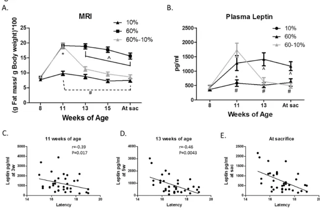
Figure 4.2: Body composition predicts latency...125
Figure 4.3: Diet did not affect tumor initiation...126
Figure 4.4: Pathological changes in the unaffected mammary gland induced by high fat diet were reversed by weight loss...127
Figure 4.5: Kinome profiling of unaffected mammary glands revealed dramatic regulation of PKD1-PKC-α-PKA-MEK3 by diet exposure...128
Figure 4.6: Protein-Protein interactions of significantly altered kinases in unaffected mammary gland of mice on 60-10% diet compared to mice on 60% diet... ...129
Figure 4.7: Tumor burden and size are not affected by diets...131
Figure 4.8: Measures of glucose intolerance were not altered by diet...132
Figure 4.9: Kinome profiling reveals significant regulation of pathways by HFD that are reversed with weight loss...133
LIST OF ABBREVIATIONS
2-DG 2-deoxyglucose 4-HNE 4-hydroxynonenal
7-AAD 7-aminoactinomycin D
AAM Alternatively activated macrophages ABCA1 ATP binding cassette transporter ABCA1 ABCG1 ATP binding cassette transporter ABCG1 ACSL Acyl-CoA synthetase
ADH Alcohol dehydrogenase ADK Adenosine kinase AKT Protein kinase B
ALDH Aldehyde dehydrogenase
AMPK 5'-AMP-activated protein kinase ANOVA Analysis of variance
ATMs Adipose tissue macrophages AUC Area under the curve BBC Basal-like breast cancer
BMDM Bone marrow derived macrophages BMI Body mass index
Bmp2K Bone morphogenetic protein-2-inducible protein kinase BMT Bone marrow transplant
CBCS Carolina Breast Cancer Study CCR2 C-C chemokine receptor type 2 CD36 Cellular differentiation
CDC Centers for Disease Control and Prevention CETP Cholesterol ester transfer protein
CIS Carcinoma in situ
CK Cytokeratin
CLS Crown-like structures CO2 Carbon dioxide
CoA Coenzyme A
CSFR1 Colony stimulating factor 1 receptor CYP2E1 Cytochrome P4502E1
DAB Diaminobenzidine DCIS Ductal carcinoma in situ
Ddr1 Epithelial discoidin domain-containing receptor 1 DIO Diet induced obesity
DMEM Eagle's minimal essential medium DTH Delayed-type hypersensitivity ECAR Extracellular acidification rates EGF1R Epidermal growth factor 1 receptor EGFR Epidermal growth factor receptor ER Estrogen receptor
ERK Extracellular signal-regulated kinases EtOH Ethanol
eWAT Epididymal white adipose FAEE Fatty acid ethyl ester Fas Fatty acids
FAT/CD36 Long-chain fatty acyl coenzyme A FATP1 Fatty acid transporter 1
FATP1-EV FATP1 empty vector FATP1-OE FATP1 overexpressing FATPs Fatty acid transport proteins FBS Fetal bovine serum
FFPE Formalin Fixed Paraffin Embedded Fn3k Ketosamine-3-kinase
GEMM Genetically engineered mouse model GLUT1 Glucose transporter 1
GLUT1-EV GLUT1 empty vector
Glut1MΦ-/- Macrophage specific Glut1 knock out GLUT1-OE GLUT1 over expresser
GSH Reduced glutathione GSSG Oxidized glutathione GTT Glucose tolerance test H&E Hematoxylin and eosin
HEPES 4-(2-hydroxyethyl)-1-piperazineethanesulfonic acid HER2 Human epidermal growth factor receptor 2
HFD High fat diet
HGF Hepatocyte growth factor HIF1α Hypoxia inducible factor 1
HMG-CoA 3-hydroxy—methylglutaryl coenzyme A i.p. Intraperitoneal
ICAM Intercellular adhesion Molecule ICAM-1 Intercellular Adhesion Molecule 1 IDC Invasive ductal carcinoma
IFN Interferon
IFNɣ Interferon gamma IGF1R IGF1 receptor
IGFs Insulin-like growth factors IHC Immunohistochemistry IL-10 Interleukin-10
IL-1β Interleukin-1β IL-6 Interleukin-6
iNOS Inducible nitric oxide synthase INSR Insulin receptor
Irak1 Interleukin-1 receptor-associated kinase 1 IRS Insulin receptor substrate
JAK Janus kinase
JNK C-Jun N-terminal Kinase
Kit Mast/stem cell growth factor receptor Kit LCCC Lineberger Comprehensive Cancer Center LCFAs Long chain fatty acids
LFA1 Lymphocyte function-associated anti-gen 1 LFD Low fat diet
Lfng Luntatic fringe LPS Lipopolysaccharide LXR Liver X receptor
MAPK Mitogen-activated protein kinase
Mark1 Serine/threonine-protein kinase MARK1 MCP-1 Monocyte chemoattractant protein 1 M-CSF Monocytes colony stimulating factor
MEK3 Specificity mitogen-activated protein kinase kinase 3 MEOS Microsomal ethanol oxidizing system
MIB Multiplexed Inhibitor Bead MMPs Matrix metalloproteinases MMTV Mouse mammary tumor virus MP1U Mouse Phase I Unit
MRI Magnetic resonance imaging MS Mass spectrometry
MΦ Macrophage
NAD+ Oxidized nicotinamide adenine dinucleotide NADH Reduced nicotinamide adenine dinucleotide NAF Normal associated fibroblasts
NF-KB Nuclear factor kappa-light-chain-enhancer of activated B cells NLRP3 NOD-like receptor family, pyrin domain containing 3
NO Nitrix oxide
OCR Oxygen consumption rate
OxLDL Oxidized low density lipoprotein PAI-1 Plasminogen activator inhibitor-1 PBS Phosphate buffered saline
PI3K Phosphatidylinositol-4,5-bisphosphate 3-kinase Pik3c3 Phosphatidylinositol 3-kinase catalytic subunit type 3 PPARs Peroxisome proliferator-activated receptor
PPP Pentose Phosphate Pathway PR Progesterone receptor
Prkaa2 5'-AMP-activated protein kinase catalytic subunit alpha-2 Prkca Protein kinase C alpha type
Prkd1 Serine/threonine-protein kinase D1 PTEN Phosphatase and tensin homolog qPCR Quantitative PCR
RPMI Roswell Park Memorial Institute medium
Salk-1 Soluble serine/threonine-protein kinase receptor R3 SAM S-adenosylmethionine
SDS Sodium dodecyl sulfate SMC Smooth muscle cells
SR-A Scavenger Receptor Class A Type I/II SREBP Steroid receptor element binding protein SREBP1 Sterol regulatory element binding protein 1 STAT3 Signal transducer and activator of transcription 3
STRING Search Tool for the Retrieval of Interacting Genes/Proteins SV40 Simian virus 40
T2D Type 2 diabetes TAG Triacylglyceride Tag T antigen
TCGA Cancer Genome Atlas
TGFα Transforming growth factor alpha TLR4 Toll-like receptor 4
TLRs Toll-like receptors
TNBC Triple-negative breast cancer TNF-α Tumor necrosis factor-α ULK3 Unc-51 like kinase 3
VEGFA Vascular endothelial growth factor A VLCFAs Very long chain fatty acids
WD Western Diet
CHAPTER I: INTRODUCTION
Significance
Lifestyle plays a very important role in both the health of individuals and the public. Obesity-related diseases like cardiovascular heart disease, stroke, type 2 diabetes and certain types of cancer are the major causes of death in the US that have modifiable risk factors, making them potentially preventable diseases1. Our genetic predisposition, which was once considered to be a determining factor for many diseases, may not be as important as we thought it was. Take cancers as an example. The study of cohorts of twins from Sweden, Denmark and Finland found that inherited genetic factors make a minor contribution to susceptibility to most types of
cancers2. The majority of cancers is considered preventable and could be avoided by adopting healthy lifestyle practices and avoiding unhealthy environmental factors3. In addition, diet can potentially modify genetic effects on diseases (the gene–environment interaction)4.
One of the leading causes of preventable death in the US is excessive alcohol
unknown if adipose tissue, an important organ in whole body metabolism, contributed to the inflammatory status after alcohol consumption and injury. Thus, part of this thesis aimed at discovering the underlying causes of systemic inflammation with alcohol exposure and focused on alterations to the adipose microenvironment.
Why is adipose tissue such an important organ to study as a contributing factor in many preventable diseases? More than one-third of U.S. adults (35%) are obese, and another one-third are overweight12. Obesity is considered to be a low-level chronic inflammatory condition that contributes to insulin resistance, type 2 diabetes, cardiovascular disease, cancer, and many other diseases13,14. It has been indicated that the infiltration of macrophages into adipose plays an important role in the inflammatory status associated with obesity15-17. Different phenotypes of macrophages contribute in varied ways to disease. Generally, classically activated macrophages (CAM), also called M1 macrophages, are found in adipose tissue of obese individuals,
contributing to inflammation; while alternatively activated macrophages (AAM), also known as M2 macrophages, are found dispersed in lean adipose tissue maintaining tissue function and homeostasis18. Importantly, M1 macrophages rely upon glucose as their main fuel, while M2 macrophages primarily use fatty acids19-21. With the recent knowledge that substrate metabolism is different in CAM versus AAM, the concept of metabolic reprogramming has arisen, wherein metabolism may drive the inflammatory response. Considering the important role of fatty acids and lipid metabolism in AAM, fatty acid transport proteins (FATPs) could be central in
macrophage-mediated inflammation and obesity22-27. While it was unknown whether
A disease tightly linked to obesity and often inflammation is breast cancer. Breast cancer alone is estimated to account for 29% (226,870) of all new cancer cases among women in the US in 2012, and it is the leading cause of cancer death among women ages 20 to 59 years28. An important advancement in the field of breast cancer research is recent stratification of breast cancer to different intrinsic subtypes based on gene expression profiles29. One of the subtypes called basal-like breast cancer (BBC) is highly aggressive with poor survival. Because it is triple negative for ER (estrogen receptor), PR (progesterone receptor) and HER2 (human epidermal growth factor receptor 2), it currently has no targeted therapy. Importantly, it is also more prevalent in premenopausal African-American women30.
Obesity is a well-known risk factor for BBC, although the mechanism of the relationship between obesity and BBC is not well known. Therefore, obesity could be an intervention target for breast cancer, especially in BBC prevention. Based on the Carolina Breast Cancer Study (CBCS), Milikan et al. estimated that up to 68% of BBC could be prevented by promoting breastfeeding and reducing abdominal adiposity31. It has not been proven, though, whether reducing weight would decrease the risk of BBC in women. Weight loss may protect against BBC onset and/or progress by modulating the microenvironment of the mammary glands32. The mechanisms need to be further explored and are investigated in this thesis.
disease onset and progress by regulating growth factors, kinases, cytokines, chemokines and possibly through other unknown pathways in the microenvironment. Therefore, our findings contribute to a growing body of evidence that the microenvironment (i.e. cellular composition, metabolism, and activation of specific kinase signaling cascades) in specific tissues contributes greatly to local and systemic pathologies.
Alcohol Consumption in the US
syndrome is also known as alcohol use disorder (AUD). Individuals must meet certain number of criteria in the Diagnostic and Statisticsal Manual of Mental Disorders (DSM) to be diagnosed with an mild, moderate or severe AUD37.
Alcohol Metabolism
Alcohol metabolism takes places primarily in the liver by three enzymatic pathways. The main pathway, which is responsible for most ethanol oxidation, is through alcohol
dehydrogenase (ADH) – aldehyde dehydrogenase (ALDH). The second pathway is the microsomal ethanol oxidizing system (MEOS) catalyzed by cytochrome P4502E1 (CYP2E1). CYP2E1 has been reported to be expressed in hepatocytes, Kupffer cells and adipocytes38. The third pathway for ethanol elimination is a nonoxidative pathway catalyzed by fatty acid ethyl ester (FAEE) synthase, resulting in the formation of fatty acid ethyl esters39. Importantly, besides in the liver, alcohol exposure involves changes in other organ systems, including intestinal barrier function, the innate immune system, and, as we and one other group has demonstrated, adipose tissue40.
Binge Drinking and Trauma
Binge drinking is the most common form of excessive drinking in the US. Alcohol
patients with burn-related injuries in the US have alcohol in their system at the time of admission, and the vast majority of those subjects are binge drinkers rather than chronic alcoholics8.
Burn injuries induce dramatic insulin resistance, hyperlipidemia, hyperglycemia and
hepatosteatosis in patients, greatly contributing to elevated morbidity and mortality47-49. Insulin resistance in liver, skeletal muscle and adipose tissue can persist even 3 weeks after a burn injury49,50. Burn injuries also drive systemic inflammation with elevations in pro-inflammatory cytokines and suppressed cell-mediated immunity, leading to multi-organ dysfunction51. Ethanol exacerbates burn-induced inflammation systemically and in the lung and adipose tissue52,53, and impairs the immune response, thus increasing a patient’s susceptibility to infection9. Clinical and laboratory studies have demonstrated that ethanol exposure prior to traumatic injury, such as a burn, markedly elevates systemic and tissue-specific inflammatory responses9,10 and is associated with poorer outcomes11. It is well established that alcohol increases the dysregulated
inflammatory and immune response caused by burns in animal models and human patients, but most of the previous focus has been on the lung and susceptibility to infection9. To date there were no studies examining the role of adipose tissue in contributing to the inflammatory response and insulin resistance associated with alcohol and burn trauma.
The Obesity Pandemic
The World Health Organization (WHO) defines overweight and obesity as excessive fat accumulation that is a risk factor to human health. A crude yet convenient population measure of obesity is the body mass index (BMI): a person’s weight (in kilograms) divided by the square of his or her height (in meters)54.
= Weigh (Ib) / Height2 (in2) * 703
In Western countries, a person with a BMI of 30 or more is considered obese, and a person with a BMI greater than or equal to 25 is considered overweight. Overweight and obesity are risk factors for many diseases, including diabetes, cardiovascular diseases and many types of cancers. The fundamental cause of obesity and overweight is an energy imbalance between calorie intake and calorie expenditure. Globally, there has been an increase in intake of energy-dense foods that are high in fat and sugar, and a decrease in physical activity due to the increasingly sedentary lifestyle of work, changing modes of transportation, and increasing urbanization55. Once
Macrophage Subtypes
The currently accepted understanding of obesity as low-level chronic inflammation that contributes to insulin resistance, type 2 diabetes, cardiovascular disease, cancer and many other diseases emerged in the early 1990s13,14. Although the mechanism is not fully understood, it has been indicated that the infiltration of macrophages into adipose tissue plays an important role in the inflammatory changes seen in obesity15-17.
Macrophages are important immune cells which reside in many tissues of the body, like microglia, Kupffer cells, osteoclasts, etc. Macrophages clear pathogens and apoptotic cells, and produce immune effector molecules58. They have a central role in protecting the host but also hinder the resolution of inflammation, which is a hallmark of numerous diseases, including atherosclerosis, obesity, tumor, asthma, etc.59. When there is tissue damage or infection, the precursors, monocytes, are rapidly recruited from the circulation to the tissue and differentiate into tissue macrophages60. Macrophages are remarkably plastic and can change their functional phenotypes depending on the environmental cues58.
CAMs are a prominent source of pro-inflammatory cytokines such as iNOS, TNFα, MCP1 and IL6. In contrast, AAMs are involved in tissue remodeling and repair68,69. Considering the important role of fatty acids and lipid metabolism in regulating macrophage biology, fatty acid transport proteins could be central in macrophage-mediated inflammation and obesity through metabolic reprogramming22-27.
Fatty Acid Transporter 1
The fatty acid transport protein (FATP) family consists of six members in humans and mice and is expressed in tissues with high levels of fatty acid uptake and active lipid metabolism,
Adapted from Johnson AR, 2012
main FATPs expressed in macrophages, with macrophage FATP1 expressed at mRNA concentrations as high as those in the total adipose tissue. FATPs have been shown to be fatty acyl-CoA synthetases with an affinity for long chain fatty acids (LCFAs) and very long chain fatty acids (VLCFAs)74,75. Acyl-CoA synthetases are enzymes that add a CoA to the fatty acid (Figure 1.2). Purified FATP-1 exhibited substrate specificity for fatty acids 16-24 carbons in length, while uptake of fatty acids shorter than 10 carbon atoms is unaffected by FATP expression70,74,76. The majority of FATP1 is found on intracellular structures like the
Whole body FATP1 deletion protected mice from fat-induced insulin resistance and intramuscular accumulation of fatty acyl-coA without alteration in adiposity87. Insulin-stimulated fatty acid uptake was completely abolished in adipocytes and greatly reduced in skeletal muscle of the FATP1 knockout mouse following a high-fat diet, while basal LCFA uptake by both tissues was unaffected88. In the same mouse model, lipids were redistributed from adipose tissue and muscle to the liver, leading to a complete protection from diet-induced obesity and insulin resistance88. FATP1 overexpression in human muscle cells led to enhanced palmitate and oleate uptake and the incorporation into triacylglycerides (TAG)89. A recent study indicated that FATP1, together with DGAT1, is part of a TAG synthesis complex that facilitates lipid droplet expansion in Caenorhabditis elegans78. However, some in vivo and in vitro studies also suggested
Jurgen Pohl. 2004
FATP1 channeled FA to oxidation by activating AMP-activated protein kinase (AMPK)90-92. Therefore, it is likely that FATP1 can transport LCFA to both lipid storage and lipid oxidation depending upon the cells, growth factors and tissues.
The role of FATP1 in insulin resistance is likely to be tissue-specific based on where and how fatty acids are stored. An increased FATP1-mediated fatty acid uptake into adipose tissue is beneficial for protecting muscle from harmful triacylglycerol accumulation, a phenomenon called “lipotoxicity”, while an increase in FATP1-mediated fatty acid uptake into muscle will contribute to insulin resistance. Macrophages can have extensive fatty acid metabolism, TG and cholesterol ester storage, especially AAMs or lipid-laden foam cells. Foam cells are fat-laden macrophages involved in fatty streak formation in atherosclerosis93. However, how FATP1 affects macrophage biology and the inflammatory response is unknown but may be important in understanding cell-specific contributions to obesity, inflammation and insulin resistance. Our research showed that when fed a high fat diet (HFD), mice with specific FATP1-/- macrophages are glucose intolerant, had greater weight gain, larger fat pads, and an increased inflammatory response in epidydimal adipose tissue and systemically when compared to control mice on HFD.
Breast Cancer
Carcinomas are cancers that arise from the epithelial component of the breast, which consists of the cells that line the lobules (milk glands) and milk ducts (thin tubes that carry milk from the lobules of the breast to the nipple). Ductal carcinoma is the most common type of breast cancer. Lobular carcinoma begins in the lobules of the breast, and ductal carcinoma originates from the milk duct. Generally ductal carcinoma can progress from atypical ductal hyperplasia (ADH) to ductal carcinoma in situ (DCIS, meaning within the ducts), and then to invasive ductal carcinoma (IDC) where the tumor invades the stroma surrounding the ducts98 (Figure 1.3). Breast cancer occurs in both men and women, but male breast cancer is rare99.
Risk factors
The strongest risk factor for breast cancer is age100. A woman’s risk of developing breast cancer increases as she gets older. Other risk factors of developing breast cancer include
Adapated from Kopans DB, 1989
inherited changes in certain genes, like Brca1 and Brca2, a family history of breast cancer, geographical location (developed country), higher breast density, menarche at early age (before age 11), menopause at older age (after age 54), a first full-term pregnancy after age 30, never having been pregnant, obesity, excessive alcohol consumption and low socioeconomic status101. There is high lifetime risk of developing breast cancer for women in the US. Approximately 12.3% of women will be diagnosed with breast cancer at some point during their lifetime, according to Cancer Statistics (2012) based on incidence data from the National Cancer Institute, the Centers for Disease Control and Prevention, and the North American Association of Central Cancer Registries102. The number of new cases of breast cancer was 124.6 per 100,000 women per year, and the number of deaths was 22.2 per 100,000 women per year in the US, using age-adjusted data based on 2007-2011 cases and deaths102. In 2011, there were an estimated 2,899,726 women living with breast cancer in the United States102.
Intrinsic subtypes based on gene expression
Breast cancer is not a single disease. Instead it is composed of a spectrum of tumor subtypes with distinct cellular origins, somatic changes, and etiologies103. In 2000, Perou’s group used a semi-unsupervised approach to identify breast cancer subtypes in a population of 40 patients with locally advanced disease treated with neoadjuvant chemotherapy104. They identified 496 genes, termed the intrinsic gene set, which showed little variance within repeated tumor sample, but high variance across different tumors, and then used this gene set for subtype
intrinsic suptypes include luminal A, luminal B, her2-enriched, basal-like, claudin-low and normal-like breast cancers105. These subtypes have been consistently identified in independent datasets using multiple different methods106-108. They are conserved across ethnic groups, and are present in preneoplasia109,110. Importantly, the intrinsic subtypes of breast cancer can predict patient relapse, overall survival, and response to endocrine and chemotherapy regimens111,112.
Basal-like breast cancer
In clinical studies, there is a group of breast tumors known as “triple-negative” due to their typical immunohistochemical (IHC) staining that is negative for ER, PR, and HER2, which are commonly scored for predictive markers in breast cancer clinics113. About 80% of triple-negative breast cancer (TNBC) is basal-like106. Likewise, a majority of basal-like breast cancers are triple-negative; about 25% of basal-like tumors are not triple-negative114. The basal-like subtype is characterized by low expression of the luminal genes and the HER2 gene cluster, high expression of the proliferation cluster, and high expression of genes called the basal cluster106. The basal gene cluster includes basal epithelial cytokeratins (CK) such as CK5, 6, 14, and 17; epidermal growth factor receptor (EGFR); c-Kit; Vimentin; P-Cadherin; Fascin; Caveolins 1 and 2; and aB-crystallin106. Importantly, it was the expression of cytokeratins 5, 6, 14, and 17 that gave rise to the term “basal-like,” as these are typically cytokeratins that are expressed within basal epithelial cells of the skin and airways103.
develop basal-like breast cancer, most basal-like breast cancers are sporadic, and the BRCA1 gene and protein appear intact in these tumors118. Other known genetic defects in basal-like tumors include a high P53 mutation rate119,120 and loss of RB1 function121,122. P53 and Rb are two tumor suppressor genes. Loss or mutation of P53 and/or Rb results in increased cell proliferation that is manifested by the high expression of the “proliferation signature”123.
Another notable association is between the basal-like subtype, race, and age. Several population-based studies have suggested that the basal-like subtype is prevalent in young women with breast cancer, especially in the African-American population120,124,125126. In the Carolina Breast Cancer Study, which is a population-based, case-control study of environmental and molecular
determinants of breast cancer risk that oversampled African American and premenopausal women120,127, basal-like breast cancer was the most common among premenopausal African-American women (27%) and least common among postmenopausal non African-African-American women (9%)31. Interestingly, this over-representation of basal-like cancers is even more prevalent in Africans in Nigeria128. These findings suggest that there may be a genetic
predisposition of Africans to basal-like tumors. The frequency of MYBL2 alleles has been found to be increased in basal-like cases versus controls129. In the Cancer Genome Atlas (TCGA) study, when tumors from different organs were analyzed based on their gene expression, basal-like breast tumors fell into their own category, indicating that basal-like breast cancer is distinct from other breast cancer subtypes130.
This indicates that basal-like and luminal subtypes require different prevention strategies based on different risks.
C3(1)-Tag Mouse Model
The C3(1)-Tag mouse model was created on FVB/N background in Jeffery Green’s lab at NCI. It was originally created for the study of prostate cancer. C3(1)-Tag mice express simian virus large T antigen (Tag) under the 5’ region of prostate steroid binding protein. SV 40 Tag can inactivate p53 and Rb, which are tumor suppressor genes, by directly or indirectly binding to them131. Researchers found that male C3(1)-Tag mice develop prostate cancer, while female ones develop breast cancer132. Green’s lab reported that, at 8 weeks old, C(3)1-Tag mice developed atypical hyperplasia of ductal epithelium; at 12 weeks ductal carcinoma in situ (DCIS) appeared; at 16 weeks female mice developed tumors and DCIS became invasive ductal carcinoma
(IDC)132,133. The SV40 Tag used in the C3(1)-Tag model inactivates p53 and RB, which also occurs in human basal-like tumors because these tumors are known to harbor p53 mutations134 (Figure 1.3&1.4). Hershkowitz et al. demonstrated that tumors from C3(1)-Tag mice 132 display characteristics consistent with human basal-like breast tumors including BRCA1-deficiency, TP53 mutant/deficiency, and high expression of Keratin 5, 17, and P-Cadherin135. Thus, for basal-like breast cancer, the C3(1)-Tag mouse is representative of human tumors.
fat pad and/or altered immune function), the C3(1)-Tag mouse has an intact stroma and requires no chemical treatment to initiate tumors.
High Fat Diet, Obesity and Breast Cancer
Obesity is associated with increased risk of cancers, including breast cancer. Increased body weight was associated with increased death rates for all cancers in a prospective cohort study of U.S. adults136. Considering the high prevalence of obesity (35.7%) and overweight (68.8%) in the US137, targeting obesity could be an effective intervention for cancer prevention. Adulthood is an important window of susceptibility for obesity-induced breast cancer138. Early epidemiologic studies indicated that obesity did not increase breast cancer risk among
premenopausal women, but did increase risk in postmenopausal women by around 50%139. Indeed, weight gain during adult life increases the risk of breast cancer among postmenopausal
Adapted from Murphy ED, 1966
women in the prospective Nurses' Health Study. However, recent advances in technology have shed light on breast cancer biology130 and specifically on obesity-induced risk. It has been
established that breast cancer has multiple subtypes29. Breast cancer can be classified based upon different intrinsic subtypes each with distinctive gene expression profiles as discussed above 29. This genomic-based classification is now being used to better understand breast cancer and can predict prognosis based on each subtype140. Therefore, early epidemiologic studies (that were not stratified based on subtype or only examined ER status) which suggested that obesity is only a risk factor in post-menopausal women, were actually based on populations heavily represented by the most prevalent breast cancer: Luminal A. Luminal A is a subtype that affects mainly older Caucasian women. In the case of BBC, several epidemiologic studies have demonstrated that obesity as measured by BMI or waist hip ratio (WHR) is associated with risk of BBC in both young (pre-menopausal) and older (post-menopausal) women31,141,142 . The prevalence of BBC is about 12.3–36.7% of all breast cancer cases143. The mechanism defining the relationship between obesity and BBC is not well known and is the subject of my studies. Of all the subtypes of breast cancer, BBC is a severe clinical problem because there are no targeted therapies, driving us to investigate potential modifiers of risk.
progression of claudin-low breast cancer compared to mice fed a control diet (10% calories from fat)148. Obesity induced by HFD was also found to promote mammary tumor progression in C57BL/6 mice with MMTV-Wnt-1 tumor cell xenografts149. Prior to our recent publications, no studies examined the effects of obesity on BBC using murine models. We previously reported that obesity significantly decreased BBC latency in C3(1)-Tag mice150.
Weight Loss and Breast Cancer
The WHO suggested that body weight and physical inactivity together account for approximately one-fifth to one-third of the most common cancers, including breast cancer151. It may be possible to decrease the risk of breast cancer by exercising and maintaining a healthy weight152-154. Michels et al. reported that weight loss of 5 kg or more since age 18, maintained for at least 4 years, was related to lower incidence of premenopausal breast cancer when compared to maintaining a stable weight155. In the Iowa Women's Health Study based on 34,000 women, maintaining ≥5% weight loss reduced post-menopausal breast cancer risk by approximately 25%156. A prospective cohort study within the Nurses' Health Study indicated that weight loss after menopause is associated with a decreased risk of breast cancer157. Furthermore, weight loss of 10% or more reduces breast cancer risk in postmenopausal overweight and obese women158. Based on Carolina Breast Cancer Study, it has been estimated that up to 68% of BBC could be prevented by promoting breastfeeding and reducing abdominal adiposity31. However, it hasn’t been proved whether reducing weight in adulthood would decrease the risk of BBC in
cancers in various animal models 157. A meta-analysis of experimental mouse models also suggested that energy restriction decreased tumorigenesis in the mammary gland160. Cleary et al. reported that intermittent calorie restriction-refeeding provided a moderate protective effect against mammary tumor development in MMTV-neu transgenic mice, which is an animal model of luminal breast cancer161. Caloric restriction using a murine xenograft model of BBC
demonstrated reduced tumor growth148. None of these studies examined weight loss in a
BBCwith a GEMM model. Green et al. demonstrated the protective benefits of exercise on BBC tumor progression, potentially through a significant exercise-induced weight loss, but the effects of exercise versus weight loss were not investigated162. Our group previously showed that in C3(1)-Tag mice made obese from weaning that were then induced to lose weight demonstrated a decrease in tumor progression that correlated with weight loss163. In sum, human and murine models demonstrate that high fat diet induced obesity is a strong but modifiable risk factor for breast cancer, although the protective effects and mechanisms of weight loss specifically on BBC must be further investigated.
The Possible Mechanisms of Obesity-induced Cancer
the stroma (the soil) is a likely hypothesis that may contribute to carcinogenesis. The
microenvironment surrounding a tumor has been realized to play an important role in tumor onset and progression32. When the microenvironment is normal, it can suppress tumorigenesis. However, changes in the microenvironment can also promote tumorigenesis and progression32 (Figure 1.5).
Modulated from Bissell MJ 2011; Sundaram S 2013.
The metabolites, growth factors, hormones and modifications of extracellular matrix in this microenvironment have critical effects on tumor cells. In fact, multiple reports from the Troester lab have demonstrated that BBC is characterized by unique epithelial-stroma
interactions, which likely play a role in the etiology164. There were numerous alterations in the gene expression profiles in the microenvironment of breast tumors compared to normal breast tissues165. BBCs are more likely to recur locally than non-BBC cancers166, even after
mastectomy with clean margins, suggesting a local field defect167 that may arise from epithelial or stromal alterations in the microenvironment.
Stroma-derived growth factors and metabolites are strongly relevant to obesity-induced cancer onset by modifying the extracellular matrix or angiogenic factors provided by the stroma. Hepatocyte growth factor (HGF) is a good example. High expression of cMet, the receptor of HGF, is significantly correlated with BBC in mouse and humans168. Our previous studies have shown that obesity upregulates HGF and cMet expression in the normal mammary gland of C3(1)-Tag mice, and this upregulation of HGF/cMet is correlated with a shortened latency150. Likewise, after life-long obesity, weight loss also decreased HGF/cMet expression in normal mammary glands163. cMet signature in 87% of BBC tumors and patients predicted worse
outcomes169. In a mutant mouse deficient for luntatic fringe (Lfng), a sugar transferase, the Perou lab demonstrated induction of BBC with amplification of the cMet locus and elevated cMet signaling170.
mammary glands of obese C3(1)-Tag mice (manuscript in prep). In addition to HGF release from immune cells like monocytes and macrophages, inflammatory cytokines release from activated macrophages contribute to breast cancer onset and development175,176.
Obesity can also disturb the microenvironment by reprogramming the kinome, or activating and interrupting growth factor/receptor signaling, such as leptin andother
mediators13,18. Leptin is a hormone produced by adipocytes in proportion to adiposity. A meta-analysis of case-control studies reported that women with breast cancer have elevated plasma leptin levels. High leptin receptor mRNA expression in breast cancer tissue was also established to predict poor prognosis in patients with high serum leptin levels. Therefore leptin may play a central role at the interface of obesity and breast cancer and may act through several pathways13. One way that leptin may drive BBC is that leptin promoted the survial of cancer stem cells (CSCs) in vivo 177, and leptin signaling is required to maintain CSC-like properties in triple negative breast cancer cells, which was shown to mediate tumor recurrence and metastasis in orthotopically transplanted mice. Leptin signaling also induces breast tumor cell growth through activation of the JAK-STAK and PI3K signaling pathway in both human and mouse cell lines. Leptin receptor deficiency in MMTV-PyMT mice was shown to attenuate tumor growth and metastasis through increasing mitochondrial respiration capacity in tumor cells and deactivating the downstream ERK and STAT3 pathways. About one fourth of all kinases may be involved in oncogenesis. Protein kinases regulate cell proliferation, survival, and metabolism and can contribute to tumor progression.
Summary
In modern society, lifestyle related factors might play a more important role in diseases compared to the genetic background. There is compelling evidence that the microenvironment can regulate disease onset and progress. This dissertation details evidence that the life style alters the microenvironment, which reflects and contributes to systemic inflammation, obesity, glucose intolerance, and basal-like breast cancer. This dissertation also addresses the gap in knowledge among obesity and cancer studies, which may shed light on mechanisms of how the
microenvironment is regulated by lifestyle factors and leads to different diseases. Considering the high prevalence of obesity in the US and the high rate of alcohol consumption (87.6% of adults), our studies will have a significant potential to develop prevention strategies for these public health issues, like breast cancer and inflammation-related diseases.
Hypothesis: Lifestyle-related factors like alcohol consumption and high fat diet contribute to varied diseases including obesity and breast cancer through altering the microenvironment.
CHAPTER П: ADIPOSE INFLAMMATION AND MACROPHAGE INFILTRATION AFTER BINGE ETHANOL AND BURN INJURY.
Qin Y, Hamilton JL, Bird MD, Chen MM, Ramirez L, Zahs A, Kovacs EJ, Makowski L.
Overview
Ethanol (EtOH) exposure prior to traumatic injury, such as a burn, elevates systemic and local inflammatory responses and increases morbidity and mortality. Adipose is a large tissue mass that is often inflamed during obesity or other stresses, which disturbs metabolic homeostasis. To date, there has been little investigation into the inflammatory response of adipose tissue after combined EtOH exposure and burn injury. Two EtOH exposure regimens were utilized to examine the role of inflammation in adipose tissue after EtOH and burn injury. Mice were either given a single or episodic binge exposure to EtOH or saline followed by scald (burn) or sham injury 30 minutes later. Twenty-four hours post injury, serum and adipose tissue were collected for assessment of inflammatory mediators. Single binge EtOH alone induced no inflammation in adipose when compared with sham vehicle-treated mice. However, single binge EtOH followed by burn injury induced significant elevations in mRNA and protein concentrations of pro-inflammatory mediators interleukin-6 (IL-6), KC, and monocyte chemoattractant protein 1 (MCP-1) compared with either insult alone or sham vehicle group. Additionally, EtOH exposure and burn injury significantly blunted inducible nitric oxide synthase (iNOS), indicating a
induced inflammatory parameters postburn were 2- to 5-fold greater than the response detected after a single exposure of EtOH, indicating EtOH-induced potentiation of burn-induced
inflammatory response. Finally, inflammatory loci and crown-like structures in adipose were significantly increased by episodic binge EtOH and burn injury. This is the first report of binge and burn-induced crown-like structure formation. Evidence presented herein suggests an important role for alcohol and burn as an additional mediator of adipose inflammation in postburn injury, a common complication in burn patients.
Introduction
Burn-induced hyperglycemia, hepatosteatosis, and insulin resistance are common complications observed in the burn patient population and are associated with poor outcomes, contributing significantly to morbidity and mortality47,48. Insulin resistance in liver, skeletal muscle, and adipose can persist even 3 weeks after burn injury49,50,178. Burn injury also drives systemic inflammation with elevations in pro-inflammatory cytokines and suppressed cell-mediated immunity, leading to multi-organ dysfunction179. One mechanism of systemic insulin resistance may be through adipose dysfunction and inflammation180. Macrophages are known to infiltrate adipose during states of metabolic stress, such as in obesity, and contribute to
animal models and patients (reviewed in9). We and others have shown previously that
neutrophils infiltrate the gut, lung, and site of injury after the combined insult181-186. While the primary role of neutrophils is to clear pathogens, they often cause damage due to production of enzymes such as elastase, reactive oxygen species, and pro-inflammatory cytokines including IL-6, IL-1β, and tumor necrosis factor alpha (TNF). Serum cytokines IL-6 and TNF and tissue
levels of KC and IL-6, are elevated in response to the dual insult of ethanol and burn injury compared to either injury alone182,186.
Chronic ethanol has been shown to drive macrophage infiltration into adipose tissue and it is associated with reduced fat mass due to upregulated lipolysis187,188. To date, it is not known how ethanol exposure combined with burn injury affects the adipose microenvironment. Using an established murine model of binge ethanol exposure and burn injury, we demonstrated that the combined insults drove systemic and adipose inflammation 24 hours post-injury.
Furthermore, we found that employing an episodic multi-day binge ethanol exposure paradigm followed by burn injury potentiated adipose inflammation and induced macrophage infiltration, indicating that binge, and especially episodic binge ethanol exposure, followed by burn drives adipose inflammation that could contribute to systemic inflammation.
Results
Single and episodic binge and burn injury drive inflammation in adipose tissue
ethanol exposure alone and increased 4-fold with burn injury compared to sham mice. IL-6 mRNA was significantly upregulated after the combined insult when compared to all other groups (P<0.001). Likewise, the adipose tissue level of IL-6 protein was unchanged after ethanol exposure alone, and was 6-fold higher in adipose tissue from burn vehicle mice compared to sham mice (Figure 2.1B). Ethanol exposure doubled the burn-induced elevation in adipose IL-6 protein (Figure 2.1B, P<0.05 burn ethanol vs. sham groups).
Further studies were conducted to determine if a more severe alcohol exposure paradigm, such as a multi-day episodic binge prior to burn would drive adipose inflammation when
compared to a single binge followed by burn. To accomplish this, mice were given ethanol (or saline) for 3 days, rested 2-4 days, and exposed again for 3 days prior to burn or sham injury. Ethanol alone did not modulate IL-6 mRNA or protein expression (Figure 2.1C&D). After episodic binge ethanol exposure and burn, adipose tissue IL-6 mRNA levels were 18-, 42- and 12-fold higher than sham vehicle, sham alcohol and burn vehicle, respectively, (Figure 2.1C, P<0.001 all groups versus burn ethanol). Significant interactions existed between burn and ethanol in single binge treatment and in episodic binge for IL-6 mRNA expressions. No significant interactions were found in single and episodic binge for IL-6 protein expression. Moreover, adipose levels of IL-6 protein reached 110 pg/mg, a value which is approximately 2 times that of burn alone and 8-fold that of sham groups (Figure 2.1D, P<0.05). Of note, IL-6 protein levels in adipose tissue were 3 times greater in tissue obtained from episodic binge and burn injury mice compared to tissue from mice exposed to a single binge and burn (Figure 2.1B
compared to Figure 2.1D).
Like IL-6, levels of the neutrophil chemokine KC were not increased after a single binge ethanol exposure, however, burn alone (in the absence of ethanol exposure) elevated KC mRNA and protein by approximately 5-fold, although this did not reach significance (Figures 2.2A&B). A statistically significant interaction between single binge and burn existed in the mRNA
ethanol treated mice (P<0.001, Figure 2.2F). This latter observation in episodic binge exposure differs from the adipose response to single ethanol exposure with and without injury where no significant differences were detected (Figure 2.2C&2.2F). No interaction was found in elastase mRNA expression in single binge, but there was a significant interaction (p<0.05) between burn and ethanol in episodic binge.
Macrophage infiltration is evident in adipose tissue after episodic binge exposure and burn
injury
We next sought to examine if macrophages were responsible for the observed elevations in adipose IL-6 levels by investigating the degree of macrophage infiltration into adipose tissue after episodic binge ethanol exposure and burn injury. We and others have demonstrated that the formation of crown-like structures is a well-documented measure of adipose tissue inflammation that correlates with insulin resistance23,180,188. Immunohistochemical (IHC) analysis was used to quantitate the number of F4/80-positive (F4/80+) crown-like structures. Representative images are shown in Figure 2.3A. No crown-like structures were detected in sham vehicle mice (Figure 2.3B). Mice exposed to ethanol or burn injury alone had detectable crown-like structures, but these measures did not reach statistical significance compared to sham vehicle mice (Figures 3B). However, the combined injury of episodic ethanol exposure and burn yielded a significant 2.5-and 4-fold increase in the crown-like structures compared to sham episodic binge, and burn vehicle, respectively (Figure 2.3B, P<0.01 both vehicle groups vs. burn ethanol, P<0.05 sham ethanol vs. burn ethanol). Sham ethanol treated adipose samples displayed an 8-fold elevation in F4/80+ inflammatory loci compared to sham vehicle (Figure 2.3C, P<0.05). There was a
P<0.01 sham vehicle vs. burn vehicle and P<0.001 sham vehicle vs. burn ethanol). No
interaction between burn and ethanol was found in episodic binge with the numbers of crown-like structures or inflammatory loci.
The expression of monocyte chemokine MCP-1 was next examined because it is a pro-inflammatory mediator demonstrated to be necessary and sufficient to drive macrophage infiltration into adipose tissue and induce crown-like structure formation180. In the absence of burn injury, mRNA and protein levels of MCP-1 were not altered by single binge ethanol alone (Figure 2.4A&B). The ethanol plus burn group displayed significant 5-fold increases in MCP-1 mRNA expression compared to both vehicle groups (Figure 2.4A, P<0.05). Burn injury alone doubled MCP-1 protein content in adipose versus sham vehicle (Figure 2.4B). MCP-1 protein was significantly increased in ethanol plus burn injured mice compared to sham treated groups (Figure 2.4B, P<0.01 sham vehicle versus burn ethanol, P<0.05 sham ethanol versus burn ethanol). Adiponectin is an anti-inflammatory adipokine that promotes insulin sensitivity 180. Nagy et al. demonstrated down regulation of adiponectin mRNA associated with chronic ethanol-induced adipose inflammation188. Single binge and burn significantly blunted
adiponectin mRNA expression by 75% (Figure 2.4C, sham vehicle (P<0.001) or burn vehicle (P<0.05 vs. burn ethanol). Episodic binge did not change MCP-1 mRNA expression in mice, nor were levels significantly upregulated after burn (Figure 2.4D). MCP-1 protein levels were elevated more than 2-fold in mice exposed to episodic binge plus burn injury versus sham vehicle controls, although this increase was not statistically significant (Figure 2.4E).
adiponectin levels (Figure 2.4F). No significant interactions between burn and ethanol were found in MCP-1 or adiponectin mRNA or protein expression with single or episodic binge treatment.
Protein levels of other pro-inflammatory (TNFα, IL-1β) or anti-inflammatory (IL-10) cytokines were not significantly different between each treatment group in single binge and burn studies (Figure 2.5 A-C). Interestingly, inducible nitric oxide synthases (iNOS) mRNA was not altered by ethanol exposure alone, but was significantly suppressed by 75% in both burn groups when compared to respective sham treated groups with greatest attenuation due to the
combination of ethanol and burn (Figure 2.5D, P<0.001 burn treated groups vs. sham treated groups). Episodic binge followed by burn did not significantly change protein levels of TNFα, IL-1β and IL-10 (Figures 2.5 E- G). Similar to acute binge exposure and burn injury, the
adipose iNOS mRNA level was not significantly altered by episodic ethanol, and was reduced by burn injury regardless of ethanol exposure. Compared to sham ethanol, burn ethanol iNOS mRNA was blunted 54% (Figure 2.5H, P<0.05). There were no significant interactions between burn and ethanol in any of the cytokines with single or episodic binge treatment.
Discussion
mortality47,49. Ethanol exacerbates burn-induced inflammation and impairs the immune response, thus increasing a patient’s susceptibility to infection9.
increasing adiponectin improves alcoholic fatty liver disease188,194. We demonstrate dramatic blunting of adiponectin after single binge and burn, which may aid in driving the
pro-inflammatory milieu. Finally, iNOS was down-regulated by single binge ethanol and burn injury. Interestingly, Syapin et al. have demonstrated that iNOS is inhibited by ethanol in glial cells195. In other tissues, burn has been shown to elevate iNOS expression or have no effect196,197. Hence, iNOS regulation by ethanol and burn in adipose needs further investigation. Taken together, our data support the findings that adipose tissue may be a source of some circulating cytokines after single binge ethanol and burn injury.
Binge drinking is not usually a single acute event; binge drinkers tend to consume alcohol in multiple binge episodes. Using an episodic binge model, we demonstrated that burn-induced inflammation was markedly elevated in mice given multiple exposures to binge levels of ethanol and that this occurred to a greater extent than following a single binge exposure. Similar to single binge, after episodic binge and subsequent burn, IL-6 and KC were elevated compared to either insult alone or sham vehicle controls. Despite elevations in KC protein in combined injury, neutrophil elastase mRNA level was dramatically reduced in burn ethanol groups relative to either insult alone. This was a surprising finding because other insults such as a 3 day exposure to high fat diet have been shown to induce neutrophil infiltration into adipose tissue190,198, which persisted for up to 90 days190. Perhaps a more detailed study of episodic binge and burn injury over a time course might capture neutrophil infiltration in response to high levels of KC. Even with dramatic increases in IL-6, KC, and MCP-1 associated with episodic binge ethanol
exposure, iNOS was blunted similar to a single binge. It is evident that while some production of pro-inflammatory cytokines or chemokines occurs in response to burn and ethanol, some
adipose inflammation as it has been shown to be both pro-inflammatory and anti-inflammatory in binge and burn injured animals, and may mediate some of the divergent effects demonstrated.
Finally, we report for the first time that F4/80+ macrophages were detected in adipose crown-like structures and inflammatory loci resulting from episodic binge ethanol exposure and burn injury, likely due to elevations in MCP-1. MCP-1 and its receptor CCR2 have been shown to mediate macrophage infiltration into adipose tissue in response to obesity180, and these
infiltrating macrophages often surround dying adipocytes forming crown-like structures180. Kang et al. have demonstrated infiltration of macrophages and production of pro-inflammatory
cytokines, IL-6, MCP-1and TNF, in adipose tissue after chronic ethanol exposure for 4
weeks188. Our data demonstrate that macrophage infiltration occurs within days after burn and ethanol exposure.
From our studies it is evident that burn injury primarily drives inflammation in adipose tissue, and ethanol exposure prior to burn potentiates this response. The dramatic increase in magnitude of response between single and episodic binge exposure suggests that the driving factor in the inflammatory response is the frequency of the ethanol exposure, since the burn injury and time of sacrifice is identical in each group. Our findings support the relevance of adipose inflammatory response to ethanol and burn insult that warrants further investigation. Like ethanol, obesity is also associated with a prolonged increase in pro-inflammatory mediators, such as IL-6 and MCP-1, an impaired immune response, and an increased susceptibility to
bacterial infection180,199. Work from our group and others over the past decade has linked adipose inflammation to obesity and insulin resistance22,23,180.
translocation into tissues to induce both organ-specific and systemic inflammation. We have previously reported on increased gut permeability and bacterial translocation in ethanol and burn models183,186,200,201. In humans and murine models, elevated morbidity and mortality result after burn injury due to inflammation secondary to intestinal permeability and septicemia183,186,201-203, which is often followed by an exaggerated alcohol-induced suppression of immune function through a lower delayed-type hypersensitivity (DTH) response and blunted lymphocyte proliferation203-206. At 6 and 24 hours post ethanol plus burn injury, gut permeability is compromised, which could account for elevated leukocyte infiltration, and IL-1β and IL-6 in mice exposed to ethanol then burn as compared to either insult alone183,186. Gut bacteria also regulate obesity susceptibility and systemic inflammation in response to high fat diet207.
Additionally, we have previously demonstrated the dependence of toll-like receptor 4 (TLR4, but not TLR2) in ethanol and burn-induced lung pathology and pro-inflammatory response189. TLR4 has also been shown to be necessary for obesity-induced inflammation208. Elevated gut
permeability and bacterial translocation after the combined insult of ethanol and burn may be responsible for the rise in lipopolysaccharide (LPS, endotoxin) burden and a TLR4 dependent inflammatory response in adipose, similar to obesity.
A second mechanism regulating ethanol and burn-induced alterations in adipose
inflammation is lipolysis and adipocyte apoptosis, which would release free fatty acids into the local microenvironment and circulation. In humans, chronic alcohol use correlated with reduced fat mass209. Burn injury also results in loss of fat mass through apoptotic cell loss210. Duffy et al. (2009) demonstrated that insulin and glucose are elevated in patients post-burn compared to healthy controls, with burn-induced increases in cytokine release from adipose tissue
rodents, single binge and burn drive a transient microvesicular steatosis, while chronic alcohol exposure also leads to reduced fat mass and adipocyte size, along with an increase in
hepatosteatosis and induction of systemic insulin resistance187,212. It has been shown that alcohol-driven lipolysis is not catecholamine-mediated213. Ethanol has been shown to increase levels of phosphatase and tensin homolog (PTEN) and suppressor of cytokine signaling (SOCS3), which are important negative regulators of insulin signaling in both liver and adipose and can lead to elevated lipolysis and cytotoxicity187,214. It is possible that saturated free fatty acids liberated by lipolysis, which are known to signal through TLRs during obesity-induced inflammation and insulin resistance215, may mediate a pro-inflammatory response after ethanol and burn exposure. Additionally, reduction in fat mass through apoptosis or lipolysis contributes to reduced insulin sensitivity because fat is redistributed from adipose to other metabolically sensitive tissues, such as the liver where hepatosteatosis ensues180. Liver IL-6 and other pro-inflammatory mediators increase with ethanol and/or burn exposure182,216. Emanuele et al. demonstrated single and/or combined injury increased hepatic ICAM-1, IL-1β, TNFα, and nuclear NF-KB217 which can lead to insulin resistance. Indeed, we have previously reported that insulin administration to rodents after ethanol and burn, improves liver inflammation and microvesicular steatosis, demonstrating further evidence of links between metabolic homeostasis and inflammatory response217.
Finally, a third mechanism linking ethanol intake to exacerbated inflammation is oxidative stress resulting from alcohol metabolism. Besides alcohol dehydrogenase (ADH), ethanol can also be metabolized through the microsomal ethanol oxidizing system (MEOS) by cytochrome P4502E1 (CYP2E1), which has been shown to lead to increased oxidative stress40. CYP2E1 is mainly expressed in the liver, but also found in the white adipose tissues218.
chronic ethanol feeding219. In our study, we failed to find a significant increase in mRNA or protein expression of CYP2E1 in adipose tissue of episodic binge, burned mice, or the combined exposure (data not shown). This may due to the type or length of exposure, as our treatment was shorter with either a one-dose single binge or episodic binge constituting a total of 6 days of ethanol exposure.
Taken together, we report for the first time that there is an inflammatory response in adipose tissue after the combined insult of ethanol and burn injury, and that this response is augmented after episodic binge relative to a single ethanol exposure. While binge drinking leads to unintentional injuries such as falls, crashes and burns, it may also lead to more insidious tissue inflammation as comorbidity with obesity leading to insulin resistance. Future studies in lean versus obese rodents could yield further mechanistic insight into burn and ethanol-induced effects on local and systemic inflammation.
Methods
Mice. Male C57BL/6 mice, 8 to 10 weeks old, were obtained from Jackson Laboratories (Bar Harbor, ME). They were housed in cages with food and water available ad libitum at the Loyola University Medical Center Animal Facility in rooms that were temperature and humidity
controlled on a 12-h light-dark cycle. All animal studies described here were performed
according to the Animal Welfare Act and the Guide for the Care and Use of Laboratory Animals, National Institutes of Health and were approved by the Loyola University IACUC. Before each experiment, mice were weighed and those weighing 22 to 27 grams were used in the studies. Murine model of ethanol exposure and burn injury. The murine model of a single binge ethanol exposure and burn injury was employed as described previously205,220 with minor
ethanol solution (1.12 g/kg) intraperitoneally (i.p.) that resulted in a blood ethanol level of 150 mg/dl at 30 minutes or saline vehicle221 or 2) for episodic binge exposure, mice were given the same dose of ethanol (or saline) daily for 3 days consecutively, rested for 2-4 days (rest time did not alter outcome, data not shown), and then given 3 additional daily exposures. Thirty minutes after ethanol exposure (or the last ethanol exposure in the case of the episodic binge paradigm), mice were anesthetized with 100 mg/kg of Ketamine and 10 mg/kg of Xylazine (Webster Veterinary, Sterling, MA), their dorsum shaved, and placed in a plastic template exposing 15% of the total body surface area of their back220 and subjected to a scald injury in a 90 to 92°C water bath for 8 seconds. As a control, sham animals were anesthetized, shaved, and immersed in room temperature water. The scald injury resulted in an insensate, full-thickness burn injury of approximately 15% total body surface area181. To compensate for fluid loss and prevent
circulatory shock, all animals received 1 mL of body temperature saline i.p. immediately after burn injury and were allowed to recover on warming pads. No other therapeutic intervention was provided as administration of anti-inflammatory or analgesic medication may introduce
confounding factors into the assessment of inflammatory responses. Twenty-four hours after burn injury, mice were sacrificed using carbon dioxide (CO2) inhalation and cervical dislocation. Blood was collected for serum isolation and measurement of cytokines. Epididymal white adipose tissue was removed. Tissue was either snap frozen in liquid nitrogen and stored at -80ºC for mRNA isolation or immunologic analysis or fixed in paraformaldehyde, paraffin-embedded, and sectioned for immunohistochemistry.