University of Pennsylvania
ScholarlyCommons
Publicly Accessible Penn Dissertations
1-1-2012
Regulation of Adamts13 Function in Hemostasis
by Cofactor and Substrate Exosite interactions
Christopher Gemil SkipwithUniversity of Pennsylvania, [email protected]
Follow this and additional works at:http://repository.upenn.edu/edissertations
Part of theBiochemistry Commons,Biophysics Commons, and thePathology Commons
This paper is posted at ScholarlyCommons.http://repository.upenn.edu/edissertations/701 For more information, please [email protected].
Recommended Citation
Skipwith, Christopher Gemil, "Regulation of Adamts13 Function in Hemostasis by Cofactor and Substrate Exosite interactions" (2012).Publicly Accessible Penn Dissertations. 701.
Regulation of Adamts13 Function in Hemostasis by Cofactor and
Substrate Exosite interactions
Abstract
ADAMTS13 (A Disintegrin And Metalloprotease with ThromboSpondin type1 repeats-13) is an enzyme that is mainly synthesized in the liver and secreted into the blood stream. In plasma, ADAMTS13 cleaves ultra large (UL) von Willebrand factor (VWF) newly released from stimulated and/or injured endothelial cells. It also cleaves soluble UL-VWF and VWF in flowing blood or at the site of growing thrombi under high shear stress. The basic enzymology and structure-function of the ADAMTS13-VWF interaction have presented many interesting questions, particularly in the context of physiological cofactors and fluid shear stress. The relatively poor understanding of this system reflects the complex assessment of VWF proteolysis, as VWF in solution exhibits a conformation that does not actively engage with ADAMTS13. It has been unclear whether force-induced unfolding of VWF is the only mechanism to enhance proteolysis by ADAMTS13, or if
interactions of ADAMTS13 and VWF with cofactors ideally position ADAMTS13 on VWF multimers for enhanced cleavage. We demonstrated that coagulation factor VIII (FVIII) and blood platelets cooperatively accelerate proteolytic cleavage of soluble VWF by ADAMTS13 through an alteration of VWF substrate conformation under physiologically relevant fluid shear stresses. In addition, we have established the critical role of the VWF propeptide and FVIII- and platelet-binding domains of VWF in regulating proteolysis of VWF by ADAMTS13 under physiologically relevant shear stress. Through site-directed mutagenesis, kinetic analyses, and peptide inhibition assays we have identified a substrate-binding exosite containing ADAMTS13 residues Arg659, Arg660 and Tyr661 that exhibits an important role in proteolytic cleavage of VWF under both non-physiological and physiological conditions. In addition, modification of this exosite region of ADAMTS13 yielded ADAMTS13 variants with reduced inhibition by autoantibodies and enhanced specific activity. Finally, we have demonstrated that infusion of ADAMTS13 and a truncated variant into
ADAMTS13-/- mice can restore the thrombus composition and kinetics of fibrin and platelet accumulation in an arterial thrombosis model. Together, these results suggest an important physiological role of cofactor binding to VWF and VWF interactions with ADAMTS13 exosites in regulating ADAMTS13 function in hemostasis.
Degree Type Dissertation
Degree Name
Doctor of Philosophy (PhD)
Graduate Group
Biochemistry & Molecular Biophysics
First Advisor Long Zheng
Keywords
ADAMTS13, Factor VIII, Thrombosis, TTP, von Willebrand Disease, von Willebrand Factor
Subject Categories
Biochemistry | Biophysics | Pathology
REGULATION OF ADAMTS13 FUNCTION IN HEMOSTASIS BY
COFACTOR AND SUBSTRATE EXOSITE INTERACTIONS
Christopher Gemil Skipwith A DISSERTATION
in
Biochemistry and Molecular Biophysics
Presented to the Faculties of the University of Pennsylvania
in
Partial Fulfillment of the Requirements for the Degree of Doctor of Philosophy
2012 Supervisor of Dissertation
_________________________
X. Long Zheng, M.D. Ph.D.
Associate Professor of Pathology and Laboratory Medicine
Graduate Group Chairperson _________________________
Kathryn Ferguson, Ph.D.
Associate Professor of Physiology
Dissertation Committee
Ronen Marmorstein, Professor of Biochemistry and Biophysics, Wistar Institute James Shorter, Ph.D., Assistant Professor of Biochemistry and Biophysics Sriram Krishnaswamy, Ph.D., Professor of Pediatrics, CHOP
John W. Weisel, Ph.D., Professor of Cell and Developmental Biology Yair Argon, Ph.D., Professor of Pathology and Laboratory Medicine, CHOP
REGULATION OF ADAMTS13 FUNCTION IN HEMOSTASIS BY
COFACTOR AND SUBSTRATE EXOSITE INTERACTIONS
COPYRIGHT
2012
iii
"It must be borne in mind that the tragedy in life doesn't lie in not reaching your goal. The
tragedy lies in having no goal to reach. It isn't a calamity to die with dreams unfulfilled, but it is a
calamity not to dream. It is not a disaster to be unable to capture your ideal, but it is a disaster to
have no ideal to capture. It is not a disgrace not to reach the stars, but it is a disgrace to have no
stars to reach for. Not failure, but low aim is sin."
- Dr. Benjamin Elijah Mays
I am nothing without those who have come before me, nor do I amount to anything
iv
ACKNOWLEDGEMENTS
My sincerest gratitude goes to my parents, to whom I owe the curiosity that has driven
my passion for science and my thirst for knowledge. My advisor, Dr. Long Zheng, manages a lab with wisdom, patience, optimism, and an especially noticeable zeal for
unmasking the mysteries of medicine. He is an asset to the scientific and medical communities and has been exceedingly supportive during my indoctrination to the world of science. The perfect complement to his constant focus on the “big picture” was the
seemingly endless knowledge of my long-time lab mates, Dr. Sheng-Yu Jin and Dr. Wenjing Cao, whose depth of technical expertise and experimental knowledge
contributed greatly to my growth as a scientist. This project would not have developed fully without the thoughtful input of my thesis committee, whose guidance helped me to understand the “big picture” about which my advisor would always speak. Many
colleagues have contributed to this work through generous donations of time, expertise, and input. These include Dr. Juan Xiao, Dr. Veronica Casina, and Christy Gandhi, whose
work preceded or complemented my own, and from whom much of my practical knowledge was gained. I am greatly indebted to Dr. Shekhar Kumar, Dr. Steven Stayrook, and my dear friend Roland Rivera-Santiago for their work and advice
regarding protein crystallography. My fellow graduate students in the Ernest E. Just Biomedical Society were a constant source of comic relief, intellectual guidance, and
v
ABSTRACT
REGULATION OF ADAMTS13 FUNCTION IN HEMOSTASIS BY COFACTOR AND SUBSTRATE EXOSITE INTERACTIONS
Christopher Gemil Skipwith X. Long Zheng, M.D., Ph.D.
ADAMTS13 (A Disintegrin And Metalloprotease with ThromboSpondin type1
repeats-13) is an enzyme that is mainly synthesized in the liver and secreted into the blood stream. In plasma, ADAMTS13 cleaves ultralarge (UL) von Willebrand factor (VWF)
newly released from stimulated and/or injured endothelial cells. It also cleaves soluble UL-VWF and VWF in flowing blood or at the site of growing thrombi under high shear stress. The basic enzymology and structure-function of the ADAMTS13-VWF interaction
have presented many interesting questions, particularly in the context of physiological cofactors and fluid shear stress. The relatively poor understanding of this system reflects
the complex assessment of VWF proteolysis, as VWF in solution exhibits a conformation that does not actively engage with ADAMTS13. It has been unclear whether force-induced unfolding of VWF is the only mechanism to enhance proteolysis by
ADAMTS13, or if interactions of ADAMTS13 and VWF with cofactors ideally position ADAMTS13 on VWF multimers for enhanced cleavage. We demonstrated that
coagulation factor VIII (FVIII) and blood platelets cooperatively accelerate proteolytic cleavage of soluble VWF by ADAMTS13 through an alteration of VWF substrate conformation under physiologically relevant fluid shear stresses. In addition, we have
vi
domains of VWF in regulating proteolysis of VWF by ADAMTS13 under physiologically relevant shear stress. Through site-directed mutagenesis, kinetic analyses,
and peptide inhibition assays we have identified a substrate-binding exosite containing ADAMTS13 residues Arg659, Arg660 and Tyr661 that exhibits an important role in
proteolytic cleavage of VWF under both non-physiological and physiological conditions. In addition, modification of this exosite region of ADAMTS13 yielded ADAMTS13 variants with reduced inhibition by autoantibodies and enhanced specific activity. Finally,
we have demonstrated that infusion of ADAMTS13 and a truncated variant into
Adamts13-/- mice can restore the thrombus composition and kinetics of fibrin and platelet
vii
TABLE OF CONTENTS
ACKNOWLEDGEMENTS ... IV
ABSTRACT ... V
LIST OF TABLES ... XI
LIST OF ILLUSTRATIONS ... XII
CHAPTER 1: INTRODUCTION ... 1
1.1 Overview ... 1
1.2 Maintenance of Hemostasis... 1
1.2.1 The vascular endothelium ... 1
1.2.2 Platelet aggregation as a hemostatic mechanism ... 1
1.2.3 The coagulation system ... 2
1.3 von Willebrand Factor Plays a Central Role in Hemostasis ... 3
1.3.1 von Willebrand Factor synthesis ... 3
1.3.2 Storage and secretion of von Willebrand Factor ... 4
1.3.3 von Willebrand Factor conformation and length ... 8
1.3.4 The effect of flow on von Willebrand Factor conformation ... 9
1.3.5 The interaction of von Willebrand Factor with other molecules ... 10
1.3.6 Molecular structure of von Willebrand Factor ... 14
1.4 von Willebrand Factor Pathophysiology ... 18
1.4.1 The deficiency or abnormality of VWF causes von Willebrand Disease ... 18
1.4.2 Genetics of von Willebrand Disease ... 19
1.4.3 Classifications of von Willebrand Disease ... 19
1.4 Structure, Evolution, and Cellular Functions of ADAMTS13 ... 20
1.4.1 ADAMTS13 is the von Willebrand Factor-cleaving protease ... 20
1.4.2 Specificity of ADAMTS13 ... 21
1.4.3 Molecular structure of ADAMTS13 ... 23
1.4 ADAMTS13 Pathophysiology ... 25
1.4.1 Deficiency of ADAMTS13 causes Thrombotic Thrombocytopenic Purpura ... 25
1.4.2 Autoantibodies in the pathogenesis of Thrombotic Thrombocytopenic Purpura ... 26
1.6 Specific Aims of the Thesis Research ... 27
viii
CHAPTER 2: PHYSIOLOGICAL COFACTORS FACTOR VIII AND
PLATELETS SYNERGISTICALLY REGULATE ADAMTS13 PROTEOLYTIC
ACTIVITY ... 37
2.1 Overview ... 38
2.2 Introduction ... 38
2.3 Materials and Methods ... 40
2.3.1 Preparation of plasma and recombinant proteins ... 40
2.3.2 Preparation of fresh and lyophilized human platelets ... 42
2.3.3 Removal of GP1bα receptor from fresh platelets by O-sialoglycoprotein endopeptidase ... 43
2.3.4 Ristocetin-induced platelet agglutination (RIPA) assay ... 43
2.3.5 Cleavage of multimeric VWF by ADAMTS13 under fluid shear stress ... 44
2.4 Results ... 45
2.4.1 Factor VIII and platelets synergistically increase cleavage of VWF by ADAMTS13 under fluid shear stress ... 45
2.4.2 Binding of FVIII to VWF is required for their synergistic effect on VWF proteolysis by ADAMTS13 under shear stress ... 50
2.4.3 Binding of platelet receptor GP1bα to VWF is also required for the synergistic effect of platelets with FVIII on VWF proteolysis by ADAMTS13 under shear stress ... 52
2.4.4 Assessing the amount of fluid shear stress generated in the assay system ... 54
2.5 Chapter Discussion ... 57
2.6 Chapter References ... 60
2.7 Supplemental Information ... 62
2.7.1 Towards a calculation of the average shear stress generated by the vortex method ... 62
CHAPTER 3: THE VWF D’D3 AND PROPEPTIDE DOMAINS PLAY A ROLE IN FACTOR VIII-MEDIATED ENHANCEMENT OF VWF PROTEOLYSIS BY ADAMTS13 ... 67
3.1 Overview ... 68
3.2 Introduction ... 68
3.3 Materials and Methods ... 70
3.3.1 Constructs ... 70
3.3.2 Preparation of plasma and recombinant VWF variants ... 70
3.3.3 Preparation of recombinant FVIII and ADAMTS13 ... 71
3.3.4 Quantitation of VWF concentrations by ELISA ... 71
3.3.5 VWF multimer analysis ... 72
3.3.6 Binding of FVIII or ADAMTS13 to type 2N VWF variants ... 72
3.3.7 Binding of type 2N VWF variants to FVIII ... 73
3.3.8 Western blotting ... 73
ix
3.3.10 Ristocetin-induced platelet agglutination ... 75
3.3.11 Ristocetin and platelet-induced cleavage of 2N VWF variants by ADAMTS13 under shear stress 76 3.4 Results ... 76
3.4.1 Multimer distribution of the expressed recombinant type 2N VWF variants ... 76
3.4.2 Binding of FVIII or ADAMTS13 to type 2N VWF variants ... 79
3.4.3 Cleavage of type 2N variants by ADAMTS13 under denaturing conditions is normal ... 81
3.4.4 Compromised proteolytic cleavage of type 2N VWF variants by ADAMTS13 in the presence of FVIII under fluid shear stress ... 83
3.4.5 Type 2N VWF variants are normal or nearly normal in ristocetin-induced platelet aggregation .... 88
3.4.6 Proteolytic cleavage of type 2N VWF/platelet complexes by ADAMTS13 in the presence of ristocetin ... 90
3.5 Chapter Discussion ... 91
3.6 Chapter References ... 97
CHAPTER 4: VWF PROTEOLYSIS BY ADAMTS13 IS EXQUISITELY CONTROLLED AT AN ADDITIONAL LEVEL BY EXOSITE BINDING ... 101
4.1 Overview ... 102
4.2 Introduction ... 103
4.3 Materials and Methods ... 105
4.3.1 Preparation of recombinant ADAMTS13 ... 105
4.3.2 Preparation of recombinant fluorescein-labeled VWF73 ... 106
4.3.3 Preparation of synthetic peptides ... 106
4.3.4 Proteolytic cleavage of fluorescein-labeled VWF73 peptide ... 107
4.3.5 Proteolytic cleavage of multimeric VWF ... 108
4.3.6 Crystallization screening and optimization ... 109
4.3.7 Data collection and structure refinement ... 109
4.3.8 Molecular modeling ... 109
4.4 Results ... 110
4.4.1 Synthetic peptides derived from the ADAMTS13 spacer domain or an isolated spacer domain of ADAMTS13 inhibits proteolytic cleavage of VWF by ADAMTS13 ... 110
4.4.3 Molecular modeling of VWF73 and RRYGEE complex ... 113
4.4.7 Molecular modeling of ADAMTS13-VWF specific interactions in the Cys-rich and Spacer domains ... 114
4.4.5 Crystallization of ADAMTS13 ... 116
4.4.6 Structure determination ... 119
4.5 Chapter Discussion ... 123
4.6 Chapter References ... 125
x
5.1 Overview ... 130
5.2 Introduction ... 131
5.3 Materials and Methods ... 133
5.3.1 Preparation of carotid artery samples and fixation for microscopy... 133
5.3.2 Immunofluorescent microscopy of carotid artery samples ... 134
5.3.3 Scanning electron microscopy of carotid artery samples ... 135
5.3.4 Quantitative analysis of micrographs by FOAMS (Fast Object Acquisition and Measurement System) ... 135
5.3.5 Focused ion beam milling and scanning-transmission electron microscopy of fixed artery sections ... 136
5.4 Results ... 137
5.4.1 Scanning electron microscopy indicates differences in the morphology and composition of WT and Adamts13-/- mice arterial thrombi ... 137
5.4.2 Kinetic analysis indicates that infusion of FL-ADAMTS13 or C-terminal truncated ADAMTS13 variant restores the kinetics of platelet/red blood cell accumulation and fibrin formation ... 140
5.4.3 An apparent increase in platelet density at the tail of thrombi in Adamts13-/- mice is corrected by infusion of FL-ADAMTS13 or C-terminal truncated ADAMTS13 variant ... 142
5.5 Chapter Discussion ... 144
5.6 Chapter References ... 145
CHAPTER 6: SUMMARY, GENERAL DISCUSSION, AND FUTURE PLANS 148 6.1 Summary ... 148
6.2 General Discussion ... 149
6.2.1 VWF binding to cofactors, and subsequent increased susceptibility to ADAMTS13 cleavage, is an autoregulatory mechanism ... 149
6.2.2 The collection of data about ADAMTS13 exosites informs the role of shear stress in the dynamic interactions taking place between ADAMTS13 and VWF ... 153
6.2.3 The co-existence of ADAMTS13 substrate exosites and inhibitory autoantibody binding sites suggests a flexibility in ADAMTS13 structure that has been utilized by nature ... 155
6.2.4 The observed autolysis of ADAMTS13 may point to other autoregulatory functions of ADAMTS13 ... 159
6.2.5 The in vivo correction of platelet and fibrin accumulation with infused ADAMTS13 suggests the importance of ADAMTS13 localization ... 160
6.3 Future Plans ... 162
6.3.1 Improvement of methods to determine kinetics of VWF proteolysis ... 162
6.3.2 Deciphering the relevance of ADAMTS13 autolysis ... 165
6.3.3 Achieving amino acid resolution of ADAMTS13 interactions ... 165
6.3.4 Rational design of ADAMTS13 for therapeutic purposes ... 166
6.4 Conclusions ... 167
xi
LIST OF TABLES
CHAPTER 1
Table 1.1. Classification of von Willebrand Disease ... 20
Table 1.2. Prevalence of autoantibodies against ADAMTS13 in patients with idiopathic TTP ... 26
CHAPTER 4
Table 4.1. Data Collection and Refinement Statistics ... 121
xii
LIST OF ILLUSTRATIONS
CHAPTER 1
Figure 1.1. The Coagulation Cascade. ... 3
Figure 1.2. Schematic representation of pro-VWF. ... 4
Figure 1.3. Biosynthesis, helical assembly, and secretion of VWF multimers. ... 5
Figure 1.4. Helical assembly of the N-terminal domains of VWF in the tubules of Weibel-Palade bodies. ... 7
Figure 1.5. Shear flow in vessels. ... 10
Figure 1.6. Molecular Structure of human factor VIII and thrombin cleavage. ... 11
Figure 1.7. Platelet surface receptors. ... 13
Figure 1.8. Crystal structures of the VWF A domains. ... 16
Figure 1.9. The VWF A2 domain. ... 17
Figure 1.10. Schematic structures of the ADAMTS13 gene and protein. ... 21
Figure 1.11. Crystal structure of the ADAMTS13-DTCS fragment. ... 24
CHAPTER 2 Figure 2.1. Initial purification of ADAMTS13-V5His. ... 42
Figure 2.2. Factor VIII and platelets synergistically accelerate cleavage of VWF by ADAMTS13 under shear stress. ... 47
Figure 2.3. Cleavage of VWF by ADAMTS13 in the presence of fixed concentration of FVIII and various concentrations of platelets under shear stress. ... 48
Figure 2.4. Cleavage of VWF by ADAMTS13 in the presence of fixed concentration of platelets and various concentrations of FVIII under shear stress. ... 49
Figure 2.5. Cleavage of VWF by ADAMTS13 in the presence of fixed concentration of FVIII-2RKR and various concentrations of platelets. ... 51
Figure 2.6. Disruption of VWF-platelet interactions inhibits cleavage of VWF by ADAMTS13 under shear stress. ... 54
Figure 2.7. Assessing the fluid shear stress generated by various vortexing assays. ... 56
xiii
Figure S2.2. Representative reading from the reaction system on the cone-plate viscometer.
... 65
CHAPTER 3 Figure 3.1. Schematic representation of type 2N mutations, and multimer distribution of variants. ... 78
Figure 3.2. Binding of FVIII or ADAMTS13 to recombinant type 2N VWF variants. ... 80
Figure 3.3. Western blotting analysis of the proteolytic cleavage of type 2N VWF variants in the presence of FVIII under denaturing conditions. ... 82
Figure 3.4. Western blotting analysis of the proteolytic cleavage of type 2N VWF variants in the presence of FVIII under fluid shear stress. ... 84
Figure 3.5. Kinetic analysis of proteolytic cleavage of type 2N VWF variants in the presence of various concentrations of FVIII under fluid shear stress. ... 86
Figure 3.6. Western blotting analysis of the proteolytic cleavage of type 2N VWF variants in the presence of various concentrations of FVIII under fluid shear stress. ... 87
Figure 3.7. Effect of type 2N mutations on ristocetin-induced platelet agglutination and VWF proteolysis by ADAMTS13. ... 89
Figure 3.8. Effect of type 2N mutations on ristocetin-induced VWF proteolysis by ADAMTS13. ... 91
Figure 3.9. Validation of the novel ELISA method for assessing proteolytic cleavage of VWF by ADAMTS13. ... 93
Figure 3.10. Proteolytic cleavage of VWF by leukocyte proteases. ... 95
CHAPTER 4 Figure 4.1. Synthetic peptides derived from ADAMTS13 spacer domain inhibit cleavage of VWF73 and multimeric VWF by ADAMTS13. ... 112
Figure 4.2. Proposed model of VWF73 and spacer domain peptide complex. ... 113
Figure 4.3. Modeling of ADAMTS13-spacer and VWF-A2 interaction. ... 115
Figure 4.4. Crystals of human FL-ADAMTS13 grown by the hanging-drop method. ... 117
Figure 4.5. Diffraction pattern obtained for human FL-ADAMTS13 crystals grown by the hanging-drop method (following optimization from Condition D). ... 118
xiv
Figure 4.7. Diffraction patterns and Wilson plots obtained for human FL-ADAMTS13
crystals grown by the hanging-drop method (following optimization from Condition F). . 120
Figure 4.8. Structure of ADAMTS13-CS and structural alignment with ADAMTS13-DTCS. ... 122
CHAPTER 5 Figure 5.1. Schematic showing preparation of carotid artery samples in C57BL/6 mice. .. 134
Figure 5.2. Immunofluorescent staining of carotid artery sections of WT mice after FeCl3 -induced thrombosis. ... 138
Figure 5.3. Scanning electron microscopy analysis of thrombi from the carotid arteries of Adamts13-/- mice after FeCl3-induced arterial thrombosis. ... 139
Figure 5.4. Time-dependent thrombus formation by scanning electron microscopy. ... 141
Figure 5.5. Focused Ion Beam milling with subsequent EM visualization of arterial thrombus sections... 143
CHAPTER 6 Figure 6.1. Schematic showing the cofactor effect of FVIII and platelets in VWF proteolysis by ADAMTS13. ... 150
Figure 6.2. Schematic showing the substrate exosite effect on enzymatic activity, as seen with ADAMTS13 and VWF. ... 154
Figure 6.3. Proposed model for in vivo cleavage of VWF by ADAMTS13. ... 155
Figure 6.4. Model of ADAMTS13 MDTCS showing surface-exposed exosites. ... 157
Figure 6.5. Sequence alignment of human ADAMTS13 TSP-1 repeats. ... 164
1
CHAPTER 1: INTRODUCTION
1.1 Overview
The coagulation system includes soluble and cellular factors that operate in concert to
prevent hemorrhage. For the coagulation system to serve the needs of the organism, the fluidity of blood must be maintained under physiologic situations and clotting must be promoted only when and where there is a threat of vascular interruption by trauma. This
process of maintenance of normal blood flow is termed hemostasis, whereby the system is maintained in a constant anticoagulant state1.
1.2 Maintenance of Hemostasis
1.2.1 The vascular endothelium
The vascular endothelium is strategically located at the interface between the tissues and blood. It is pivotal for protecting against vascular injury and maintaining blood fluidity.
Disruption of the vascular endothelium results in loss of the anticoagulant state. In addition to the loss of anticoagulant function, disruption of the endothelium exposes the subendothelial cell matrix, which contains collagen, von Willebrand factor (VWF) and
other substances to which circulating platelets bind1,2,3. Trauma also directly stimulates the expression of procoagulant activity by endothelial cells3.
1.2.2 Platelet aggregation as a hemostatic mechanism
Platelet aggregates bound to the subendothelium serve to reinforce the effect of
2
is physically unstable and is often insufficient by itself to permanently arrest bleeding4, which is why the process of clot formation is critical. Activated platelets accelerate the
rate at which clotting occurs compared with reaction rates in plasma by provide binding sites for the clotting proteins, concentrating and orienting them on the cell surface in a
way that enhances their efficient interaction4,5. Activated platelets also promote redistribution of anionic phospholipids from the inner to the outer cell membrane where they serve as important cofactors for several clotting reactions5.
1.2.3 The coagulation system
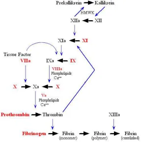
The coagulation system is composed of a series of proteins, most of which are synthesized in the liver and circulate in plasma primarily as single chain, inactive precursors (Figure 1.1)6. Once the coagulation system has been activated, several of these proteins are
converted into two-chain, active enzymes. In turn, these enzymes act upon other specific coagulation protein precursors converting them into active enzymes or into active
cofactors. Ultimately, the enzyme thrombin is generated, which cleaves fibrinogen to fibrin6,7. Polymerized fibrin is the predominant constituent of the visible clot. Cell surfaces accelerate coagulation reactions dramatically by binding and orienting enzymes,
substrates and cofactors7. These cell surfaces may include injured or activated endothelium, activated monocytes/macrophages, exposed smooth muscle cells, or
3
Figure 1.1. The Coagulation Cascade. The coagulation cascade can follow alternative routes
depending on the initiating factor. The extrinsic pathway is initiated by tissue factor and involves calcium ions and factor VII. In the intrinsic pathway, factors XII, XI, IX and VIII are activated by exposure to subendothelial collagen or foreign surfaces. Both pathways lead to the activation of factor X and proceed along the common pathway, involving factors V, II, I and XIII, to the formation of a fibrin clot.
1.3 von Willebrand Factor Plays a Central Role in Hemostasis
1.3.1 von Willebrand Factor synthesis
VWF is synthesized in all vascular endothelial cells, and in developing megakaryocytes7,8. A 275 kDa monomer of pro-VWF, consisting of the
inter-4
monomer disulfide bonds in the C-terminal cysteine-knot (CK) domain9. After transport to the Golgi (pH ~ 6.2), pro-VWF dimers form homo-polymers or multimers through an
additional disulfide bond near the amino terminus of the mature subunit, and the propeptide is then cleaved from pro-VWF by furin7,8. It is also in the Golgi that N-linked
glycans are processed and O-linked glycans are added9.
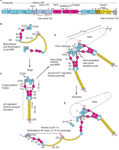
Figure 1.2. Schematic representation of pro-VWF. From Zheng and Sadler79. Binding sites are
indicated for collagen, FVIII, GP1b and integrin αIIbβ3. The cleavage site (Tyr1605-Met1606) and inter-subunit disulfide bonds (S-S) are also shown.
1.3.2 Storage and secretion of von Willebrand Factor
The multimeric VWF is stored in the Weibel-Palade bodies of endothelial cells and α
-granules of platelets. A small fraction of VWF multimers is constitutively secreted from endothelial cells9. It has been shown in vitro (with Ca2+ and pH 6.2) that D1-D2 monomers and D’D3 dimers can assemble into helices with the same dimensions and
three-dimensional shape as VWF tubules in Weibel-Palade bodies10,11. The ultimate purpose of this method of assembly is likely to overcome the challenges of synthesis of a
5
Figure 1.3. Biosynthesis, helical assembly, and secretion of VWF multimers. Modified from
6
VWF dimers linked by disulfide bonds near the N-terminal end in the D3 domain are assembled onto the ends of growing helical tubules in the Weibel-Palade bodies.
Pro-VWF dimers that are nearby have growing tubule ends that place one D’D3 domain adjacent to the D’D3 domain of the previously assembled pro-VWF dimer (Figure 1.3).
The role of this helical assembly is to encourage inter-dimer disulfide bond formation and enable furin cleavage between the D2 and D’D3 domains12. It has been shown that the organization of VWF dimers in the Weibel-Palade bodies is quite peculiar (Figure 1.4)11.
Whereas DNA forms structures with two molecules per helix, the VWF helices only contain one multimer11. Similar to DNA, however, there is 2-fold symmetry, which
7
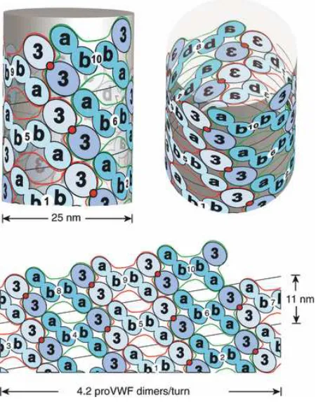
Figure 1.4. Helical assembly of the N-terminal domains of VWF in the tubules of
Weibel-Palade bodies. Modified from Huang et. al.11. Helical assembly is shown as progressing from
bottom to top. Each successive pro-VWF dimer is numbered and shown alternately as lighter domains outlined in red or darker domains outlined in green. Inter-dimer disulfide crosslinks form at the 2-fold symmetry axis between D’D3 domains (red circles). The D1 and D2 domains are denoted a and b.
Upon stimulation, VWF is secreted from endothelial cells as ultra-large (UL)-VWF multimers that form string-like structures attached to the endothelial cell surface13,
perhaps through interaction with P-selectin14. Orderly coiling of VWF in Weibel-Palade bodies is paramount to proper unraveling of VWF during secretion15. The first step in secretion involves formation of a narrow constriction between the plasma membrane and
8
expansion in Weibel-Palade body width and an increase in spacing between the tubules10. A reasonable conclusion is that dissociation of the D1-D2 pro-domain, coupled with
unraveling of VWF multimers, proceeds at the VWF tubules closest to the secretion pore12. Given this method of VWF secretion, the process may proceed such that multiple
VWF multimers are released in parallel. It has been shown that VWF molecules can self-associate16, and VWF fibrils extend from stimulated endothelial cells in the flow direction17.
1.3.3 von Willebrand Factor conformation and length
Electron microscopy of VWF at pH 7.4 reveals either compact or extended structures18,19. Atomic force microscopy (AFM) has demonstrated that, under shear flow, VWF adopts an extended conformation20. The domain organization can be described as variably-sized
beads spaced close together on a flexible string. This structure is interrupted by thin interdomain segments, such as O-glycosylated segments on either side of the A1 domain
(Figure 1.3)12. An estimate of the monomer size is about 60-70 nm based on the average spacing observed between domains18. VWF is highly flexible at pH 7.419, permitting dynamic, rapid changes in multimer shape under flow conditions.
SDS-agarose of VWF suggests that larger multimers consist of 14-20 dimers18. Electron microscopy shows longer VWF multimers that can consist of 30 dimers18. Under shear
flow, fluorescent-labeled VWF molecules can extend up to 15 µm21. Combining these measurements with the previously-mentioned monomer size of 60-70 nm, one can estimate that there are 60–250 monomers per multimer12. It has been reported that there is
9
electron microscopy11. Thus, a 5 µm long Weibel-Palade body tubule would yield a 250 µm long molecule of VWF, which is highly concordant with the observation of
100-1000 µm VWF strings after secretion from endothelial cells in vitro13.
1.3.4 The effect of flow on von Willebrand Factor conformation
The conformation of VWF after release is governed by fluid dynamics in vascular systems. The most common type of flow encountered by VWF is shear flow. In shear
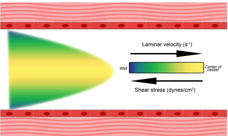
flow, the rate of fluid flow increases from the wall toward the center of the vessel (Figure 1.5). This system exhibits laminar flow, which dictates that the velocity of each lamina
increases from negligible at the wall to maximal at the center of the vessel (Figure 1.5)22,23. Conversely, the shear rate is highest at the wall. The shear flow experienced by VWF is a combination of rotational flow and elongational flow23. Rotational flow causes
particles to tumble, whereas elongational flow causes particles to adapt elongated conformations22. VWF multimers have a compact shape18,19,21,26 however in flow VWF
10
Figure 1.5. Shear flow in vessels. In shear flow, the rate of fluid flow increases from the wall
toward the center of the vessel. This system exhibits laminar flow, where the velocity of each lamina increases from negligible at the wall to maximal at the center of the vessel. The shear rate is highest at the wall.
This remarkable behavior of VWF under flow makes it such that VWF is most extended where its hemostatic functions are required. Because shear stress is greatest at the vessel wall, VWF which has been recruited to the sub-endothelial matrix experiences forces that
are much greater than free-flowing VWF12,14, suggesting that the forces experienced are solely elongational26.
1.3.5 The interaction of von Willebrand Factor with other molecules
Coagulation Factor VIII
VWF binds factor VIII (FVIII), a clotting cofactor, and stabilizes it in the circulation.
11
are organized as A1-A2-B-A3-C1-C2 (Figure 1.6). The mature protein has a molecular weight of 280 kDa composed of a light chain and a heavy chain28. The light chain has a
molecular weight of 80 kDa and is made up of domains A3-C1-C2, while the heavy chain is composed of domains A1-A2-B and has a more heterogeneous composition with
molecular weights varying from 90 – 200 kDa28.
Figure 1.6. Molecular Structure of human factor VIII and thrombin cleavage. Schematic
representation of the domain structure of FVIII. The heavy chain composed of A1–A2 domains is linked to a heterogeneously processed B domain of variable length. The light chain is composed of A3–C1–C2 domains. The three acidic regions are denoted as a1, a2, and a3. Upon activation by thrombin, FVIII is released from VWF, after which thrombin cleaves FVIII after Arg residues 372, 740, and 1689. Dissociation of FVIII from VWF is facilitated by the cleavage after Arg-1689. Cleavage after Arg-740 releases the dispensable B chain while cleavage after Arg-372 allows the A1 domain to separate from the A2 domain.
FVIII circulates in plasma at levels of approximately 200 ng/ml27,28. In vivo, a 1:50 ratio
12
through a non-covalent complex which persists until the FVIII is activated by thrombin28. The VWF-bound FVIII is protected from being inactivated by activated factor IX,
activated factor X and activated protein C29,30 and antibodies against FVIII31.
Upon activation by thrombin, FVIII is released from VWF27, after which thrombin
cleaves FVIII after Arg residues 372, 740, and 168932. Dissociation of FVIII from VWF is facilitated by the cleavage after Arg-168932. Cleavage after Arg-740 releases the dispensable B chain while cleavage after Arg-372 allows the A1 domain to separate from
the A2 domain28,32. Activated FVIII (FVIIIa) accelerates the rate of FX activation by FIXa and eventually leads to the formation of a blood clot.
Platelets
VWF also binds platelets (Figure 1.7) through interaction with the platelet receptor
glycoprotein Ibα of the GP1b-IX-V receptor complex in the VWF A1 domain33,34,35, and interactions of the VWF C-domains with integrins αIIbβ336,37 and αVβ338,39. The
ligand-binding site for VWF is located within the 45-kDa N-terminal region of GP1bα34,35. The GP1bα receptor is expressed on the platelet surface in a functional state that requires no prior activation to bind VWF. After initial platelet arrest due to GP1ba-VWF interaction,
intracellular signaling occurs and the platelets become activated34. This leads to conformational change and engagement of further receptors and the presentation of
13
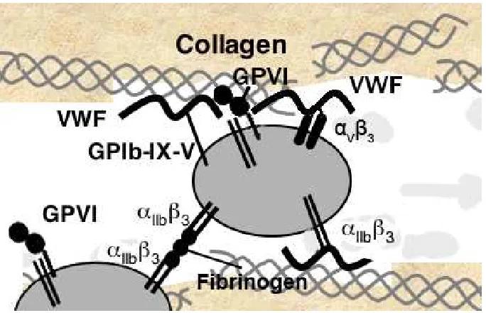
Figure 1.7. Platelet surface receptors. Normal hemostasis in response to vascular injury is
initiated by exposure of subendothelial matrix, allowing VWF and collagen access to their platelet receptors, GPIb-IX-V and GPVI. These interactions lead to rapid elevation of cytosolic Ca2+, cytoskeletal rearrangements, and activation of αIIbβ3 that binds VWF or fibrinogen and
mediates platelet aggregation.
There is a strong correlation between the effects of hydrodynamic flow on VWF
elongation and platelet aggregation. Studies have shown that VWF adsorbed to the wall of a flow chamber adheres to platelets through GPIbα37. The binding site for GP1bα in the A1 domain appears to be hidden or cryptic under static conditions, and exposed by
flow12,33,34. The D’D3 domain has been shown to shield recognition of platelets by the A1 domain40. VWF length may potentiate binding to GP1bα on platelets by enhancing shear
or elongational flow-dependent extension of VWF. The avidity of GP1bα to VWF is also modulated by flow37. Multivalent binding sites are better exposed in the extended conformation of VWF, as opposed to its compacted conformation. Furthermore, platelets
14
elongational force on VWF. This results in a positive feedback mechanism for amplifying hemostasis.
Collagen
VWF has also been shown to bind sub-endothelial collagens through the VWF A1 and A3 domains41. VWF mediates the adhesion of platelets to exposed subendothelium by forming a bridge between collagen, heparin-like glycosaminoglycans and other
components of the subendothelium. Several types of fibrillar collagen are bound by VWF in vitro42, and the major collagen binding site is within VWF domain A341, while a
second site may be present in the A1 domain43. Non-fibrillar collagen type VI binds domain A1 rather than domain A342. In addition, monoclonal antibodies can prevent VWF binding to fibrillar collagen or subendothelial connective tissue, but not both41,43.
VWF can be deposited on collagen substrates under static conditions41, however it has also been shown that the shear thresholds for VWF extension and deposition on collagen
substrates are superimposable21, indicating that, unlike platelet binding, VWF binding to collagen may not be dependent on hydrodynamic flow.
1.3.6 Molecular structure of von Willebrand Factor
The current understanding of the organization and boundaries of domains in VWF
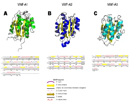
remains imperfect. Only the 3 A domains (A1, A2, A3) are well characterized with determined crystal structures44,45,46. The VWF A domains are the defining members of the VWA protein fold and family47. They have a central hydrophobic β-sheet with 6 β
15
domains are also found in integrins as ligand-binding αI and βI domains44,45, in complement components47, and in intracellular proteins with diverse functions47. Some
VWF A-like domains contain a metal-ion dependent adhesion site, but those in VWF do not12,47. VWF A-like domains in integrins and complement undergo substantial
conformational changes that regulate affinity for ligand47. Conformational change in VWF A domains has not yet been observed. VWF A1 and integrins differ substantially in the location of ligand binding sites on the surfaces of the VWF A-like domains. Most
modules in extracellular proteins are all-β44,45, and have their N- and C-terminal ends at opposite ends of the domain. In contrast, VWF A-like domains are α/β, and their N- and
16
Figure 1.8. Crystal structures of the VWF A domains. Panels A-C. Ribbon diagrams for
17
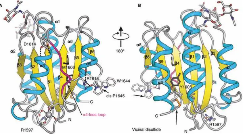
Unique among VWF domains, A2 lacks a long-range disulfide bond (Figure 1.7) and,
therefore, is unprotected from unfolding by tensile force applied along the length of VWF multimers47. Two 1.9 Å crystal structures of A246,48 have revealed surprising evolutionary
adaptations to the function of the VWF A2 domain as a “shear bolt” domain (Figure 1.9), and how mutations destabilize A2 in von Willebrand Disease (VWD)46,48. The wild-type mammalian A2 structure reveals two N-glycosylation sites, as well as a vicinal disulfide
bond46 whereas the mutant E. coli A2 structure reveals a Ca2+-binding loop48.
Figure 1.9. The VWF A2 domain. Adapted from Emsley et. al.47 and Zhang et. al.48 Ribbon
18 1.4 von Willebrand Factor Pathophysiology
1.4.1 The deficiency or abnormality of VWF causes von Willebrand Disease
von Willebrand Disease (VWD) is an inherited bleeding disorder that is caused by deficiency or dysfunction of VWF. Defects in VWF can cause bleeding by impairing
platelet adhesion or by reducing the concentration of FVIII. VWD is a relatively common cause of bleeding, but the prevalence varies considerably among studies and depends strongly on the case definition that is used. VWD prevalence has been estimated in
several countries on the basis of the number of symptomatic patients seen, and the values range from roughly 23 to 110 per million population (0.0023–0.01%)49.
The patient who led to the discovery of VWD was a 5-year-old girl who was seen by Dr. Erik von Willebrand50. He ultimately assessed 66 members of her family and reported, in 1926, that this was a previously undescribed bleeding disorder that differed from
hemophilia and exhibited mucocutaneous bleeding, autosomal inheritance, prolonged bleeding times, and normal clotting time50. In the 1950s, it became clear that a “plasma
factor” was decreased in these patients, and the factor was called “von Willebrand factor”49. As cryoprecipitate and commercial FVIII concentrates were developed, it was recognized that both VWF and antihemophilic factor (FVIII) purified together.
When immunoassays were developed, persons who had VWD were found to have reduced factor VIII-related antigen (FVIIIR:Ag) (which we now refer to as VWF antigen
19
Furthermore, a deficiency of VWF resulted in increased FVIII clearance because of the reduced VWF carrier protein.
1.4.2 Genetics of von Willebrand Disease
Since the 1980s, molecular and cellular studies have defined HA and VWD more precisely. Persons who had VWD had a normal FVIII gene on the X chromosome, but some were found to have an abnormal VWF gene on chromosome 1251. Variant forms of
VWF were recognized in the 1970s, and these variations are now recognized as the result of synthesis of an abnormal protein51. Gene sequencing identified many of these persons
as having a VWF gene mutation. The genetic causes of milder forms of low VWF are still under investigation, and these forms may not always be caused by an abnormal VWF gene. In addition, acquired disorders may result in reduced or dysfunctional VWF.
1.4.3 Classifications of von Willebrand Disease
VWD is classified into three major categories: partial quantitative deficiency (type 1), qualitative deficiency (type 2) and total deficiency (type 3) (Table 1.1)52,53. Type 2 VWD is divided further into four variants (2A, 2B, 2M and 2N) on the basis of details of the
phenotype. Before publication of the 1994 revised classification of VWD53, VWD subtypes were classified using roman numerals (types I, II, and III), generally
corresponding to types 1, 2 and 3 in the 1994 classification, and within type II several subtypes existed (designated by adding sequential letters of the alphabet, i.e. II-A through II-I). Most of the latter VWD variants were amalgamated as type 2A in the 1994
20
created. In addition, a new subtype (2M, with “M” representing “multimer”) was created to include variants with decreased platelet-dependent function but no significant decrease
of higher molecular weight VWF multimers54. Subtype 2N VWD was defined, with “N” representing “Normandy”, where the first individuals were identified, with decreased
FVIII because of VWF defects of FVIII binding54.
Table 1.1. Classification of von Willebrand Disease
Type Description
1 Partial quantitative deficiency of VWF
2 Qualitative VWF defect
2A Decreased VWF-dependent platelet adhesion with selective deficiency of high-molecular-weight multimers
2B Increased affinity for platelet GPIbα
2M Decreased VWF-dependent platelet adhesion without selective deficiency of high-molecular-weight multimers
2N Markedly decreased binding affinity for FVIII
3 Complete deficiency of VWF
1.4 Structure, Evolution, and Cellular Functions of ADAMTS13
1.4.1 ADAMTS13 is the von Willebrand Factor-cleaving protease
Newly released UL-VWF is not only large in size, but also functionally hyperactive. It binds platelets with increased affinity55 and causes abnormal platelet adhesion and
21
achieved by proteolytic cleavage of VWF by a plasma metalloprotease, ADAMTS13 (A
Disintegrin and Metalloprotease with Thrombospondin type1 repeats-13)13. The
translated product consists of 1,427 amino acid residues with an estimated molecular weight of 145 kDa58. It comprises a signal peptide, a propeptide, a metalloprotease
domain (Met), a disintegrin domain (Dis), first thrombospondin type 1 repeat, a Cys-rich domain and a spacer domain (Figure 1.10). The C-terminus of ADAMTS13 has seven additional TSP1 repeats and two CUB domains (Figure 1.9)58.
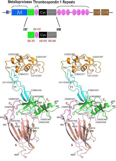
Figure 1.10. Schematic structures of the ADAMTS13 gene and protein. Modified from
Zheng et. al.58 ADAMTS13 consists of a signal peptide (S), propeptide (P) and a metalloprotease domain, followed by a proximal C-terminal region including disintegrin (Dis), first TSP1 (1), Cys-rich (Cys) and spacer domains. The middle and distal C-terminal regions have the 2nd to 8th TSP1 repeats and two CUB domains, respectively.
1.4.2 Specificity of ADAMTS13
The catalytic subunit, known as the metalloprotease domain (Met), contains a characteristic HEXXHXXGXXHD sequence that coordinates Zn2+ or Ca2+ binding58.
22
as GST-VWF7359,60 and FRETS-VWF7359,61). The ancillary domains within the proximal C-terminal region of ADAMTS13 including the disintegrin domain (Dis), the
first TSP1, the Cys-rich domain (Cys) and the spacer domain (Spa) appear to be required for recognition and cleavage of VWF under static/denaturing conditions59,61.
Removal of either domain within the proximal C-terminal region significantly reduces the ability of ADAMTS13 to cleave VWF and reduces the specificity59,61.
Cleavage of peptide substrates, such as GST-VWF73 and FRETS-VWF73, requires
even fewer domains of ADAMTS1359, suggesting that the interaction between ADAMTS13 and full-length VWF is quite different from that between ADAMTS13 and
peptide substrates. Regardless of the kind of substrate used, it has been demonstrated that the Cys/Spa region of ADAMTS13 is critical for recognition of VWF, partly through its binding to the amino acid residues (Glu1660-Arg1668) in the VWF-A2
domain60.
The role of the more distal C-terminal domains (such as TSP1 repeats and CUB domains)
of ADAMTS13 in recognition of VWF, however, remains controversial. Peptides or the fragment derived from the first CUB domain of ADAMTS13 inhibited cleavage of endothelial cell bound UL-VWF multimers62, but another study showed that an
ADAMTS13 mutant truncated after Spa was active in cleavage of endothelial cell bound UL-VWF using the same assay63. Under physiologically relevant conditions, the
cooperative activity between the TSP1 5-8 and CUB domains appeared to be critical for recognition of soluble VWF under fluid shear stress64, suggesting that binding of the middle and distal C-terminal domains of ADAMTS13 may be required for further
23
Spa. Homozygous mutations found in hereditary TTP resulted in truncation of ADAMTS13 four amino acid residues after Spa65, further supporting the role of the
middle and distal C-terminal domains in ADAMTS13 function in vivo.
1.4.3 Molecular structure of ADAMTS13
To date, crystal structures of the M and D domains of three human ADAMTS-family proteins, ADAMTS166, ADAMTS467 and ADAMTS567, have been reported. The first
crystal structure of exosite-containing fragments of ADAMTS13 was reported by Akiyama et. al68. The structures of the ADAMTS13-DTCS fragment, obtained at 2.6-Å
and 2.8-Å resolution, revealed linearly distributed exosites that recognize distinct substrate regions (Figure 1.11)68.
The ADAMTS13 cleavage site at residues Tyr1605 and Met1606 lies in a very inaccessible
site in the folded A2 domain, situated in the center of the β4-strand, sandwiched between amphipathic α-helices and loops on either side (Figure 1.9)46. The regions around the
cleavage site, as well as between the cleavage site and the C-terminus, are poorly packed46. Multiple side chain conformations are visible for cleavage site residues Met1606 and Leu1603, whereas disordered side chains are not seen in similar buried positions in the
crystal structure of the VWF A1 and A3 domains (Figure 1.8)44,45,46. Cys1669 and Cys1670 are linked in a vicinal disulfide bond at the C-terminus of the A2 domain (Figure 1.9)46.
Strikingly, all A2 interactions occur in this C-terminal portion, and this region is bounded by the β4-strand and the α6-helix, which contain the key recognition sites for ADAMTS13, which have been defined using peptide substrates60,69,70,71. Deletion of
24
Figure 1.11. Crystal structure of the ADAMTS13-DTCS fragment. Adapted from Akiyama
25 1.4 ADAMTS13 Pathophysiology
1.4.1 Deficiency of ADAMTS13 causes Thrombotic Thrombocytopenic Purpura
Thrombotic Thrombocytopenic Purpura (TTP), first described by Eli Moschcowitz in
192473, is a potentially fatal syndrome. The clinical manifestations of this disease are thrombocytopenia, microangiopathic hemolytic anemia, neurological symptoms and
signs, and various degrees of renal functional abnormalities74. TTP is caused by the inability to cleave VWF due to hereditary8,75,76,77 or acquired deficiency13,14,55 of plasma ADAMTS13 activity.
Approximately 5% of all TTP cases are caused by hereditary mutations of ADAMTS138. The mutations are present throughout the gene, causing changes in
amino acid composition, truncation of protein and impaired secretion78,79,80. Most cases of adult TTP are caused by autoantibodies against ADAMTS13. In the earliest reports, inhibitors of ADAMTS13 were found in 65% to 96% of patients with acquired
(idiopathic) TTP with severe deficiency (less than 5∼10% of normal activity)74. In later
prospective studies, a lower prevalence of anti-ADAMTS13 autoantibodies (31-44%) was reported (Table 1.2)81,82. Assessment of binding activity by ELISA has indicated
that autoantibodies can be found in almost all patients with acquired (idiopathic) TTP who exhibit a severe deficiency of plasma ADAMTS13 activity83. Autoantibodies
26
Table 1.2. Prevalence of autoantibodies against ADAMTS13 in patients with idiopathic TTP
Authors, year81,82,83,84,85 Number of
positive/investigated % positive
Furlan et. al., 1998 20/24 83%
Tsai et. al., 1998 26/39 67%
Tsai et. al., 2001 31/41 76%
Veyradier et. al., 2001 30/59 51%
Rich et. al., 2002 26/29 90%
Study et. al., 2003 31/50 62%
Coppo et. al., 2004 17/31 55%
Zheng et. al., 2004 7/20 44%
1.4.2 Autoantibodies in the pathogenesis of Thrombotic Thrombocytopenic Purpura
Despite of the importance of autoantibodies in pathogenesis of TTP, and its presence correlates with more relapsing disease and poorer prognosis than its absence82,86,87, the
binding epitopes and functional consequences of many autoantibodies against various domains of ADAMTS13 remain poorly understood. A limited number of published studies demonstrated that the major binding epitopes of autoantibodies are localized to
Cys and Spa82,83,86,87,88.
Using recombinant ADAMTS13 and variants expressed in mammalian cells, our lab
also showed that approximately 30-45% of TTP patients harbored antoantibodies recognizing the middle TSP1 2-8 repeats and distal CUB domains82. Cys and Spa have been shown to play a critical role in the recognition and cleavage of a peptidyl substrate
27
roles of the middle and distal C-terminal domains remain unclear. Understanding the biological and pathological significance of these autoantibodies against the middle and
distal C-terminal domains will provide novel insight into the pathogenesis of TTP.
1.6 Specific Aims of the Thesis Research
The long-term goal is to understand of the biology of VWF processing by ADAMTS13, and thereby the pathophysiology of thrombotic complications. ADAMTS13 cleaves
native VWF relatively poorly, and its preference for cleavage of the larger multimers is considered to lie in their greater susceptibility to deformation under shear stress or under
flow. The specific hypothesis behind this thesis research is that physiological cofactors form ternary complexes with VWF and ADAMTS13 and, in addition to shear stress, cause the required conformational change of VWF to accelerate proteolytic processing by
ADAMTS13 and that the ability of ADAMTS13 to recognize and cleave VWF is governed by exosite interactions with VWF.
Chapter 2 demonstrates that both FVIII and platelets can serve as cofactors for
enhancing VWF proteolysis by ADAMTS13 by synergistically altering VWF conformation under shear stress. This rate-enhancing effect of both platelets and FVIII on
ADAMTS13-mediated VWF proteolysis depends on the specific high affinity interactions between FVIII/platelets and VWF.
Chapter 3 demonstrates that a naturally-occurring type 2N VWF variant that exhibits significantly reduced FVIII binding is compromised in proteolytic cleavage by ADAMTS13
in the presence of FVIII, proportional to the defects of their FVIII binding. These results
28
regulating VWF proteolysis by ADAMTS13 and suggest an evolutionary advantage by
limiting VWF proteolysis in cases of VWD. Currently, the developed assays for detecting ADAMTS13 cleavage of VWF require simultaneous cleavage of scissile bonds in adjacent subunits, greatly underestimating the true catalytic efficiency of ADAMTS13.
Thus, Chapter 3 also describes a novel ELISA-based assay detects the N-terminal peptide (i.e. EQAPNVY) resulting from cleavage after the Tyr residue occurring
anywhere along a VWF multimer, which dramatically increases the sensitivity of the ADAMTS13-mediated VWF cleavage assay.
Chapter 4 examines the specific exosite interactions that govern binding to the Spa
domain of ADAMTS13 and their effects on VWF cleavage and binding of inhibitory anti-ADAMTS13 antibodies. This work, as part of two published papers, was primarily
done by others in the lab. Here, I describe my contributions to the project. Following is a description of the crystallization, screening, data collection, and structure determination for an autolysis product of ADAMTS13, the ADAMTS13-CS fragment.
Chapter 5 explores the in vivo function of ADAMTS13, specifically the composition of
Adamts13-/- mice thrombi, as well as the differential composition at the head and tail of
the thrombus. We determined that infusion of recombinant FL-ADAMTS13 or a
truncated ADAMTS13 variant into Adamts13-/- mice restored the kinetics of platelet/red blood cell accumulation and fibrin formation to those observed in wild-type mice. Our findings, revealing the apparent difference in thrombus composition, provide novel
29
Chapter 6 concludes with a discussion regarding the implications of the results described
in the preceding chapters as well as a comprehensive analysis of future works in the field
of cofactor- and exosite interaction-mediated VWF proteolysis by ADAMTS13.
1.7 Chapter References
1. Milstone, JH. The chain reaction of the blood clotting mechanism in relation to the theory of hemostasis and thrombosis. Blood 1949; 4: 1290-7.
2. Hoffman M. Remodeling the blood coagulation cascade. J Thromb Thrombolysis 2003; 16: 17-20.
3. Caen JP, Rosa JP. Platelet-vessel wall interaction: from the bedside to molecules.
Thromb Haemost 1995; 74:18-24.
4. Jurk K, Kehrel BE. Platelets: physiology and biochemistry. Semin Thromb Hemost 2005; 31: 381-92.
5. Tanaka KA, Key NS, Levy JH. Blood coagulation: hemostasis and thrombin regulation. Anesth Analg 2009; 108: 1433-46.
6. Becker RC. Cell-based models of coagulation: a paradigm in evolution. J Thromb
Thrombolysis 2005; 20: 65-8.
7. Sadler JE. von Willebrand factor. J Biol Chem 1991; 266: 22777-80.
8. Wagner DD, Marder VJ. Biosynthesis of von Willebrand protein by human endothelial cells: processing steps and their intracellular localization. J Cell Biol 1984; 99: 2123-30.
9. Wagner DD. Cell biology of von Willebrand factor. Annu Rev Cell Biol 1990; 6: 217-46.
10.Berriman JA, Li S, Hewlett LJ, Wasilewski S, Kiskin FN, Carter T, Hannah MJ, Rosenthal PB. Structural organization of Weibel-Palade bodies revealed by cryo-EM of vitrified endothelial cells. Proc Natl Acad Sci USA 2009; 106: 17407-12.
30
12.Springer TA. Biology and physics of von Willebrand factor concatamers. J Thromb
Haemost 2011; 9: 130-43.
13.Dong JF, Moake JL, Nolasco L et al. ADAMTS-13 rapidly cleaves newly secreted ultralarge von Willebrand factor multimers on the endothelial surface under flowing conditions. Blood 2002; 100: 4033-9.
14.Padilla A, Moake JL, Bernardo A et al. P-selectin anchors newly released ultra-large von Willebrand factor multimers to the endothelial cell surface. Blood 2004; 103: 2150-6.
15.Zenner HL, Collinson LM, Michaux G, Cutler DF. High-pressure freezing provides insights into Weibel-Palade body biogenesis. J Cell Sci 2007; 120: 2117-25.
16.Ruggeri ZM, Mendolicchio GL. Adhesion mechanisms in platelet function. Circ Res 2007; 100: 1673-85.
17.Dong JF, Moake JL, Nolasco L, Bernardo A, Arceneaux W, Shrimpton CN, Schade AJ, McIntire LV, Fujikawa K, Lopez JA. ADAMTS-13 rapidly cleaves newly secreted ultralarge von Willebrand factor multimers on the endothelial surface under flowing conditions. Blood 2002; 100: 4033-9.
18.Fowler WE, Fretto LJ, Hamilton KK, Erickson HP, McKee PA. Substructure of human von Willebrand factor. J Clin Invest 1985; 76: 1491-500.
19.Slayter H, Loscalzo J, Bockenstedt P, Handin RI. Native conformation of human von Willebrand protein. J Biol Chem 1985; 260: 8559-63.
20.Siedlecki CA, Lestini BJ, Kottke-Marchant KK, Eppell SJ, Wilson DL, Marchant RE. Shear-dependent changes in the three-dimensional structure of human von Willebrand factor. Blood 1996; 88: 2939-50.
21.Schneider SW, Nuschele S, Wixforth A, Gorzelanny C, Alexander-Katz A, Netz RR, Schneider MF. Shear-induced unfolding triggers adhesion of von Willebrand factor fibers. Proc Natl Acad Sci USA 2007; 104: 7899-903.
22.Lumley, JL. Drag reduction by additives. Ann Rev Fluid Mech 1967; 1: 367-84. 23.Smith DE, Babcock HP, Chu S. Single-polymer dynamics in steady shear flow.
Science 1999; 283: 1724-7.
31
25.Steppich DM, Angerer JI, Sritharan K, Schneider SW, Thalhammer S, Wixforth A, Alexander-Katz A, Schneider MF. Relaxation of ultralarge VWF bundles in a microfluidic-AFM hybrid reactor. Biochem Biophys Res Commun 2008; 369: 507-12. 26.Alexander-Katz A, Schneider MF, Schneider SW, Wixforth A, Netz RR.
Shear-flow-induced unfolding of polymeric globules. Phys Rev Lett 2006; 97: 138101.
27.Vlot AJ, Koppelman SJ, van den Berg MH, Bouma BN, Sixma JJ. The affinity and stoichiometry of binding of human factor VIII to von Willebrand factor. Blood 1995; 85: 3150-7.
28.Lenting PJ, Van Mourik JA, Mertens K. The life cycle of coagulation factor VIII in view of its structure and function. Blood 1998; 92: 3983-96.
29.Fay PJ, Coumans JV, Walker FJ. von Willebrand factor mediates protection of factor VIII from activated protein C-catalyzed inactivation. J Biol Chem 1991; 266: 2172-7. 30.Koppelman SJ, van HM, Vink T et al. Requirements of von Willebrand factor to
protect factor VIII from inactivation by activated protein C. Blood 1996; 87: 2292-2300.
31.Dasgupta S, Repesse Y, Bayry J et al. VWF protects FVIII from endocytosis by dendritic cells and subsequent presentation to immune effectors. Blood 2007; 109: 610-12.
32.Fay PJ, Beattie TL, Regan LM, O'Brien LM, Kaufman RJ. Model for the factor VIIIa-dependent decay of the intrinsic factor Xase. Role of subunit dissociation and factor IXa-catalyzed proteolysis. J Biol Chem 1996; 271: 6027-32.
33.Huizinga EG, Tsuji S, Romijn RA, Schiphorst ME, de Groot PG, Sixma JJ, Gros P. Structures of glycoprotein Iba and its complex with von Willebrand factor A1 domain. Science 2002; 297: 1176-9.
34.De Luca M, Facey DA, Favaloro EJ, Hertzberg MS, Whisstock JC, McNally T, Andrews RK, Berndt MC. Structure and function of the von Willebrand factor A1 domain: analysis with monoclonal antibodies reveals distinct binding sites involved in recognition of the platelet membrane glycoprotein Ib-IX-V complex and ristocetin-dependent activation. Blood 2000; 95: 164-72.
32
36.Girma JP, Kalafatis M, Pietu G, et al. Mapping of distinct von Willebrand factor domains interacting with platelet GPIb and GPIIb/IIIa and with collagen using monoclonal antibodies. Blood 1986; 67: 1356-66.
37.Savage B, Saldivar E, Ruggeri ZM. Initiation of platelet adhesion by arrest onto fibrinogen or translocation on von Willebrand factor. Cell 1996; 84: 289-97.
38.Huang J, Roth R, Heuser JE, Sadler JE: Integrin αvβ3 on human endothelial cells binds von Willebrand factor strings under fluid shear stress. Blood 2008, 113: 1589-97.
39.Varga-Szabo D, Pleines I, Nieswandt B: Cell adhesion mechanisms in platelets.
Arterioscler Thromb Vasc Biol 2008, 28: 403-12.
40.Ulrichts H, Udvardy M, Lenting PJ, Pareyn I, Vandeputte N, Vanhoorelbeke K, Deckmyn H. Shielding of the A1 domain by the D’D3 domains of von Willebrand factor modulates its interaction with platelet glycoprotein Ib-IX-V. J Biol Chem 2006; 281: 4699-707.
41.Cruz MA, Yuan H, Lee JR, Wise RJ, Handin RI. Interaction of the von Willebrand factor (VWF) with collagen. Localization of the primary collagen-binding site by analysis of recombinant VWF A domain polypeptides. J Biol Chem 1995; 270: 10822-7.
42.Bernardo A, Bergeron AL, Sun CW, Guchhait P, Cruz MA, López JA, Dong JF. Von Willebrand factor present in fibrillar collagen enhances platelet adhesion to collagen and collagen-induced platelet aggregation. J Thromb Haemost 2004; 2:660-9.
43.Vanhoorelbeke K, Depraetere H, Romijn RA, Huizinga EG, De Maeyer M, Deckmyn H. A consensus tetrapeptide selected by phage display adopts the conformation of a dominant discontinuous epitope of a monoclonal anti-VWF antibody that inhibits the von Willebrand factor-collagen interaction. J Biol Chem 2003; 278: 37815-21.
44.Bienkowska J, Cruz M, Atiemo A, Handin R, Liddington R. The von willebrand factor A3 domain does not contain a metal ion-dependent adhesion site motif. J Biol
Chem 1997; 272: 25162-7.
45.Emsley J, Cruz M, Handin R, Liddington R. Crystal structure of the von Willebrand Factor A1 domain and implications for the binding of platelet glycoprotein Ib. J Biol
Chem 1998; 273: 10396-401.
33
47.Springer TA. Complement and the multifaceted functions of VWA and integrin I domains. Structure 2006; 14: 1611-6.
48.Zhou M, Dong X, Baldauf C, Chen H, Zhou Y, Springer T, Lu X, Zhong C, Grater F, Ding J. A novel calcium-binding site of von Willebrand factor A2 domain regulates its cleavage by ADAMTS13. Blood 2011; 117: 4326-31.
49.Sadler JE, Mannucci PM, Berntorp E. Impact, diagnosis and treatment of von Willebrand disease. Thromb Haemost 2000; 84: 160-74.
50.Von Willebrand EA. Hereditary pseudohaemophilia. Hemophilia 1999; 5: 223-31. 51.Nichols WL, Hultin MB, James AH, Manco-Johnson, MJ, Montgomery RR, Ortel
TL, Rick ME, Sadler JE, Weinstein M, Yawn BP. von Willebrand Disease (VWD): Evidence-based diagnosis and management guidelines, The National Heart, Lung, and Blood Institute (NHLBI) Expert Panel Report (USA). Hemophilia 2008; 14: 171-232.
52.Sadler JE, Budde U, Eikenboom JC. Working Party on von Willebrand Disease Classification. Update on the pathophysiology and classification of von Willebrand disease: a report of the Subcommittee on von Willebrand factor. J Thromb Haemost 2006; 4: 2103-14.
53.Sadler JE. Subcommittee on von Willebrand Factor of the Scientific and Standardization Committee of the International Society on Thrombosis and Haemostasis. A revised classification of von Willebrand disease. Thromb Haemost 1994; 71: 520-5.
54.Castaman G, Federici AB, Rodeghiero F, Mannucci PM. Von Willebrand’s disease in the year 2003: towards the complete identification of gene defects for correct diagnosis and treatment. Haematologica 2003; 88: 94-108.
55.Arya M, Anvari B, Romo GM et al. Ultralarge multimers of von Willebrand factor form spontaneous high-strength bonds with the platelet glycoprotein Ib-IX complex: studies using optical tweezers. Blood 2002; 99: 3971-7.
56.Moake JL, Chow TW. Increased von Willebrand factor (VWF) binding to platelets associated with impaired VWF breakdown in thrombotic thrombocytopenic purpura.
J Clin Apher 1998; 13: 126-32.