*Corresponding author: Akher Ali
International Journal of
Current Multidisciplinary Studies
Available Online at
Vol. 5, Issue,
RISK FACTOR ASSOCIATED WITH RETINOPATHY OF PREMATURITY:
Akher Ali
1,2
Department of Paediatrics, Guaha
3
Regional Institute of Ophthalmology, Gauhati Medical College and Hospital
A R T I C L E I N F O
Received 13th March, 2019 Received in revised form 11th
April, 2019
Accepted 8th May, 2019 Published online 28th June, 2019
Keywords:
ROP, RFL, NICU, CPAP
INTRODUCTION
Retinopathy of prematurity (ROP) is a vasoproliferative disorder of the retina among premature babies. ROP begins to develop between 32 and 34 weeks after conception, regardless of gestational age at delivery and has two distinct phases. During the acute first phase, the normal vasculogenesis of the retina is disturbed by the relative hyperoxia of the extrauterine environment. This causes vaso-obliteration and non vascularization of some areas of the anterior retina. The subsequent hypoxia causes a second chronic phase, characterized by the proliferation of vasc
arteriovenous shunt formation, occasionally leading to involution or permanent cicatricial changes and visual impairment. It is the major cause of preventable blindness in infants.Spectrum of ROP is broad and ranges from a spontaneously recovering stage to a vision threatening sequelae. In infants with birth weight less than 1000 grams, the risk of ROP is 82%, and 9.3% of them are potentially under the risk of blindness.1
Copyright © 2019 Akher Ali et al., This is an open
use, distribution, and reproduction in any medium, provided the original work is properly cited.
Akher Ali
International Journal of
Current Multidisciplinary Studies
Available Online at http://www.journalijcms.com
, Issue,06(A), pp.1009-1014,JUNE,201
RESEARCH ARTICLE
RISK FACTOR ASSOCIATED WITH RETINOPATHY OF PREMATURITY:
A HOSPITAL BASED STUDY
Akher Ali
1., Rukeya Begum
2and Farhana Rahman
Department of Paediatrics, Guahati Medical College and Hospital
Regional Institute of Ophthalmology, Gauhati Medical College and Hospital
A B S T R A C T
Background: Retinopathy of prematurity (ROP), which was previously called as Retrolental Fibroplasia (RFL), is a vaso-proliferative disorder of the retina. Preterm low birth weight infants are more prone for this disease those are exposed to large amount of Oxygen.
Objectives: Identify the risk factor which could influence the development of ROP. To study the incidence of ROP in preterm infants with a gestational age of
of less than 1500 grams admitted to NICU for a period of 1 year. To
prospective observational study, conducted at NICU, Gauhati Medical College, Guwahati, from 1st July 2017 to 30thJune 2018. Result: Out of 347 admission to NICU, 122 satisfied inclusion criteria. 20 babies developed any stage of ROP and
ROP were termed as NON ROP. The mean birth weight among ROP was calculated 1178.70 ±309.513gm and NON ROP was1288.69±148.447gm .Overall incidence of ROP in this study was 16.39%. Statistical analysis showed Birth weight (p<0.
age(p<0.016).Oxygen supplementation(p=0.000),Apnea(p<0.001),Sepsis(p=0.009),Anemia needing blood transfusion(p=0.0329), CPAP(P=0.000) were found risk factor for developing ROP. However Surfactant therapy, Mechanical Ventilation and Photot
insignificant. Conclusion: The timely retinal screening in high important to prevent the development of ROP and its complications.
Retinopathy of prematurity (ROP) is a vasoproliferative disorder of the retina among premature babies. ROP begins to develop between 32 and 34 weeks after conception, regardless of gestational age at delivery and has two distinct phases. During the acute first phase, the normal vasculogenesis of the isturbed by the relative hyperoxia of the extrauterine obliteration and non-vascularization of some areas of the anterior retina. The subsequent hypoxia causes a second chronic phase, characterized by the proliferation of vascular and glial cells, arteriovenous shunt formation, occasionally leading to involution or permanent cicatricial changes and visual impairment. It is the major cause of preventable blindness in infants.Spectrum of ROP is broad and ranges from a ly recovering stage to a vision threatening sequelae. In infants with birth weight less than 1000 grams, the risk of ROP is 82%, and 9.3% of them are potentially under the
The pathogenic process involved in causation of ROP is multifactorial. It is attributed to many possible risk factors like prematurity, hyperoxia, sepsis, necrotizing enterocolitis, intraventricular hemorrhage (IVH), low birth weight (LBW), prolonged exposure to Oxygen, severity of neonatal illnesses, severe respiratory distress requiring mechanical ventilation, shock, hypoxia, prolonged ventilatory support, need for blood transfusion, acidosis, anemia, high ambient light and vitamin E deficiency.The present study was conducted in Gauhati Medical College, Neonatal Intensive Care Unit (NICU) with the following aims and objectives: To identify the risk factor which could influence the development of ROP. To study the incidence of ROP in preterm infants with a gestational age of 32 weeks or a birth weight of less than 15
the Neonatal Intensive Care Unit (NICU), for a period of 1 year.
MATERIAL AND METHODS
Study Design: A prospective observational study.
This is an open access article distributed under the Creative Commons Attribution License, which permits unrestricted use, distribution, and reproduction in any medium, provided the original work is properly cited.
Current Multidisciplinary Studies
,2019
RISK FACTOR ASSOCIATED WITH RETINOPATHY OF PREMATURITY:
Farhana Rahman
3ti Medical College and Hospital
Regional Institute of Ophthalmology, Gauhati Medical College and Hospital
Retinopathy of prematurity (ROP), which was previously called as Retrolental proliferative disorder of the retina. Preterm low birth weight infants are more prone for this disease those are exposed to large amount of Oxygen. : Identify the risk factor which could influence the development of ROP. To study the incidence of ROP in preterm infants with a gestational age of ≤ 32 weeks or a birth weight of less than 1500 grams admitted to NICU for a period of 1 year. To Methods: This was a prospective observational study, conducted at NICU, Gauhati Medical College, Guwahati, : Out of 347 admission to NICU, 122 satisfied inclusion criteria. 20 babies developed any stage of ROP and 102 babies who did not develop ROP were termed as NON ROP. The mean birth weight among ROP was calculated 1178.70 ±309.513gm and NON ROP was1288.69±148.447gm .Overall incidence of ROP in this study was 16.39%. Statistical analysis showed Birth weight (p<0.001), Gestational age(p<0.016).Oxygen supplementation(p=0.000),Apnea(p<0.001),Sepsis(p=0.009),Anemia 0.0329), CPAP(P=0.000) were found risk factor for developing ROP. However Surfactant therapy, Mechanical Ventilation and Phototherapy were found The timely retinal screening in high-risk preterm infants is important to prevent the development of ROP and its complications.
The pathogenic process involved in causation of ROP is torial. It is attributed to many possible risk factors like prematurity, hyperoxia, sepsis, necrotizing enterocolitis, intraventricular hemorrhage (IVH), low birth weight (LBW), prolonged exposure to Oxygen, severity of neonatal illnesses, ry distress requiring mechanical ventilation, shock, hypoxia, prolonged ventilatory support, need for blood transfusion, acidosis, anemia, high ambient light and vitamin E deficiency.The present study was conducted in Gauhati Medical nsive Care Unit (NICU) with the following aims and objectives: To identify the risk factor which could influence the development of ROP. To study the incidence of ROP in preterm infants with a gestational age of ≤ 32 weeks or a birth weight of less than 1500 grams admitted to the Neonatal Intensive Care Unit (NICU), for a period of 1 year.
MATERIAL AND METHODS
A prospective observational study.
IJCMS
Place of Study: The study was conducted at Neonatal Intensive Care Unit (NICU) under the Department of Paediatrics Gauhati Medical College and Hospital.
Duration of Study:Study was carried out for a period of one year from 1st of july 2017 to 30th june 2018.
Study Population: All babies admitted in NICU, Gauhati Medical College and Hospital who were ≤ 32 weeks gestation or with birth weight <1500 grams during study period were screened.
Consent and Ethical Clearance:Informed consent of parents was taken after explaining in detail about procedure involved in the present study. Ethical clearance was obtained.
Inclusion Criteria
Babies with birth weight <1500 gm
Babies born at ≤32 weeks of gestation
Selected preterm infants with a birth weight between 1500 and 2000g or gestational age of more than 32 weeks with sickness like-
need of cardiorespiratory support
prolonged oxygen therapy
apnea of prematurity
anemia needing blood transfusion and neonatal sepsis or believed by their attending pediatrician or neonatologist to be at high risk.
Exclusion Criteria
Infants who died before sufficient number of eye examinations could be done to diagnose ROP
Infants who were lost to follow up before sufficient number of eye examinations could be done to either rule out ROP or see the progression/regression of established ROP
Congenital cataract, hazy cornea, abnormal anterior chamber.
Consent not given
Preparation and Precautions
During examination, all the precautions were taken as per the AAP 2013 guidelines.2 Babies were fed at least one hour before examination to avoid vomiting and aspiration. Hand washing was done and asepsis maintained before examination. Trained person in neonatology was available throughout the procedure in anticipation of any complications.
Dilatation of the pupil:Pupils were dilated with Phenylephrine 2.5% and Tropicamide 0.5%. One drop of Tropicamide was instilled every 10-15 minutes for 4 times starting 1 hour before the scheduled time for examination. This was followed by Phenylephrine, one drop just before examination.
Instruments used: Indirect ophthalmoscope with 20D lens, pediatric wire speculum, scleral indenter.
First Examination and follow up
The first indirect ophthalmoscopic examination was performed in NICU at 3-4 weeks of chronological age or 32 weeks post conceptional age whichever was later by ophthalmologist. If no ROP was detected at initial examination the infants were
re-evaluated every 2 weeks until complete vascularization of retina.
Procedure
The Screening of ROP involves indirect ophthalmoscopy using 20D or 28/30D lens by an experienced ophthalmologist. First the anterior segment of the eye is examined to look for tunica vasculosa lentis, pupillary dilation, and lens / media clarity; followed by the posterior pole to look for plus disease; followed by sequential examination of all clock hours of the peripheral retina. A sclera depressor is often used to indent the eye externally to examine areas of interest, rotate and stabilize the eye. Ophthalmological notes should be made after each ROP examination, detailing zone, stage and extent in terms of clock hours of any ROP and the presence of any pre-plus or plus disease. These notes should include a recommendation for the timing of the next examination (if needed) and be kept with the baby’s record .Details of ROP were recorded in the proforma as per International Classification of ROP (ICROP) as shown in figure below-
Statistical Methods: A prospective analysis was done on the data available to identified risk factors associated with ROP and Non ROP infants. The Excel , INSTAT.EXE, SSPS software (Version 21.0) were used for data entry and analysis. P value <0.05 is statistically significant.
RESULTS AND OBSERVATIONS
Of the total 347 admission to NICU, 122 babies (35.15%) satisfied the inclusion criteria and were enrolled in present study. Neonates who developed any stage of ROP were considered as ROP and the neonates without ROP were considered as NON ROP.
Table 1 Incidence of Rop Among Study Subjects (N = 122)
In the present study, the overall incidence of ROP was 16.36% (20 babies). Among these 15 babies(75.00%) had stage 1 ROP, 1 baby(5.00%) had stage 2, 1 baby(5.00%) had stage 3, 1 baby(5.00%) had stage 1+, 1 baby(5.00%) had stage 2+, 1 baby(5.00%) had stage 3+ respectively.
Number Percentage
Non ROP 102 83.60%
ROP 20 16.39%
Stage 1 15 75.00% Stage 2 1 5.00% Stage 3 1 5.00% Stage 1+ 1 5.00% Stage 2+ 1 5.00% Stage3+ 1 5.00%
Table 2 Analysis of Neonatal Risk Factors And Rop
Parameter Response ROP NON
ROP P Value
Birth weight in grams
Mean 1178.70 1288.69 <0.001(Significant) Standard Deviation 309.513 148.447
Gestational age (in weeks)
Mean 29.25 30.73 <0.016(Significant) Standard Deviation 1.618 1.260
Oxygen supplementation in
days
Yes 19 45 0.000(Significant)
No 1 57
Apnea Yes 13 19 <0.001(Significant)
No 7 83
Sepsis Yes 7 12 0.009(Significant)
No 13 90
Anemia needing blood transfusion
Yes 5 6 0.0329
(Significant)
No 15 96
Cpap Yes No 13 7 21 81 0.000(Significant)
Mechanical ventilation
Yes 2 3 0.145(Not Significant)
No 18 99
Surfactant therapy Yes 5 19
0.512 (Not Significant)
No 15 83
Phototherapy Yes 4 22 0.876 (Not Significant)
DISCUSSION
Incidence Rop: The overall incidence of ROP in the present study was 16.39%. The overall incidence of ROP found in various Indian studies and from various international studies were 17.5% to 51.9% and 10.0% to 45.4% respectively, 3, 4 , 5,6
Table 3 Incidence of Rop In Various Indian Studies
Author / Year Gestational Age(In
Weeks) Birth Weight(In Grams) Incidence of ROP Maheshwari
R/19963 <35 <1500 20%
Patil J / 19977 <32 <1250 21.7%
Gupta VP/20048 ≤32 ≤1700 21.7%
Chaudhari
S/20099 ≤32 <1500 22.3%
Balakrishnan U/
201610 ≤34 ≤1750 18.45
Kumar N /201711 <35 <1500 16.00%
Present study ≤32 <1500 16.39%
Table 4 Showing Incidence of Rop In Various International Studies International Studies Gestational Age(In Weeks) Birth weight(In Grams) Incidence
Nair P /200312 ≤32 ≤1500 25.4% Fortes Filho
JB/200913 <32 <1500 24.2%
Lomuto
CC/201014 <32 <1500 26.2%
Mitsiakos
G/201615 <32 <1500 15.06%
Yau GS/201616 <32 <1500 18.5%
Significant Risk Factors of Various Studies
Following risk factors were discussed with other studies as follows-
Birth weight and gestation
Table 5
Author/year Parameter N0N ROP ROP P value
n % n %
Rao KA/201317 Birth weight(in
grams
≤999 14 14 <0.001 1000-1249 49 23 0.001 1000-1500 85 17 0.12 >1500 73 7 -
Gestational age (weeks)
≤30 38 21 <0.001 31-32 78 29 0.001
>32 105 11 -
Shivaprasad B /201418
Birth weight(in
grams)
<1000 5 5.7% 5 38.5% 0.001 1001-1500 42 48.3% 6 46.2% 1501-1750 40 46.0% 2 15.4%
Gestational age(weeks)
24-28 1 1.1% 4 30.8% 0.000 28-33 47 54.0% 8 61.5% 33-35 39 44.8% 1 7.7%
Vijayalaxmi Gagandeep/201619
Birth weight(in
grams
<1000(3) 1 2
0.002
1000-1500(149) 119 30 >1500(42) 41 1 Gestational
age(weeks)
≤32 100 28
0.01 >32 61 5
Present Study Birth weight(in
grams)
<1000 2 1.96% 6 30% <0.001 1000-1499 89 87.25% 12 60% 1500-2000 11 10.78% 2 10%
Gestational age(weeks)
27 0 2 0.030
28 4 6 0.0041
29 15 4 0.7422
30 20 4 1.000
31 32 2 0.165
32 25 1 0.125
33 6 1 1.000
Other risk Factors Are Discussed With Various Studies As Follows
Oxygen Therapy
Table 6 a
Author/year
Oxygen therapy
ROP NON ROP
N % N % P
value
Rekha S/19964 46 54 0.005
Chaudhuri S/20099 64.5% 39.7% 0.031
Shivaprasad. B/201418 10 76.9% 35 40.2% 0.013
Kumar N/201711 8 25 0.027
Vasavada D/201720 45 83% 140 63% 0.004
Ahuja AA/ 201821 40 12 0.01
Present Study 19 45 0.000
Duration of oxygen therapy
Table 6 b
Author/year
Mean duration of Oxygen(in
days) P value
ROP NON ROP
Shah VA /200522 55.8±75.6 10.4±22.1 0.0001
Freitas AM /201823 27 6 <0.001
Bas AY/201824 65±53 10±23 <0.001
Present study 7.74±2.725 1.50±2.062 <0.001
Table 6c
Author/year
ROP NON ROP P
value Mean of
Maximum
SPO2(%)
Standard Deviation
Mean of Maximum
SPO2(%)
Standard Deviation
Shetty
SP/201525 97.429 3.780 99.212 1.152 0.01
Present
Study 95.26% 1.195 93.56% 2.062 0.001
Apnea
Table 7
Author/year
Apnea
ROP NON ROP P value
n % n %
Rekha S/19964 0.001
Chaudhuri S/20099 38.4% 10.7% 0.0001
Rao KA/201317 10 16 0.03
Shivaprasad
B/201418 6 46.2% 7 8.0% <0.001
Sneha R/201426 7 19.4% 4 7.4% 0.108
Kumar N/201711 1 4 0.797
Present Study 13 19 <0.001
Sepsis
Table 8
Author /year
Sepsis
ROP NON ROP P value
n % n %
Rekha S/19964 0.04
VAShah/200522 42 25.5% 32 8% 0.0001
Chaudhuri S/20099 22.0% 11.4% 0.001
Rao KA/201317 7 19 0.03
Kapoor R/201427 13 35.1% 24 64.9% 0.0001
Sneha R/201426 17 31.4% 18 50% 0.122
Shivaprasad B/201418 6 46.2% 8 9.2% <0.001
Kumar N/201711 6 26 0.68
Freitas AM/201823 165 83.5% 285 71.6% 0.001
Present Study 7 35% 12 11.76% 0.009
Anemia Needing Blood Transfusion
Table 9
Author/year
Blood transfusion
ROP NON ROP
n % n % P value
Al-Essa M/199928 55 93% 56 79% 0.025
Chaudhuri S/20099 23.6% 14.2% 0.125
Rao KA/201317 13 17 0.002
Present Study 5 25% 6 5.88% 0.0329
CPAP
Table 10
Author/year
CPAP
ROP NON
ROP
P value
n % n %
Chaudhuri S/20099 68.9% 67.4% 0.578 Shivaprasad
B/201418 3 23.1% 15 17.2% 0.000 Present Study 13 65% 21 20.59% <0.001
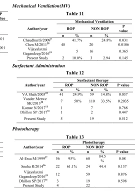
Mechanical Ventilation(MV)
Table 11
Author/year
Mechanical Ventilation
ROP NON ROP P
value
n % n %
ChaudhuriS/20099 41.7% 24.8% 0.031
Chen M/201129 48 20 0.0106
Vijayalaxmi
Gagandeep/201619 5 16 0.365
Present Study 2 10.0% 3 2.94 0.145
Surfactant Administration
Table 12
Author/year
Surfactant therapy
ROP NON ROP P value
n % n %
VA Shah/200522 41 24.9% 59 14.8% 0.037
Vander Merwe
SK/201330 7 50% 110 33.5% 0.2035
Kumar N/201711 1 7 0.768
Dhillon SP /201731 1 2 0.447
Present Study 5 19 0.512
Phototherapy
Table 13
Author/year
Phototherapy
ROP NON ROP
n % n % P value
Al-Essa M/199927 56 95% 60 84.5
% 0.08 Sneha R/201426 22 61.1% 24 44.4 0.137
Vijayalaxmi
Gagandeep/201619 12 59 0.876
Dhillon SP/201731 3 19 0.598
Present Study 4 22
The present study was discussed with various other studies as mentioned in above tables.The present study correlates well with other studies. Mechanical Ventilation, Surfactant therapy and phototherapy were not found statistically significant risk factors of ROP in present study.
CONCLUSION
The incidence of ROP in present study was 16.39%. A treatment was performed in 20% of ROP cases. ROP remains a major complication in pre-mature newborns despite all the advances that have been made in recent years. The excellence in neonatal care, screening and early treatment of ROP are keys to prevent vision loss induced by this disease. It is mandatory to do ophthalmological check up of those newborns who have satisfied the criteria.
In present study the most significant risk factors to development of ROP were low birth weight, low gestational age, need for oxygen therapy, neonatal sepsis, apnea of prematurity, need of cardiopulmonary support (CPAP), and anemia needing blood transfusion. However, phototherapy, use of surfactant, MV were not seen significantly associated with ROP in present study.
References
Incidence and early course of retinopathy of prematurity. Ophthalmology 1991 Nov; 98(11):1628-40.
2. American Academy of Pediatrics Section on Ophthalmology. Screening examination of premature infants for retinopathy of prematurity. Pediatrics. 2012 Dec 31:peds-2012.
3. Maheshwari R, Kumar H, Paul VK, Singh M, Deorari AK, Tiwari HK. Incidence and risk factors of retinopathy of prematurity in a tertiary care newborn unit in New Delhi. NatlMed J India 1996;9:211-4 4. Rekha S, Battu RR. Retinopathy of prematurity:
incidence and risk factors. Indian Pediatr 1996;33:999-1003
5. Charan R, Dogra MR, Gupta A, Narang A. The incidence of retinopathy of prematurity in a neonatal care unit. Indian journal of ophthalmology. 1995 Jul 1;43(3):123.
6. Blair BM, Hallmoran HS, Panly TH, Stevens JL. Decreased incidence of retinopathy of prematurity. J AAPOS. 2001;5(2):118-22.
7. Patil J, Deodhar J, Wagh S, Pandit AN. High risk factors for development of Retinopathy of Prematurity. Indian Pediatrics 1997;34:1024-7. 8. Gupta VP, Dhaliwal U, Sharma R, Gupta P, Rohatgi J.
Retinopathy of prematurity-risk factors. The Indian Journal of Pediatrics. 2004 Oct 1;71(10):887-92. 9. Chaudhari S, Patwardhan V, Vaidya U, Kadam S,
Kamat A. Retinopathy of prematurity in a tertiary care center--incidence, risk factors and outcome. Indian pediatrics. 2009 Mar 1;46(3).
10. Balakrishnan U, Shaik SJ, Manian N, Muthukumar M, Thomas M, Amboiram P, Ninan B, Chandrasekaran A, Ramaswamy S. Screening based on incidence of severe retinopathy of prematurity in a tertiary care center in India: are Indian infants different?. International Journal of Contemporary Pediatrics. 2016 Dec 21;3(3):847-53.
11. Kumar N, Kaushik SL, Grover N, Sharma RL. Retinopathy of prematurity: incidence and risk factors: a hospital based study from Shimla, Himachal Pradesh, India. International Journal of Research in Medical Sciences. 2016 Dec 19;5(1):56-61.
12. Nair P, Ganesh A, Mitra S, Sham S, Ganguly. Retinopathy of Prematrity inVLBW and extreme LBW babies. Indian J Paediatrics 2003;70(4):303-6. 13. Fortes Filho JB, Eckert GU, Valiatti FB, Costa MC,
Bonomo PP, Procianoy RS. Prevalence of retinopathy of prematurity: an institutional cross-sectional study of preterm infants in Brazil. Revista Panamericana de Salud Pública. 2009;26:216-20.
14. Lomuto CC, Galina L, Brussa M, Quiroga A, Alda E, Benitez AM, Bouzas L, Dinerstein NA, Erpen N, Falbo J, Manzitti J, Marinaro S, Nieto R, Sepulveda T, Visintin P. Epidemiology of retinopathy of prematurity in public services from Argentina during 2008. Arch Argent Pediatr. 2010;108(1):24–30. 15. Mitsiakos G, Papageorgiou A. Incidence and factors
predisposing to retinopathy of prematurity in inborn
infants less than 32 weeks of gestation. Hippokratia. 2016 Apr;20(2):121.
16. Yau GS, Lee JW, Tam VT, Liu CC, Yip S, Cheng E, Chu BC, Yuen CY. Incidence and risk factors of retinopathy of prematurity from 2 neonatal intensive care units in a Hong Kong Chinese population. The Asia-Pacific Journal of Ophthalmology. 2016 May 1;5(3):185-91.
17. Rao KA, Purkayastha J, Hazarika M, Chaitra R, Adith KM. Analysis of prenatal and postnatal risk factors of retinopathy of prematurity in a tertiary care hospital in South India. Indian journal of ophthalmology. 2013 Nov;61(11):640.
18. Shivaprasad B , Usha. H. N, Kishore Baindur .Study on Incidence and Risk Factors of Retinopathy of Prematurity.Sch. J. App. Med. Sci., 2014; 2(6A):1962-1966.
19. Vijayalaxmi gagandeep, Arun P. Incidence and risk factors for retinopathy ofprematurity in a teritiary care neonatal unit. TJPRC: International Journal of General Pediatrics and Medicine (TJPRC: IJGPM) 2016 Dec, Vol. 1, Issue 1, 45-50.
20. Vasavada D, Sengupta S, Prajapati VK, Patel S. Incidence and risk factors of retinopathy of prematurity in Western India–Report from A Regional Institute of Ophthalmology. Nepalese Journal of Ophthalmology. 2017 Jul;9(2):112-20.
21. Ahuja AA, Reddy YC, Adenuga OO, Kewlani D, Ravindran M, Ramakrishnan R. Risk factors for retinopathy of prematurity in a district in South India: a prospective cohort study. Oman journal of ophthalmology. 2018 Jan;11(1):33.
22. Shah VA, Yeo CL, Ling YL, Ho LY. Incidence, risk factors of retinopathy of prematurity among very low birth weight infants in Singapore. Ann Acad Med Singapore. 2005 Mar 1;34(2):169-78
23. Freitas AM, Mörschbächer R, Thorell MR, Rhoden EL. Incidence and risk factors for retinopathy of prematurity: a retrospective cohort study. International journal of retina andvitreous. 2018 Dec;4(1):20. 24. Bas AY, Demirel N, Koc E, Isik DU, Hirfanoglu İM,
Tunc T. Incidence, risk factors and severity of retinopathy of prematurity in Turkey (TR-ROP study): a prospective, multicentre study in 69 neonatal intensive care units. BritishJournalofOphthalmology. 2018 Mar 26:bjophthalmol-2017.
25. Shetty SP, Shetty J, Amin H, Shenoy RD. The incidence, risk factors and outcome of retinopathy of prematurity at a tertiary care centre in south India. IOSR Journal of Dental and Medical Sciences (IOSR-JDMS). 2015;1(14):77-83.
26. Sneha R, Shankar P. A clinical study on incidence of retinopathy of prematurity changes in preterm infants and associated risk factors in a tertiary centre. Journal of Evolution of Medical and Dental Sciences. 2014 Mar 10;3(10):2603-8.
from a tertiary care hospital in Delhi. International Journal of Medicine and Public Health. 2014;4(4). 28. Al-Essa M, Azad RV, Rashwan N. Rate of and risk
factors associated with retinopathy of prematurity: a prospective study from Kuwait. Medical Principles and Practice. 1999;8(2):115-8.
29. Chen M, Çitil A, McCabe F, Leicht KM, Fiascone J, Dammann CE, Dammann O. Infection, oxygen, and immaturity: interacting risk factors for retinopathy of prematurity. Neonatology. 2011;99(2):125-32.
30. Van der Merwe SK, Freeman N, Bekker A, Harvey J, Smith J. Prevalence of and risk factors for retinopathy of prematurity in a cohort of preterm infants treated exclusively with non-invasive ventilation in the first week after birth. South African Medical Journal. 2013;103(2):96-100.
31. Dhillon SP, Kumar A, Rani A, Kaur P, Pannu MS. Incidence and Risk Factors of Retinopathy of Prematurity (ROP) in Neonates of Weight 1.5 to 2 kg. Int. J. Curr. Res. Med. Sci. 2017;3(7):38-44.