Ibrahim Uygun
1, Mehmet H. Okur
1, Yilmaz Arayici
1, Aysenur Keles
2,
Hayrettin Ozturk
3, Selcuk Otcu
1Magnetic Compression Ostomy for Simple Tube
Colostomy in Rats – Magnacolostomy
Kolostomia z zastosowaniem systemu magnetycznego u szczurów
1 Department of Pediatric Surgery and Pediatric Urology, Medical Faculty of Dicle University, Diyarbakir, Turkey 2 Department of Pathology, Medical Faculty of Dicle University, Diyarbakir, Turkey
3 Department of Pediatric Surgery and Pediatric Urology, Medical Faculty of Abant Izzet Baysal University, Bolu, Turkey
Abstract
Background. Magnetic compression anastomoses (magnamosis) have been previously described for gastrointesti-nal, biliary, urinary, and vascular anastomoses.
Objectives. Herein, the authors report the creation of a magnetic compression colostomy (magnacolostomy) using a simple technique in rats.
Material and Methods Animals were randomized into two groups (n = 8, each): a magnetic colostomy (MC) group and a control surgical tube colostomy (SC) group. In the MC group, the first magnetic ball (3 mm) was rectally introduced into the rat colon. The second magnetic ball (4 mm) was placed subcutaneously into the left quadrant, and the two magnetic balls strongly coupled. On postoperative day 20 for the MC group and postoperative day 10 in the SC group, the rats were sacrificed and the colostomies evaluated macroscopically, histopathologically, and for mechanical burst testing.
Results. From the macroscopic evaluation, two rats failed to form the colostomy canal due to colostomy catheter and magnetic ball removal. In the remaining rats, evidence of complications were not observed. Two rats in the MC group displayed mild adhesion and all rats in the SC group displayed moderate adhesion. No significant differences between the burst pressures were observed. However, a significant difference (p < 0.001) between the procedure times of the MC (4.13 ± 1.00 minutes) and SC groups (14.25 ± 2.05 minutes) was evident.
Conclusions: Magnacolostomy is an easy and effective procedure in the rat model and presents a safe, minimally invasive alternative to current tube colostomy procedures such as antegrade continence enemas, percutaneous endoscopic, and colostomy/cecostomy in humans (Adv Clin Exp Med 2012, 21, 3, 301–305).
Key words: colostomy, magnacolostomy, magnetic compression anastomosis, magnostomy.
Streszczenie
Wprowadzenie. Zespolenie z uciskiem magnetycznym używa się w chirurgii przewodu pokarmowego, dróg żół-ciowych, dróg moczowych i naczyń.
Cel pracy. Opracowanie kolostomii z uciskiem magnetycznym za pomocą prostej techniki u szczurów.
Materiał i metody. Zwierzęta podzielono losowo na dwie grupy (n = 8): grupę z magnetyczną kolostomią (MC) i grupę kontrolną z jednolufową kolostomią (SC). W grupie MC pierwszą magnetyczną kulę (3 mm) wprowadzono doodbytni-czo do jelita grubego szczura. Drugą kulę magnetyczną (4 mm) umieszdoodbytni-czono podskórnie w lewej ćwiartce, a pola magne-tyczne obu kul były silnie sprzężone. W 20. dobie pooperacyjnej grupy MC i 10. dobie po operacji w grupie SC szczury uśmiercano i oceniano kolostomie makroskopowo, histopatologicznie oraz wykonano mechaniczną próbę rozrywania.
Wyniki. Według makroskopowej oceny u 2 szczurów kanał kolostomii nie wytworzył się z powodu usunięcia cew-nika kolostomii i kuli magnetycznej. U pozostałych szczurów powikłań nie obserwowano. U 2 szczurów w grupie MC stwierdzono łagodną przyczepność, a u wszystkich szczurów w grupie SC umiarkowaną przyczepność. Nie stwierdzono istotnych różnic między ciśnieniem pęknięcia. Stwierdzono jednak istotną różnicę (p < 0,001) między czasem zabiegu w grupie MC (4,13 ± 1,00 minuty) i grupie SC (14,25 ± 2,05 minuty).
Wnioski. Magnakolostomia jest łatwym i skutecznym postępowaniem w modelu szczurzym i oferuje bezpieczną, minimal-nie inwazyjną alternatywę dla obecnych zabiegów kolostomii, takich jak płukaminimal-nie jelita grubego od strony jego proksymal-nego odcinka, przezskórna endoskopowa kolostomia/cekostomia u ludzi (Adv Clin Exp Med 2012, 21, 3, 301–305).
Słowa kluczowe: kolostomia, magnakolostoma, zespolenie z uciskiem magnetycznym, magnostomia.
Adv Clin Exp Med 2012, 21, 3, 301–305 ISSN 1899–5276
OrIgINAl PAPErS
Colostomy is frequently performed in patients for the palliative or curative management of nu-merous conditions [1]. In addition, for antegrade continence enema, the tube large bowel ostomy procedures including appendicostomy, cecostomy, colostomy, and sigmoidostomy, have been widely used in the management of patients with fecal in-continence and chronic constipation [2]. Various techniques of tube colostomy have been developed [1–4]. Despite its strong safety record, tube colos-tomy procedures are associated with an overall major and minor complication rate of 16–52%, in-cluding procedural complications, stenosis, leak-age, infection, failure, prolapse, perforation, organ injury, and tube problems [1–4].
Magnetic compression anastomoses (magna-mosis) has previously been described for gastroin-testinal, biliary, urinary, and vascular anastomoses [5–15]. In this study, the authors report the first creation of a magnetic compression colostomy (magnacolostomy) using a simple technique in rats.
Material and Methods
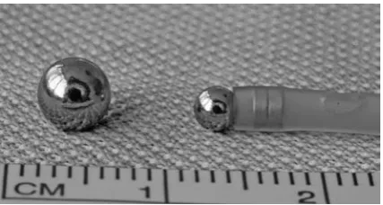
Two spherical neodymium-iron-boron mag-nets coated with chrome (N35 strength, outer di-ameter 4 mm and N35 strength, outer didi-ameter 3 mm) were used for each rat colostomy (Fig. 1).
The magnetic insertion catheter was specially designed for insertion of the intracolonic mag-net through the rectum to the rat colon. An 8-F feeding tube was cut from its tip and two small cylindrical neodymium-iron-boron magnets (N35
strength, outer diameter 2 mm, thickness 1 mm) were inserted 2 mm proximally to couple the magnet balls, but allow simple uncoupling when required (Fig. 1).
Sixteen adult male Wistar-Albino rats weigh-ing 290–370 g were used in this study. All experi-mental protocols were reviewed and approved by the Institutional Animal Care and Use Committee of Dicle University (approval no. 2011/33).
The rats were housed individually in cages and allowed free access to standard rat chow and water prior to the experimental procedure. The animal rooms were windowless and under controlled tem-perature (22 ± 2ºC) and lighting conditions. The animals were fasted overnight prior to the experi-ments and were allowed free access to water. The rats were anesthetized by intraperitoneal injection of ketamine (Ketalar Flakon, Pfizer Ilaclari ltd. Sti., Istanbul) at a dose of 75 mg/kg of body weight.
The animals were randomized into two groups
(n = 8, each): a magnetic colostomy (MC) group,
where rats were subjected to the magnet insertion procedure; and a surgical colostomy (SC) group, including rats subjected to the surgical tube colos-tomy procedure. The procedure times were noted for each group.
In the MC group, following animal anestheti-zation in the supine position, the magnetic inser-tion catheter coupled with the first magnetic ball (3 mm) was introduced rectally approximately 8-cm proximally from the anus into the colon in the MC group. Following colon insertion of the first magnet, a small midline skin incision (5 mm) was performed. The subcutaneous tissue was dis-sected through the left quadrant by clamp. The second magnet ball (4 mm) was then placed sub-cutaneously into the left quadrant and the two magnetic balls strongly coupled. The magnetic in-sertion catheter was then easily removed.
In the SC group, a laparotomy was performed through a lower midline incision measuring 3 cm. The subcutaneous tissue was dissected through the left quadrant by clamp. A 5-F polyurethane cathe-ter, as a tube colostomy cathecathe-ter, was inserted sub-cutaneously into the colon through the abdominal wall. The catheter was secured with a purse-string suture using 6-0 Vicryl (Ethicon Inc., Somerville, NJ), and the colon was fixed to the abdominal wall with two primary sutures using the same suture material. The proximal end of the catheter was cut and occluded with a tying 3-0 Vicryl (Ethicon Inc.). The catheter was fixed to the abdominal fas-cia with the same suture. The fasfas-cial layer of the abdominal wall was continuously closed with the same suture.
In all animals, the abdominal skin was closed with an interrupted suture using 3-0 Vicryl
(Ethi-Fig. 1. Spherical magnets (outer diameter 4 and 3 mm) coated with chrome and magnetic insertion catheter produced from an 8-F feeding tube and two small cylindrical magnets inserted into its tip to pull the spherical 3 mm magnet
con Inc.). The animals were allowed food and wa-ter immediately afwa-ter the operation and recovery from anesthesia.
On postoperative day 20 after the formation of a full tubularization in the MC group, and day 10 after completion of wound healing in the SC group, the rats were deeply anesthetized by an in-traperitoneal injection of ketamine at a dose of 100 mg/kg body weight. External magnetic balls with internal magnets and colostomy catheters were carefully disengaged through the abdominal wall (Fig. 2). Colostomies were examined to determine the patency of the ostomy and calibrated externally with feeding tubes. Subsequently, all animals un-derwent laparotomy and exploration of the abdo-men for evidence of leakage, adhesion, intestinal obstruction, peritonitis, an incorrect colostomy side, or other complications. All colostomy and colonic segments within the abdominal wall were then dissected and totally excised.
For mechanical burst testing, one catheter was inserted through the end of the colon and the sec-ond was secured by suturing. The other end of the colon was clamped. The catheter was connected to a calibrated pressure-measuring circuit with a sy-ringe for instillation of 0.9% normal saline solu-tion. The colostomy opening was occluded from the abdominal wall through compression with a clamp. Saline was injected into the colon while monitoring pressure changes. Continuous infu-sion of the saline solution was administered in-tracolonically until failure of the colostomy on the peritoneal surface or the surrounding colon dem-onstrated leakage or bursting. The point of leakage or bursting was recorded as the burst pressure of the colostomy.
The tissue was fixed in 10% formalin. Colosto-mies were evaluated with hematoxylin and eosin, and Masson’s trichrome stain.
The Mann-Whitney U test was used to de-termine differences in the procedure times and bursting pressures between the groups. Data was analyzed using SPSS 15.0 for Windows (SPSS Inc.,
Chicago, Il, USA). A p value ≤ 0.05 was
consid-ered statistically significant.
Results
Sixteen rats underwent successful operations. There were no episodes of intestinal obstruction or food intolerance. Based on the macroscopic evaluation, the colostomy canal did not form in only two rats due to the colostomy catheter (one rat in the SC group) and all magnetic balls (one rat in the MC group) were removed. In all other rats, evidence of leakage, intestinal obstruction, perito-nitis, and incorrect colostomy side were not ob-served, but a single rat of the MC group displayed mild adhesion and all rats in the SC group dis-played moderate adhesion (Fig. 3). One magnetic and one surgical colostomy were damaged during harvesting. Burst pressure data was obtainable for the remaining 12 (6 magnetic and 6 surgical) co-lostomies (Fig. 4). The average burst pressure for the MC group was 150 mm Hg (SD 47 mm Hg, SEM 19 mm Hg), and for SC was 152 mm Hg (SD
Fig. 2. External and internal magnet balls were disen-gaged through the abdominal wall (magnetic anasto-mosis) of rat
Ryc. 2. Zewnętrzna i wewnętrzna kula magnetyczna zostały rozłączone przez powłoki brzuszne szczura (magnetyczne zespolenie)
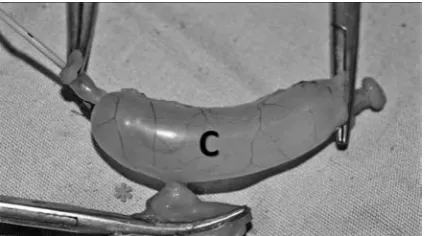
Fig. 3. Macroscopic evaluation of the magnetic colos-tomy. The colostomy canal (asterisk) was examined to determine patency of the ostomy and calibrated externally (arrow) with a feeding tube (t). (C – colon, P – peritoneum of the abdominal wall)
29 mm Hg, SEM 12 mm Hg). Of these, only 3 rats in the MC group and 1 in the SC group failed at the colostomy site. All other burst failures occurred at the corpus of the colon. No significant differences were evident between the burst pressures of the SC and MC groups (p = 0.937). However, a significant difference (p < 0.001) between the procedure times of the groups was observed (mean ± SD, SEM; MC group: 4.13 ± 1.00, 0.35 minutes versus SC group: 14.25 ± 2.05, 0.73 minutes).
Histological analysis showed evidence of epi-thelization across the colostomy within the sub-mucosa between the colonic and abdominal walls, which displayed lower inflammation compared with the surgical colostomies.
Discussion
Enteric magnets were first described by Cope and colleagues who created anastomoses through compression necrosis in animals and eventually humans [5, 6]. Erdmann et al. used magnet tech-nology to create sutureless vascular anastomosis [14]. Magnets in the medical world have been in-creasingly utilized as a device to treat several dis-eases such as biliary duct and urethral strictures, malignant gastric outlet obstruction, esophageal atresia and pectus excavatum, and other gastroin-testinal diseases requiring gastroingastroin-testinal by-pass anastomoses [6, 7, 11–13, 15, 16]. This study is the first to describe the use of magnetic compression colostomy in an animal model.
To date, for all experimental studies regard-ing magnamosis, large animals (pigs and dogs) have been used [5, 8–10, 14]. Herein, the authors used a rat model and utilized spherical magnetic balls for magnamosis. The magnetic balls pos-sess a smooth round shape and the ability to roll and move, meaning all contact surfaces to the
tis-sues are identical. Thus, insertion, coupling, and removal of the magnetic balls is simple and non-invasive. The authors used the magnetic balls for tube colostomies required at the small opening. Magnetic technology producing strong and deli-cate magnets has rapidly advanced since the 1980’s [17]. This study may initiate further magnetic compression ostomy research for all ostomies including gastrostomy, esophagostomy, jejunos-tomy, cecosjejunos-tomy, cyctosjejunos-tomy, and ureterosjejunos-tomy, which all can be performed using magnets.
Tube colostomy procedures are frequently performed in patients using open surgical, laparo-scopic, percutaneous, endolaparo-scopic, and fluoroscop-ic procedures [1–4]. These techniques require gen-eral or local anesthesia, special, expensive medical equipment, and X-ray exposure for fluoroscopic techniques. These procedures also display major and minor complications. An advantage of the magnacolostomy technique is the lack of require-ment for these procedures coupled with a low oc-currence of complications in all rats observed.
Pichakron et al. reported that magnamosis (gastrojejunostomy and jejunojejunostomy) is im-mediately patent and develops strength equal to or greater than that of hand-sewn or stapled anasto-moses over 3 to 10 days in a pig model [8]. In this study, the authors observed that the anastomosis between the colon and abdominal wall was com-plete at 20 days due to the thick abdominal wall. Thus, the current technique was not applied in pa-tients requiring immediate colonic decompression. Jamshidi et al. and Pichakron et al. indicated that magnamosis is effective in the pig model and may present a safe, effective, minimally invasive alter-native to current anastomotic strategies in humans that is compatible with endoscopic and natural or-ifice approaches [8, 10]. The magnacolostomy also may be an easy and minimally invasive alternative to current tube colostomy procedures in humans in the future.
Present observations regarding magnacolosto-my are handicapped by the limited number of rats in this report. Nonetheless, these results suggest that magnacolostomy is minimally invasive, safe, and technically easy, and equivalent to the tube co-lostomies created by surgical techniques. This study suggests that magnetic ball compression distal tube colostomy can be created via a completely rectal tube approach using blindly inserted colonic mag-nets with developed magnetic insertion devices. The authors believe that, in the future, magnaco-lostomy for cecostomy and appendicostomy will be applied using fluoroscopy in animals and humans.
In this study, colostomies were created in all animals using magnetic balls, all the balls were blindly inserted into the distal colon, and no
com-Fig. 4. Mechanical burst pressure testing of the colos-tomy (asterisk) (C – colon)
plications occurred. With the future of interven-tion focusing on non-invasive procedures, this technique may be applicable in the outpatient set-ting due to its lack of requirement for sedation, an-esthesia, fluoroscopy, and endoscopy for insertion of the rectal tube. The magnacolostomy is effective
in the rat model and may be a safe, minimally in-vasive alternative to some colostomy procedures such as antegrade continence enemas and percuta-neous endoscopic colostomy in humans. However, further investigations should be performed to de-velop magnacolostomy in animals and humans.
References
[1] Borkowski S:Pediatric stomas, tubes, and appliances. Pediatr Clin North Am 1998, 45, 1419–1435.
[2] Rawat DJ, Haddad M, Geoghegan N et al.:Percutaneous endoscopic colostomy of the left colon: a new tech-nique for management of intractable constipation in children. gastrointest Endosc 2004, 60, 39–43.
[3] Rodriguez L, Flores A, Gilchrist BF et al.: laparoscopic-assisted percutaneous endoscopic cecostomy in chil-dren with defecation disorders (with video). gastrointest Endosc 2011, 73, 98–102.
[4] Donkol RH, Al-Nammi A: Percutaneous cecostomy in the management of organic fecal incontinence in chil-dren. World J radiol 2010, 2, 463–467.
[5] Cope C: Evaluation of compression cholecystogastric and cholecystojejunal anastomoses in swine after peroral and surgical introduction of magnets. J Vasc Interv radiol 1995, 6, 546–552.
[6] Cope C, Clark TW, Ginsberg G et al.: Stent placement of gastroenteric anastomoses formed by magnetic com-pression. J Vasc Interv radiol 1999, 10, 1379–1386.
[7] van Hooft JE, Vleggaar FP, Le Moine O et al.: Endoscopic magnetic gastroenteric anastomosis for palliation of malignant gastric outlet obstruction: a prospective multicenter study. gastrointest Endosc 2010, 72, 530–535.
[8] Pichakron KO, Jelin EB, Hirose S et al.: Magnamosis II: Magnetic compression anastomosis for minimally inva-sive gastrojejunostomy and jejunojejunostomy. J Am Coll Surg 2011, 212, 42–49.
[9] Myers C, Yellen B, Evans J et al.: Using external magnet guidance and endoscopically placed magnets to create suture-free gastro-enteral anastomoses. Surg Endosc 2010, 24, 1104–1109.
[10] Jamshidi R, Stephenson JT, Clay JG et al.: Magnamosis: magnetic compression anastomosis with comparison to suture and staple techniques. J Pediatr Surg 2009, 44, 222–228.
[11] Zaritzky M, Ben R, Zylberg GI et al.: Magnetic compression anastomosis as a nonsurgical treatment for esopha-geal atresia. Pediatr radiol 2009, 39, 945–949.
[12] Takamizawa S, Yamanouchi E, Muraji T et al.: MCrA of an anastomotic stenosis after esophagoesophagostomy for long gap esophageal atresia: a case report. J Pediatr Surg 2007, 42, 769–772.
[13] Itoi T, Kasuya K, Sofuni A et al.: Magnetic compression anastomosis for biliary obstruction: review and experi-ence at Tokyo Medical University Hospital. J Hepatobiliary Pancreat Sci 2011, 18, 357–365.
[14] Erdmann D, Sweis R, Heitmann C et al.: Side-to-side sutureless vascular anastomosis with magnets. J Vasc Surg 2004, 40, 505–511.
[15] Stepanov EA, Erokhin AP, Nikolaev VV et al.: Treatment of short urethral strictures in children using magnets. Urol Nefrol 1989, 3, 8–11.
[16] Harrison MR, Curran PF, Jamshidi R et al.: Magnetic mini-mover procedure for pectus excavatum II: initial findings of a Food and Drug Administration-sponsored trial. J Pediatr Surg 2010, 45, 185–192.
[17] Harrison MR: What if? Why not? J Pediatr Surg 2010, 45, 1–10.
Address for correspondence:
Ibrahim Uygun
Department of Pediatric Surgery and Pediatric Urology Medical Faculty of Dicle University
21280 Diyarbakir Turkey
Tel.: +90 412 237 8250 Mobile: +90 505 413 0944
E-mail address: [email protected] Conflict of interest: None declared received: 4.01.2012