GATA2 is required for lymphatic vessel valve
development and maintenance
Jan Kazenwadel, … , Hamish S. Scott, Natasha L. Harvey
J Clin Invest. 2015;
125(8)
:2979-2994.
https://doi.org/10.1172/JCI78888
.
Heterozygous germline mutations in the zinc finger transcription factor GATA2 have recently
been shown to underlie a range of clinical phenotypes, including Emberger syndrome, a
disorder characterized by lymphedema and predisposition to myelodysplastic
syndrome/acute myeloid leukemia (MDS/AML). Despite well-defined roles in
hematopoiesis, the functions of GATA2 in the lymphatic vasculature and the mechanisms
by which GATA2 mutations result in lymphedema have not been characterized. Here, we
have provided a molecular explanation for lymphedema predisposition in a subset of
patients with germline GATA2 mutations. Specifically, we demonstrated that
Emberger-associated GATA2 missense mutations result in complete loss of GATA2 function, with
respect to the capacity to regulate the transcription of genes that are important for lymphatic
vessel valve development. We identified a putative enhancer element upstream of the key
lymphatic transcriptional regulator PROX1 that is bound by GATA2, and the transcription
factors FOXC2 and NFATC1. Emberger GATA2 missense mutants had a profoundly
reduced capacity to bind this element. Conditional Gata2 deletion in mice revealed that
GATA2 is required for both development and maintenance of lymphovenous and lymphatic
vessel valves. Together, our data unveil essential roles for GATA2 in the lymphatic
vasculature and explain why a select catalogue of human GATA2 mutations results in
lymphedema.
Research Article
Development
Vascular biology
Find the latest version:
Introduction
The discovery that GATA2 mutations underlie Emberger syn-drome — a condition characterized by primary lymphedema and predisposition to myelodysplastic syndrome/acute myeloid leukemia (MDS/AML) (1) — revealed key roles for this zinc fin-ger transcription factor in the lymphatic vasculature (2–4). Our initial investigations determined that GATA2 protein levels were most prominent in lymphatic vessel valves. This observation, coupled with our demonstration that GATA2 regulates genes important for lymphatic vessel valve development, prompted us to investigate whether GATA2 mutations result in lymphedema due to defective valve development and/or function. While sev-eral earlier studies implicated GATA2 in vascular development (5–8), the lack of an obvious vascular phenotype in Gata2–/– mice
prior to their demise around E10.5 has precluded in depth analy-ses of Gata2 function in vasculogenesis and angiogenesis. SNPs in GATA2 have been associated with coronary artery disease (9–11), implying a role for GATA2 in arterial development, while ablation of gata2a in zebrafish affects morphogenesis of the dorsal aorta (12). Recent studies in which Gata2 was deleted in hematopoietic and endothelial cell compartments (13), or was disrupted due to mutation of an enhancer element important for endothelial Gata2 expression (14), suggested that Gata2 is important for vascular integrity and for efficient separation of the blood and lymphatic vascular networks (13, 14). The depen-dence of these phenotypes on endothelial versus hematopoietic GATA2 was not, however, dissected in these studies.
The lymphatic vasculature plays crucial roles in the return of interstitial fluid to the bloodstream, absorption of dietary lipids, and trafficking of immune cells. The formation of valves in collect-ing lymphatic vessels is a key event durcollect-ing maturation of the lym-phatic vasculature and is crucial for lymph to be efficiently returned to the bloodstream; defects in lymphatic valve formation contrib-ute to aberrant lymphatic function in lymphedema syndromes (15). Recent studies have begun to dissect the genes and cellular events important for valve morphogenesis in lymphatic vessels.
Heterozygous germline mutations in the zinc finger transcription factor GATA2 have recently been shown to underlie a range of clinical phenotypes, including Emberger syndrome, a disorder characterized by lymphedema and predisposition to myelodysplastic syndrome/acute myeloid leukemia (MDS/AML). Despite well-defined roles in hematopoiesis, the functions of GATA2 in the lymphatic vasculature and the mechanisms by which GATA2 mutations result in lymphedema have not been characterized. Here, we have provided a molecular explanation for lymphedema predisposition in a subset of patients with germline GATA2 mutations. Specifically, we demonstrated that Emberger-associated GATA2 missense mutations result in complete loss of GATA2 function, with respect to the capacity to regulate the transcription of genes that are important for lymphatic vessel valve development. We identified a putative enhancer element upstream of the key lymphatic transcriptional regulator PROX1 that is bound by GATA2, and the transcription factors FOXC2 and NFATC1. Emberger GATA2 missense mutants had a profoundly reduced capacity to bind this element. Conditional Gata2 deletion in mice revealed that GATA2 is required for both development and maintenance of lymphovenous and lymphatic vessel valves. Together, our data unveil essential roles for GATA2 in the lymphatic vasculature and explain why a select catalogue of human GATA2 mutations results in lymphedema.
GATA2 is required for lymphatic vessel valve
development and maintenance
Jan Kazenwadel,1 Kelly L. Betterman,1 Chan-Eng Chong,2 Philippa H. Stokes,3 Young K. Lee,2 Genevieve A. Secker,1 Yan Agalarov,4 Cansaran Saygili Demir,4 David M. Lawrence,5,6 Drew L. Sutton,1 Sebastien P. Tabruyn,1 Naoyuki Miura,7 Marjo Salminen,8 Tatiana V. Petrova,4,9 Jacqueline M. Matthews,3 Christopher N. Hahn,2,10 Hamish S. Scott,2,5,6,10 and Natasha L. Harvey1,10
1Centre for Cancer Biology, University of South Australia and SA Pathology, Adelaide, South Australia, Australia. 2Centre for Cancer Biology, University of South Australia and
Department of Molecular Pathology, SA Pathology, Adelaide, South Australia, Australia. 3School of Molecular Bioscience, University of Sydney, Sydney, New South Wales, Australia. 4Department of Oncology, University Hospital of Lausanne and Department of Biochemistry, University of Lausanne, Lausanne, Switzerland.
5Australian Cancer Research Foundation (ACRF) Cancer Genomics Facility, Centre for Cancer Biology, University of South Australia and SA Pathology, Adelaide, South Australia, Australia.
6School of Molecular and Biomedical Bioscience, University of Adelaide, Adelaide, South Australia, Australia. 7Department of Biochemistry, Hamamatsu University School of Medicine, Hamamatsu, Japan. 8Department of Veterinary Biosciences, University of Helsinki, Helsinki, Finland. 9École Polytechnique Fédérale de Lausanne (EPFL), Swiss Institute for Experimental Cancer Research (ISREC),
Lausanne, Switzerland. 10School of Medicine, University of Adelaide, Adelaide, South Australia, Australia.
Related Commentary: p. 2924
Authorship note: Jan Kazenwadel and Kelly L. Betterman contributed equally to this
work.
Conflict of interest: The authors have declared that no conflict of interest exists. Submitted: September 4, 2014; Accepted: May 28, 2015.
a transcription factor heptad that includes SCL, LYL1, LMO2, RUNX1, ERG, and FLI-1 (44). ChIP-Seq studies performed across a range of cell types demonstrate that GATA2 has distinct sites of chromatin occupancy and transcriptional targets in each cell type in which it acts, suggesting tissue-specific transcriptional interactions (8, 24, 44–46). The zinc fingers of GATA2 serve at least 2 key roles: the C-terminal zinc finger mediates binding to WGATAR motifs (47, 48), whereas the N-terminal zinc finger mediates protein-pro-tein interactions with transcriptional cofactors and stabilizes DNA binding (37). Many of the heterozygous GATA2 mutations iden-tified in patients result in either premature truncation of the pro-tein or impact the C-terminal zinc finger by introducing missense alterations to this critical region of the protein. We (3) and others (49) have suggested that a complete loss of function of one GATA2 allele is the key factor that predisposes to lymphedema onset, given the clinical heterogeneity in symptoms observed in patients with GATA2 mutations. Most GATA2 missense mutations, including the prevalent T354M mutation (more than 50 to date), do not correlate with lymphedema. Our hypothesis that complete heterozygous loss of GATA2 function underlies lymphedema was complicated by the description of 3 Emberger syndrome patients with missense muta-tions R361L, C373R (2), and R396Q (4). This finding prompted us to investigate further the mechanisms by which GATA2 Emberger mutations impact on GATA2 function.
Here, we sought to determine the specific role of Gata2 in lymphatic vascular development, with a particular emphasis on valve development, by selectively inactivating Gata2 in the lym-phatic vasculature. To investigate the mechanisms by which only a select catalogue of GATA2 mutations result in lymphedema, we assessed the effects of missense GATA2 Emberger mutations on the structure and binding to DNA of the GATA2 C-terminal zinc finger. Our work demonstrates that Gata2 plays key roles during lymphovenous and lymphatic vessel valve formation and provides molecular evidence that Gata2 “null” haploinsufficiency under-lies the propensity to develop lymphedema, through regulation of genes, including Prox1 and Foxc2, that are important for lymphatic vessel development and valve development.
Results
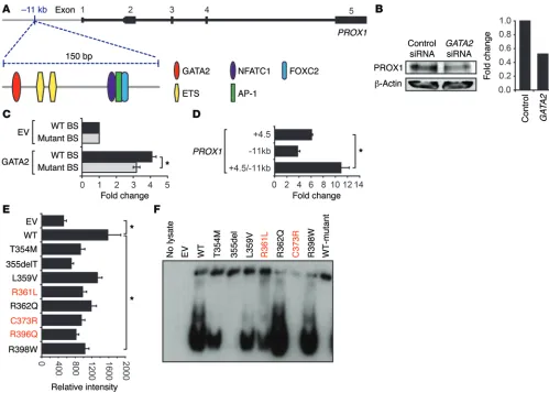
GATA2 binds to and transactivates PROX1 regulatory elements. Our earlier work determined that siRNA-mediated GATA2 knock-down in primary embryonic mouse LECs resulted in reduced PROX1 levels (3). To investigate whether GATA2 regulation of Prox1 is mediated directly, we searched for consensus WGATAR binding sites in a 4-kb region of the first intron of Prox1, approxi-mately 4.5 kb downstream from the transcription start site (Prox1 +4.5 kb), previously shown to be important for SOX18-mediated Prox1 expression (50). Five consensus WGATAR sites conserved between mouse and human were present in this region (Supple-mental Figure 1A; supple(Supple-mental material available online with this article; doi:10.1172/JCI78888DS1), and we showed that GATA2 is able to drive reporter gene expression from this element (Supplemental Figure 1B). We next compared the transcriptional activity of an allelic series of germline GATA2 mutants found in Emberger syndrome (R361L, C373R, and R396Q), together with those found in patients with hematological malignancies but no reported lymphedema (hereafter referred to as hematological Initiation of valve development is marked by the appearance of
clusters of cells, often near vessel branchpoints, that exhibit high levels of the transcription factors PROX1, FOXC2, and GATA2 (16–18). These valve-forming cells reorient themselves with respect to the longitudinal axis of the vessel, extend into the vessel lumen, and form elongated valve leaflets composed of a bilayer of endothelial cells sandwiching an extracellular matrix core com-posed largely of Fibronectin-EIIIA (FN-EIIIA), laminin-α5, and EMILIN1 (19–21). Genes identified to be important for lymphatic vessel valve development include the transcription factors FOXC2 and NFATC1 (15, 20, 22), the transmembrane ligand ephrinB2 (23), integrin-α9 and its ligands FN-EIIIA (19) and Emilin1 (21), gap junction proteins connexin37 (CX37) and connexin43 (CX43) (16, 24), NOTCH1 (25), SEMA3A together with receptor compo-nents NRP1 and PLEXINA1 (26, 27), angiopoietin2 (28, 29), TIE1 (30), and BMP-9 (31). Though the signals that initiate valve devel-opment are in large part enigmatic, the location of valves predom-inantly in regions of disturbed flow suggested that mechanical stimuli including shear stress might be important valve-initiating stimuli. Indeed, human lymphatic endothelial cells (hLECs) sub-jected to oscillatory shear stress (OSS) in vitro exhibit elevated lev-els of FOXC2 and Connexin37 (CX37), activation of NFATC1, and a change in their morphology from an elongated to cuboidal shape (20), as is observed in lymphatic endothelial cells within valve ter-ritories in vivo (20). The finding that venous valves share expres-sion of lymphatic valve markers including PROX1, ephrinB2, and integrin-α9 — together with a dependence on ephrinB2 and integ-rin-α9 signalling for their development — suggests that common genetic pathways direct valve development in both lymphatic ves-sels and veins (32).
Another specialized population of valve endothelial cells com-prises the lymphovenous valves (LVVs) (33, 34). These valves are located at the junction of the jugular and subclavian veins with the thoracic and right lymphatic ducts and function, together with platelets (35), to partition the lymphatic vasculature from the blood vasculature. Endothelial cells of the LVVs express PROX1, FOXC2, integrin-α9, and integrin-α5 (33, 36), providing further evidence that valve development in distinct vascular compartments relies on common pathways. Unlike the valves within lymphatic ves-sels, however, the LVVs comprise one leaflet derived from venous endothelial cells and one derived from lymphatic endothelial cells. These valves are exquisitely sensitive to changes in Prox1 dosage for their development (33); whereas lymphatic vessel valves form in Prox1 heterozygous mice, LVVs do not. Little is known regarding the mechanisms by which these specialized valves develop. Some of the initiating signals may be distinct from those controlling lym-phatic vessel valve development, given that the flow patterns and shear stress to which these venous cells are exposed are likely dif-ferent to those encountered by lymphatic vessels.
(Supplemental Figure 1D). Immunoblotting of nuclear lysates was performed to ensure that comparable levels of WT and mutant GATA2 protein were present in all conditions tested in this assay (Supplemental Figure 2). Together, these data suggest that sus-ceptibility to lymphedema might be directed by differential capacity of GATA2 mutants to bind and regulate the expression of genes important for development and function of the lymphatic vasculature, rather than hematopoiesis.
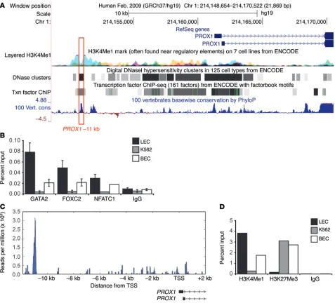
To investigate in more detail the sites bound by GATA2 in the vicinity of the PROX1 locus, we analyzed data deposited in the ENCODE database (http://genome.ucsc.edu./encode). A number of studies, including 2 undertaken in human microvascular endo-thelial cells (HMVECs) (24) and human umbilical vein endoendo-thelial cells (HUVECs) (8), demonstrated prominent GATA2 binding to a region 11 kb upstream of the first, noncoding exon of PROX1. Scanning of the DNA sequence in this peak region revealed a highly conserved region of approximately 150 nucleotides con-mutants: germline T354M, 355del, R398W, somatic L359V, and
[image:4.585.44.543.53.410.2]R362Q), with the transcriptional activity of WT GATA2 (Sup-plemental Figure 1B). In all cases, the ability of GATA2 mutants to drive reporter gene expression mediated by the Prox1 +4.5 kb element was reduced compared with WT. The ability of GATA2 to bind to each of the 5 consensus sites in the Prox1 +4.5 kb ele-ment was then assessed using Western blot combined with elec-trophoretic mobility shift assays (WEMSA) (51). The fourth of 5 sites, located proximal to the SOX A site, displayed highest levels of binding by GATA2 (Supplemental Figure 1C), likely due to the presence of 2 overlapping GATA sites in this region. The GATA2 Emberger mutants and 355del (lacking the majority of the C-ter-minal zinc finger) almost completely lost the capacity to bind this site in Prox1 +4.5 kb (Supplemental Figure 1D). In contrast, the germline GATA2 T354M mutation — found in MDS/AML but not to date in any patients with lymphedema — and several other hematological mutants retained the ability to bind this site
ment (containing GATA sites 4 and 5, see Methods for details) act-ing as a promoter. This addition resulted in a significant increase in luciferase activity, demonstrating that the Prox1 –11 kb element is capable of acting as an enhancer (Figure 1D). We next investigated the capacity of the GATA2 mutants to drive reporter gene expres-sion from the –11 kb enhancer element. All mutants demonstrated substantially reduced ability to drive reporter gene expression (Fig-ure 1E). The relative affinity of WT and mutant GATA2 binding to the GATA site in PROX1 –11 kb was then assessed using WEMSA (51) (Figure 1F). As observed with the Prox1 +4.5 kb GATA site (Sup-plemental Figure 1D), the binding of Emberger syndrome mutants R361L and C373R to the PROX1 –11 kb GATA site was substantially reduced, especially in the case of C373R, where binding was com-pletely abolished (Figure 1F). In contrast, apart from the 355del mutant, the hematological GATA2 mutants retained demonstrable binding to the PROX1 –11 kb GATA site.
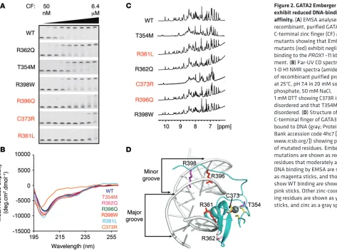
GATA2 Emberger mutants exhibit reduced DNA-binding affinity. The binding of GATA2 mutants to the PROX1 –11 kb GATA site was next investigated using EMSA and recombinant purified C-termi-nal zinc finger constructs (Figure 2A). EMSAs demonstrated that the hematological GATA2 mutant R362Q does not significantly affect binding to DNA, whereas the hematological GATA2 mutants T354M and R398W bound with moderately reduced affinity (Fig-ure 2A). More strikingly, all 3 Emberger mutants R396Q, C373R, and R361L exhibited much lower levels of binding to DNA, with gel-shifts evident only at very high concentrations of GATA2 (Fig-taining a WGATAR motif, together with consensus binding sites
for key transcriptional regulators of lymphatic vessel valve devel-opment, FOXC2 and NFATC1 (ref. 22, Figure 1A, and Supplemen-tal Figure 3). Moreover, hallmarks of an active enhancer element, including a DNaseI hypersensitivity site and a histone H3 at lysine 4 (H3K4Me1) ChIP peak, were evident in this region (see below). Treatment of adult human dermal lymphatic microvascular endo-thelial cells HMVEC-dLyAd (hLEC) with GATA2 siRNA con-firmed that GATA2 knockdown results in substantially reduced PROX1 levels in primary adult hLECs (Figure 1B). Together, these data raised the likelihood that the –11 kb region harbors an enhancer element important for regulating PROX1 expression and suggested that GATA2 binding to this element might be important for controlling PROX1 transcription.
To establish whether GATA2 binding is capable of driving reporter gene expression from the –11 kb element, an 832 bp frag-ment encompassing the WGATAR motif was cloned into pGL4.12 (Promega) and transfected into HEK293 cells together with an expression construct containing WT GATA2. Luciferase expression was substantially elevated in GATA2 transfected cells compared with cells transfected with a vector control (Figure 1C). Mutation of the GATA site from GATA to CTTA reduced luciferase expres-sion by approximately 25% (Figure 1C), demonstrating that GATA2 binding to this site is important for optimal enhancer activity. The –11 kb element was also cloned distally into the enhancer site of pGL4.12, together with a subcloned region of the Prox1 +4.5 kb
[image:5.585.41.514.53.405.2]minal zinc finger from folding and binding to DNA. The T354M mutation appears to be molten globule–like with high levels of sec-ondary but poorly packed tertiary structure. This smaller disrup-tion to folding allows the protein to bind DNA, probably through a binding and folding mechanism, albeit with reduced affinity. The structure of the GATA2 C-terminal zinc finger has not been deter-mined. However, the zinc finger domains of GATA1–3 are highly conserved, with all mutated residues being identical (Supplemen-tal Figure 5). A homology model of the GATA2 C-terminal zinc fin-ger (Supplemental Figure 5), based on GATA3 bound to DNA (52) indicates that the Emberger R361L and R396Q mutations likely disrupt binding to DNA by mutation of key residues that interact with the major and minor grooves, respectively (Figure 2D). R362 makes some minor interactions with phosphates on the backbone ure 2A). The purity of GATA2 WT and Emberger zinc fingers was
assessed by SDS-PAGE analysis prior to use in EMSA assays (Sup-plemental Figure 4). Folding of these mutants was assessed by far-UV circular dichroism (CD) spectroscopy and 1D H1-NMR
spec-troscopy (Figure 2, B and C) to assess secondary-structure content and overall fold, respectively. The CD data indicates that all mutants apart from C373R have WT-like levels of secondary struc-ture. Both the CD and NMR spectra for C373R are characteristic of a largely disordered protein domain with a blue-shifted mini-mum in the CD spectrum and poor peak dispersion in the NMR spectrum. T354M also shows some disruptions to structure in the NMR spectrum with peak dispersion intermediate between that of the WT and C373R proteins. These data indicate that mutation of a zinc-coordinating residue, C373, prevents the GATA2
[image:6.585.58.535.55.487.2]of DNA, but this interaction is fairly solvent exposed, and muta-tion to polar glutamine likely has very little effect on DNA binding. Based on data for GATA3-DNA interactions, R398 should not play a direct role in binding to WGATAR sites (52) (but is involved in binding to pseudo-palindromic CTACTGATA sites through bind-ing in the minor groove), so moderate loss of DNA bindbind-ing likely arises from loss of long-range electrostatic interactions between the positively charged arginine sidechain and the negatively charged DNA. Overall, these data support the hypothesis that sub-stantial losses in PROX1 –11 kb binding, through mutation of key structural or DNA-interacting residues in the C-terminal zinc fin-ger of GATA2, correlate with lymphedema.
The PROX1 –11 kb locus is differentially regulated in lymphatic compared with blood vascular endothelial cells. We next investi-gated the binding of GATA2 to the PROX1 –11 kb region in both adult human dermal lymphatic microvascular endothelial cells (hLEC) and adult human dermal blood microvascular endothelial cells (hBECs) using ChIP. Hallmarks of an active enhancer ele-ment, including a DNaseI hypersensitivity site and an H3K4Me1 ChIP peak, were evident in this region (Figure 3A). Substantial occupancy of GATA2 at the –11 kb site was obvious in hLECs, using both ChIP (Figure 3B) and ChIP-Seq approaches (Figure 3C). ChIP experiments also detected GATA2 binding, though to a
lesser extent, at the PROX1 –11 kb region in hBECs (Figure 3B). In contrast, no significant occupancy at this site was detected in ery-throleukemic K562 cells (Figure 3B), which express GATA2 but not PROX1. Given our observation that consensus sites for FOX and NFAT transcription factors lie in close proximity to the GATA site in PROX1 –11 kb, we next employed ChIP to investigate the occu-pancy of chromatin by FOXC2 and NFATC1 in hLECs, hBECs, and K562 cells. As with GATA2, marked occupancy of the PROX1 –11 kb region by both FOXC2 and NFATC1 was observed in hLECs, and to a lesser extent hBECs, but not in K562 cells (Figure 3B). Given that FOXC2 and NFATC1 have been shown to physically associate and cooperatively regulate transcription (22), we inves-tigated potential protein-protein interactions between GATA2, NFATC1, and FOXC2 using coimmunoprecipitation. We con-firmed an interaction between FOXC2 and NFATC1 in HEK293 cells ectopically expressing these proteins, but no interaction was detected between GATA2 and FOXC2, nor between GATA2 and NFATC1 (Supplemental Figure 6).
[image:7.585.34.399.54.441.2]With the exception of the embryonic cardinal veins (53), LVVs (33), and venous valves (32), substantial levels of PROX1 are not detected in blood vascular endothelial cells. We reasoned that reduced binding of GATA2, FOXC2, and NFATC1 at PROX1 –11 kb in hBECs compared with hLECs was not sufficient to explain
why PROX1 is generally silent in hBECs. To investigate the mech-anisms by which the PROX1 –11 kb region may contribute to turn-ing PROX1 “on” in LECs and “off” in BECs, we investigated the status of chromatin in the PROX1 –11 kb region in hLECs, hBECs, and K562 cells. ChIP for a marker of active chromatin, H3K4Me1, demonstrated substantial levels of this histone mark at PROX1 –11 kb in hLECs, much lower levels in hBECs, and none in K562 cells (Figure 3D), consistent with the transcription factor analysis. Con-versely, no association of the repressive histone mark histone H3 on lysine 27 (H3K27Me3) was detected at PROX1 –11 kb in hLECs, while this mark was present in hBECs and K562 cells (Figure 3D). Together, these data provide further evidence that the PROX1 –11 kb region harbors an enhancer element important for regulating PROX1 expression in the vasculature and suggest that chromatin modifications that act to silence this region are important in keep-ing PROX1 switched off in blood vessels.
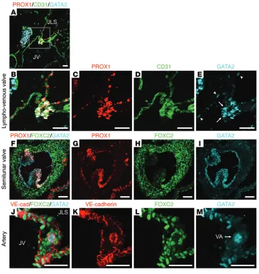
GATA2 is present at prominent levels in lymphatic vessel valves, LVVs, cardiac valves, and arteries. In addition to lymphedema, other cardiovascular phenotypes described in patients harboring GATA2 mutations or polymorphisms include venous thromboses, culture negative endocarditis (49), and susceptibility to coronary artery disease. Given that our early work documented high levels of GATA2 in lymphatic vessel valves (3), we analyzed GATA2 lev-els in LVVs and cardiac valves. Prominent levlev-els of GATA2 were observed in the endothelial cells that comprise LVVs (Figure 4, A–E). In comparison to the low level of GATA2 protein obvious in the endothelial cells lining the jugular vein and the lymph sacs (Figure 4E, arrowheads), GATA2 levels in the cells comprising the valve leaflets was greatly elevated (Figure 4E, arrows). GATA2 was also obvious, together with prominent PROX1 and more restricted FOXC2 staining, in semilunar valves of the embryonic heart (Fig-ure 4, F–I). Taken together, these data suggest that — like PROX1, FOXC2, and NFATC1 — GATA2 marks valve endothelial cells across distinct vascular compartments. GATA2 protein was also obvious in arterial endothelial cells at a discernibly higher level than in veins and lymphatic vessels, though at a lower level than that present in valve endothelial cells (Figure 4, J–M).
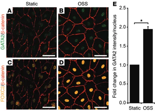
GATA2 levels are elevated in response to oscillatory flow. To investigate the mechanisms by which GATA2 is elevated in valves, we assessed the effects of exposing hLECs to oscillatory
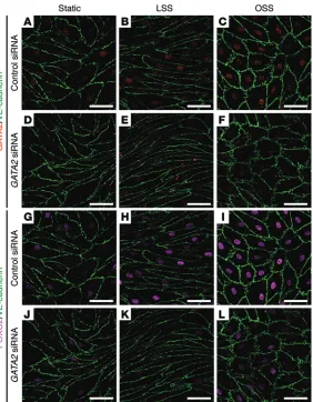
flow in vitro. Previous work established that subjection of hLECs to OSS, reflective of the turbulent flow pattern characteristic of vessel branchpoints, promoted the acquisition of many of the cellular characteristics of valve-forming cells (16). These fea-tures include cytoskeletal remodeling and adoption of a cuboidal rather than elongated cell shape, activation of calcineurin/NFAT signaling, and elevation of CX37 levels. Moreover, acquisition of these characteristics was found to be dependent on PROX1 and FOXC2 (16). Our previous work demonstrated that reduction of Gata2 levels in primary mouse LECs (mLECs) resulted in greatly diminished levels of both Prox1 and Foxc2, suggesting that GATA2 might lie upstream of these key transcription factors in the hier-archy of events required for the initiation of valve development (3). Taking these data into account, we reasoned that GATA2 may be responsive to mechanical stimuli including shear stress and hypothesized that elevated GATA2 levels may be required for the induction of FOXC2 levels mediated by OSS. Exposure of hLECs to OSS for 48 hours resulted in elevated GATA2 mRNA levels and significantly higher levels of GATA2 protein in the nuclei of hLECs (Figure 5, A, B, and E, and Figure 6, A and C). Accord-ingly, hLECs adopted a characteristic cuboidal morphology in response to OSS, and the levels of nuclear FOXC2 were markedly elevated (Figure 5, C, D, and Figure 6, G–I). To assess whether the elevation in FOXC2 levels in response to OSS was dependent on GATA2, hLECs were treated with control (Figure 6, A–C, and G–I) or GATA2 siRNA (Figure 6, D–F, and J–L) and exposed to laminar (Figure 6, B, E, H, and K) or oscillatory (Figure 6, C, F, I, and L) flow. Strikingly, FOXC2 levels were much lower in GATA2 siRNA–treated cells exposed to OSS (Figure 6L) compared with control siRNA–treated cells (Figure 6I). These data suggest that OSS, typical of turbulent flow at vessel branchpoints, may be one of the stimuli by which GATA2 levels are elevated in developing valves and reveal that elevation of FOXC2 levels in response to OSS is dependent on GATA2.
[image:8.585.41.303.54.242.2]GATA2 is required for lymphatic vascular development. In order to assess the requirement for GATA2 for lymphatic vascu-lar development and, in particuvascu-lar, for valve development, we utilized a number of approaches to conditionally delete Gata2 in the vasculature. The first was to delete Gata2 in vascular and hematopoietic compartments by crossing Tie2-Cre mice (54)
compared with I, L, and M). To investigate the underly-ing cause of enlarged jugular lymph sacs, we quantified proliferating lymphatic endothelial cells in the jugular lymph sacs of GataΔEC embryos and their control
litter-mates; the number of Ki67-positive, proliferating lym-phatic endothelial cells was elevated approximately 4-fold in E13.5 GataΔEC embryos (Supplemental Figure
7). To investigate the nature of the blood-filled lym-phatic phenotype, we analyzed the structure of LVVs, which function together with platelet clotting to prevent blood from entering the lymphatic vasculature (33, 35). At E13.5, the leaflets of the LVVs appeared multilayered in GataΔEC embryos compared with their control
coun-terparts (Figure 7, O and P, compared with J and K), suggesting that structural abnormalities in LVV devel-opment might permit the entry of blood to the jugular lymph sacs. In addition, the levels of PROX1 observed in the venous leaflets of the LVVs (PROX1-positive, Podoplanin-negative, Endomucin-low cells) were sub-stantially lower in GataΔEC embryos compared with
their control counterparts (Figure 7, P compared with K, arrows), suggesting that the identity and function of venous valve endothelial cells might be affected. Poten-tial defects in platelet clotting as a result of Gata2 dele-tion in both hematopoietic and endothelial compart-ments in these mice might also contribute to the failure of effective separation of the blood and lymphatic vas-cular networks. Blood-filled jugular lymph sacs and dermal lymphatic vessels (Figure 7, Q and R, compared with L and M) and focal hemorrhages (Figure 7N) were also observed in the skin of GataΔEC embryos compared
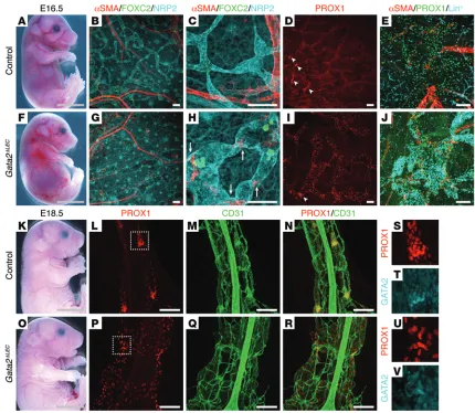
with control (Figure 7I). These data reveal that Gata2 is not required for the initiation of Prox1 expression, nor for the specification and early migration of lymphatic endothelial progenitor cells, but is required for normal LVV development and effective separation of the blood vasculature from the lymphatic vasculature.
We next investigated the consequences of selective Gata2 deletion in the lymphatic vasculature by crossing Prox1-CreERT2
mice (56) with Gata2fl/fl mice and inducing Gata2 deletion at a
range of embryonic stages (55) (Gata2ΔLEC). To compare the
con-sequences of selectively deleting Gata2 in the lymphatic vascula-ture to pan-endothelial and hematopoietic deletion, we adminis-tered tamoxifen at E10.5, E11.5, and E12.5 and analyzed embryos at E13.5 (Figure 8, A–D), or administered tamoxifen at E10.5 and E11.5, analyzing embryos at E14.5 (Figure 8, E–L). Using both regimes, Gata2ΔLEC embryos exhibited pronounced edema
(Fig-ure 8, C and I, arrows) and enlarged jugular lymph sacs. The phe-notype of blood-filled jugular lymph sacs and dermal lymphatic vessels was, however, most prominent with the E10.5, E11.5, and E12.5 tamoxifen regime. Due to the extremely edematous nature of the embryos, it was not possible to obtain sections of suffi-ciently intact morphology for the analysis of LVV development, though it is presumable that the blood-filled lymphatic phenotype would likely occur as a result of defective LVV development. In the Gata2ΔLEC embryos that were administered tamoxifen at E10.5 and
E11.5 and analyzed at E14.5, though edema was obvious, blood-with Gata2fl/fl mice (55) (GataΔEC). Given our finding that GATA2
binds to the PROX1 –11 kb element and regulates PROX1 expres-sion, we investigated whether Gata2 is required for the initiation of Prox1 expression in lymphatic endothelial progenitor cells in the cardinal veins. As Prox1 expression is initiated in the cardi-nal veins at approximately E9.75, Tie2-Cre–mediated deletion should provide an ideal model with which to target Gata2 prior to the stage at which Prox1 is first turned on. Analysis of GataΔEC
embryos at E11.5 revealed no striking vascular defects compared with control embryos (Figure 7, A and E). Moreover, PROX1 was apparent in lymphatic endothelial progenitor cells in the cardi-nal veins (Figure 7, F–H), and the exit of progenitor cells from the veins to form the initial lymphatic vascular plexus was not obvi-ously perturbed (Figure 7, F–H) compared with control counter-parts (Figure 7, B–D).
In accordance with previous studies, deletion of Gata2 in vas-cular and hematopoietic compartments resulted in embryonic demise around E14. E13.5 GataΔEC embryos were edematous; had
[image:9.585.40.322.53.415.2]dramatically enlarged, blood-filled lymph sacs; and exhibited multiple areas of hemorrhage (Figure 7, N, Q, and R, asterisks,
Figure 6. OSS induced elevation of FOXC2 is dependent on GATA2. hLECs were trans-fected with control or GATA2 siRNA and cultured under static (A, D, G, and J) conditions, or subjected to laminar shear stress (LSS; B, E, H, and K) or OSS (C, F, I, and L) for 48 hours (4 dyn/cm2, 1/4 Hz). Immunostaining revealed reduced nuclear FOXC2 levels in
at E16.5 revealed that GataΔLEC embryos exhibited edema,
blood-filled dermal lymphatic vessels, and pooling of blood in the jugular region: phenotypes not observed in control littermates (Figure 9, A and F). Whole-mount immunostaining of embryonic skin illus-trated that the dermal lymphatic vessels were bulbous, irregular in shape, and blood filled (Figure 9, B–E and G–J). In addition, ectopic association of vascular smooth muscle cells (SMCs) with dermal lymphatic vessels was observed in GataΔLEC embryos
(Figure 9H, arrows), a phenotype reminiscent of that observed in Foxc2–/– mice (15). Accordingly, FOXC2 levels appeared much
lower in the lymphatic vessels of GataΔLEC embryos compared
with their control counterparts (Supplemental Figure 8). Analysis of our GATA2 ChIP-Seq data revealed a prominent GATA2 bind-filled lymphatics were not apparent, and accordingly, no striking
abnormalities in LVV development were observed (Figure 8, E–L). In the majority of cases, GATA2 remained detectable in at least a few cells in the LVV region in of E14.5 embryos that had been administered tamoxifen at E10.5 and E11.5 (Figure 8K). Taken together, these data suggest that administration of tamoxifen at E12.5 could be a crucial determinant of Gata2 excision efficiency in the LVV compartment.
[image:10.585.79.510.53.426.2]To investigate the consequences of Gata2 deletion on lym-phatic vessel valve development, which is initiated in the dermal lymphatic vasculature at approximately E16.5, tamoxifen was administered to pregnant females at E12.5, E13.5, and E14.5, and embryos were analyzed at E16.5 and at E18.5. Analysis of embryos
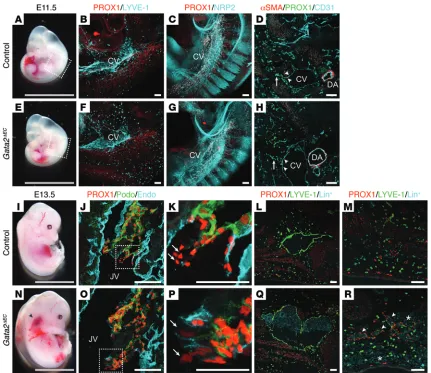
Figure 7. Lymphatic vessel hyperplasia in Gata2ΔECembryos. E11.5 Gata2ΔECembryos appear phenotypically normal (A and E). Whole-mount
immunos-taining for PROX1 (red) and LYVE-1 or NRP2 (cyan) demonstrated that PROX1-positive lymphatic endothelial progenitor cells are specified and migrate away from the cardinal veins of Gata2ΔECembryos (F and G). Immunostaining of transverse sections for α smooth muscle actin (αSMA; red), PROX1 (green),
and CD31 (cyan) confirmed PROX1 expression in lymphatic endothelial progenitor cells of the cardinal veins of Gata2ΔECembryos (D and H; arrowheads) and
migration of these cells from the veins (D and H, arrows). Boxed regions in A–E correspond to areas shown in B, C, F, and G. E13.5 Gata2ΔECembryos exhibit
severe edema, pooling of blood in the jugular region (N, arrowhead), and focal dermal hemorrhages (R, asterisks), compared with control littermates (Cre-negative;Gata2fl/fl; I and M). LVVs appeared multi-layered (O) with reduced PROX1 levels in the venous leaflets (Podoplanin-negative, Endomucin-
low) of Gata2ΔEC embryos (P, arrows) compared with controls (J and K). PROX1 levels in lymphatic leaflets (Podoplanin-positive, Endomucin-negative)
of Gata2ΔEC embryos were not substantially altered (O and P). Jugular lymph sacs (Q) and dermal lymphatic vessels (R, arrowheads) were enlarged and
blood-filled in Gata2ΔEC embryos, phenotypes not observed in control embryos (L and M). Magnified images of boxed regions in J and O are shown in K and
GataΔLEC mesenteries. Likewise, lymphatic vessels appeared
bul-bous, and valve development appeared to be perturbed in the skin of E18.5 GataΔLEC embryos (Supplemental Figure 10). Taken
together, our data illustrate that Gata2 is essential for lymphatic vessel valve development to be initiated.
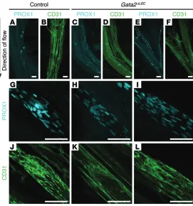
To assess the consequences of Gata2 deletion on later stages of valve maturation, we next administered tamoxifen to pups at P4. Assessment of valve morphology and PROX1 levels in the valves of mesenteric collecting vessels at P8 revealed substantially reduced PROX1 levels in the valves of GataΔLEC pups (Figure 10, C, D, E, F,
H, I, K, and L) compared with controls (Cre-negative;Gata2fl/fl,
Fig-ure 10, A, B, G, and J). In addition, cells within the valve territories appeared more disorganized (Figure 10, C, E, H, and I). Investiga-tion of β-galactosidase levels in the mesenteric lymphatic vessel valves of P8 Prox1-CreERT2 Gata2fl/+ ROSA26R mice administered a
single dose of tamoxifen at P4 confirmed efficient Cre activity in lymphatic vessel valves (Supplemental Figure 11, A–P). Moreover, PROX1 levels were reduced in the valves of Prox1-CreERT2 Gata2fl/+
ROSA26R mice (Supplemental Figure 11, G and O) and the cells in which β-galactosidase levels were highest correlated with the most obvious reduction in PROX1 levels (Supplemental Figure 11, F–H, and N–P). To investigate later-stage consequences of Gata2 dele-tion on the mesenteric lymphatic vasculature, pups administered tamoxifen at P4 were analyzed at P10. The phenotypes of dysmor-ing peak 17 kb downstream of the FOXC2 gene in hLECs
(Sup-plemental Figure 9A), and the ability of GATA2 to drive reporter gene expression from this element was confirmed in luciferase assays (Supplemental Figure 9B). Together with our previous data demonstrating that Gata2 knockdown results in decreased Foxc2 levels in primary embryonic mLECs (3), these data suggest that GATA2 regulates Foxc2 expression in the lymphatic vasculature and that the +17 kb element is potentially an important site for GATA2-mediated control of Foxc2.
The number of clusters of PROX1- and FOXC2-high cells at lymphatic vessel branchpoints in the skin, indicative of initial valve–forming territories, also appeared reduced in GataΔLEC
embryos at E16.5 (Figure 9, D and I, arrowheads), suggesting that the initiation of valve development might be affected as a result of Gata2 deletion. To investigate a potential defect in valve development more closely, we assessed lymphatic vessel development in the mesentery of GataΔLEC embryos and their
control littermate counterparts (Cre-negative;Gata2fl/fl) at E18.5.
Striking defects in lymphatic vessel structure were observed in GataΔLEC embryos at this stage of development; lymphatic
[image:11.585.34.372.48.430.2]vessels appeared bulbous and were poorly organized (Figure 9, K–V). Moreover, in contrast to control littermates in which tightly organized valve-forming territories were obvious (Figure 9, L, P, and S–V), no sign of valve development was apparent in
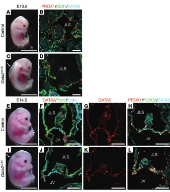
Figure 8. Lymphatic vascular phenotypes in E13.5 and E14.5 Gata2ΔLEC embryos. Prox1-
CreERT2 Gata2fl/+ male mice were crossed with
Gata2fl/fl female mice, and tamoxifen was
administered at E10.5, E11.5, and E12.5, fol-lowed by analysis at E13.5 (A–D), or E10.5 and 11.5, followed by analysis at E14.5 (E–L). E13.5
Gata2ΔLEC embryos exhibit striking edema (C,
arrow) and greatly enlarged, blood-filled jugu-lar lymph sacs (arrowhead, C and D), compared with control littermates (Cre-negative; Gata2fl/fl, A and B). At E14.5, Gata2ΔLEC embryos
exhibit edema (I, arrow). Immunostaining of coronal E14.5 tissue sections demonstrated no obvious morphological abnormalities in LVVs of Gata2ΔLEC mice (J–L) compared with
ducts of GataΔEC/+ mice 1 hour following injection (Figure 12, A
and B), though in 2 of 9 GataΔEC/+ mice, Evans Blue was barely
detectable. In 2 cases, blood was observed in the thoracic ducts of adult GataΔEC/+ mice (Figure 12C). Quantification of thoracic
duct areas in a cohort of mice revealed that the thoracic ducts of adult GataΔEC/+ mice were significantly larger in calibre than their
control counterparts (Figure 12, A–D). Taken together, these data illustrate alterations in lymphatic vessel structure and deficiencies in lymphatic vascular transport function in adult GataΔEC/+ mice.
Discussion
Here, we identify a crucial role for Gata2 in lymphatic vascu-lar development and, in particuvascu-lar, demonstrate that Gata2 is required for the formation and maintenance of lymphovenous and lymphatic vessel valves. Gata2-deficient lymphatic vessels appear bulbous, are aberrantly invested with vascular SMCs and phic valves and bulbous lymphatic vessels were substantially more
severe at this stage, suggesting a progressive decline in valve orga-nization following Gata2 deletion (Figure 11). Like at P8, PROX1 levels were decreased in some valve territories of GataΔLEC pups at
P10, though intriguingly in other cases, PROX1 levels remained high in valves and were elevated in the lymphangion regions of mesenteric vessels. In all cases, the cells within valve territories were markedly disorganized (Figure 11, J–L, and N–P). We hypoth-esize that the elevation of PROX1 levels observed at P10 is a sec-ondary effect of disrupted flow and vessel distension that occurs as a consequence of the loss of valve function.
To investigate the capacity of the lymphatic vasculature to efficiently transport lymph in the setting of Gata2 deficiency, we injected adult GataΔEC/+ mice with Evans Blue dye and
[image:12.585.77.507.53.427.2]moni-tored the transport of dye from the footpads to the thoracic duct. In the majority of cases, Evans Blue was visible in the thoracic
Figure 9. Lymphatic vessel valve development is perturbed in Gata2ΔLEC embryos. Prox1-CreERT2 Gata2fl/+ male mice were crossed with Gata2fl/fl female mice,
and tamoxifen was administered at E12.5, E13.5, and E14.5. E16.5 Gata2ΔLEC embryos exhibit blood-filled dermal lymphatic vessels and pooling of blood in
the jugular region (F), phenotypes not observed in control littermates (Cre-negative;Gata2fl/fl, A). Whole-mount immunostaining of skin from E16.5 Gata2ΔLEC
embryos revealed enlarged, blood-filled dermal lymphatic vessels (G–J) that were ectopically associated with vascular SMCs (H, arrows), phenotypes not observed in control littermates (B–E). Reduced numbers of PROX1-high valve territories (I, arrowheads) were obvious in E16.5 Gata2ΔLEC embryos compared
with littermate controls (D, arrowheads). Gata2ΔLEC embryos appeared indistinguishable from control littermates at E18.5 (K and O). Whole-mount
immu-nostaining of E18.5 mesenteries demonstrated bulbous mesenteric lymphatic vessels in Gata2ΔLEC embryos (P–R), lacking organized valve forming territories
ing analyses revealed very low levels of GATA2 protein throughout the majority of the embryonic vasculature. Notably higher protein levels were obvious in arterial endothelial cells and, most prominently, in the cells that comprise lymphovenous and lymphatic vessel valves. One explanation for the divergence between Gata2 mRNA and protein levels could be differential posttranscriptional/ posttranslational regulation in distinct vascular beds; indeed, our earlier work documenting that miR-451 levels are elevated in primary embryonic BECs compared with LECs (3) provides one potential mechanism by which this could be accomplished. An important role for GATA2 in arteries may underlie the hemorrhagic phenotype of Gata2ΔEC mice described by us and others, as well as the
association of GATA2 SNPs in patients with early-onset coronary artery disease (9–11). Targeted mutation of gata2a in zebrafish has recently been demonstrated to result in defective morphogenesis of the dorsal aorta, resulting in arterial-venous shunts and suggesting that arterial defects could also contribute to the vascular phe-notype observed in Gata2ΔEC mice. To date, however, we
have not observed any striking abnormalities in dorsal aorta morphology (12).
Conditional deletion of Gata2 throughout the vascu-lar and hematopoietic compartments using either +9.5 kb enhancer-driven (13) or VE-cadherin Cre lines (43) has been reported to result in both hematopoietic and vas-cular defects, including anaemia, hemorrhage, edema, and blood-filled lymphatic vessels. Targeted deletion of the Gata2 +9.5 kb regulatory element, essentially ablat-ing Gata2 in the endothelial and hematopoietic lineages, results in similar phenotypes (14). Our documentation of aberrant LVV formation in Gata2ΔEC mice likely explains the
phe-notypes of edema and blood-filled lymphatic vessels observed in the mutant mice reported in earlier studies, though we can’t at present discount a potential contribution from hematopoietic cell defects to this phenotype. On the basis of published work, the lineage most likely to contribute to blood-filled lymphatics is the megakaryocyte/platelet lineage, recently documented to func-tion together with the LVV to prevent the entry of blood cells to the lymphatic vasculature (35), and a lineage in which Gata2 has documented roles (45, 58).
Intriguingly, blood-filled lymph sacs and lymphatic vessels were prominent in E13.5 GataΔLEC embryos in which Gata2
exci-sion was induced by the administration of tamoxifen at E10.5, E11.5, and E12.5, but were not obvious in E14.5 GataΔLEC embryos
in which Gata2 excision was induced at E10.5 and E11.5. Accord-ingly, no prominent defects in LVV formation were observed in these E14.5 GataΔLEC mice. Though disruption to tissue
morphol-ogy in E13.5 GataΔLEC embryos has restricted rigorous analysis of
LVV morphology, we expect that either the structure or function of this valve is impaired, resulting in the blood-filled lymphatic phenotype. The presence of edema in Gata2ΔLEC embryos at both
E13.5 and E14.5, even in the absence of blood-filled jugular lymph sacs, is suggestive of lymphatic vessel dysfunction and a potential role for Gata2 in the lymphatic vasculature prior to the initiation of lymphatic vessel valve development. Our analyses of Gata2 exci-exhibit dysmorphic lymphatic vessel valves (Figure 13).
Under-pinning this phenotype, we identify putative enhancer elements bound by GATA2 at both PROX1 and FOXC2 loci and reveal that neither of these transcription factors is elevated in prospective valve-forming territories in the absence of Gata2. Intriguingly, our data suggest that GATA2 binding at the PROX1 –11 kb locus is not required for the initiation of PROX1 expression in lymphatic endo-thelial progenitor cells in the cardinal veins but may be important as a rheostat to “dial up” PROX1 levels in valve-forming endothe-lial cells. We furthermore demonstrate that GATA2 is mechano- responsive and is elevated in hLECs in response to OSS, providing a mechanism by which GATA2 levels may be increased to initiate the process of valve development. Our data provide new insight to the mechanisms by which only a select catalogue of GATA2 mutations result in primary lymphedema by revealing that mis-sense mutations from Emberger patients, but not those found in hematological disorders in the absence of lymphedema, result in near complete loss of GATA2 function with respect to their ability to regulate the expression of genes important for lymphatic vessel valve development, including Prox1.
Though enigmatic to date, work from multiple studies has implicated GATA2 in vascular development. An enhancer ele-ment located in the fourth intron of Gata2, also known as the +9.5 element (57), has been reported to drive reporter gene expression uniformly through the vasculature (5), though our
immunostain-Figure 10. Gata2 is required for lymphatic vessel valve maintenance. Gata2ΔLEC and
littermate control (Cre-negative;Gata2fl/fl) pups were injected with tamoxifen at P4.
Whole-mount immunostaining of mesenteries at P8 with PROX1 (cyan) and CD31 (green) demonstrated reduced PROX1 levels in lymphatic vessel valves of Gata2ΔLEC
[image:13.585.40.311.54.344.2]marker of repressed, inactive chromatin (60) — was not detected at PROX1 –11 kb in hLECs but was prominent in both hBECs and K562 cells. Taken together, these data suggest that GATA2 may be poised at the PROX1 –11 kb enhancer in BECs and that yet-to-be-identified chromatin remodeling/transcription factors are important for switching this enhancer to the “on” state in LECs. An important answer to the question of how PROX1 transcription is temporally and spatially controlled will come from defining the relative contribution of the –11 kb element compared with other potential enhancer elements.
How is it that GATA2 levels are distinctly higher in valve-forming territories? Our data suggest that at least one mechanism responsible for the elevation of GATA2 levels in lymphatic vessel and LVV valves is mechanical in nature and mediated by OSS, though additional stimuli are likely involved in regulating GATA2 levels within the lymphatic vasculature and between distinct vas-cular endothelial compartments. Established regulators of GATA2 transcription include GATA2 itself, as well as GATA1, reported to repress GATA2 expression in hematopoietic cells (37). GATA2 is both positively and negatively regulated by the Notch signalling pathway; NOTCH1/RBJκ is required to initiate Gata2 expres-sion in hematopoietic stem cells in the embryonic aorta-gonad- mesonephros region (61), while the Notch-induced gene Hes1 subsequently negatively regulates Gata2 in hematopoietic stem cells of the AGM, controlling the production of functional HSC (62). NOTCH1 function has recently been shown to be impor-tant for lymphatic vessel valve development; loss of NOTCH1 results in fewer valves, disrupted reorientation of valve endothe-lial cells, and reduced levels of valve markers, including ITGα9 and FN-EIIIA (25). Whether or not Notch signalling is important sion in the LVV region of E13.5 and E14.5 GataΔLEC embryos
sug-gests that tamoxifen administration at E12.5 might be critical for efficient Gata2 deletion in the LVV using the Prox1-CreERT2 line. In
support of this possibility, our analysis of β-galactosidase levels in the LVV region of E14.5 Prox1-CreERT2 ROSA26R embryos
follow-ing one tamoxifen administration at E12.5 revealed that the major-ity of cells in the LVV region were labeled.
[image:14.585.41.408.55.343.2]While a number of recent studies have contributed to our understanding of how PROX1 transcription is controlled, there remains a dearth of knowledge in this arena. Our data identify GATA2 as an important transcriptional regulator of PROX1 and reveal a novel enhancer element 11 kb upstream of the first, non-coding exon of PROX1 that is bound by GATA2, FOXC2, and NFATC1. Given our data demonstrating that Prox1 expression is still initiated in lymphatic endothelial progenitor cells in the cardinal veins in the absence of Gata2, we hypothesize that the binding of Gata2 to the –11 kb enhancer element is not required to “switch on” Prox1, but rather is important for “dialling up” Prox1 levels in valve-forming cells. There seems little doubt that tran-scriptional cofactors, in addition to GATA2, are required to coor-dinate PROX1 transcription differentially in LECs and BECs, since we detected binding of GATA2 at the PROX1 –11 kb locus in both cell types, though at a greater magnitude in hLECs. In support of this hypothesis, ChIP studies identified differences in the chro-matin architecture of the PROX1 –11 kb locus in hLECs compared with hBECs. Monomethylation of H3K4Me1, a mark indicative of active or poised enhancer elements (59), was associated with the PROX1 –11 kb locus in hLECs and, to a lesser extent, in hBEC; however, it was not present in K562 cells, an erythroid cell line negative for PROX1. In contrast, trimethylation of H3K27Me3 — a
Figure 11. Gata2 deletion results in degeneration of lymphatic vessel valves and lymphatic vessel disten-sion. Gata2ΔLEC and littermate control
(Cre-negative; Gata2fl/fl) pups were
injected with tamoxifen at P4. Whole-mount immunostaining of mesen-teries at P10 with PROX1 (cyan) and CD31 (green) demonstrated severely dysmorphic lymphatic vessel valves and distended lymphatic vessels in Gata2ΔLECmesenteries (C–H, J–L, and
cular endothelial cells remains to be established and is an avenue of research we are actively pursuing. At the posttranslational level, GATA2 is reportedly subject to control by phosphorylation, acetyla-tion, and sumoylation (37), though whether any of these modifica-tions has an impact on the high levels of nuclear GATA2 present in valve-forming territories remains to be determined.
Our assessment of the impact of germline GATA2 mutations found in Emberger syndrome on protein structure, compared with those found only in hematological disorders (germline or somatic), has provided insight to the reasons why some but not all germline GATA2 mutations result in lymphedema. Analysis of the structure of the GATA2 zinc fingers containing Emberger muta-tions R361L, C373R, and R396Q, together with their binding to the GATA site in the PROX1 –11 kb enhancer element, revealed that these mutations exhibit a near complete ablation of DNA binding due to alteration in key DNA-binding residues or severe disruption to protein folding. In contrast, GATA2 mutants associ-ated with hematological disorders but not lymphedema retained some capacity to bind the PROX1 –11 kb enhancer. This finding is in agreement with hypotheses proposed by us (3) and others (49), suggesting that effectively null haploinsufficiency of GATA2 is the critical factor predisposing to lymphedema onset. We don’t rule out the possibility, however, that additional insults such as infection, defective immune cell trafficking resulting in inflam-mation, and/or injury may contribute to lymphedema onset in patients. It remains enigmatic that mutations such as 355del, essentially loss of function in all assays tested, are not to date associated with lymphedema. Additional phenotypes reported in patients carrying GATA2 mutations/deletions that may reflect a role for GATA2 as a general regulator of valve development and function include venous thromboses and culture-negative for the control of Gata2 levels in valve endothelial cells remains
to be assessed. Other signalling axes that regulate Gata2 expres-sion include BMP signalling, required to induce Gata2 and specify ventral identity in Xenopus (63), and retinoic acid (RA) signalling, shown to impact on the transcriptional activity of GATA2 by virtue of a direct interaction between the zinc fingers of GATA2 and the DNA-binding domain of RA receptor α (RARα) (64). Of interest, BMP9 has recently been shown to control lymphatic vessel valve formation (31), and our own recent work revealed aberrant devel-opment of LVVs in Cyp26b1–/– mice in which RA signalling is
ele-vated (65). Potential regulation of GATA2 by both of these path-ways will be the subject of future investigation.
Recent large-scale ChIP-Seq studies analyzing the binding of multiple transcription factors across distinct hematopoietic cell types have revealed key insights to the identity of transcription-factor complexes and target genes important for programming cell identity (44, 45). Such studies in hematopoietic cell lineages have revealed that GATA2 works cooperatively with transcription factors, including RUNX1, SCL/TAL1, FLI1, LYL1, LMO2, and ERG (44, 45). The identity of GATA2 transcriptional cofactors in endothelial cells and, in particular, in lymphatic versus blood
vas-Figure 12. Lymphatic vascular defects in adult Gata2ΔEC mice. Adult
heterozygous Gata2ΔEC/+ mice injected with Evans Blue dye exhibited
col-lecting lymphatic vessels of substantially larger caliber (B–D) than con-trols (A). Thoracic duct area was measured using ImageJ in control (n = 5) and heterozygous Gata2ΔEC/+(n = 6) adult mice (D). *P < 0.05, by 2-tailed
Student’s t-test. Reduced transport of Evans Blue dye to the thoracic duct and blood within the thoracic duct (C; arrow) were also observed in
[image:15.585.87.228.56.332.2]Gata2ΔEC/+ mice. Scale bars: 1 mm. TD, thoracic duct.
[image:15.585.300.539.557.739.2]ChIP-Seq data accession. ChIP-Seq data has been deposited in the European Nucleotide Archive (ENA), accession number PRJEB9436 (http://www.ebi.ac.uk/ena/data/view/PRJEB9436).
Statistics. Analysis of luciferase activity of all GATA2 mutants compared with WT (shown in Figure 1E and Supplemental Figure 1B) was performed using 1-way ANOVA with Dunnett’s multiple-compar-isons test. For all other statistical analyses, P values were calculated using the 2-tailed Student’s t test. P values of less than 0.05 were con-sidered statistically significant.
Study approval. Experiments using mice were approved and con-ducted in accordance with the SA Pathology/CALHN Animal Ethics Committee and Australian National Health and Medical Research Council (NHMRC) guidelines.
Acknowledgments
We thank Sylvia Tichborne, Chris Brown, Kelly Wicks, and staff at the SA Pathology Animal Facility for animal husbandry; Guillermo Oliver for Prox1-CreERT2 mice; Leigh Coultas for Tie2-Cre mice;
Mat Francois for the pGL2BasicProx1-4kbintron1 construct; Junia Melo for K562 cells; Stuart Pitson for HEK293 cells; and John Pimanda, Guillermo Oliver, and Brian Black for helpful discus-sions. This work was supported by project grants from the NHMRC (APP1061365, to N.L. Harvey and H.S. Scott; APP1004651, to J.M. Matthews), South Australian Health & Medical Research Institute Beat Cancer Project (to N.L. Harvey and H.S. Scott), and Swiss National Science Foundation (CRII3 141811, to T.V. Petrova). N.L. Harvey is supported by an ARC Future Fellowship. J.M. Matthews is a Senior Research Fellow and HSS, a Principal Research Fellow of the NHMRC. C.S. Demir was supported by the People Programme (Marie Curie Actions) of the European Union’s Seventh Framework Programme FP7 (2007–2013) under REA grant agreement 317250.
Address correspondence to: Natasha Harvey, Centre for Cancer Biology, UniSA and SA Pathology, PO Box 14, Rundle Mall, Ade-laide, South Australia, 5000, Australia. Phone: 61.8.8222.3569; E-mail: [email protected].
endocarditis (49). Since many of the genes and cellular events important for lymphatic vessel valve development are also impor-tant for venous valve development (32), it stands to reason that venous valve development and/or function may also be affected in patients with GATA2 mutations, a scenario that may contribute to venous thromboses. Likewise, our demonstration that Gata2 is also present in cardiac valves suggests that GATA2 mutations may potentially result in defective cardiac valve development, one of the factors underlying culture-negative endocarditis. An impor-tant role for a second GATA family member, Gata4, has been doc-umented during atrioventricular valve development in mice (66) and mutations in GATA4 have been associated with human atrial and ventricular septal defects and pulmonary valve stenosis (67).
In conclusion, our study defines a crucial role for Gata2 in lym-phatic vascular development and reveals that Gata2 is essential for the morphogenesis of lymphovenous and lymphatic vessel valves. We provide new insight to the mechanisms by which selected GATA2 mutations result in lymphedema in human patients, demonstrating that complete loss of transcriptional activity with respect to the control of genes important for lymphatic vessel valve development underlies this phenotype. Our work has identified a potentially “valve selective” enhancer element 11 kb upstream of the PROX1 locus that is bound by GATA2, FOXC2, and NFATC1 in endothelial cells and that is, in general, “on” in lymphatic endo-thelial cells and “poised” in blood vascular endoendo-thelial cells, at the epigenetic level. Our work sets the stage for future investigations to define the precise mechanisms, in terms of the transcriptional cofactors and target genes of GATA2, that are important for the process of valve development and has the potential to uncover new targets to which the design of novel agents able to promote valve development and/or function could be designed.
Methods
For a full description of Methods, see Supplemental Methods online. Mice. Prox1-CreERT2 (56) Gata2fl/fl (55), Tie2-Cre (54), and ROSA26R Cre reporter (68) mice have been previously described.
1. Mansour S, et al. Emberger syndrome- primary lymphedema with myelodysplasia: report of seven new cases. Am J Med Genet A. 2010;152A(9):2287–2296.
2. Ostergaard P, et al. Mutations in GATA2 cause primary lymphedema associated with a predis-position to acute myeloid leukemia (Emberger syndrome). Nat Genet. 2011;43(10):929–931. 3. Kazenwadel J, et al. Loss-of-function germline
GATA2 mutations in patients with MDS/AML or MonoMAC syndrome and primary lymphedema reveal a key role for GATA2 in the lymphatic vas-culature. Blood. 2012;119(5):1283–1291. 4. Ishida H, et al. GATA-2 anomaly and clinical
phenotype of a sporadic case of lymphedema, dendritic cell, monocyte, B- and NK-cell (DCML) deficiency, and myelodysplasia. Eur J Pediatr. 2012;171(8):1273–1276.
5. Khandekar M, et al. A Gata2 intronic enhancer confers its pan-endothelia-specific regulation.
Development. 2007;134(9):1703–1712.
6. Mammoto A, et al. A mechanosensitive transcrip-tional mechanism that controls angiogenesis.
Nature. 2009;457(7233):1103–1108.
7. Pimanda JE, et al. Endoglin expression in blood and endothelium is differentially regulated by modular assembly of the Ets/Gata heman-gioblast code. Blood. 2008;112(12):4512–4522. 8. Linnemann AK, O’Geen H, Keles S, Farnham PJ,
Bresnick EH. Genetic framework for GATA fac-tor function in vascular biology. Proc Natl Acad
Sci U S A. 2011;108(33):13641–13646.
9. Muiya NP, et al. A study of the role of GATA2 gene polymorphism in coronary artery disease risk traits. Gene. 2014;544(2):152–158.
10. Connelly JJ, et al. GATA2 is associated with famil-ial early-onset coronary artery disease. PLoS
Genet. 2006;2(8):e139.
11. Liu YH, et al. Gene polymorphisms associ-ated with susceptibility to coronary artery disease in Han Chinese people. Genet Mol Res. 2014;13(2):2619–2627.
12. Zhu C, et al. Evaluation and application of modu-larly assembled zinc-finger nucleases in zebrafish.
Development. 2011;138(20):4555–4564.
13. Lim KC, et al. Conditional Gata2 inactivation
results in HSC loss and lymphatic mispatterning.
J Clin Invest. 2012;122(10):3705–3717.
14. Johnson KD, et al. Cis-element mutated in GATA2-dependent immunodeficiency governs hematopoiesis and vascular integrity. J Clin
Invest. 2012;122(10):3692–3704.
15. Petrova TV, et al. Defective valves and abnormal mural cell recruitment underlie lymphatic vascu-lar failure in lymphedema distichiasis. Nat Med. 2004;10(9):974–981.
16. Sabine A, Petrova TV. Interplay of mechanotrans-duction, FOXC2, connexins, and calcineurin signaling in lymphatic valve formation. Adv Anat
Embryol Cell Biol. 2014;214:67–80.
17. Bazigou E, Makinen T. Flow control in our ves-sels: vascular valves make sure there is no way back. Cell Mol Life Sci. 2012;70(6):1055–1066. 18. Koltowska K, Betterman KL, Harvey NL, Hogan
BM. Getting out and about: the emergence and morphogenesis of the vertebrate lymphatic vas-culature. Development. 2013;140(9):1857–1870. 19. Bazigou E, et al. Integrin-alpha9 is required for