OF POTATO VIRUS Y
A thesis submitted in partial fulfilment
of the requirements for the degree
of
Doctor of Philosophy in Molecular Biology
in the
University of Canterbury
by
Joanne M. Hay
University of Canterbury (1989)
Chapter Page
Abbreviations iI
Abstract
1 An introduction to potyviruses and a thesis overview 2
2 Virus purification and cloning of the coat protein gene 7
3
DNA sequence analysis of the PVVN coat protein gene40
4
Plant transformation and the molecular analysis of transformants 825 Thesis review
130
Acknowledgements
133
References
134
AI AIMV APS ATP A.tumefaciens BAP BMV bp BPB BSA BYMV CaMV cDNA CIP CI CMV CPMV CP ddjdATP ddjdCTP ddjdGTP ddjdTIP DEPC DIECA DMSO DNA DTE E.coli EDTA ELISA GAa HC IPTG JGMV kb ABBREVIATIONS amorphous inclusion alfalfa mosaic virus ammonium persulphate adenosine triphosphate Agrobacterium tumefaciens 6-benzylaminopurine bromes mosaic virus base pairs
bromophenol blue bovine serum albumin bean yellow mosaic virus cauliflower mosaic virus
complementary deoxyribonucleic acid calf intestinal al kaline phosphatase cytoplasmic inclusion
cucumber mosaic virus cow pea mosaic virus coat/capsid protein
dideoxy jdeoxyadenosine triphosphate , dideoxy/deoxycytidine triphosphate
dideoxy jdeoxyguanosine triphosphate dideoxy jdeoxythymidine triphosphate diethylpyrocarbonate dlethyldithiocarbamic acid dimethylsulphoxide deoxyribonucleic acid dithioerythritol Escherichia coli ethylenediaminetetra-acetic acid enzyme-linked immunosorbant assay gibberellic acid
M-MLV murine myeloid leukemia virus
NAA naphthaleneacetic acid
NI nuclear Inclusion
NOS nopaline synthase
NPT II neomycin phosphotransferase
OCS octoplne synthase
PEG polyethylene glycol
PEBV pea early browning virus
PeMV pepper mottle virus
poly (A) polyadenylate
PPV plum pox virus
PRY papaya ringspot virus
PWV passionfruit woodiness virus
PTA phosphotungstic acid
PYX potato virus X
PVY potato virus Y
RF replicative form
RNA ribonucleic acid
RUBISCO ribulose bisphosphate carboxy oxygenase
SCMV sugarcane mosaic virus
SDDC diethyldithiocarbamic acid
SDS sodium dodecyl sulphate
SMV ' soybean mosaic virus
TCA trichloroacetic acid
T~DNA transfer deoxyribonucleic acid
TEM transmission electron microscopy
Temed N,N,N',N'-tetramethylethylenedlamine
TEV tobacco etch virus
TMV tobacco mosaic virus
TRV tobacco rattle virus
TVMV tobacco vein mottle virus
VPg viral genome linked protein
Aspects from the coat protein from Potato Virus Y (PVY) were Investigated. Polyadenylated, full-length RNA was isolated from purified preparations of two strains of the virus, pvyN and pvyC. The RNA was used as a template to produce double stranded complementary DNA (cDNA) which was susequently cloned into the plasmid vector pUC19. Of the resultant pvyC -derived clones, two recombinant p/asmids (pVYC5 and pVYC11) were analysed by DNA hybridlsation and DNA sequencing. PVYC11 contained a viral cDNA insert, while pVYC5 did not.
A recombinant clone containing pvyN cDNA sequences (pVYN27) was characterised by DNA sequencing. The 3'-terminal1134 nucleotide sequence coded for the coat protein gene and contained one large open reading frame capable of encoding a protein of 264 amino acid
residues with a combined molecular weight of 29 631. The ten amino-terminal amino acids of the protein were confirmed by amino acid sequencing.
Transcriptional fusions encoding the pvyN coat protein gene and either the CaMV 35S promoter or the mannopine synthase promoter were inserted into an Agrobacterium binary vector encoding the NPT II gene. These were mobilised into two species of Agrobacterium (A. tumefaciens, LBA4404 and A. rhlzogenes, A4T) and transformed into the genomes of Nicot/ana plumbagin/folia and Solanum tuberosum. The transgenicity of regenerated tobacco plants was confirmed by 1. the presence of the chimaeric pvyN coat protein gene as demonstrated by DNA hybridisation, 2. expression of the NPT II gene was demonstrated by the regeneration of plants on media containing kanamycin at normally inhibitory concentrations, and 3. demonstration that progeny from transformed plants inherited the NPT II gene in a Mendelian manner. No
CHAPTER ONE
AN INTRODUCTION TO POTYVIRUSES AND A THESIS OVERVIEW
1.1. POTYVIRUSES
The potato Y viruses, or potyviruses, are the largest known group of plant viral pathogens (Matthews, 1981; Hollings and Brunt, 1981; Edwardson, 1974). They infect a wide variety of host plants including both monocotyledonous and dicotyledonous species. Most potyviruses have a narrow host range and this host specialisation minimises interspecific competition among members (Shukla and Ward, 1988a). Dependent on the definition of a distinct potyvirus, there are estimated to be more than 100 different members of this group (Edwardson, 1974; Francki et al., 1985; de Bokx and Huttinga, 1981). Economic losses attributed to potyviral infection can be significant (de Bokx and Huttinga, 1981).
All potyviruses share a number of common features (Dougherty and Carrington, 1988). The virus particles are flexous rods approximately 700 to 900 nm long, and 12 to 15 nm in diameter. Potyviral genomes are mono partite and consist of a single stranded, positive sense infectious RNA of approximately 10,000 nucleotides, which constitutes 5% of the virion weight (Hill and Benner, 1976; Hinostroza-Orihuela, 1975; Allison et al., 1986; Brakke and van Pelt, 1970; Domier et al., 1986). The RNA genomes are 3'-polyadenylated and have a 5'-terminal genome-linked protein (VPg) (Hari, 1979, 1981). All potyviral genomes contain a protein which
aggregates in the cytoplasm during infection to form characteristic 'pin-wheel' or 'scroll-shaped' Inclusion bodies (Edwardson. 1974; Christie and Edwardson, 1977). Potyviruses can be
mechanically transmitted, but most are transmitted by aphids in a non-persistent, non-circulative manner; transmission in seed and by other insect vectors has been reported (de Bokx and Huttinga, 1981). The closest relatives of the potyvirus group are the plant comoviruses and the animal picornaviruses, each of which possess RNA genomes that are linked to VPg molecules and encode large polyproteins (Carrington et al., 1989).
nucleotide sequence data and biochemical analyses of gene products (Dougherty and Carrington, 1988).
NI
TEV
87HC
VGg
I
aI
? ? CI b I
CP
.1
31I
56 50 70
49
58 3011111
II
POLY A28 53
42
702\-
52
5629
[image:7.558.86.466.124.250.2]TVMV
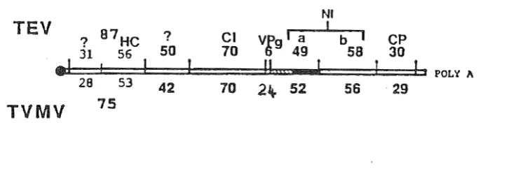
75Figure 1.1. Proposed genetic maps for TEV and TVMV (after Dougherty and Carrington, 1988). The molecular weights (x 103) of the putative cleavage products are present for each virus. The following clstrons are Identified: coat protein (CP), nuclear Inclusion proteins (Nla & Nib), cytoplasmic Inclusion protein (CI), helper component (He) and VPg.
Gene products are Initially expressed as a single, large polyprotein precursor that undergoes posHranslational proteolytic processing, analogous to picornavlruses (Dougherty, 1983). The polyproteins which are produced have a molecular weight of approximately 340 kd (Allison
et
al., 1986; Domieret
al., 1986; Hellmannet
al., 1983, 1986; Vance and Beachy, 1984; Yeh and Gonsalves, 1985). Intermediate cleavage products and individual mature viral proteins arise from the rapid proteolytic processing of the polyprotein.The major structural potyviral protein is the coat protein and it has been mapped proximal to the 3'-terminus (Allison et a/., 1985b, 1986; Domler et al., 1986; Dougherty and Hiebert, 1980c; Dougherty et al., 1985, Ravelonandro et al., 1988; Hay et ai, 1989). It is the major component of the virion and envelopes the genomic material with approximately 2000 monomeric units (Hollings and Brunt, 1981). Each potyvirus contains a single type of capsid protein monomer, ranging in size from 30 to 45 kd (Allison et al., 1985a). Heterogeneity in the apparent capsid protein size is a common feature of potyviruses owing to degradation during purification and/or storage (Shukla and Ward, 1988a). The amino acid sequences of a number of different
on a separate RNA species (Dougherty and Carrington, 1988). In TEV, there is evidence that the coat protein participates In the aphid transmission phenotype (Pirone and Thornbury, 1983).
A genome-linked VPg protein is found covalently attached to the S'-terminal nucleotide of potyviral RNA from TEV (Hari, 1981) and TVMV (Shahabuddin et al., 1988) virus preparations. The TVMV VPg is unusually large, 24 kd; for TEV the protein is 6 kd. A role for the VPg in replication can be inferred from studies of other viruses with similar gene organisation and expression (Dougherty and Carrington, 1988). The protein could conceivably be exposed externally on the virion and may have a potential role In aphid transmission (Dougherty and Carrington, 1988).
All potyviruses code for a cytoplasmic pin-wheel Inclusion protein with similar molecular weights (65 to 75 kd) (Dougherty and Hiebert, 1980b; Edwardson, 1974). These aggregates consist of protein monomers and their morphology is virus-specific. No particular function has been conclusively assigned to this protein although it has been hypothesised to have a role in replication. The aggregates have also been observed to associate with the plasmodesmata (Edwardson, 1974) which implicates a role for this protein In the cell-to-cell movement of the virus. The CI protein may have multi-functional domains and play an integral role in both of these viral processes (Dougherty and Carrington, 1988).
A cytoplasmic amorphous Inclusion body (AI) has been detected in a number of, but not all, potyvirus-infected plants. When formed, the polypeptide is large in size (51 to 56 kd) and is quite stable (de Mejia et al., 1985a). Biochemical and immunological evidence exist that equate the Al and the helper component (de Mejia et al., 1985b). The HC is a virus-encoded protein necessary for insect transmission (Berger and Pirone, 1986; Hiebert et al., 1984; Pirone and Thornbury, 1983; Thornbury et al., 1985; Thornbury and Pirone, 1983). However, purified AI
bodies do not have HC activity suggesting that the inclusion-bound form of the protein is inactive. Alternatively, the HC activity may be associated with a processed or modified form of the
inclusion protein (Dougherty and Carrington, 1988).
All potyviruses code for two proteins which aggregate in equimolar amounts (Dougherty and Hiebert, 1980b; Knuhtsen et al., 1974). Together they comprise the nuclear inclusion body, although only a limited number of potyvirus infections cause stable NI aggregates to form (Edwardson, 1974). NI bodies vary in shape and size depending on the specific virus isolate. The two NI protein genes have been mapped adjacent to one another (Allison et al., 1986; Hellmann et al., 1986), and are approximately 58 and 49 kd, respectively. The 49 kd protein is a virus-encoded proteinase. It releases itself by an autocatalytic mechanism and functions in
trans
at three additional cleavage sites within the carboxy terminus of the TEV polyprotein (Carrington and Dougherty, 1987; Carrington et al., 1988). On the basis of sequence homology studies, the 58 kd NI protein may be an RNA-dependent RNA polymerase (viral replicase) (Allison et al., 1986; Domler et al., 1987). The 3' positioning of the replicase to the VPg-proteinase cluster Is
The major cell-free translation prod ucts of TEV and TVMV RNA are proteins of 87 kd and 75 kd, respectively, and have been predicted to be encoded by sequences proximal to the 5' terminus of the open reading frame (Dougherty and Hiebert, 1980c; Hellmann et al., 1980). These are thought to be precursors which are processed to produce proteins approximately 30 kd and 54 kd (Hellmann et al., 1983, 1985; Hiebert et al., 1984; Thornbury et al., 1985). The smaller protein has been implicated in the cell-to-cell movement of the virus (Domier et al., 1987). The remaining 54 kd portion has tentatively been identified as the aphid transmission HC and AI protein (Hellmann et al., 1985; Hiebert et al., 1984; Thornbury et al., 1985). In TEV the proteinase responsible for the cleavage of this 5' precursor from the polyprotein has been identified and localised to the carboxy terminal half of the 56 kd HC (Carrington et al., 1989). The proteinase appears to be released by an autocatalytic mechanism and cleavage In vitro occurs at a
dipeptide which differs from those recognised by the 49 kd proteinase. Carrington et al. suggest the HC and the proteinase have separate domains within the 56 kd protein. Neither the site of cleavage between the 31 and 56 kd proteins nor the activity responsible for the cleavage in TEV have been identified. R. E. Rhoads (pers. comm.) observed that for TVMVtwo separate
proteolytic activities are responsible for the maturation of the HC. One is encoded by the 5'-terminal gene (34 kd cistron) and mediates the HC/34 cleavage, and one is encoded by the HC cistron and mediates the HC/42 cleavage.
The gene located 3' to the sequence encoding the TEV 87 kd or the TVMV 75 kd 'precursor', is predicted to code for a protein with a molecular weight of approximately 50 kd (Domier et al., 1987; Dougherty and Hiebert, 1980c). No specific function has been associated with this protein (Dougherty and Carrington, 1988).
There is no information available for the replicative cycle of potyviral RNA or on the manner of encapsidation.
1.2. THESIS OVERVIEW
Potato virus Y (PVY) is a member of the potyvirus group (Matthews, 1981) and causes economically important diseases world wide in potato. tomato, pepper and tobacco crops. Because of its prevalence in potato and tobacco crops in New Zealand and because few studies have been made of this 'type-member' for the potyvirus group, PVY was selected as the subject for this study. The initial aim of this study was to clone and sequence the PVY coat protein gene. Subsequently, the coat protein gene was to be expressed in host plants Solanum tuberosum and Nicotiana plumbaginifolia, with a view toward generating PVY-resistant plants.
Chapter one outlines some general properties of the potyvirus group and the current models for potyviral genome organisation and expression.
Chapter three reports the sequencing and subsequent analysis of the 3' -terminal 1134 nucleotldes of the local isolate of pvyN. A predicted amino acid sequence for the coat protein is inferred from the nucleotide sequence.
Chapter four reports the construction of chimaeric coat protein genes and their insertion into host plants Nlcotiana tabacum and Solanum tuberosum, via the Agrobacterium binary vector for plant transformation. The presence of the PVY coat protein gene was examined by Southern blot analysis and DNA slot blot hybridisation. Expression of the gene in vivo was analysed by Northern and Western blots. The question of coat protein-induced 'genetically engineered cross protection' was addressed.
Chapter five summarizes the findings made by this study.
CHAPTER TWO
VIRUS PURIFICATION AND CLONING OF THE COAT PROTEIN GENE
2.1. INTRODUCTION
2.1.1. SYMPTOMOLOGY
Potato Virus Y (PVY) is the type member of the potyvirus group (Matthews, 1981). and on the basis of symptom development on indicator plants comprises of three main groups of strains: PVVO (common strain), PVVN (tobacco veinal strain) and PVVC (stipple streak strain) (de Bolo:: and Huttinga, 1981). Two of these, PVVc and PVVO, have long been identified in New Zealand, but PVVN was isolated from potato only three years ago (Fletcher, 1986).
A PVVN strain was first isolated in 1935 from tobacco growing near potato plants, in the United States of America (Smith and Dennis, 1940). Subsequently strains have been found all around the world, primarily in tobacco and potato crops. Although isolates from different countries were not found to be identical and often differed markedly in their physical characteristics, they were clearly related (Klinkowski and Schmelzer, 1960).
Differences in symptom expression in hosts depends on the strain of virus, cultivar of host plant and environmental conditions. Generally, primary infection by PVVO strains induces severe systemic crinkle symptoms, leaf drop and premature death in potato cultivars; and yellowing, systemic mottling and leaf drop in tobacco. 'Potato cultivars susceptible to PVVC strains show systemic mosaic, necrosis and stipple streak symptoms on the petioles and stems during primary infection, and may develop necrosis on the tubers during infection in the second year (secondary infection). Symptoms similar to those Induced by PVVO are observed for tobacco hosts. PVVN strains cause necrotic rings and spots on the leaves of potato cultivars. The primary symptoms are usually mild, while those developing in the second year are more obvious. These extend from mosaic and streak on the leaves, to leaf drop and may lead to premature death of the plant. Severe systemic necrotic lesions, often following the main veins, are observed on the leaves of infected tobacco. Primary veins become brown and leaves collapse against the stem. The stems also show necrosis, especially near the base (Klinkowski and Schmelzer, 1960).
The PVVN strains are highly aphid transmissable and all strains can be transmitted mechanically.
reduced tuber size by 12-14% on average, and yield by 14-20%. De Bokx and Huttinga (1981)
quoted decreases of potato yields in the order of 10-80%, depending on host and viral strains. They also observed that the virus was transmitted to plant tubers during secondary infection. Infection of tobacco by PVY decreased yield by approximately 30% (Sievert, 1978), with pvyN strains being particularly destructive and often causing complete crop failure. PVY also infects pepper and tomato cultivars which may result in heavy crop losses. The destructive effect of PVY is compounded when found In conjunction with other viruses, ego potato leafroll virus and potato virus X (Edwardson, 1974). Hence, infection by PVY potentially incurs enormous annual financial losses to the agricultural sector in many countries throughout the world.
Nicotiana tabacum cvs. Samsun and White Burley are good indicator plants for differentiating strains of PVY; pyyC infection results in vein clearing and slight epinasty of the leaves, and pvyN produces necrotic lesions on the leaf and stem. The ease and rapidity of Infection, and clarity of symptoms, contributed to Nicotiana tabacum varieties being good sources of virus. These varitles were used as laboratory host plants for purification and subsequent experimentation.
2.1.2. CLONING
Recombinant DNA methods allow the isolation and amplification of single genes from whole genomes. They also facilitate the modification of genes which can then be re-introduced into cells for expression of RNA or protein rtVu, 1987). Such methods are useful for studying plant viruses, in particular RNA viruses. A copy of DNA (cDNA) complementary to the RNA viral genome can be made using reverse transcriptase. (Hull and Davies, 1983). The cDNA can then be inserted into a prokaryote for replication and amplification. This approach contributes to an understanding of the complex problems of RNA virus evolution, the induction of disease symptoms, the function of plant viral sequences, the nature of virulence. and has contributed to the development of methods for reducing infection of plants.
cDNA has been used for sequencing the whole genomes of tobacco etch virus (TEV) (Allison et al .• 1986) and tobacco vein mottling virus (TVMV) (Domier et al., 1986) and has led to a study of the relatedness of different virus species using nucleotide and amino acid sequence comparisons. Studies on the genetic organisation of plant viruses have been facilitated by using cDNA clones to the complete or partial genome, ego pepper mottle virus (PeMV) (Dougherty et
al., 1985) and cowpea mosaic virus (CMV) 010s et al., 1984).
By recombining segments of viral genomes as DNA. phenotypes can be mapped
physically to specific regions. Two early examples of this application are to the RNA viruses aB coliphage (Taniguchi et al., 1978) and poliovirus (Rancaniello and Baltimore, 1981).
in vitro from full-length cDNA clones of a number of plant RNA viruses including CPMV, brome mosaic virus (BMV) (Ahlquist et al., 1984) and tobacco mosaic virus (rMV) (Meshi et al., 1986). cDNA fragments have been used as probes in nucleic acid hybrldisation for identifying plant viruses, ego citrus tristeza virus (Rosner et al., 1984) and bean yellow mosaic virus (BYMV) (Abu-Samah and Randles, 1981). Rosner et al. (1986) cloned fragments of PVY to use as a diagnostic probe, via molecular hybrldisation, for PVY Infection.
The production of virus resistant plants using genetic engineering technology may potentially contribute to the prevention of plant diseases. For example, cDNA encoding the coat protein of TMV and alfalfa mosaic virus (AMV) has been expressed in plants via the
Agrobacterium tumefaciens Ti plasmid, and has been shown to induce virus resistance (Powell-Abel et a/., 1986; Nelson et al., 1987; van Dun et al., 1988).
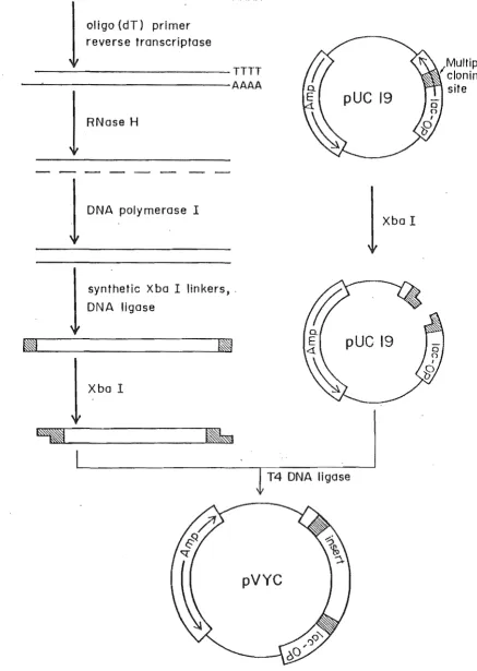
The first reports of molecular cloning of cDNAs were by Rougeon and Mach (1976) and Efstratiadis et a/ .• (1976). Since then, the techniques have been increasingly refined to provide versatile tools for the molecular analysis of eukaryote and prokaryote genes, examples of which have been described. Gubler and Hoffman (1983) modified the method of Okayama and Berg (1982) and combined classical ollgo(dT)-prlmed first strand synthesis with the novel Rnase H-DNA polymerase I second strand synthesis. The production of double stranded cH-DNA became a simple two-step process. This procedure is useful because it permits synthesis from mRNAs which are present in low concentrations. It also eliminates the S1-nuclease mediated cleavage of hairpin loops generated by previous synthesis procedures, which usually removed important 5'-terminal sequences. D'Alessio et al. (1987) further modified the protocol by developing a 'one tube' system for the synthesis of cDNA from RNA by reverse transcription. A single strand of DNA complementary to the RNA template is sythesised from a primer by an RNA dependent DNA polymerase (reverse transcriptase). The primer is provided by a poly(dT) oligonucleotide
annealed to the RNA 3'-poly(A) tall. The cDNA is made double stranded with DNA polymerase I from
E.
coli and the primer required by this enzyme is provided by short fragments of RNA generated by RNase H digestion of the cDNA-RNA heteroduplex (Gubler and Hoffman, 1983). The strategy of D'Alessio et al. is outlined in Fig. 2.1. and was used In this study. The aim was to produce complementary DNA to two strains of pvy, pvyN andpvy
C. In this study, synthetic Xballinkers were attached to the double stranded cDNA and the cDNA was inserted into the plasmid cloning vector, pUC19.The pUC series of plasmids are widely used as cloning vectors. They encode ampicillin resistance and a portion of the ,a-galactosidase gene from the E. coli lac operon, Including the promoter, which complements the portIon encoded in
E.
coli hosts such as the JM83 strain. The pUC plasmids also contain a polyllnker within the ,a-galactosidase encoding region, they1
oligo (dT) primer
reverse transcriptase
---TTTT
---AAAA
1
RNase H
1
DNA polymerase I
synthetic Xba I linkers, .
DNA ligase
pUC 19
Xba I
pUC 19
T4 DNA ligase
pVYC
figure 2.1.
Strategy for cloning the
pyyNcoat protein gene.
[image:14.560.41.479.53.669.2]isopropylthiogalactoside (IPTG), the chromogenic j3-galactosidase substrate, 5-bromo-4-chloro-3-indolyl-j3-D-galactoside (Xgal), is hydrolysed to bromochloroindole and confers a blue colour to the infected colony. If an insert is cloned into the polylinker, the a-peptide coding region is interrupted and no functional j3-galactsidase can be produced, hence the recombinant colony is white.
2.2. MATERIALS AND METHODS
2.2.1. VIRUS ISOLATES AND PROPAGATION
Materials
pvyN inoculum - isolated from Solanum tuberosum (Canterbury) and donated by
J.
Fletcher (Plant Diseases Division, Department of Industrial and Scientific Research, (PDD, DSIR), Lincoln, New Zealand), (Fletcher, 1986).pvyC inoculum - isolated from Solanum tuberosum cv. Lichte Rode Star and donated
b~
J.
A. de Bokx, (Research Institute for Plant Protection, Wageningen, The Netherlands).Methods
Fresh or freeze-dried inoculum was homogenised in an oven-baked mortar and pestle in four to five volumes (wjv) of Yarwoods buffer (0.5% potassium phosphate, 0.5% bentonite; Yarwood 1968). Both strains of the virus were propagated in Nicotiana tabacum cv. White Burley by mechanical inoculation. Four to six week old plants were dusted with carborundum powder (400
+
mesh) on two to three large leaves per plant and rubbed with the inoculum. Excess inoculum and carborundum were Immediately rinsed off with distilled water to prevent dessication.Plants were grown in insect-free growth rooms or glasshouses at 180 -20oC .. Symptoms developed 10-21 days after inoculation, depending on the strain, and infection was confirmed using a commercial Enzyme Linked Immuno:,orbent Assay (ELISA) kit (Boehringer Mannheim). Samples were assayed according to the manufacturers instructions and the intensity of the colour reaction assessed visually. A value from - to
+ + +
was assigned.2.2.2. VIRUS PURIFICATION
Four protocols for the purification of PVY were used including those of Hamilton and Nichols (1978), R.Forster (pers. comm.) and Dougherty and Hiebert (1980a). The highest yield of intact virus particles were purified using a procedure published by Reddick and Barnett (1983) with modifications to the initial extraction buffer.
in a Sorvall GS-3 rotor. The resulting pellets were re-extracted in 0.75 volumes of phosphate buffer and 0.4 volumes of chloroform and centrifuged as before.
The virus was concentrated from the combined supernatants by adding 4% polyethylene glycol (PEG) (w/v) and 0.25M NaCI and stirring for one hour at 40C. The PEG precipitate was collected by centrifugation at 8000 rpm for 15 minutes and resuspended in 50.0 mls of
phosphate/urea buffer (0.5M potassium phosphate pH 7.0, 1 M urea) containing 1 % Triton X-1 00. The suspension was stirred for two hours at 40C and then centrifuged at 8000 rpm for 15 minutes in a Sorvall SS-34 rotor. The supernatant was subjected to a second PEG precipitation by stirring with 4% PEG (w Iv) and 0.25M NaCI for one hour at 40C, and the crude virus pellet was collected by centrifugation at 8000 rpm for 15 minutes.
Equilibrium centrifugation in caesium sulphate (Cs2S04) followed immediately: the virus pellet was resuspended In 7.5 ml of phosphate/urea buffer containing 1.5g of Cs2S04, and layered onto two 0.8 ml cushions of 53% (w /w) Cs2S04 in 20mM Tris-HCl pH 7.5 In two Beckman SW55 ultraclear centrifuge tubes. The tubes were topped up with phosphate/urea buffer and centrifuged at 30,000 rpm at 50C for 16 hours in a Beckman SW55 rotor. The
opalescent virus band was removed from the Cs2S04 using a peristaltic pump, diluted five times in 20 mM Tris-HCI pH 7.0, and dialysed against one litre of 20mM Trls-HCI pH 7.0 for four hours with a buffer change after one hour.
The resulting suspension was cleared by centrifugation at 10,000 rpm for 10 minutes in a Sorvall SS34 rotor and the virus collected from the supernatant by high speed centrifugation at 50.000 rpm for 1.5 hours In a Beckman SW55 rotor. The glassy pellet was resuspended in 250 ILl of 20mM Tris-HCI pH 7.0.
The presence of virus was ascertained by transmission electron microscopy (TEM) using 2% phosphotungstic acid (PTA) pH 7.0 as a negative stain. PVY concentrations were determined spectrophotometrically using a coefficient of 2.3 rng ml-1 at 260nm or by the dye-binding method of Bradford (1976). A spectrophotometric profile between 220nm and 320nm was produced.
Whole virus was stored in allquots at -200C.
2.2.3. RNA ISOLATION
Freshly purified virus was dissociated as described by Brakke and van Pelt (1970). Virus particles were dissolved In a small volume of disruption buffer (100mM Trls-HCI pH 9.0, 1mM EDTA, 1% sodium dodecylsulphate (SDS). 0.1 mg mr1 bentonite) containing 10 ILg mr1 proteinase K (pre-incubated at 370C for 10 minutes). After gentle mixing, the mixture was incubated at room temperature for 15 to 30 minutes.
Sucrose Gradient Centrifugation (Brakke and van Pelt, 1970)
the top of one gradient, the tubes topped with paraffin oil to prevent collapse and centrifuged at 25,000 rpm for 13 hours at 140C.
The tubes were pierced using a Beckman fraction recovery system and one ml fractions were collected. The absorbance of each fraction was measured at 260nm and the peak fractions pooled and precipitated. The RNA was resuspended to 1 mg
mr1
in double glass distilled (dd)H20
and stored at -ao°C in 10 J.d aliquots.The length and size distribution of the resulting RNA was determined by gel electrophoresis in 1% agarose gels containing 1xTBE (section 2.2.13.).
2.2.4. SYNTHESIS OF COMPLEMENTARY DNA
Double stranded complementary DNA (cDNA) was synthesised from 5 ug of PVY RNA using the single tube reaction described by d'Alessio et a/. (1987).
First Strand Synthesis
A 50 1'1 reverse transcription reaction was prepared containing 5 pg of PVY RNA heat treated to 650C for 10 minutes and quick chilled, plus 50mM Tris-HCI pH 8.3, 75mM KCI, 10mM
dlthioerythritol (DTE), 3mM MgCI2, 500 I'M each of dATP, dGTP, dCTP and dTTP, 50 pg mr1 oligo (d1)12-18 primer and 10,000 units ml·1 M-MLV reversetranscriptase.
After the enzyme was added, the reaction mixture was kept on ice and a 10 1'1 aliquot transferred to a separate tube containing 10 pCi of [a-35SjdCTP (1000 Ci mmor1) or 7.6 pCi of [methyl-3HjdTTP (47 Ci mmor1). A one 1'1 sample of this labelled reaction was immediately removed, TCA precipitated (section 2.2.11) and counted to provide a 'time zero' ('to') product. Both tubes were incubated at 370C for one hour; the reaction was stopped by placing the tubes on ice. A one 1'1 sample was removed from the labelled reaction, TCA precipitated and counted to determine the yield of first strand product at 'too'.
Second Strand SyntheSiS
The tube containing 40 1'1 of unlabelled first strand reaction was diluted to a final volume of 320 1'1 containing 25mM Trls·HCI pH 8.3, 100mM KCI, 5mM MgCI2, 5mM DTE, 250 I'M each of dATP, dGTP, dCTP, dTTP, 500 cpm/pmole [a.35SjdCTP (or 250 cpm/pmole [methyl.3HjdTTP), 250 units mr 1
E.
coli DNA polymerase I and 8.5 units mr 1 E. coli RNase H. The mixture wasincubated for two hours at 160C and synthesis was terminated by the addition of 10 1'1 of 0.5M EDTA.
1 mM EDTA) or 5 JLI of 1x T4 polynucleotide kinase buffer (section 2.2.5.) in preparation for ligation of linkers, and stored at 40C.
Portions of single and double stranded cDNAs were analysed by 1 % agarose gel electrophoresis and the remainder was used for cloning.
2.2.5. CLONING OF COMPLEMENTARY DNA INTO A PLASMID VECTOR
Blunt End ligation of Synthetic linkers
Synthetic XbaJ linkers were supplied in S'-hydroxyl, single stranded form (Collaborative
Research). They were re-annealed by heating to 650C for 10 minutes and allowed to cool slowly to room temperature. The double stranded linkers were phosphorylated at their S'-hydroxyl ends as follows: one JLg of double stranded Xballinkers was incubated at 370C for one hour in 10 JLI of kinase buffer (10mM DTE, 10mM Tris-HC! pH 7.S, 1 mM ATP, 10mM MgCI2, 200 ug m,-1
acetylated bovine serum albumin (BSA» and two units of T4 polynucleotide kinase (Maniatis
et
al., 1982).This reaction was added to 10 JLg of cDNA in 5 JLI of 1x kinase buffer for blunt end ligation. After the addition of two units ofT4 DNA ligase and 0.4 p.1 of 17.7mM ATP, the reaction was Incubated at 220C for 6-10 hours. The linker ligation reaction was stopped by the addition of 2 JLI of 0.5M EDTA pH 8.0, and extracted once with phenol:chloroform:isoamyl alcohol, (25:24:1). The organic phase was back-extracted once with 20 JLI of TE and the combined aqueous phases precipitated with ethanol. The dried pellet was resuspended in 100 JLI of buffer (SOmM Tris-HCI pH 8.0, 10mM MgCI2, 50mM NaCI), 100 units of Xbal restriction endonuclease were added and incubated at 370C for four hours. The reaction was stopped by adding 2 JLI of O.SM EDTA,
extracted once with phenol/chloroform and the excess linkers removed by gel filtration on a spun Sephadex G-SO column (section 2.2.10.). Five JLI samples were taken from the eDNA after spinning through the column, TCA precipitated and counted to determine losses of cDNA.
The linkered cDNA eluate was ethanol precipitated and resuspended in 10 JLI of TE pH 7.0 for ligation Into vector. A one JLI sample was counted to determine the final yield.
Preparation of Vector for ligation
Ten JLg of plasmid vector pUC19 were made linear in a 100 JLI reaction by digestion with
Xbal
at its single recognition sequence in the multiple cloning site.to 100 ILl in STE (10mM Trls-HCI pH 8.0, 100mM NaCl, 1mM EDTA) and 0.5% SDS. The CIP was inactivated by heating the reaction mix to 650C for 15 minutes, extracted twice with
phenol/chloroform and the DNA precipitated In ethanol. The plasmid DNA was resuspended in 10 ILl TE pH 7.5 in preparation for ligation to the cDNA.
Ligation of Vector and eDNA
Vector pUC19 DNA and linker treated cDNA were incubated at 160C overnight In a 10 ILl ligation reaction (50mM Tris-HCI pH 7.5, 10mM MgCI2, 10mM DTE, 1mM spermidine, 1mM ATP, 100/ug
mr 1 BSA) with one Weiss unit of T 4 DNA ligase. /
A total of 120 ng of DNA, and a vector to insert ratio of 5:1 in the 10 ILl reaction was used. A control reaction containing no cDNA was included to determine the efficiency of the
dephosphorylation of pUC19.
Tra nsformation
E.
coli strain JM83 cells were made competent and transformed with the pUC19/cDNA ligation reaction using a modification of the method published by Hanahan (1983). Ten ml of SOB (2% (w/v) tryptone, 0.5% yeast extract, 10mM NaCI, 10 mM MgS04, 10mM MgCI2, 2.5mM KCI) were inoculated with a single colony of JM83 and incubated overnight at 370C with agitation. One ml of this culture was used to inoculate 10-30 mls of SOB In a 250 ml flask and allowed to incubate at 370C with shaking until an absorbance of 0.45-0.55 at 550 nm (A550) was reached. The cells were collected in a pre-chilled 50.0 ml polypropylene centrifuge tube, placed on Ice for 10-15 minutes and pelleted at 2500 rpm for 12 minutes at 40C In a Sorvall SS34. The cells were resuspended in TFB buffer (10mM KMES pH,6.2, 100mM RbCI, 45mM MnCI2.4H20,10mMCaCI2.2H20) at one third of the initial volume with gentle vortexing and placedon ice for 15 minutes. The cells were pelleted as before and resuspended in TFB buffer at 1/12.5 of the initial volume of cells. Fresh dimethyl sulphoxide (DMSO) was added to 3.5% and the cells left on Ice for 5 minutes. DTE was added to 7.5%, gently swirled and the cells incubated on ice for a further 10 minutes. A second portion of DMSO was added and the cells iced for 5 minutes. 210 ILl samples of competent cells were placed in pre-chilled eppendorf tubes and the 10 ILlllgation mixtures added, gently mixed and incubated on ice for 30 minutes. The tubes were heat pulsed at 42°C for 90 seconds and placed on ice for 1-2 minutes. 800
ILl
of SOC (S08 containing 20mM glucose) was added to each tube and allowed to shake slowly at 370C for one hour. The cells were pelleted in a microfuge for 5 minutes, resuspended in 200 ILl of SOB and spread on lM plates (1% tryptone, 0.5% yeast extract, 10mM NaCl, 10mM MgS04.7H20, 1.5% (w/v)2.2.6. COLONY SCREENING
Colonies that were ampicillin resistant and lac· (unable to metabolise Xgal) were screened for insert size by making mini-preparations of the recombinant plasmid DNA using a rapid alkaline extraction procedure (Birnboin and Doly, 1979), followed by digestion with Xbal and analysis of Insert size by electrophoresis through 1 % agarose gels.
Colonies to be screened were dispersed in one ml of LB media (1 % tryptone, 0.5% yeast extract, 1 % NaCI) containing 50 I'g mr 1 ampicillin, in eppendorf tubes and incubated overnight at 370C. The cells were pelleted in a microfuge, iced and resuspended in 1001'1 of pre-lysis buffer (50mM glucose, 25mM Tris-HCI pH 7.6, 1 mM EDTA) by vigorous vortexing. Cells were lysed by adding egg white lysozyme to 2.5 mg mr 1 and were incubated at room temperature for 15 minutes. 2001'1 of freshly prepared lysis solution (200mM NaOH, 1 % SDS) were added to the cells, mixed gently by inversion and allowed to incubate for 5 minutes at room temperature. 150 1'1 of acidifying buffer (3M NaOAc pH 4.8) were then added, mixed by Inverting the tubes 30 times and incubated on ice for 30 minutes. Insoluble debris was pelleted by microfugation for 15 minutes and the supernatant decanted into a new tube. One ml of ethanol was added and the tubes centrifuged at room temperature for 3-4 minutes. After drying the tubes to remove residual ethanol, the pellets were resuspended in neutralising buffer (0.1 M NaOAc, 50mM Tris-HCI pH 8.0) by vigorous vortexing and incubated on ice for 2-3 minutes. This was centrifuged to precipitate any remaining proteins and SDS, the supernatant retained and the nucleic acids precipitated in 700 1'1 of ethanol. The tubes were centrifuged for five minutes, the ethanol poured off and the pellet dried and resuspended in 50 1'1 TE pH 7.5 containing 10 I'g ml-1 of RNase A.
Ten 1'1 samples were loaded on a 1 % agarose gel to visualise the recombinant plasmids. Twenty 1'1 samples were digested with 10 units of Xbal restriction endonuclease for 30 minutes at 370C (section 2.2.5). Xbal/BamHI restriction endonuclease double digests were incubated in high salt co.nditions with 10 units each of Xbal and BamHI and digested for 30 minutes at 370C. Where Xbal/Seal double digests were needed, samples were digested with 10 units of Seal and 6mM Tris-HCI pH 7.5, 6mM MgCI2, 150mM NaCl, 0.1 mg mr 1 BSA for 30 minutes at 370C, then adjusted to high salt buffer conditions. Ten units of Xbal were added and allowed to digest for a further 30 minutes. These samples were analysed on a 1 % agarose gel for cDNA insert lengths.
2.2.7. DOT BLOT HYBRIDISATION
Probe Preparation
An internal Clal restriction endonuclease digested fragment from pVYN27 was purified on a 6% polyacrylamide gel as described (section 3.2.1.). Twenty-five ng of fragment were labelled with [a-32P]dCTP using the Amersham Multiprime DNA labeling system as described in section 4.2.6.
DNA Slot Blot Hybridisation
Twelve pg of purified plasmid DNA and 5 pg of crudely prepared whole plant DNA from PVY-infected and unPVY-infected Nicotiana tabacum was blotted onto the 'B' side of a piece of
Genescreen Plus membrane in a 200 pi volume of 0.125N NaOH, 0.125x SSC (0.01875M NaCI, 0.001875M Na citrate) using a Bio-dot SF blotting apparatus (Biorad). Non-recombinant pUC19, pBR322, pGQ101, recombinant pVYN27 and unlabelled Clal fragment isolated from pVYN27 were used as controls.
The membrane was neutralised, hybridised and washed as described in section 4.2.7. The membrane was sealed in a plastic bag and used to expose an X-ray film at -800C for three days as described by Maniatis et al. (1982).
2.2.8. 50S-POLYACRYLAMIDE GEL ELECTROPHORESIS
One pg of pvyN whole virus was mixed with an equal volume of Laemmli (1970) sample buffer (0.0625M Tris-HCI pH 6.8, 2% SDS, 10% glycerol, 5% 2-p-mercaptoethanol, 0.001% bromophenol blue) and heated for three minutes in boiling water before loading onto the gel. Electrophoresis was performed on a 10% polyacrylamide separating gel (375mM Trls-HCI pH 8.8, 10%
acrylamide:bls (29.2:0.8), 0.1 % SDS, 0.1 % ammonium persulphate, 15 pi N,N,N' -tetra methylethylene dlamine (TEMED)) and run in electrophoresis buffer (0.025M Tris-HCI pH 8.3, 0.192M glycine, 0.1 % SDS) according to the method of Laemmli. The Biorad markers used were low-range molecular weight protein markers and were phosphorylase b (97,400 d), bovine serum albumin (66,200 d), ovalbumin (42,699 d), bovine carbonic anhydrase (30,000 d), soybean trypsin inhibitor (21,500 d) and lysozyme (14,400 d). Following electrophoresis, the gel was stained with either a coomassie blue stain (0.125% Coomassie Blue R-250, 50% methanol, 10% acetic acid) for half an hour and destained in 5% methanol, 10% acetic acid, or with the more sensitive silver stain (Switzer et a/., 1979; Oakley et a/., 1980).
2.2.9. PHENOL/CHLOROFORM EXTRACTION AND ETHANOL PRECIPITATION (MANIATIS et a/., 1982)
Nucleic acid solutions were deproteinised by mixing with an equal volume of phenol saturated with 0.1 M Tris-HC] pH 8.0. Phases were separated by spinning In a microfuge for five minutes at 13,000 rpm, and the upper aqueous phase collected. The organic phenol layer was
phenol:chloroform:iso-amyl alcohol, 25:24:1) was added to further deproteinise, followed once by two volumes of chloroform (chloroform:iso-amyl alcohol, 24:1).
Nucleic acids were precipitated from the pooled aqueous solution by adding 0.1 volume of 3M NaOAc pH'5.0 (to 0.3M), and two volumes of cold, 100% ethanol (to 70%). The solutions were incubated either overnight at 40C or for 15 minutes at -800C, and the nucleic acids pelleted by centrifugation at 13,000 rpm for 10 minutes at 40C. The supernatant was poured off and traces of ethanol removed by drying the pellet in vacuo. If the pellet showed white salt residues, the pellets were gently washed with a further aliquot of cold ethanol and pelleted as before.
For quantitative recovery of less than microgram quantities, DNA was precipitated by adding 0.5 volumes of 7.5M NH40Ac (to 205M) and 2.5 volumes of 100% ethanol (to 70%) {Crouse and Amorese, 1987}. The precipitate was allowed to form overnight at 40C and collected by microfugation for 30 minutes. The pellet was dried and washed as above.
2.2.10. SPUN SEPHADEX G-50 COLUMN (MANIATIS et al., 1982)
A spun Sephadex G-50 column was used for size fractionation of DNA samples. A one ml disposable plastic syringe plugged with a disc of porous polyethylene was filled with a slurry of G-50 beads (O.lg G-50 dissolved in STE). The column was spun in a glass conical centrifuge tube for four minutes at 3400 rpm in a bench top centrifuge to pack the column to a volume of 0.9' ml. The column was rinsed by adding 100 J,l1 of STE and re-centrifuging; this was repeated twice more. The DNA was then added in a 100 J,l1 volume, centrifuged as before and the effluent collected in a capless eppendorf tube in the bottom of the glass centrifuge tube.
2.2.11. TRICHLOROACETIC ACID (TCA) PRECIPiTATION (MANIATIS et al., 1982) The yields of a number of reactions were calculated by measuring the radioactivity of small samples of ~he labelled DNA. Labelled DNA was separated from unincorporated labelled nucleotides by precipitation in excess cold 10% TCA and 0.1 % Na pyrophosphate, and collected by filtration onto glass fibre filters (What man GF
/C).
The filters were rinsed with cold 5% TCA and dried at 800C for 15-30 minutes. The dried discs were placed in plastic scintillation vials containing 5.0 mls of scintillation fluid (toluene/POPOP /PPO). Samples were counted in a Philips PW4540 scintillation counter on the appropriate channel and yields were calculated based on the known specific activities for each reaction.2.2.12. PREPARATION OF BENTONITE
suspension was centrifuged at 3500 xg for 15 minutes. The final pellet was resuspended in 30.0 ml of sterile 50mM Tris-HCI pH 7.6, giving a final concentration of bentonite as 16 mg mr1. This can be stored indeflnatelyat 40C.
2.2.13. GEL ELECTROPHORESIS
The lengths of various RNA and DNA species were estimated by gel electrophoresis. One percent agarose mini-gels made in 1x TBE (89nM Tris-borate, 89mM boric acid, 8mM EDTA) were used for analysis of viral RNA, single stranded and double stranded cDNA and for screening recombinant plasmids.
RNA gels were run and prepared in ribonuclease- (RNase) free conditions. Ten to twenty 141 samples were mixed with 5x sample buffer (50% glycerol, 0.25% bromophenol blue in 5x TBE) and loaded onto each lane. Molecular weight markers were one JJg of 1 kb ladder (BRL) and
electrophoresis was run in 1 x TBE at 5 V cm -1. After the blUe dye was seen to have migrated approximately one third to one half the way down the gel. it was removed and stained in a 0.5 JJg mr1 solution of ethidlum bromide for 15 minutes. Gels were then examined on a short wavelength ultraviolet transilluminator and photographed with Polaroid 667 film for ten §econd exposures.
2.2.14. ENZYME TREATMENT
Various RNA and DNA preparations were examined for composition by treatment with either DNase or RNase A.
DNase Treatment
Twenty JJI of nucleic acid was incubated with 2 I4g of RNase-free DNase (BRL) and 50mM NaOAc pH 6.5, 10mM MgCI2, 2mM CaCI2, for one hour at 370C.
RNase A Treatment
A one mg mr1 stock of RNase A (Sigma) was heated to 1000C for 15 minutes and allowed to cool slowly to room temperature. Two JJg were added to 20 JJI of nucleic acid In 20mM Tris-HCI pH 7.5 and Incubated for 30 minutes at room temperature.
2.2.15. STERIUTY
2.3. RESULTS
2.3.1. VIRUS PROPAGATION
Nicotiana tobacum plants inoculated with PVVN developed typical severe veinal necrotic
symptoms on leaves and necrotic lesions on the stem, accompanied by chlorotic yellowing of the leaves after 11-14 days. Plants inoculated with PVVC developed mild mottling on the leaves and some veinal clearing around the primary and secondary leaf veins 14-21 days after Inoculation. (Plates 2.1. and 2.2.).
Samples of leaves Inoculated with PVV
N
were analysed by ELISA and revealed ari increase in viral titre in systemically infected leaves from day 1-10. No virus was detected in directly inoculated leaves Until day 6. By day 10, the concentration of virus appeared similar in both directly inoculated and systemically infected leaves. (Table 2.1.).Table 2.1. Colour development in ELISA tests on PVVN systemically infected and directly inoculated leaves from N. tabacum.
2 3 4 5
Dlr. inoculated
-
.
-
-
-Systemic + + + + ++
2.3.2. VIRUS PURIFICATION
DAYS 6
+
++
7
++
++
8
++
+++
9 10
+++ +++
+++ +++
Four potyviral purification protocols were tested for final PVV yield. All four procedures used an initial extraction In buffered solution containing a number of antioxidants and organic solvents, followed by a low speed centrifugation to separate plant debris and organic and aqueous
phases, with the virus partitioning into the aqueous phase.
The methods published by Hamilton and Nichols (1978) and Dougherty and Hiebert (1980a) use low salt buffers for the Initial extraction of virus from infected material: 0.1 M Trls-Hel pH 9.0 and 20mM Hepes pH 7.0, respectively. Methods by Reddick and Barnett (1983) and R. Forster (pers. comm.) use high salt buffers: 0.5M KP04 pH 7.0, 1M urea and 0.5M KP04, respectively. Samples taken from the Initial homogenate before and after low speed
[image:26.558.55.494.354.481.2]The method giving the best overall recovery was that described by Reddick and Barnett (1983) and this was used for all pyyN and pyyC purifications. Virus was purified from the aqueous phase of the initial homogenate by a series of PEG precipitations; viral pellets were resuspended in buffers containing Triton X-100; the virus was finally purified by isopycnic centrifugation in a Cs2SO 4 gradient. These steps in particular, facilitated the purification of a virus prone to aggregation. The virus formed an opalescent band in the Cs2S04 gradient (Plate 2.3.) and after dialysis and high speed centrifugation, the final viral pellets were clear.
Whole virus was electrophoresed on a 10% SDS polyacrylamide gel and was stained with coomassie blue or silver stain (Plate 2.4.). The gels showed two protein bands at approximately 34 and 31 kilodaltons (kd) by comparison with protein markers. The larger was in good
agreement with published pyy coat protein molecular weights (de Bokx and Huttinger, 1981, Shukla et al., 1986). The preparations were shown to be free of contamination by other proteins. This was supported by an observed A
260:A280 ratio of 1 :3, which is in agreement with the published value of 1.22 for pure pyy (de Bokx and Huttinga, 1981). Examination of a PTA stained preparation using a transmission electron microscope showed the virus particles to be slighty aggregated and to have a modal length of 750 nm (Plate 2.5.).
Yields were estimated as 1-5 mg kg-1 of infected plant material, based on 1 mg ml-1 of purified pyy having an absorbance at 260 nm (A
260) of 2.3 (de Bokx and Huttlnga, 1981). An example of an absorbance profile for pyyC is shown in Fig. 2.2.
2.3.3. RNA ISOLATION
A minimum of 5 mg of purified virus was required before detectable RNA could be isolated. In most RNA preparations, the A260 readings of fractions collected from the sucrose density gradients revealed a major absorbance peak, as shown in Fig. 2.3. The A260/A280 of the pooled fractions of pyyC RNA, from the major peak in Fig. 2.3., was 1.7. The integrity of the isolated viral RNA is shown in Plate 2.6. and was shown to be sensitive to RNase A and resistant to DNase (Plate 2.6., lanes 2-5). Some degradation of the RNA was seen, but high molecular weight RNA species were observed to be present by comparison with the DNA 1 kb ladder marker, ie. 8-10,000 base pairs.
2.3.4. COMPLEMENTARY DNA SYNTHESIS AND CLONING
97400
66200
42699
30000
97400
66200
42699
30000
Plate 2.4. An SOS-PAGE on pvyN. The gels are stained with (A) coomassie blue and (8) silver
,.--.,.
E
c
... (J)u
c
0 ..0L.-a
(f) ..0 00.4
0.3
0.2
0.1
0.0+---+---~~~-4---+~~--~210 230 250 270 290 310 330 350
[image:33.558.104.479.23.356.2]wavelength
Figure 2.2. Ultraviolet absorbance spectrum of purified
pyyC whole virus.
E
c
o
(()C'l
+Jo
(J)u
c
o
..0L.-a
(f) ..0o
0 . 3 0 . , - - - ' - - - 0
0.25
f-0.20+
0.15
+
O.OO+---~,~----+-:----~f---~:----~,'
o
5
10
15
20
25
1.0 ml fractions
[image:33.558.115.489.430.747.2]Plate 2.6. Electrophoresis of pvyC RNA in a 1 % agarose gel. lane 1. BRL 1 kb DNA ladder. 2. 1 pg pvyC RNA treated with DNase. 3. 0.5 pg pvyC RNA treated with DNase. 4. 1 P9 pvyC RNA
Reaction Total cpm Yield Efficiency
First strand 5000 260 ng 4%
Second strand 1000 253 ng 97%
LInker ligation:
Spun G-50 column 2261 249ng 98%
Ethanol precipitation 787.5 15.6 ng 6%
[image:35.558.57.487.78.214.2]Double stranded DNA total cpm were calculated as 't120'-'tO' cpm, for [3 H]-dTIP at 250 cpm/pmol. Reactions containing either no M-MLV reverse transcriptase (in the first strand reaction) or E. coli DNA polymerase
r
(in the second strand reaction), and no oligo(dT) primer (in the first strand) were Included as controls.Table 2.3. Colonies produced on Amp +, Xgal + plates after transformation of JM83.
Transforming DNA
PW eDNA/5' -OH pUC19a Rellgated 5'-OH pUC19b pUC19°
Untransformed JM83d
Lac- (white)
14
Lac+ (blue)
30 12 :;>2000
a. 100 ng 5'-dephosphorylated pUC19 ligated with 20 ng Xbal IInkered PW·cDNA b. 100 ng rellgated 5'-dephosphorylated pUC19
c. 1 ng uncut pUC19 d. No DNA added
The efficiency of the first strand synthesis reaction was invariably low (approximately 5%) while the efficiency of the second strand reaction was usually between 90-100%. Losses of TCA precipitable counts during the cloning procedure are also summarized in Table 2.2.
The numbers of colonies resulting from transformation of PVVC cDNA/pUC19 into
E.
coli [image:35.558.78.485.349.525.2]2.3.5. COLONY SCREENING
The fourteen lac- colonies were screened for the presence of inserts in the Xbal site of pUC19 (Plates 2.7. and 2.8.). Two colonies contained inserts of significant length: pVYC5 (2.4 kb) and pVYC11 (840 kb). The Insert in pVYC5 was approximately the same length as pUC19 (2686 base pairs) and it was difficult to resolve the doublet on an agarose mini-gel (Plate 2.9., lane 6). To confirm the length of the insert a dOUble digestion using endonucleases Xbal and Seal was done. The Xbal cDNA insert was cleaved into two fragments of 1889 and 506 base pairs (bp) (Plate 2.9., lane 7). The pVYC11 insert appeared to be flanked by a single Xbal site (Plate 2.9., lane 8) and was resolved from the plasmid by a double digestion with BamHI and Xbal (Plate 2.9., lane 9). After re-streaking onto fresh ampicillin/X-gal LM plates, pVYC5 and pVYC11 were purified in large scale for further analysis.
2.3.6. DNA SLOT BLOT HYBRIDISATION
An autoradiograph showing hybridisation of a probe containing pvyN coat protein sequences to selected plasmids is presented in Plate 2.10. The 32P-labelled Clal fragment of pVYN27
2.6
kb
Plate 2.1. Electrophoresis of plasmids prepared from lac- colonies. Lane 1. 1 J-L9 pUC19. 2. 1 J-L9 pBR322. 3-11. pVYC1-pVYC9.
4.3
kb
2.6
kb
1 2 3 4 5 6
2036
1018
o
"
"
"
pVYC11 pUC19(3) Infected pvyN
"
"
"
--pVYN27 pBR322 Uninfected
.
"
"
"
pUC19(1 ) Infected pvyG Cia I fragment
~-"
"
"
Plate 2.10. (A) Analysis of selected plasmids probed with the PVYN27 cDNA insert. (8) Key to
2.4. DISCUSSION
Strains pvyN and pvyG were propagated in the laboratory host Nlcot/ana tabacum. When Inoculated with pvyN, symptoms generally gave a goOd indication of potyvirus levels in the host plant. Only leaves with well developed veinal necrosis were included for virus purification. Leaves which were yellowing or without symptoms were omitted to reduce contamination by secondary metabolites, including phenolics, and to increase the yield of virus from the preparation. N. tabacum plants inoculated with pvyG gave less easily defined symptoms. In comparison with pvyN, the infection took longer to establish and yields were generally much lower, making this a more difficult strain with which to work. It was often necessary to test plants for pvyG infection using the ELISA assay, to determine whether the virus concentration was suitable for purification.
Delgado-Sanchez and Grogan (1966) observed that directly inoculated leaves were a better source of virus than systemically infected tissues, and detected a decline in virus titre in the host plant sap 10 days after inoculation. However, ELISA assays performed on leaves which were either directly inoculated or systemically infected by pvyN, revealed a similar concentration of virus after 8-10 days. Therefore, all leaves showing positive viral infection, either by ELISA or symptom development, were used for purification.
The major difficulties encountered in purifying large quantities of potyviruses can be attributed to their relatively low concentrations in infected sap (Edwardson, 1974), and to the tendency of virus particles to aggregate causing losses during low speed centrifugation steps (de Bokx and Huttinga, 1981). The highest yield of purified potyvirus reported was 40 mg kg,1 of leaf material (MCDonald et al., 1976; Leiser and Richter, 1978 (in de Bokx and Huttinga, 1981 )), but yields in the order of 2-10 mg kg-1 are more commonly observed (Stace-Smith and Tremaine, 1970).
(1966) suggested prevented discolouration of the homogenate and helped prevent aggregation by acting as chelating agents. Clarification of the supernatant with chloroform aided purification by partitioning the virus into the aqueous layer. Polyethylene glycol precipitations are a general method for concentrating biological macrostructures, and were useful in concentrating virus particles from aqueous phases. Standard density gradient procedures such as sucrose gradients, which differentiate particles on the basis of mass (and not density) proved inappropriate for concentrating PVY. Equilibrium centrifugation In Cs
2SO 4' however, successfully banded the virus, Including aggregates as well as individual particles. Reddick and Barnett's procedure gave a clean virus preparation as indicated by inspection of the last high speed centrifugation pellet using TEM, and spectrophotometric data.
Two polypeptides were observed by SDS-PAGE. There have been a number of reports of heterogeneity of the coat protein by such analysis, including BYMV, PVY, TEV and pea mosaic virus. All are shown to migrate as two bands with the slower moving unit being 32-34 kDa and the faster being 26-28 kDa (Hiebert and MCDonald, 1973; Hill et a/., 1973; Gough and Shukla, 1981). The probable reason for this is a partial degradation of the polypeptide by proteolytic enzymes. Recently, Shukla and Ward (1988a) suggested that degradation initially involved the removal of the amino- and carboxy- termini of the coat protein and that this may occur during storage at 4°C. Results for pvyN and pvyG support this observation.
RNA was Isolated from whole virus by the method of Brakke and van Pelt (1970) using a buffer containing SDS which disrupted the virus, and proteinase K, which degraded proteins including ribonucleases. In experiments not presented, in vitro synthesised RNA labelled with [3H]-UTP showed higher yields when purified from a sucrose gradient (31 %) compared with phenol extraction and ethanol precipitation (12%). In vitro RNA purified by sucrose gradient centrifugation was also less degraded than that prepared by phenol/chloroform extraction (results not .presented). Hence the final purification of PVY RNA was on a sucrose gradient. Hinostroza-Orihuela (1975) and Hari (1981), however, both reported infectivity of potyviral RNA after phenol/chloroform extraction. pvyG RNA tended to sediment over several zones in the sucrose gradient (Fig. 2.3.) and smearing of the RNA on agarose gels indicated a number of degradation products. However high molecular weight RNA species (approximately 10,000 bp) were consistently observed. It was assumed that the quality of the virus particles prepared affected the quality of RNA recovered, and that broken or partially degraded virus particles contributed to the smaller RNA species seen on the gels. Also, the use of a non-denaturing gel system meant that secondary structure which alters the electrophoretic mobility of RNA species, was not abolished. A substantial degree of secondary structure has been indicated for other potyviral RNA, and Hill and Benner (1976) observed a decrease in electrophoretic mobility between 30-40% after formaldehyde treatment of a number of potyvirus RNAs.
Isolate any detectable RNA. Typically 5 mg or more of freshly purified virus were needed and this posed a problem when isolating pvyG RNA. The disease produced by this strain never appeared to be as severe as that produced by pvyN, and whole virus yields of pvyG were consistently low.
Currently there Is great interest in cloning cDNA copies of RNA virus genomes.
Applications of recombinant technology such as 'genetically engineered cross-protection' have important implications in the applied biology of RNA viruses and their hosts. In this study purified RNA was used as a template for cDNA synthesis. The cDNA which was synthesised to PVY RNA, however, was not representative of the whole genome. It was assumed that oligo(dT) priming of first strand cDNA would increase the likelihood of cloning the 3'-terminal sequences of PVY RNA. As this study was interested in cloning the coat protein gene which has been shown to be encoded at the 3'-terminus in other potyviruses (Dougherty et al., 1985; Allison et al., 1985a), cloning of the whole PVY genome, although desirable, was not necessary. It was also assumed that oligo(dT) priming would reduce the likelihood of degraded or internal RNA species being us.ed as templates for cDNA synthesis.
Fellowes (1988) observed that the labelling of reactions with radioactive nucleotides was only useful for estimating yields if the specific activity of the stock solution was not greatly diluted. 1000 cpmjpmol of [a:-35SjdCTP resulted in one labelled nucleotide being incorporated every 8000 nucleotides. This possibly contributed to the apparent low yield of first strand cDNA as determined by TCA precipitable counts, where a 10,000 nucleotide RNA template was used.
'Blunt ending' of pvyG double stranded cDNA by T4 DNA polymerase I in preparation for the ligation of synthetic Xballinkers was omitted on the assumption that coli DNA polymerase I
used in the second strand synthesis, would produce a fairly large population of blunt ended molecules. A loss of 94% was observed betWeen linker ligation ahd the final ethanol precipitation of the second strand cDNA. The final yield was 6% of the second strand initially synthesised, and the losses s,eemed primarily to be associated with the ethanol precipitation steps.
T. Turpen (in press) comments on a number of technical problems accentuated by cloning large RNAs. These include distinguishing cloning artifacts from authentic cDNA, for example, the ligation of two cDNA fragments into the vector, or failing to recover small internal restriction fragments which could complicate further analysis. Small changes due to sequence variation in the RNA population and error in first strand synthesis by the reverse transcriptase may also affect the biological activity of the product cDNA. Considerable difficulty is often encountered In obtaining terminal5'-end sequence Information. The production of many independent
overlapping clones of each region, previously mapped by restriction enzymes, was one
Most of the recombinant plasm Ids screened for pvyG cDNA inserts were less than 500 base pairs, and consequently were too short to encode the full coat protein gene sequence. The absence of longer cDNAs may have been due to the fragmentation of either the RNA template or the cDNA. Colonies containing the plasmids pVYC5 and pVYC11 were streaked out and
rescreened for ampicillin resistance and ,a-galactosidase activity. Recombinants pVYC5 and pVYC11 contained Inserts of approximately 2400 and 800 nucleotides, respectively.
One ofthe recombinant clones produced from pvyN RNA by Fellowes (1988) was pVYN27. Hybridisation of a cDNA probe transcribed from pvyN RNA to this recombinant plasmid and to pvyN RNA, but not to non-recombinant plasmids, indicated that pVYN27 contained sequences from pvyN• Subsequent sequence analysis of pVYN27 (Chapter 3.0) confirmed that the clone contained the 3'-sequence of the coat protein gene, and so this clone was used to probe recombinant plasmids pVYC5 and pVYC11. While pVYC11 gave a positive signal, pVYC5 did not. This suggested that pVYC5 did not contain PVY coat protein sequences but possibly sequences from elsewhere In the genome. This could not be discounted as internal
Xbal sites were not protected by methylation prior to digestion with Xbal. Such sequences are rare in TEV and TVMV, and Turpen (in press) did not observe digestion of cDNA made to PVY RNA with Xbal. Also, as they occur approximately every 46 (4096) nucleotides, an Xbal site would be unlikely in this cDNA sequence. Further investigations into the origin of the pVYC5 inserted sequence were made by DNA sequencing (Chapter 3.0).
All further experiments reported in this study use the pVYN27 clone derived from PWN RNA. The clone from this strain was used because it encoded a longer cDNA sequence than pVYC11 and was more likely to encode the whole PVY coat protein gene; the PeMV coat protein gene is 1130 nucleotides long (Dougherty et al., 1985). More significantly, pvyN Is the most recent PVY isolate found In Ne,w Zealand, it Is a local Isolate, and is highly aphid transmissable. pvyN therefore poses a greater threat to current New Zealand potato and tobacco plantations, as well as pepper and tomato crops, and is consequently of greater agricultural economic value.
CHAPTER THREE
DNA SEQUENCE ANALYSIS OF THE PVVN COAT PROTEIN GENE
3.1. INTRODUCTION
3.1.1. GENERALINTRODUCTION
The first part of this introduction discusses dideoxy DNA sequencing. The second section relates the application of DNA sequencing to potyviruses and outlines some of the results gained from this.
As early as 1970, knowledge of nucleic acid enzymology and chemistry was sufficiently
advanced to anticipate the development of modern rapid methods of DNA sequencing (Hindley, 1983). The development of gel electrophoretic techniques, particularly the extraordinary resolving power of polyacrylamide gels run under denaturing conditions, and the application of recombinant DNA techniques and gene cloning to isolate, identify and amplify the DNA in question, have provided the means for DNA sequencing to become a reality (Hindley,1983). These methods also had implications for sequencing cDNAs produced by reverse transcription of RNA species and cloned into M13 (Hamlyn et al., 1978; Houghton et aI, 1980 (in Hindley, 1983)).
Currently there exist a plethora of sequencing methods, all of which have certain advantages. The majority can be divided into two broad groups for generating labelled oligonucleotides from which DNA sequences can be deduced: the primed synthesis approach and the chemical method. Both of these protocols depend on analytical polyacrylamide gel electrophoresis to resolve oligonucleotides which vary in length by a single nucleotide. The other prerequisite is the availability of microgram quantities of small defined fragments of DNA.
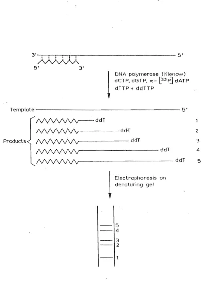
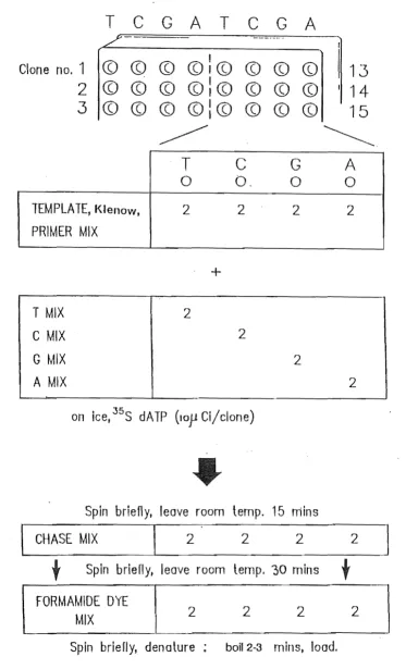
Detection Is via the incorporation of a radioactively or fluorescently labelled nucleotide residue into the DNA molecule which is to be sequenced. Two methods have established themselves as central to sequencing: the dideoxy chain terminator method of Sanger et al. (1977) and the chemical DNA sequencing method of Maxam and Gilbert (1977).
3.1.2. PRIMED SYNTHESIS DNA SEQUENCING