of Cells to Copper Excess
Graphical Abstract
Highlights
d
Expression level of the essential mitochondrial ferredoxin
determines Cu resistance
d
Copper targets the FeS domain of yeast Yah1
d
Copper action at Yah1 affects downstream, essential
FeS-protein function
d
The function in copper resistance is conserved in the human
ferredoxin, Fdx2
Authors
Cindy Vallie`res, Sara L. Holland,
Simon V. Avery
Correspondence
[email protected]
In Brief
Vallie`res et al. identify the conserved
ferredoxin Yah1 (Fdx2 in humans) as an
important target of copper excess in
cells. Copper targets the FeS domain of
the ferredoxin, needed for the essential
process of FeS-cluster biogenesis. This
knowledge could help in tackling
copper-related diseases.
Vallie`res et al., 2017, Cell Chemical Biology24, 1–10
Mitochondrial Ferredoxin Determines
Vulnerability of Cells to Copper Excess
Cindy Vallie`res,1Sara L. Holland,1and Simon V. Avery1,2,*
1School of Life Sciences, University of Nottingham, University Park, Nottingham NG7 2RD, UK 2Lead Contact
*Correspondence:[email protected] http://dx.doi.org/10.1016/j.chembiol.2017.08.005
SUMMARY
The essential micronutrient copper is tightly
regu-lated in organisms, as environmental exposure or
homeostasis defects can cause toxicity and
neuro-degenerative disease. The principal target(s) of
cop-per toxicity have not been pinpointed, but one key
effect is impaired supply of iron-sulfur (FeS) clusters
to the essential protein Rli1 (ABCE1). Here, to find
up-stream FeS biosynthesis/delivery protein(s)
respon-sible for this, we compared copper sensitivity of
yeast-overexpressing candidate targets.
Overex-pression of the mitochondrial ferredoxin Yah1
pro-duced
copper
hyper-resistance.
55Fe
turnover
assays revealed that FeS integrity of Yah1 was
particularly vulnerable to copper among the test
pro-teins. Furthermore, destabilization of the FeS domain
of Yah1 produced copper hypersensitivity, and
YAH1
overexpression rescued Rli1 dysfunction. This
cop-per-resistance function was conserved in the human
ferredoxin, Fdx2. The data indicate that the essential
mitochondrial ferredoxin is an important copper
target, determining a tipping point where plentiful
copper supply becomes excessive. This knowledge
could help in tackling copper-related diseases.
INTRODUCTION
Micronutrients that are essential for life create a dilemma for all organisms. There is a need to balance adequate supply against deleterious effects that inevitably can arise from excess (Heffern et al., 2016; Renwick, 2006). The trace element copper is essen-tial for function of diverse enzymes such as cytochromec oxi-dases and superoxide dismutases. Copper homeostasis is tightly regulated as Cu is highly toxic in excess. Copper homeo-stasis defects or elevated environmental Cu exposure can result in displacement of other essential metals from cellular constitu-ents, inappropriate protein binding, or provocation of stress from reactive oxygen species (ROS) due to the metal’s redox activity, among other reported effects (Macomber and Imlay, 2009; Pena et al., 1999). Copper toxicity has been widely described in different organisms. In humans, alterations in Cu levels or Cu-dependent functions have been associated with the pathogen-esis of neurodegenerative disorders such as Wilson’s disease
(Bandmann et al., 2015; Kaplan and Maryon, 2016). In addition to Cu transporters and Cu-requiring enzymes, metalloproteins such as Cu-metallothioneins help to buffer free Cu in cells. How-ever, when such defenses are inadequate, a key question re-mains: which molecular target(s) may be the principal ‘‘Achilles’ heel’’ of organisms, accounting for the inhibitory action of copper? Different mechanisms of Cu toxicity have been pro-posed as aforementioned, but the specific target(s) remain elusive. Identification of primary cause(s) (rather than effects) of Cu action could open new opportunities for combating Cu-related disease.
Stressors and drugs commonly have essential-protein targets. These are identifiable by the essential function being sensitive to the agent in question, with knockdown of the protein producing a sensitive phenotype and overexpression conferring resistance (Avery, 2011). Iron-sulfur (FeS) clusters are protein cofactors that are among the most ROS-sensitive structures in biology (Imlay, 2006; Jang and Imlay, 2007). FeS proteins play roles in fundamental cellular processes such as the tricarboxylic acid (TCA) cycle, amino acid biosynthesis, respiratory chain, DNA synthesis and repair, mRNA translation, and FeS-protein biogen-esis itself. Several FeS proteins are notoriously ROS labile, commonly those with surface exposed FeS clusters. FeS clus-ters are also susceptible to displacement of Fe by metals such as Cu, Ag, and Hg (Brancaccio et al., 2017; Macomber and Imlay, 2009; Tan et al., 2017; Xu and Imlay, 2012). To date, most studies of ROS- and/or Cu-sensitive FeS proteins have focused on (conditionally-) nonessential FeS proteins such as aconitase, isopropylmalate isomerase, and fumarase (Foster et al., 2014; Jang and Imlay, 2007, 2010; Macomber and Imlay, 2009; Tan et al., 2017). Work with yeast showed that Cu stress also impairs function of the essential FeS protein Rli1 (ABCE1) (Alhebshi et al., 2012). Rli1 is highly conserved across eukaryotes and archaea, being required for ribosome biogenesis and matu-ration, translation initiation and termination, and ribosome recy-cling (Nurenberg and Tampe, 2013). Loss of Rli1 function and growth inhibition caused by Cu was due to defective FeS-cluster supply to Rli1 (after incorporation into Rli1, the FeS clusters were relatively stable) (Alhebshi et al., 2012). This indicated that a pri-mary target accounting for inhibition lay upstream of Rli1, within the FeS-cluster synthesis or delivery pathway.
In eukaryotic cells, FeS clusters are synthesized and inserted into apoproteins by the mitochondrial iron-sulfur cluster (ISC) assembly machinery and the cytosolic iron-sulfur protein assem-bly (CIA) machinery (Figure 1A). These pathways are well conserved from yeast to humans (Lill, 2009; Rouault, 2012; Shef-tel et al., 2010). Mitochondrial ISC assembly comprises more
than 15 components.De novoFeS-cluster synthesis on the scaf-fold protein Isu1 requires sulfur and iron donors, the cysteine desulfurase complex Nfs1-Isd11, and frataxin (Yfh1), respec-tively, as well as proteins required for electron transfer, namely the ferredoxin reductase Arh1 and the reduced ferredoxin Yah1 (Lill et al., 2014). Subsequently, the nascent cluster is trans-ferred to mitochondrial apoproteins or exported to the cytosol. Here, maturation of cytosolic and nuclear FeS proteins such as Rli1 is supported by the CIA machinery, which includes the Cfd1-Nbp35 scaffold complex, and Nar1 and Cia1, which direct FeS clusters to apoproteins (Lill, 2009).
The lability of FeS clusters to oxygen and Cu (Chillappagari et al., 2010; Djoko and McEwan, 2013; Jang and Imlay, 2007; Macomber and Imlay, 2009) suggests that all steps in the biogenesis pathway are potentially Cu susceptible. Neverthe-less, the component proteins are compartmentalized in the mitochondria and the cytoplasm, between which Cu will not be evenly distributed. Furthermore, FeS-cluster orientation in different proteins affects accessibility of Cu and ROS (Imlay, 2014; Jang and Imlay, 2007). Accordingly the FeS clusters of Rli1 itself proved Cu stable whereas cluster supply to Rli1 was not (Alhebshi et al., 2012). Those results led to the aim of this study, to characterize a primary target of Cu toxicity in cells. Here, we pinpoint the essential mitochondrial ferredoxin, Yah1, the functional ortholog of bacterial ferredoxins and human Fdx2 (Sheftel et al., 2010). The mitochondrial ferredoxin fulfilled the key criteria of a primary Cu target described above, indi-cating that this protein’s Cu-labile FeS clusters account for downstream effects of Cu on essential FeS-protein function and on associated growth defects of cells faced with an excess of this micronutrient.
RESULTS
FeS Biosynthesis/Delivery Proteins that Confer Copper Resistance
Previous work suggested that a key target of Cu toxicity may reside in the FeS-cluster biosynthesis and delivery pathway, up-stream of the cytosolic FeS-recipient protein Rli1 (Alhebshi et al., 2012). A small panel of proteins representing diverse locations and functions in the pathway were analyzed here: Cfd1 and Nar1 of the CIA machinery, and Isu1, Yah1, and Yfh1 of the ISC assembly machinery. Where a protein is the major target of an agent’s inhibitory action, increasing the protein’s expres-sion should confer resistance while knockdown should produce sensitivity (Avery, 2011). We also reasoned that proteins located close in a pathway to a major target would confer partial resis-tance if their activity can compensate some lost FeS flux through the target. We compared Cu resistance mediated by the above candidates, overexpressed through use oftet-promoter con-structs in cells incubated without doxycycline to give maximal expression (Alhebshi et al., 2012) (Figures 1andS1). Throughout this work, Cu was supplied at concentrations either moderately or barely inhibitory to growth of the wild-type, depending on whether the assay was for increased resistance (i.e.,Figure 1) or sensitization, respectively, in the test strain(s). (In vitroassays of cluster turnover [below] required much lower Cu concentra-tions as these were with purified proteins and in simple buffers, less prone to Cu complexation [Avery et al., 1996; Macomber
and Imlay, 2009].) Copper toxicity was rescued by overexpres-sion of Isu1 and Yfh1, and particularly Yah1, of the ISC assembly machinery (Figures 1B and 1C). Isu1 and Yfh1 gave compara-tively small increases in Cu resistance, but overexpression of Yah1 altered the cell doubling time from 7 hr to 3 hr at 0.6 mM Cu(NO3)2. Overexpression of the CIA pathway proteins Nar1 and Cfd1 had no protective effect. As the expression levels of each gene differed (Figure S1), we also considered the growth-rate effects after normalizing for relative degree of gene overex-pression (Figure 1D). Here again, Cu resistance was highest in Isu1- and Yah1-overexpressing cells, whereas the effect of Yfh1 appeared more marginal considering its relatively high level of overexpression. The Yah1 and Isu1 proteins conferring Cu resistance are at the mitochondrial root of FeS biogenesis. The mitochondrial weakness of the BY-yeast background used here (seeDiscussion) has certain advantages for detecting mito-chondria-targeting actions relevant to FeS biogenesis. Whereas mitochondrial genes generally are overrepresented among the Cu-sensitive set of BY-yeast deletion mutants (Bleackley et al., 2011), many individual mitochondrial proteins of course do not confer Cu resistance while many cytosolic proteins do. The latter include cytosolic Rli1 (Alhebshi et al., 2012), discussed above. Therefore, any key role for cytosolic Nar1 or Cfd1 should be simi-larly detectable, but our data suggested Yah1 and Isu1 as the lead candidates. We took forward Yah1 and Isu1 for further investigation, alongside Nar1 and Rli1 as cytosolic controls.
The Essential Mitochondrial Ferredoxin Undergoes Iron Turnover in High-Copper Conditions
We hypothesized that a primary Cu target in the FeS biogenesis/ delivery pathway would have Cu-labile FeS cluster(s). We there-fore compared the loss of cluster from Yah1 and Isu1 during Cu treatment by monitoring the amount of55Fe in the proteins. This method provides a faithful and sensitive determination of FeS cluster in the proteins in vitroand in vivo(Pierik et al., 2009). Yfh1 was not tested, as it gave the smallest resistance to Cu (above) and radiolabeled Fe cannot be detected in the protein. We used Nar1 as a control as its overexpression did not confer Cu resistance, as well as the pathway endpoint Rli1, which has relatively stable FeS clusters (Alhebshi et al., 2012). Test proteins were expressed with hemagglutinin (HA) tags and immunopre-cipitated from cells preincubated with 55FeCl3. Fe turnover from each55Fe-labeled, immunoprecipitated protein was com-pared according to55Fe release duringin vitroincubations with a Cu(NO3)2/ascorbic acid system (Alhebshi et al., 2012; Ma-comber and Imlay, 2009). Results for each protein are presented as a ratio of55Fe retention in the Cu versus minus-Cu conditions (data for the individual conditions are given inFigure S2). Only Yah1 showed a marked (80%) Cu-dependent turnover of 55
Fe (Figure 2A). The other proteins showed some variable leakage of 55Fe in the absence of Cu in vitro(Figure S2), but the preloaded55Fe only of Yah1 was susceptible to Cu addition. We also investigated Fe turnoverin vivo, to reflect more closely the physiological situation within cells. Cells preloaded with 55FeCl
3 during culturing were subsequently incubated in 55
rate. The mild Cu treatment did not cause a significant55Fe turn-over from Rli1, Nar1, or Isu1. In contrast, Cu caused a50% turnover of55Fe from Yah1. This decrease was not attributable to a decrease in the level of immunoprecipitated Yah1 protein (Figure S3). As reasoned earlier, mild rescue of Cu toxicity by Isu1 (Figures 1B and 1C) but absence of FeS turnover in Isu1 (Figure 2) could indicate that this protein’s pathway proximity to Yah1 allows it to compensate partly for any decreased FeS flux due to Yah1 dysfunction. It is also possible that Isu1 may sta-bilize Yah1 from damage. As Yah1 is essential for FeS biogen-esis, the Cu-dependent50% loss of iron from the [2Fe-2S] cluster of Yah1 could be expected to have downstream conse-quences. Decreased expression of Yah1 causes decreased
55
Fe incorporation to Rli1 (Kispal et al., 2005) and other cytosolic FeS proteins (Lange et al., 2000; Paul et al., 2015) (this would not be expected to affect the presentin vivoassays of Cu-depen-dent55Fe turnover with the cytosolic proteins, as the high level of Yah1 overexpression more than compensates for 50% Cu-dependent loss of upstream Yah1-holoprotein;Figure S1). The data suggest that a Cu-sensitive weak point in the FeS syn-thesis pathway may occur at Yah1.
Yah1-Dependent Resistance to Copper
[image:4.603.97.507.103.471.2]Having established that Yah1 overexpression increases Cu resistance (Figures 1B and 1C), we tested whether decreased functional Yah1 is sufficient to produce Cu sensitivity. Absence
Figure 1. Influence of FeS Biosynthesis/Delivery Proteins on Cellular Copper Resistance
(A) Simplified scheme showing FeS-cluster biogenesis and transfer to extra-mitochondrial proteins (e.g., Rli1) in the yeast model. Proteins tested in this study are shown in color. In blue are proteins of the ISC machinery: Isu1, scaffold; Yfh1, iron donor; Yah1, electron donor. In pink are proteins of the CIA machinery: Cfd1, scaffold; Nar1, cluster transfer from the scaffold complex to apoproteins. Rli1 is an essential destination protein.
(B)S. cerevisiaeBY4741 transformed with high-copytetbearing plasmid, empty or overexpressing proteins of interest, were cultured in YNB medium sup-plemented or not with 0.6 mM Cu(NO3)2. Doxycycline was excluded to give maximal expression. Mean data are shown from triplicate independent growth experiments ± SEM.
(C) Data shown are calculated from maximum growth rates (mmax= ln2/g, where g is cell doubling time) determined during exponential growth, from plots presented in (B). *p < 0.05, **p < 0.01, ****p < 0.0001 according to Student’s t test, two tailed.
of Yah1 (byYAH1deletion) causes viability loss, consistent with a putative target of toxicant action. We expressed ayah1allele (CR5 mutant), which has decreased Yah1-related function ( Bar-ros et al., 2002). The CR5 mutant was Cu sensitive (Figure 3). Copper exposure and genetic defects in FeS-cluster status can provoke mitochondrial Fe hyperaccumulation, including in Yah1-depleted cells (Foster et al., 2014; Lange et al., 2000). We tested Cu sensitivity in two such FeS-cluster assembly mu-tants with Fe hyperaccumulation phenotypes, namely isu1D
(Ramazzotti et al., 2004) andssq1D(Knight et al., 1998). Neither mutant was Cu sensitive (Figure S4), countering the possibility that Fe accumulation in Yah1-depleted cells may be the cause of their Cu sensitivity (the hint of a delayed lag-phase exit in thessq1Dmutant reflected one outlying replicate curve, not re-produced in independent replicates). As Cu is redox active and can provoke oxidative stress, we also considered whether any effect of Yah1 activity on ROS levels might be a cause of Yah1-dependent Cu resistance. However, the level of Yah1 expression did not markedly alter ROS levels measured with the ROS probe DHE, in either the absence or presence of Cu (Figure S5A).
In contrast to the observed rescue of Cu sensitivity via overex-pression of Yah1 (Figures 1B and 1C) or of pro-oxidant action via Rli1 (Alhebshi et al., 2012; Laleve et al., 2016; Vallieres and Avery, 2017), increased expression of nonessential FeS proteins is known potentially to exacerbate ROS stress. This is because of the increased pool of labile FeS which, following turnover (e.g., ROS mediated), leads to the accumulation of free Fe and further potential for ROS stress via Fe-catalyzed Fenton chemis-try (Keyer and Imlay, 1996; Liochev and Fridovich, 1999). This ROS stress may be further aggravated by upregulation of Fe up-take in response to the eroding FeS status (Chillappagari et al., 2010; Foster et al., 2014; Rutherford et al., 2005). In the condi-tions used above (Figure 1), such potential detrimental conse-quences of increased Fe release from overexpressed Yah1 during Cu stress (Figure 2) appeared to be outweighed by the benefits (from Yah1 overexpression) of any rescue of essential Yah1 function. We hypothesized that this balance might be
reversed under conditions more permissive for Fe-catalyzed ROS stress. Therefore, we overexpressed YAH1 in a sod2D
background, defective for scavenging of mitochondrial superox-ide, which may fuel Fe-catalyzed ROS formation and further stimulate upstream FeS-cluster turnover (Imlay, 2006; Irazusta et al., 2006; Jang and Imlay, 2007). We also tested a methionine sulfoxide reductase mutant (mxr1D,mxr2D or ‘‘mxrD’’) which shows impaired FeS-cluster integrity, suggested to result from elevated superoxide (Sideri et al., 2009). In these mutants,
YAH1 overexpression did not confer Cu resistance. Indeed, high Yah1 expression produced Cu hypersensitivity in sod2D
cells (Figure 4). We infer that the Cu resistance that results fromYAH1overexpression in the wild-type (Figure 1) requires lower (wild-type) levels of mitochondrial superoxide, because higher superoxide may exacerbate the toxic consequences of (Cu-dependent) Fe release from Yah1.
Yah1-dependent Cu resistance seen in the wild-type back-ground appeared not to be related to any suppression of Fet3-dependent Fe uptake (FET3is upregulated in response to low FeS-cluster status [Rutherford et al., 2005]), asFET3expression in Cu-treated cells was not affected by Yah1 overexpression (Figure S5B). Similarly, Yah1 overexpression did not affect expression of Mrs3 or Mrs4, which mediate Fe transport across the inner mitochondrial membrane (Figure S5C). The Cu-trans-porting ATPase encoded by CCC2 is the yeast ortholog of humanATP7AandATP7B, mutations in which cause Cu homeo-stasis defects linked to Menkes’ and Wilson’s diseases, respec-tively. Such defects are also associated with mitochondrial oxidative damage (Rossi et al., 2004), raising the question of whether mitochondrial ferredoxin may protect against such con-sequences also of internal Cu imbalance (i.e., without excess Cu addition). However, the yeast ccc2D mutant did not exhibit elevated mitochondrial ROS (Figure S5D) and is known to have a Cu-limited phenotype (Voskoboinik et al., 2001).
The CR5 mutant described above (Figure 3) has several randomly generated mutations in Yah1, and it is not known which are responsible for the associated phenotypes (Barros et al., 2002). Hypothesizing that the FeS cluster of Yah1 is the protein’s
Figure 2. 55Fe Turnover during Copper
Treatment of Key Proteins Involved in FeS-Cluster Biosynthesis and Delivery
Yeast expressing HA-tagged constructs of the specified proteins, under tetcontrol from high-copy plasmids, were cultured without doxycycline to maximize expression; this overexpression gave yields of between 0.63106
cpm55
Fe (g cells) 1 (for Isu1-HA) and 1.63106cpm55Fe (g cells) 1(for Yah1-HA) in immunoprecipitates of the proteins after labeling as follows (the value for the back-ground was <0.1).
(A) HA-tagged,55
Fe-labeled proteins were immu-noprecipitated from protein extracts of cells preincubated with 180mCi L1 55
FeCl3. Copper-resistant 55
Fe retention by the proteins was calculated from55
Fe determinations after subsequent 10-min incubations with 350mM ascorbate/100mM histidine (Macomber and Imlay, 2009) supplemented or not with 12.5mM Cu(NO3)2(Cu had effect at low concentrations in thesein vitroconditions).
(B) After preloading cells with55Fe (supplied at 180mCi L 1, in the absence of Cu),55FeCl3was removed from the medium and cells incubated for 1 hr with or without 1 mM Cu(NO3)2, before immunoprecipitation of the HA-tagged proteins and55Fe quantification.
***p < 0.001, ****p < 0.0001 according to Student’s t test, two tailed, by comparison with data for Rli1 (which retains55
Fe in the Cu condition [Alhebshi et al., 2012]). All values are means from at least three independent experiments ± SEM.
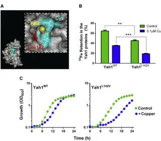
Cu-vulnerable target, we reasoned that specific mutations to further destabilize the cluster should confer Cu hypersensitivity. There are no standard approaches for manipulating FeS-cluster lability within proteins. We exploited knowledge from the cyano-bacteriumAnabaena variabilis. Its two ferredoxins have similar activities in electron transfer, but FdxH1 is relatively stable whereas FdxH2 is oxygen labile, with the residue at position 77 critical for this difference: the longer side chain of a leucine (in FdxH1) than valine (FdxH2) inhibits access of oxygen to the cav-ity at the FeS cluster (Singh et al., 1999, 2001). Based on sequence alignments, we identified a leucine (Leu-142) in the yeast mitochondrial ferredoxin (CysX5CysX2CysXNLeuXCys) corresponding to Leu-77 of FdxH1 (CysX4CysX2CysXNLeuXCys) (the Cys residues are those coordinating the FeS cluster). Leu-142 is also in the cavity leading to the FeS cluster (Figure 5A) (Webert et al., 2014). We replaced Leu-142 of Yah1 with a valine by site-directed mutagenesis, and comparedin vitro55Fe turn-over in the wild-type and mutant Yah1. The Yah1L142Vmutant showed significantly greater loss of55Fe than the wild-type pro-tein, even in the absence of Cu (Figure 5B). (Since the assay sys-tem did not strictly exclude oxygen, the observation for the minus-Cu condition is consistent with the L142V mutation allow-ing increased access of oxygen to the FeS cluster [Singh et al., 2001]; seeDiscussion.) Yeast modified to express Yah1L142Vin place of wild-type Yah1 exhibited normal growth under standard conditions (Figure 5C). However, the Yah1L142Vmutant was Cu sensitive, with an extended lag phase and exponential-phase cell doubling time 1.3-fold slower than the control strain at 1 mM Cu(NO3)2 (Figure 5C). Therefore, destabilization of the Yah1 FeS-cluster produced a Cu-sensitive phenotype. The data collectively supported the hypothesis that Yah1 is a key target of Cu action.
Rli1 Function Depends on Yah1 Expression Level during Copper Stress
When Yah1 expression is decreased, the level of55Fe incorpo-rated to the essential cytosolic protein Rli1 is decreased (Lange et al., 2000). Therefore, perturbation of Yah1 function could explain the impaired Rli1 activity associated with Cu stress pre-viously suggested to arise from targeting of an upstream FeS
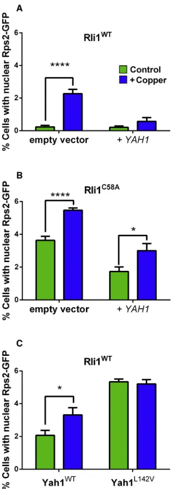
biogenesis or transfer step (Alhebshi et al., 2012). We tested the impact of Yah1 on Rli1 function during mild Cu stress using the principal in vivo assay for Rli1 function: nuclear export of the small ribosomal subunit Rps2. Nuclear export of fluores-cence in cells expressing Rps2-GFP is a sensitive indicator of FeS-dependent Rli1 activity (Alhebshi et al., 2012; Kispal et al., 2005). As reported previously (Alhebshi et al., 2012), Cu expo-sure increased the proportion of wild-type cells exhibiting nu-clear accumulation of Rps2-GFP, i.e., defective Rli1 function. However, YAH1 overexpression under the same condition rescued75% of nuclear Rps2-GFP export activity (Figure 6A).
YAH1 overexpression also partly rescued nuclear Rps2-GFP export in cells expressing Rli1C58A(in place of the wild-type protein) (Figure 6B), a labile Rli1 construct associated with decreased export activity and Cu sensitivity (Alhebshi et al., 2012; Barthelme et al., 2007). Conversely, nuclear accumulation of Rps2-GFP was increased in the Yah1L142V-expressing strain (Figure 6C). This was in keeping with the FeS instability of the mutant Yah1 (Figure 5B), expected to perturb downstream FeS supply to destination proteins. In conclusion and combined with the other results, Yah1 fulfilled the anticipated criteria of a key direct protein target of Cu toxicity.
The Human Ferredoxin, Fdx2, Confers Copper Resistance
Human cells possess two mitochondrial ferredoxins, Fdx1 and Fdx2 (annotated as ‘‘Fdx1L’’). Fdx1 and Fdx2 are both sequence
Figure 4. Copper Resistance with Increased Yah1 Expression Is Abolished in Strains Defective for Antioxidant Defense
S. cerevisiae mxrDandsod2Dmutants transformed with thetet bearing plasmid, either empty (ev) or overexpressingYAH1, were cultured in YNB medium supplemented or not with 0.6 mM Cu(NO3)2. Doxycycline was excluded to give maximalYAH1expression. SEMs from triplicate independent growth experiments are smaller than the dimensions of the symbols.
Figure 3. Decreased Functional Yah1 Sensitizes Cells to Copper
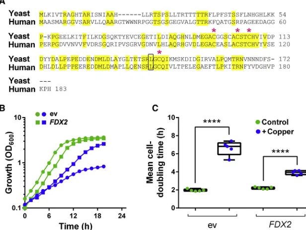
orthologs of yeast Yah1, but only Fdx2 can complement essen-tial Yah1 activities (Sheftel et al., 2010). Fdx2 has 73% similarity and 50% identity with yeast Yah1, including the Leu-142 residue close to the FeS-cluster cavity (Figure 7A). Like Yah1, Fdx2 is essential for FeS-cluster biogenesis (Shi et al., 2012). To indicate whether Fdx2 can be an important target of Cu toxicity, we over-expressed the human protein in yeast (using a tet-FDX2
construct with the mitochondrial targeting sequence ofSOD2).
FDX2overexpression conferred resistance to Cu, decreasing the yeast cell doubling time from7 hr to 4 hr in the presence of 1.1 mM Cu(NO3)2(Figure 7B). Therefore, dependency of Cu resistance on yeast Yah1 is functionally conserved in the human ferredoxin.
DISCUSSION
This work points to the essential, conserved mitochondrial ferre-doxin as an important target of Cu toxicity in cells. Toxicity of this essential micronutrient can arise from defects in homeostasis or environmental exposures (Bandmann et al., 2015; Pena et al., 1999; Renwick, 2006). Previously described molecular targets of Cu toxicity include cytochromecbiogenesis (Durand et al., 2015), oxidative damage to cell constituents such as membrane lipids (Howlett and Avery, 1997), nucleotide synthesis (Johnson et al., 2015), and FeS-protein integrity or biogenesis (Alhebshi et al., 2012; Brancaccio et al., 2017; Foster et al., 2014; Ma-comber and Imlay, 2009; Tan et al., 2017). Here, Yah1 proved to be a Cu-sensitive weak point of the FeS-cluster biogenesis/ delivery pathway in yeast and, as expected for a key target (Avery, 2011), disabling the protein produced Cu-sensitive phe-notypes while overexpression of Yah1 or human Fdx2 conferred resistance. We propose that impairment of Yah1 function
ex-plains downstream loss of Rli1 function and associated growth inhibition reported previously (Alhebshi et al., 2012). It was FeS-cluster supply to Rli1, which has essential roles in protein synthesis (Nurenberg and Tampe, 2013), that was defective in Cu-exposed cells (Alhebshi et al., 2012). The activities of some nonessential FeS proteins such as aconitase are also Cu sensi-tive (Foster et al., 2014; Macomber and Imlay, 2009), but these do not account for growth inhibition. Yah1 is required for normal FeS supply to FeS proteins, including Rli1 (Kispal et al., 2005). Here, Yah1 overexpression restored essential function of Rli1 (in ribosome subunit export) under Cu stress, while expression of Yah1L142Vwith defective FeS stability impaired Rli1 function. The results reveal Yah1 as a new Cu target, accounting for loss of essential Rli1 activity and associated growth inhibition under conditions of Cu excess.
[image:7.603.60.381.94.374.2]Despite its important functions in biogenesis and assembly of FeS clusters, mitochondrial ferredoxin is one of the less well studied proteins of this pathway. This may be partly due to the fact this protein was linked only recently to human disease (mito-chondrial myopathy), unlike Yfh1 (Friedreich’s ataxia) and Isu1 (myopathy), among others (Spiegel et al., 2014). There are of course broader consequences of defective FeS biogenesis. Yah1 is also involved in biosynthesis of respiratory-chain components such as heme A (Barros et al., 2002) and coenzyme Q (Pierrel et al., 2010), essential for respiratory growth (but not re-spiro-fermentative growth as used here). FeS clusters also occur in several respiratory complexes. In HeLa cells, depletion of hu-man Fdx2 causes a decrease in complex I and IV activities ( Shef-tel et al., 2010). Decreased activities also of complexes I, II, and III were described in cells with decreased Fdx2 content due to a mutation disrupting the ATG translation-initiation site, which is linked to mitochondrial myopathy (Spiegel et al., 2014), and
Figure 5. Construction and Expression of a Yah1 Mutant with Defective FeS Integrity, Conferring Copper Sensitivity
(A) Location of the mutated L142V residue (in red) within the reduced yeast ferredoxin. The location of the Cys residues coordinating the FeS cluster is shaded blue. Figure prepared using the co-ordinates of PDB: 2MJE.
(B) HA-tagged proteins were immunoprecipitated from protein extracts of cells preincubated with 55
FeCl3.55Fe retention by the proteins was calcu-lated from55
Fe determinations before and after 10 min of incubation with 350 mM ascorbate/ 100mM histidine supplemented or not with 0.1mM Cu(NO3)2. **p < 0.01, ***p < 0.001 according to Student’s t test, two tailed. All values are means from three replicate determinations ± SEM. (C)yah1Dcells expressing either wild-typeYAH1 or YAH1L142V from single-copy plasmids were cultured in YNB medium supplemented or not with 1 mM Cu(NO3)2. SEMs from triplicate independent experiments are smaller than the dimensions of the symbols.
defects in respiratory chain complex activities are known to be associated with other human disorders (Meunier et al., 2013). Therefore, besides downstream effects on Rli1, effects of Cu on respiratory chain function (Djoko and McEwan, 2013; Durand et al., 2015; Hosseini et al., 2014) could also be partly mediated through its effects on mitochondrial ferredoxin.
The principal genetic background used in this study (BY4741) is the same as used in the previous Rli1 work (Alhebshi et al., 2012) and widely elsewhere, being a core background for con-struction of yeast genomic resources (Winzeler et al., 1999). The BY4741 background carries a mutation in the mitochon-drion-related Hap1 transcription factor (Gaisne et al., 1999) and so may give more sensitive detection of factors targeting mitochondrial functions, as anticipated in this study. Accord-ingly, whereas Yah1 overexpression readily increased Cu resis-tance in the BY4741 background, this particular phenotype was not detectable against a Hap1-proficient W303 background (C.V. and S.V.A., unpublished data). We could assign this differ-ence to Hap1, as complementation with wild-type Hap1 sup-pressed the resistance phenotype in BY4741. Moreover, using target inactivation as a more sensitive approach than overex-pression (Avery, 2011), achieved in the present study with both the Yah1-CR5 and Yah1L142Vmutants (Figures 3,5, and6), we did observe Yah1-dependent Cu resistance also in the W303 background. Therefore, Yah1-dependent Cu resistance is not specific to BY4741, although this background facilitates its detection.
Copper has been reported to bind to solvent-exposed FeS clusters of dehydratase enzymes, displacing Fe (Macomber and Imlay, 2009). The Yah1L142V mutant that we constructed here and proved Cu labile was based on previous observations with cyanobacterial ferredoxins, in which a critical amino acid residue (Val versus Leu) localized in the cavities leading to these proteins’ clusters was considered to determine relative oxygen accessibility and the proteins’ respective oxygen labilities (Singh et al., 2001). A similar cavity occurs in Yah1 (Webert et al., 2014). As well as Cu, the Yah1L142Vmutation sensitized cells to the pro-oxidant paraquat (Figure S6) and appeared to increase the lability of the Yah1 FeS cluster even without added Cu, consis-tent with increased oxygen access. Elevated Fe turnover was accentuated in the presence of Cu, suggesting increased access also of Cu. After reaching an FeS cluster, Cu can bind the sulfur atoms and thereby displace the catalytic Fe atoms (Macomber and Imlay, 2009). This mechanism (versus FeS turnover via Cu-catalyzed ROS formation) is consistent with the present results, particularly considering the discussion below.
[image:8.603.88.258.98.581.2]ROS such as hydrogen peroxide or superoxide can damage FeS clusters through targeting the catalytic Fe atoms in the pro-teins (Flint et al., 1993; Jang and Imlay, 2007). Here, in marked contrast to Cu, YAH1 overexpression caused hypersensitivity to the superoxide-generating agent paraquat (Figure S6). Two considerations may explain these differing outcomes for Cu and paraquat. First, Cu can degrade FeS clusters more extensively than ROS in vivo (Macomber and Imlay, 2009). Therefore, should Yah1 retain partial function during ROS action, as does Rli1 in its [3Fe-4S]+ state (Barthelme et al., 2007), further degradation by Cu should result in greater Yah1 dysfunction with associated retardation of cell growth, restor-able byYAH1overexpression. Second, iron released from FeS
Figure 6. Rli1 Function Depends on Yah1 Expression Level during Copper Stress
(A)S. cerevisiaeBY4741 transformed with pRS315-RPS2-eGFPand a high-copy tetplasmid, either empty or overexpressingYAH1(+YAH1), was cultured with or without 0.35 mM Cu(NO3)2(which affected cell doubling time <15%). Doxycy-cline was excluded. Cells with nuclear Rps2-GFP were enumerated after 4 hr. (B) Cells expressing Rli1C58A
in place of wild-type Rli1 and transformed as above were incubated for 4 hr in the absence or presence of 0.25 mM Cu(NO3)2 (this lower Cu concentration reproduced the mild <15% effect on doubling time in this Cu-sensitive mutant).
(C)yah1Dcells expressing either wild-typeYAH1orYAH1L142Vfrom single-copy plasmids were incubated for 4 hr in the absence or presence of 0.35 mM Cu(NO3)2.
clusters can participate in Fenton chemistry to aggravate oxida-tive damage. Excess superoxide (e.g., paraquat generated) should exacerbate these effects of Fe released from overex-pressed Yah1 or other FeS protein (Keyer and Imlay, 1996; Lio-chev and Fridovich, 1999). In contrast, Cu and Fe may primarily be replacing each other. The suggestion that the level of intro-duced superoxide may determine the different outcomes for paraquat and Cu is supported by the reversal of Yah1 protection against Cu toxicity seen in asod2Dmutant (Figure 4). This also showed that despite the potential for decreased Sod2 function through Cu mismetallation (Culotta et al., 2006), sufficient Sod2 activity is evidently retained in Cu-exposed wild-type cells to establish the different phenotypes versus sod2D cells. Regarding maintenance of FeS supply (from Yah1 through to Rli1) under oxidative conditions, its importance is underscored by the report of two non-FeS proteins, Yae1 and Lto1, which facilitate FeS assembly specifically on Rli1 (Paul et al., 2015); these proteins are essential only during aerobic growth.
Our construction of the Yah1L142Vmutant with its unstable FeS cluster should prove a useful tool beyond this study. A mutant with increased FeS stability could also be very useful, but increasing the stability of a naturally evolved FeS cluster is a challenging task. We did attempt two approaches to obtain a Cu-resistant Yah1, but neither specific substitution of oxida-tion-sensitive amino acids near the FeS cluster (e.g., M129L) (Figure S7) nor random mutagenesis ofYAH1followed by selec-tion for resistant transformants (not shown) successfully pro-duced increased Cu resistance. Perhaps the FeS cluster is already as stable as it can be without adversely affecting Yah1 function. This would not be unexpected from an evolutionary perspective (Imlay, 2006).
What is it about Yah1 that appears to make its Fe(S) content especially unstable to Cu stress? Subcellular localization to the mitochondrion could explain differences versus some of the cytosolic FeS proteins we testedin vivo, but similar differences between the test proteins persisted during in vitroassays of
55
FeS turnover (Figure 2A). Solvent exposure of FeS clusters near the protein surface is a common explanation for the ROS labilities of dehydratases (Flint et al., 1993; Liochev and Frido-vich, 1999), and a similar relationship was proposed for these en-zymes’ Cu sensitivities (Macomber and Imlay, 2009). The FeS clusters of Rli1 are predicted to be shielded from solvent (Karcher et al., 2008) and are also resistant to Cu damage ( Alheb-shi et al., 2012). In contrast, HemN, a bacterial coproporphyrino-gen III oxidase involved in heme biosynthesis under anaerobic conditions, has a buried cluster but is susceptible to damage by Cu (Azzouzi et al., 2013; Djoko and McEwan, 2013). The explanation in this case is the presence of a large cleft, which allows coproporphyrinogen III to access the active site and may also allow Cu to enter (Azzouzi et al., 2013). Similarly, the cysteines involved in binding the FeS cluster of Yah1 are buried in the protein, although a cavity that may give Cu direct access to the cluster is present (Webert et al., 2014).
SIGNIFICANCE
[image:9.603.62.370.105.336.2]The results show that the FeS content of the essential pro-tein Yah1 is Cu labile and that this can account for associ-ated downstream growth-inhibitory effects of Cu in cells. The ferredoxin that is the focus of this investigation is well conserved in bacteria and eukaryotic mitochondria. We successfully inferred structure/function relationships from comparison of the yeast and cyanobacterial ferredoxins in this study. Furthermore,the mature form of the human ferre-doxin Fdx2 has 50% identity and 73% similarity with the yeast protein. Fdx2 complements Yah1 functionally including,we showed,in determining the Cu resistance of cells. The other mitochondrial ferredoxin of humans,Fdx1, is not a functional ortholog of Fdx2 or Yah1 (Sheftel et al., 2010) but nonetheless also has an important role in FeS-cluster biogenesis(Shi et al., 2012),so we cannot discount that it may also contribute to Cu resistance. As discussed
Figure 7. The Human Ferredoxin, Fdx2, Con-fers Copper Resistance
(A) Sequence alignment of human Fdx2 and Yah1 fromS. cerevisiae. Conserved residues are high-lighted in yellow. Asterisks show cysteines involved in the FeS-cluster binding. Leu-142 is framed in black.
in this article,the new knowledge generated here could have implications for therapy of Cu-related diseases. It also shows how vulnerability of a specific cellular function can determine when plentiful supply of an essential micronu-trient becomes excessive.
STAR+METHODS
Detailed methods are provided in the online version of this paper and include the following:
d KEY RESOURCES TABLE
d CONTACT FOR REAGENT AND RESOURCE SHARING
d EXPERIMENTAL MODEL AND SUBJECT DETAILS B Yeast Strains and Growth Conditions
d METHOD DETAILS B Strains and Plasmids
B Yeast Culturing and Toxicity Assays
B 55
Fe-Labelling and Turnover
B Assay of Nuclear Rps2-eGFP Export
B Western Blotting
B RNA Extraction and Quantitative RT-PCR (qRT-PCR)
B ROS Accumulation
d QUANTIFICATION AND STATISTICAL ANALYSIS
SUPPLEMENTAL INFORMATION
Supplemental Information includes seven figures and can be found with this article online athttp://dx.doi.org/10.1016/j.chembiol.2017.08.005.
AUTHOR CONTRIBUTIONS
C.V. and S.L.H. performed the experiments. All authors participated in the design of the study, interpretation of data, and writing of the manuscript. S.V.A. conceived the study. All authors read and approved the final manuscript.
ACKNOWLEDGMENTS
This work was supported by the Biotechnology and Biological Sciences Research Council [BB/I000852/1]. We thank Rhiannon Evans (University of Oxford) for helpful discussions, and Matt Kokolski and Roxane Raulo (Univer-sity of Nottingham) for expert technical help.
Received: May 27, 2016 Revised: March 2, 2017 Accepted: August 1, 2017 Published: August 31, 2017
REFERENCES
Alhebshi, A., Sideri, T.C., Holland, S.L., and Avery, S.V. (2012). The essential iron-sulfur protein Rli1 is an important target accounting for inhibition of cell growth by reactive oxygen species. Mol. Biol. Cell23, 3582–3590.
Avery, S.V. (2011). Molecular targets of oxidative stress. Biochem. J.434, 201–210.
Avery, S.V., Howlett, N.G., and Radice, S. (1996). Copper toxicity towards
Saccharomyces cerevisiae: dependence on plasma membrane fatty acid composition. Appl. Environ. Microbiol.62, 3960–3966.
Azzouzi, A., Steunou, A.S., Durand, A., Khalfaoui-Hassani, B., Bourbon, M.L., Astier, C., Bollivar, D.W., and Ouchane, S. (2013). Coproporphyrin III excretion identifies the anaerobic coproporphyrinogen III oxidase HemN as a copper target in the Cu+
-ATPase mutant copA
-of Rubrivivax gelatinosus. Mol. Microbiol.88, 339–351.
Bandmann, O., Weiss, K.H., and Kaler, S.G. (2015). Wilson’s disease and other neurological copper disorders. Lancet Neurol.14, 103–113.
Barros, M.H., Nobrega, F.G., and Tzagoloff, A. (2002). Mitochondrial ferre-doxin is required for heme A synthesis inSaccharomyces cerevisiae. J. Biol. Chem.277, 9997–10002.
Barthelme, D., Scheele, U., Dinkelaker, S., Janoschka, A., Macmillan, F., Albers, S.V., Driessen, A.J., Stagni, M.S., Bill, E., Meyer-Klaucke, W., et al. (2007). Structural organization of essential iron-sulfur clusters in the evolution-arily highly conserved ATP-binding cassette protein ABCE1. J. Biol. Chem.
282, 14598–14607.
Bleackley, M.R., Young, B.P., Loewen, C.J.R., and MacGillivray, R.T.A. (2011). High density array screening to identify the genetic requirements for transition metal tolerance inSaccharomyces cerevisiae. Metallomics3, 195–205.
Brancaccio, D., Gallo, A., Piccioli, M., Novellino, E., Ciofi-Baffoni, S., and Banci, L. (2017). [4Fe-4S] cluster assembly in mitochondria and its impairment by copper. J. Am. Chem. Soc.139, 719–730.
Cadwell, R.C., and Joyce, G.F. (1992). Randomization of genes by PCR muta-genesis. PCR Methods Appl.2, 28–33.
Chillappagari, S., Seubert, A., Trip, H., Kuipers, O.P., Marahiel, M.A., and Miethke, M. (2010). Copper stress affects iron homeostasis by destabilizing iron-sulfur cluster formation inBacillus subtilis. J. Bacteriol.192, 2512–2524.
Culotta, V.C., Yang, M., and O’Halloran, T.V. (2006). Activation of superoxide dismutases: putting the metal to the pedal. Biochim. Biophys. Acta1763, 747–758.
Djoko, K.Y., and McEwan, A.G. (2013). Antimicrobial action of copper is ampli-fied via inhibition of heme biosynthesis. ACS Chem. Biol.8, 2217–2223.
Durand, A., Azzouzi, A., Bourbon, M.L., Steunou, A.S., Liotenberg, S., Maeshima, A., Astier, C., Argentini, M., Saito, S., and Ouchane, S. (2015). c-Type cytochrome assembly is a key target of copper toxicity within the bac-terial periplasm. MBio6, e01007–01015.
Elliott, N.A., and Volkert, M.R. (2004). Stress induction and mitochondrial local-ization of Oxr1 proteins in yeast and humans. Mol. Cell. Biol.24, 3180–3187.
Flint, D.H., Tuminello, J.F., and Emptage, M.H. (1993). The inactivation of Fe-S cluster containing hydrolyases by superoxide. J. Biol. Chem. 268, 22369–22376.
Foster, A.W., Dainty, S.J., Patterson, C.J., Pohl, E., Blackburn, H., Wilson, C., Hess, C.R., Rutherford, J.C., Quaranta, L., Corran, A., et al. (2014). A chemical potentiator of copper-accumulation used to investigate the iron-regulons of
Saccharomyces cerevisiae. Mol. Microbiol.93, 317–330.
Gaisne, M., Becam, A.M., Verdiere, J., and Herbert, C.J. (1999). A ’natural’ mu-tation inSaccharomyces cerevisiaestrains derived from S288c affects the complex regulatory geneHAP1(CYP1). Curr. Genet.36, 195–200.
Gietz, R.D., and Woods, R.A. (2002). Transformation of yeast by lithium ace-tate/single-stranded carrier DNA/polyethylene glycol method. Methods Enzymol.350, 87–96.
Giorgini, F., Guidetti, P., Nguyen, Q.V., Bennett, S.C., and Muchowski, P.J. (2005). A genomic screen in yeast implicates kynurenine 3-monooxygenase as a therapeutic target for Huntington disease. Nat. Genet.37, 526–531.
Halliwell, S.C., Smith, M.C., Muston, P., Holland, S.L., and Avery, S.V. (2012). Heterogeneous expression of the virulence-related adhesin Epa1 between in-dividual cells and strains of the pathogenCandida glabrata. Eukaryot. Cell11, 141–150.
Heffern, M.C., Park, H.M., Au-Yeung, H.Y., Van de Bittner, G.C., Ackerman, C.M., Stahl, A., and Changa, C.J. (2016). In vivo bioluminescence imaging re-veals copper deficiency in a murine model of nonalcoholic fatty liver disease. Proc. Natl. Acad. Sci. USA113, 14219–14224.
Hosseini, M.-J., Shaki, F., Ghazi-Khansari, M., and Pourahmad, J. (2014). Toxicity of copper on isolated liver mitochondria: impairment at complexes I, II, and IV leads to increased ROS production. Cell Biochem. Biophys.70, 367–381.
Imlay, J.A. (2006). Iron-sulphur clusters and the problem with oxygen. Mol. Microbiol.59, 1073–1082.
Imlay, J.A. (2014). The mismetallation of enzymes during oxidative stress. J. Biol. Chem.289, 28121–28128.
Irazusta, V., Cabiscol, E., Reverter-Branchat, G., Ros, J., and Tamarit, J. (2006). Manganese is the link between frataxin and iron-sulfur deficiency in the yeast model of Friedreich ataxia. J. Biol. Chem.281, 12227–12232.
Jang, S., and Imlay, J.A. (2007). Micromolar intracellular hydrogen peroxide dis-rupts metabolism by damaging iron-sulfur enzymes. J. Biol. Chem.282, 929–937.
Jang, S., and Imlay, J.A. (2010). Hydrogen peroxide inactivates theEscherichia coliIsc iron-sulphur assembly system, and OxyR induces the Suf system to compensate. Mol. Microbiol.78, 1448–1467.
Johnson, M.D.L., Kehl-Fie, T.E., and Rosch, J.W. (2015). Copper intoxication inhibits aerobic nucleotide synthesis in Streptococcus pneumoniae. Metallomics7, 786–794.
Kaplan, J.H., and Maryon, E.B. (2016). How mammalian cells acquire copper: an essential but potentially toxic metal. Biophys. J.110, 7–13.
Karcher, A., Schele, A., and Hopfner, K.P. (2008). X-ray structure of the complete ABC enzyme ABCE1 fromPyrococcus abyssi. J. Biol. Chem.283, 7962–7971.
Keyer, K., and Imlay, J.A. (1996). Superoxide accelerates DNA damage by elevating free-iron levels. Proc. Natl. Acad. Sci. USA93, 13635–13640.
Khozoie, C., Pleass, R.J., and Avery, S.V. (2009). The antimalarial drug quinine disrupts Tat2p-mediated tryptophan transport and causes tryptophan starva-tion. J. Biol. Chem.284, 17968–17974.
Kispal, G., Sipos, K., Lange, H., Fekete, Z., Bedekovics, T., Janaky, T., Bassler, J., Aguilar Netz, D.J., Balk, J., Rotte, C., et al. (2005). Biogenesis of cytosolic ribosomes requires the essential iron-sulphur protein Rli1p and mitochondria. EMBO J.24, 589–598.
Knight, S.A.B., Sepuri, N.B.V., Pain, D., and Dancis, A. (1998). Mt-Hsp70 ho-molog, Ssc2p, required for maturation of yeast frataxin and mitochondrial iron homeostasis. J. Biol. Chem.273, 18389–18393.
Kryukov, G.V., Kumar, R.A., Koc, A., Sun, Z.H., and Gladyshev, V.N. (2002). Selenoprotein R is a zinc-containing stereo-specific methionine sulfoxide reductase. Proc. Natl. Acad. Sci. USA99, 4245–4250.
Laleve, A., Vallieres, C., Golinelli-Cohen, M.P., Bouton, C., Song, Z.H., Pawlik, G., Tindall, S.M., Avery, S.V., Clain, J., and Meunier, B. (2016). The antimalarial drug primaquine targets Fe-S cluster proteins and yeast respiratory growth. Redox Biol.7, 21–29.
Lange, H., Kaut, A., Kispal, G., and Lill, R. (2000). A mitochondrial ferredoxin is essential for biogenesis of cellular iron-sulfur proteins. Proc. Natl. Acad. Sci. USA97, 1050–1055.
Lill, R. (2009). Function and biogenesis of iron-sulphur proteins. Nature460, 831–838.
Lill, R., Srinivasan, V., and Muhlenhoff, U. (2014). The role of mitochondria in cytosolic-nuclear iron-sulfur protein biogenesis and in cellular iron regulation. Curr. Opin. Microbiol.22, 111–119.
Liochev, S.I., and Fridovich, I. (1999). Superoxide and iron: partners in crime. IUBMB Life48, 157–161.
Macomber, L., and Imlay, J.A. (2009). The iron-sulfur clusters of dehydratases are primary intracellular targets of copper toxicity. Proc. Natl. Acad. Sci. USA
106, 8344–8349.
Meunier, B., Fisher, N., Ransac, S., Mazat, J.P., and Brasseur, G. (2013). Respiratory complex III dysfunction in humans and the use of yeast as a model organism to study mitochondrial myopathy and associated diseases. Biochim. Biophys. Acta1827, 1346–1361.
Nurenberg, E., and Tampe, R. (2013). Tying up loose ends: ribosome recycling in eukaryotes and archaea. Trends Biochem. Sci.38, 64–74.
Paul, V.D., M€uhlenhoff, U., St€umpfig, M., Seebacher, J., Kugler, K.G., Renicke, C., Taxis, C., Gavin, A.-C., Pierik, A.J., and Lill, R. (2015). The deca-GX3
pro-teins Yae1-Lto1 function as adaptors recruiting the ABC protein Rli1 for iron-sulfur cluster insertion. eLife4, e08231.
Pena, M.M., Lee, J., and Thiele, D.J. (1999). A delicate balance: homeostatic control of copper uptake and distribution. J. Nutr.129, 1251–1260.
Pierik, A.J., Netz, D.J.A., and Lill, R. (2009). Analysis of iron-sulfur protein maturation in eukaryotes. Nat. Protoc.4, 753–766.
Pierrel, F., Hamelin, O., Douki, T., Kieffer-Jaquinod, S., Muhlenhoff, U., Ozeir, M., Lill, R., and Fontecave, M. (2010). Involvement of mitochondrial ferredoxin and para-aminobenzoic acid in yeast coenzyme Q biosynthesis. Chem. Biol.
17, 449–459.
Ramazzotti, A., Vanmansart, V., and Foury, F. (2004). Mitochondrial functional interactions between frataxin and Isu1p, the iron-sulfur cluster scaffold pro-tein, inSaccharomyces cerevisiae. FEBS Lett.557, 215–220.
Renwick, A.G. (2006). Toxicology of micronutrients: adverse effects and un-certainty. J. Nutr.136, 493S–501S.
Rossi, L., Lombardo, M.F., Ciriolo, M.R., and Rotilio, G. (2004). Mitochondrial dysfunction in neurodegenerative diseases associated with copper imbal-ance. Neurochem. Res.29, 493–504.
Rouault, T.A. (2012). Biogenesis of iron-sulfur clusters in mammalian cells: new insights and relevance to human disease. Dis. Model Mech.5, 155–164.
Rutherford, J.C., Ojeda, L., Balk, J., Muhlenhoff, U., Lill, R., and Winge, D.R. (2005). Activation of the iron regulon by the yeast Aft1/Aft2 transcription factors depends on mitochondrial but not cytosolic iron-sulfur protein biogenesis. J. Biol. Chem.280, 10135–10140.
Sheftel, A.D., Stehling, O., Pierik, A.J., Elsasser, H.P., Muhlenhoff, U., Webert, H., Hobler, A., Hannemann, F., Bernhardt, R., and Lill, R. (2010). Humans possess two mitochondrial ferredoxins, Fdx1 and Fdx2, with distinct roles in steroidogenesis, heme, and Fe/S cluster biosynthesis. Proc. Natl. Acad. Sci. USA107, 11775–11780.
Shi, Y.B., Ghosh, M., Kovtunovych, G., Crooks, D.R., and Rouault, T.A. (2012). Both human ferredoxins 1 and 2 and ferredoxin reductase are important for iron-sulfur cluster biogenesis. Biochim. Biophys. Acta1823, 484–492.
Sideri, T.C., Willetts, S.A., and Avery, S.V. (2009). Methionine sulphoxide re-ductases protect iron-sulphur clusters from oxidative inactivation in yeast. Microbiology155, 612–623.
Singh, B.B., Curdt, I., Jakobs, C., Schomburg, D., Bisen, P.S., and Bohme, H. (1999). Identification of amino acids responsible for the oxygen sensitivity of ferredoxins from Anabaena variabilis using site-directed mutagenesis. Biochim. Biophys. Acta1412, 288–294.
Singh, B.B., Curdt, I., Shomburg, D., Bisen, P.S., and Bohme, H. (2001). Valine 77 of heterocystous ferredoxin FdxH2 in Anabaena variabilisstrain ATCC 29413 is critical for its oxygen sensitivity. Mol. Cell. Biochem.217, 137–142.
Spiegel, R., Saada, A., Halvardson, J., Soiferman, D., Shaag, A., Edvardson, S., Horovitz, Y., Khayat, M., Shalev, S.A., Feuk, L., et al. (2014). Deleterious mutation inFDX1L gene is associated with a novel mitochondrial muscle myopathy. Eur. J. Hum. Genet.22, 902–906.
Tan, G., Yang, J., Li, T., Zhao, J., Sun, S., Li, X., Lin, C., Li, J., Zhou, H., Lyu, J., et al. (2017). Anaerobic copper toxicity and iron-sulfur cluster biogenesis in
Escherichia coli. Appl. Environ. Microbiol.83, e00867-17.
Vallieres, C., and Avery, S.V. (2017). The candidate antimalarial drug MMV665909 causes oxygen-dependent mRNA mistranslation and synergises with quinoline-derived antimalarials. Antimicrob. Agents Chemother.http://dx. doi.org/10.1128/AAC.00459-17.
Voskoboinik, I., Mar, J., Strausak, D., and Camakaris, J. (2001). The regulation of catalytic activity of the Menkes copper-translocating P-type ATPase—role of high affinity copper-binding sites. J. Biol. Chem.276, 28620–28627.
Webert, H., Freibert, S.A., Gallo, A., Heidenreich, T., Linne, U., Amlacher, S., Hurt, E., Muhlenhoff, U., Banci, L., and Lill, R. (2014). Functional reconstitution of mitochondrial Fe/S cluster synthesis on Isu1 reveals the involvement of ferredoxin. Nat. Commun.5, 5013.
Winzeler, E.A., Shoemaker, D.D., Astromoff, A., Liang, H., Anderson, K., Andre, B., Bangham, R., Benito, R., Boeke, J.D., Bussey, H., et al. (1999). Functional characterization of the S. cerevisiaegenome by gene deletion and parallel analysis. Science285, 901–906.
STAR
+
METHODS
KEY RESOURCES TABLE
REAGENT or RESOURCE SOURCE IDENTIFIER Antibodies
Mouse HA tag monoclonal antibody ThermoFisher Scientific Cat#26183; RRID:AB_10978021 Goat anti-mouse IgG (H+L) poly-HRP polyclonal
secondary antibody
ThermoFisher Scientific Cat#32230; RRID:AB_1965958 anti-HA beads Sigma-Aldrich A2095
Bacterial and Virus Strains
Escherichia coliXL1-blue competent cells ThermoFisher Scientific Cat#50-125-058
E. coliNEB5-alpha competent cells New England Biolabs Cat#C2987 Chemicals, Peptides, and Recombinant Proteins
Phusion DNA polymerase New England BioLabs Cat#M0530L hygromycin B PanReac AppliChem Cat#A2175,0005 Yeast Nitrogen Base without amino acids Formedium Cat#CYN0402 YPD (yeast extract peptone + 2% glucose) Avery lab N/A
55FeCl
3 Perkin-Elmer Cat#NEZ043001MC
Amplification Grade DNase I Sigma-Aldrich Cat# AMPD1-1KT Dihydroethidium Sigma-Aldrich Cat#37291 MitoSOXTMRed Thermo Fisher Scientific Cat#M36008
Critical Commercial Assays
Q5 Site-Directed Mutagenesis Kit New England BioLabs Cat#E0552S Bradford assay kit Bio-Rad Cat#500-0006 Electrochemiluminescence HRP kit Thermo Fisher Scientific Cat#32209 Experimental Models: Organisms/Strains
Saccharomyces cerevisiaeBY4741 and isogenic single-gene deletion strains
Euroscarf, Frankfurt N/A
S. cerevisiaeW303 Barros et al. (2002)
S. cerevisiae mxr1D/mxr2D Kryukov et al. (2002) N/A
S. cerevisiae RLI1C58A Alhebshi et al. (2012) N/A
S. cerevisiaeW303yah1::URA3 Barros et al. (2002) N/A
S. cerevisiaeW303yah1::hphNT1 Avery lab. N/A Recombinant DNA
pYAH/ST1 Barros et al. (2002) N/A pYAH/CR5 Barros et al. (2002) N/A pCM190-YAH1-HA Avery lab. N/A pRS315-YAH1 Avery lab. N/A pRS315-YAH1-HA Avery lab. N/A pRS315-YAH1L142V-HA Avery lab. N/A pRS315-YAH1M129V-HA Avery lab. N/A
pCM190-RLI1-HA Avery lab. N/A pCM190-NAR1-HA Avery lab. N/A pCM190-CFD1-HA Avery lab. N/A pCM190-ISU1-HA Avery lab. N/A pCM190-YFH1-HA Avery lab. N/A pCM190-YAH1-HA Avery lab. N/A pCM190-YAH1L142V-HA Avery lab. N/A pCM190-hFDX2-MTSSOD1-HA Avery lab. N/A
CONTACT FOR REAGENT AND RESOURCE SHARING
Further information and requests for resources and reagents should be directed to and will be fulfilled by the Lead Contact, Simon Avery ([email protected]).
EXPERIMENTAL MODEL AND SUBJECT DETAILS
Yeast Strains and Growth Conditions
Saccharomyces cerevisiaeBY4741 (MATa;his3-1;leu2-0;met15-0;ura3-0) was the principal strain background used throughout the work, and from which isogenic strains were derived as detailed below.S. cerevisiaeW303 (MATa;ade2-1;his3-1;leu2-3;112trp1-1;
ura3-1;yah1::URA3) was used only for expression of theyah1tsallele (CR5). Yeast strains were routinely cultured at 30 C with rotary aeration in YPD broth (Khozoie et al., 2009) or in YNB broth [0.69% yeast-nitrogen base without amino acids (Formedium), 2% (w/v) D-glucose], supplemented as required for plasmid selection with amino acids, adenine or uracil. Where necessary, media were solidified with 2% (w/v) agar (Sigma-Aldrich, St. Louis, MO).
METHOD DETAILS
Strains and Plasmids
Saccharomyces cerevisiaeBY4741 (MATa;his3-1;leu2-0;met15-0;ura3-0) and the isogenicsod2D,isu1D,ssq1Dandccc2Dstrains were from Euroscarf (Frankfurt, Germany). An mxr1D/mxr2D (msra/bD) isogenic with BY4741 was constructed previously (mxr1::URA3;mxr2::KanMX4) (Kryukov et al., 2002). A yah1Dmutant in the W303 background (MATa; ade2-1;his3-1;leu2-3;
112trp1-1;ura3-1;yah1::URA3) transformed with single copy plasmids expressing either wild-typeYAH1(LEU2marker) or ayah1ts
allele (CR5;TRP1marker) were kind gifts from A. Tzagoloff (Barros et al., 2002). TheRLI1C58Amutant was constructed previously (isogenic with BY4741;MATa;leu2-0;met15-0;ura3-0;RLI1C58A::HIS3) (Alhebshi et al., 2012).
To construct mutant versions ofYAH1, a fragment encompassing theYAH1open reading frame (ORF), together with native pro-moter (500 bp) and terminator (300 bp), was amplified from yeast genomic DNA and ligated between theHindIII-BamHI sites of pRS315 (see below). A C-terminal HA tag was added by site-directed mutagenesis using the Q5 Site-Directed Mutagenesis Kit (New England BioLabs) in conjunction with Phusion DNA polymerase. The Leu-142 or Met-129 codons were replaced with a valine codon by site-directed mutagenesis using the Q5 kit with pRS315-YAH1-HAas the PCR template. TheYAH1L142Vand YAH1M129V
bearing fragments were cloned into pRS315 between theHindIII andBamHI sites. To express these constructs in cells, first theURA3
marker of theyah1Dmutant (complemented with plasmid borneYAH1) was replaced byhphNT1, with transformants selected on YPD agar supplemented with 150mg ml-1hygromycin B (PanReac AppliChem). This new strain was transformed with
pCM190-YAH1-HA(see below) and the originalYAH1-bearing plasmid removed by repeated subcloning on leucine-containing YNB medium. The resultant strain was transformed with pRS315-YAH1-HA, pRS315-YAH1L142V-HAor pRS315-YAH1M129V-HA, and
pCM190-YAH1-HAwas removed by plasmid shuffling on 5-fluoroorotic acid. A random mutagenesis was also performed, using the PCR tech-nique (Cadwell and Joyce, 1992). The resultant library was used to transform theyah1Dmutant containing pRS315-YAH1followed by a selection of any resistant transformants on agar supplemented with Cu(NO3)2.
For overexpression of FeS proteins, the ORFsNAR1,CFD1,ISU1,YFH1,YAH1,YAH1L142VorFDX2(with added mitochondrial targeting sequence (MTS) from SOD2(Elliott and Volkert, 2004)) were placed under the control of the tetO promoter in the pCM190 vector and C-terminally tagged with the HA epitope, as described previously for pCM190-RLI1-HA(Alhebshi et al., 2012).FDX2 was amplified from humanFDX2cDNA cloned in pCMV-SPORT6 (Dharmacon). ORFs were ligated between the
NotI-PstI sites (forCFD1,ISU1,YFH1,YAH1),NotI-SbfI sites (FDX2,YAH1L142V) orBamHI-PstI sites (NAR1) of pCM190. A pCMHIS vector was constructed by inserting theHIS3marker between theSfoI andEcoRI sites of pCM190.YAH1-HAwas inserted in the new plasmid between theNotI andSbfI sites. This plasmid and the pCMHISempty-vector were used to transform themxr1D/mxr2D
strain. All DNA cloning and genetic manipulations were inEscherichia coliXL1-Blue cells (Invitrogen) or high-efficiency NEB 5-alpha competentE. colifor mutagenesis (New England BioLabs). Yeast transformations were by the lithium acetate method (Gietz and Woods, 2002).
Continued
REAGENT or RESOURCE SOURCE IDENTIFIER
pCMV-SPORT6-FDX2 Dharmacon Cat#MHS6278-202800851
pCMHIS Avery lab. N/A
Yeast Culturing and Toxicity Assays
ExperimentalS. cerevisiaecultures were inoculated from overnight starter broth-cultures grown from single colonies, and cultured to exponential phase (OD6002.0) in YNB broth medium at 30C, 120 rev min-1. Culture samples were diluted to OD6000.1, and 300-ml aliquots transferred to 48-well plates (Greiner Bio-One, Monroe, NC) before addition or not of Cu(NO3)2or paraquat (Sigma-Aldrich, St. Louis, MO). Cultures were incubated with shaking in a BioTek Powerwave XS microplate spectrophotometer, as described pre-viously (Khozoie et al., 2009).
55
Fe-Labelling and Turnover
Forin vitroanalysis of iron turnover, 200 ml culture that had been pre-incubated for 3 h with55FeCl3(180mCi L-1) (from Perkin-Elmer) was harvested by centrifugation (1500 xg, 5 min). Cells were washed and resuspended in lysis buffer (400ml oxygen-free 50mM phosphate buffer, pH 7.4, 3% (v/v) glycerol, 5mM PMSF (Sigma-Aldrich, St. Louis, MO), EDTA-free protease inhibitor cocktail (Roche, Indianapolis, IN)) together with 500ml of glass beads, diameter 425-600mm. Samples were vortexed at maximum speed three times for 1 min, interspersed with three 1-min cooling periods on ice, before centrifugation at 16,000 xgfor 5 min. Protein in the supernatant was determined with a Bradford assay kit (Bio-Rad, Hercules, CA). Protein (500ml) was mixed with 80ml anti-HA beads (A2095; Sigma-Aldrich) for 1 h at 4C. Beads were washed four times with lysis buffer. Aliquots of the beads were incubated aerobically for 10 min at room temperature with 350mM sodium L-ascorbate and 100mM histidine (Alhebshi et al., 2012; Macomber and Imlay, 2009) in the absence or presence of Cu(NO3)2. Beads were collected by centrifugation, suspended in 5 ml scintillation fluid (Emulsifier Safe; Perkin Elmer-Cetus, Waltham, MA), and bead-associated55Fe measured with a Packard Tri-Carb 2100TR liquid scintillation analyzer (Meriden, CT). The measurements of iron turnover from the test proteinsin vivowere performed exactly as described previously (Alhebshi et al., 2012).
Assay of Nuclear Rps2-eGFP Export
Cells transformed with plasmid pRS315-RPS2-eGFP(LEU2marker) or pRS316-RPS2-eGFP(URA3marker) (kindly donated by E. Hurt, University of Heidelberg) were examined for cytosolic and/or nuclear localization of fluorescence, as described previously. Cells were viewed with a GX L3201-LED microscope.
Western Blotting
For Western blotting, proteins were separated by electrophoresis on 10% (w/v) NuPAGE Bis-Tris gels (Life Technologies) before transfer to nitrocellulose membrane (GE Healthcare). Immunodetection of Yah1-HA was with a mouse anti-HA primary antibody (1:5000 dilution; Thermo Scientific) and poly horseradish peroxidase (poly HRP)-conjugated goat anti-mouse antibody (1:5000 dilu-tion; Thermo Scientific). Yah1-HA was detected with an electrochemiluminescence HRP kit (Pierce) and imaged using a Chemidoc XRS (Bio-Rad). Protein-band intensities were quantified with ImageJ software.
RNA Extraction and Quantitative RT-PCR (qRT-PCR)
mRNA from specified genes was quantified by qRT-PCR exactly as described previously (Halliwell et al., 2012), except that RNA was isolated by the ‘‘hot phenol’’ technique then treated with Amplification Grade DNase I (Sigma-Aldrich, St. Louis, MO), and 25 ng cDNA with 175 nM gene-specific primers (sequences available on request) were used in the PCR reactions. PCRs were carried out for 40 cycles; denaturation at 95C for 15 s, annealing/extension at 60C for 30 s. Melting-curve analysis confirmed a single PCR prod-uct. Amplification was quantified from a standard curve constructed from reactions with defined genomic DNA concentrations.
ROS Accumulation
ROS accumulation in cells was determined with the fluorescent probe dihydroethidium (DHE) (Giorgini et al., 2005) or for mitochon-dria-specific assay with MitoSOXTMRed (Thermo Fisher Scientific). Samples of yeast culture (1.5 ml at OD6002.0) were centrifuged, washed and then incubated in 100ml PBS, 5mM DHE or MitoSOX for 30 min at 30C, 120 rev min-1. Cells were harvested by centri-fugation and resuspended in 500ml PBS before analysis of cellular DHE or MitoSOX fluorescence with a Beckman Coulter FC500 cytometer equipped with a 488 nm laser. Emitted fluorescence was collected with either 625/26 (DHE) or 575/25 (MitoSOX) band pass filters.
QUANTIFICATION AND STATISTICAL ANALYSIS