METHODOLOGY
Sensitive and high throughput
quantification of abscisic acid based
on quantitative real time immuno-PCR
Yi Su
1,2, Wei Li
1,3, Zhigang Huang
1,2, Ruozhong Wang
1,2, Weigui Luo
1, Qing Liu
1,2, Jianhua Tong
1and Langtao Xiao
1,2*Abstract
Background: Abscisic acid (ABA) functions as a stress phytohormone in many growth and developmental pro-cesses in plants. The ultra-sensitive determination of ABA would help to better understand its vital roles and action mechanisms.
Results: We report a new sensitive and high throughput quantitative real time immuno-PCR (qIPCR) method based on biotin–avidin linkage system for ABA determination in plants. ABA monoclonal antibody (McAb) coated on the inner surface of PCR well pretreated with glutaraldehyde. The pre-prepared probe complex, including biotinylated McAb, biotinylated DNA and streptavidin linker, was convenient for high throughput operations. Finally, probe DNA was quantified by real-time PCR. The detectable ranges were from 10 to 40 ng/L with a limit of detection (LOD) of 2.5 fg. ABA contents in plant sample were simultaneously analyzed using LC–MS/MS to validate the qIPCR method. The results showed that qIPCR method has good specificity and repeatability with a recovery rate of 96.9%.
Conclusion: The qIPCR method is highly sensitive for ABA quantification for actual plant samples with an advantage of using crude extracts instead of intensively purified samples.
Keywords: qIPCR, ABA, Biotin, Avidin
© The Author(s) 2018. This article is distributed under the terms of the Creative Commons Attribution 4.0 International License (http://creat iveco mmons .org/licen ses/by/4.0/), which permits unrestricted use, distribution, and reproduction in any medium, provided you give appropriate credit to the original author(s) and the source, provide a link to the Creative Commons license, and indicate if changes were made. The Creative Commons Public Domain Dedication waiver (http://creat iveco mmons .org/ publi cdoma in/zero/1.0/) applies to the data made available in this article, unless otherwise stated.
Background
The phytohormone abscisic acid (ABA) is a sesquit-erpenoid which plays important functions on many bio-processes in plants [1–3]. As the basic support-ing technology for action mechanism study, highly sensitive ABA quantification in trace plant sample is therefore eagerly required by present intensive ABA studies because of its increasing importance in plant science and agriculture. Before 1970s, the epidermal strip stomatal opening test has been employed as the bioassay according to ABA-mediated inhibition of the light-induced stomata opening [4, 5]. Afterwards, enzyme linked immunosorbent assay (ELISA) has been
employed to quantify several plant growth substances including ABA since the late 1960s [6]. Commercial phytohormonal ELISA kits are still available today [7, 8], and the limit of detection (LOD) is around the nanogram leve [9, 10]. Radioimmunoassay (RIA) has been employed to quantify ABA with better reproduc-ibility [11, 12]. However, specific Liquid Scintillation Counter (LSC) is needed to measure radioactivity and an official license for researchers is usually required to deal with isotopes in RIA. Along with the progress in instrumental technology, gas chromatography (GC) [13], high performance liquid chromatography (HPLC) [14], gas chromatography–mass spectrometry (GC–MS) [15] and liquid chromatography–mass spec-trometry (LC–MS) [16] have successively contributed to ABA quantification. At present, the most popu-lar and widely recognized quantification method for ABA is tandem mass spectrometry because of its high
Open Access
*Correspondence: [email protected]
sensitivity [17–19]. In addition, several electrochemi-cal sensors have been developed to quickly quantify ABA according to the electrochemical properties of ABA molecule [10, 20, 21]. Phytohormone-inducible promoter-reporter systems such as pAtHB6T::LUC and ProRAB18::GFP have been constructed and applied in ABA quantification to sketchily reveal its distribution in plant tissues [22, 23]. Although the existing quanti-fication methods can supply some choices for phyto-hormonal quantification, highly sensitive tandem mass method needs complicated operation and high costs, and the sensitivities of other available methods are usually limited. Therefore, new method in ABA quan-tification both with easy operation and high sensitivity is still urgently needed.
Immuno-polymerase chain reaction (IPCR) has been studied for quantification since 1990s [24]. The method can rapidly and specifically magnify the detectable signals of analytes, thus allows highly effi-cient quantification in samples containing target mol-ecules at low concentration since it integrates the advantages of both PCR and immunoassay [25]. PCR has been proved to be a highly sensitive, specific and efficient technique for DNA detection and theoreti-cally capable to sense a single nucleotide [26]. ELISA based on antibodies allows analysis for a broad range of organic molecules, but it has much lower sensitivity compared to PCR. Thus, IPCR has been established by integrating PCR and ELISA.
IPCR is performed in various modes depending on the aim of the experiment. Similar to ELISA, the main reaction manners include the direct, indirect, sand-wich, indirect sandwich and competitive [27–30]. Using PCR as the signal amplification system, the sen-sitivity of IPCR was significantly enhanced comparing with ELISA [31]. Along with the technical develop-ment, the introduction of quantitative real time PCR (QRT-PCR) instruments enables IPCR to perform high throughput analysis. IPCR including qIPCR were widely utilized in medical and environmental fields for the quantification of organic-molecule/organism, such as antibodies, proteins, toxins, nucleic acids and path-ogens [25, 27, 32, 33]. Regretfully, no report has been presented in phytohormone quantification through IPCR. Therefore, we developed a qIPCR method using biotin–avidin system for sensitive and high through-put ABA quantification in trace plant sample. After strategy design and condition optimization, this ABA qIPCR could simultaneously perform accurate and high throughput analysis of ABA even in a 96-well plate. The limit of detection (LOD) could reach 2.5 fg, to the best of our knowledge, a sensitivity close to that of LC–MS/MS.
Materials and methods
Reagents and buffers
ABA monoclonal antibody (McAb) was provided with a titer of 1:1000 and showed fine specificity in our pre-vious work [34]. Biotinylation primer and McAb were performed by Sangon Biotech (Shanghai, China). DNA polymerase and TransStart Green qPCR SuperMix kit was ordered from TransGen Biotech (Beijing, China).
2H-ABA was purchased from Olchemim Ltd.
(Olo-mouc, Czech Republic). Bovine serum albumin (BSA) and streptavidin were ordered from Sigma-Aldrich Co. LLC (USA). Analytical-grade glutaraldehyde was pur-chased from Sangon Biotech (Shanghai, China). Agarose gel DNA extraction kit was purchased from Dingguo Biotech (Beijing, China). Washing buffers included TBS (tris-buffered saline, pH 7.5), PBS (phosphate-buffered saline, pH 7.4), and PBST (phosphate-buffered saline plus Tween, pH 7.5). Coating buffer was 50 mmol/L carbonate buffer (pH 9.5).
Amplification and purification of biotinylated probe DNA
Biotinylated double-stranded DNA (250 bp) with no homologous sequence in plant was generated by PCR amplification using pUC19 as the template through for-ward primer (biotin-5′-TAT GCA GTG CTG CCA TAA CCA TGA -3′) and reverse primer (5′-ATT GTT GCC GGG AAG CTA GAG TAA GTA GTT -3′). The reaction mixture, in a total volume of 100 μL, contained 10 μL 10 × PCR
buffer, 2 μL dNTP (5 μmol/L), 2 μL primer (10 μmol/L), 2.5 U Taq polymerase, and 10 pg template pUC DNA. PCR conditions were 4 min at 94 °C followed by 30 cycles of 30 s at 94 °C, 30 s at 55 °C, and 20 s at 72 °C. The PCR product labeled with biotin was analyzed on 1.5% aga-rose gel stained with ethidium bromide and then purified by an agarose gel DNA extraction kit. To obtain highly purified biotinylated DNA, HPLC purification were employed.
PCR tube/plate preparation and ABA monoclonal antibody coating
in the bottom of PCR plate and incubated for 2 h at 4 °C. Then, the McAb solution in PCR tube/plate was removed and the tube/plate was washed three times with washing buffer. About 100 μL 1% bovine serum albumin (BSA) was added to each well and followed by incubation for 60 min at 4 °C to block the residual adsorption sites and 5 times of washing with PBS. For the coating efficiency cal-culation, the protein in the removed McAb solution were detected by using Bio-Rad Protein Assay Dye Reagent Concentrate (Cat No. 500-0006) according to the proto-cols in the user manual.
Probe complex preparation
The probe complex was consisted of biotinylated McAb, biotinylated DNA and streptavidin linker. A certain rate of three components was mixed in PBS. After incubat-ing for 2 h at 4 °C, the probe complex solution was ultra-filtrated at 5000 g for 15 min at 4 °C through 100 kD ultra-filter tube (EMD Millipore UFC910024) to remove the unlinked probe DNA. The ultra-filter was washed three times using PBS through centrifugation at 5000g
for 15 min at 4 °C and then the solution in ultra-filter tube was pipetted into another 2 mL centrifugal tube and stored at − 20 °C. Moreover, the amount of linked/ unlinked probe DNA was quantified by Eppendorf Bio-Photometer plus for the linking efficiency evaluation.
Binding kinetics analysis of biotin‑McAb and biotin‑DNA with avidin
One fmol avidin and different mass (1–4 fmol) of biotin-McAb and biotin-DNA was mixed in 50 µL PBS, then incubated for 2 h at 4 °C. The mix was ultra-filtrated at 5000 g for 15 min at 4 °C through 100 kD ultra-filter tube (EMD Millipore UFC910024) and the unlinked probe DNA was existed in effluent. The amount of unlinked biotin-DNA was quantified by Eppendorf BioPhotometer plus for the binding kinetics analysis.
qIPCR mix and running program
ABA sample (1 μL), probe complex (1 μL) and ddH2O
(8 μL) were added into McAb coated PCR plate and incubated for 2 h at 4 °C. Then, the solution in coated PCR plate-wells were removed and the plate-wells were washed three times with PBS. Finally, a 10 μL reaction solution was made. Specific primers (5′-CCG GTT CCC AAC GAT CAA G-3′ and 5′-AAC CGC TTT TTT GCA CAA CAT-3′, each 1 μL), a certain volume of compo-nents of real-time PCR kit and ddH2O (up to 10 μL) were
piped into PCR plate. Real-time PCR was performed on StrataGene Mx3000p Real-time PCR system (USA). The following programs were employed: pre-denaturing for 10 min at 95 °C, then amplifying for 40 cycles including
denaturing for 30 s at 95 °C, annealing for 30 s at 56 °C and extending for 10 s at 72 °C.
Plant materials and sampling
Arabidopsis and rice (Oryza sativa) seeds were surface sterilized by 70% alcohol and 5% NaClO (v/v), washed 3 times, and placed in the dark for 48 h at 4 °C to synchro-nize germination. The seedlings were grown in Murashige and Skoog (MS) medium [35] solidified with 0.4% (w/v) phytagel (Sigma-Aldrich Co. LLC, USA). Then both seed-lings were placed vertically in a growth chamber under 16 h of light and 8 h of dark at 22 °C and 32 °C respec-tively. About 100 mg fresh tissues of rice and Arabidop-sis seedlings were collected in 2 mL centrifuge tube and were immediately frozen in liquid nitrogen before storage in − 86 °C ultra-low temperature freezer. Additionally, to collect Arabidopsis stem and flower, 10-day old seedlings were planted into pots and growth in green house under 16 h of light and 8 h of dark at 22 °C to mature stage. Sampling was identical to that of seedlings.
ABA extraction and determination through LC–MS/MS
Plant tissue was ground in liquid nitrogen and then 1 mL 80% methanol was used to extract ABA for 4 h in dark at 4 °C. Centrifugation was performed to remove solid impurities at 15,000 g for 10 min. Dried extract was dis-solved in 200 µL of sodium phosphate solution (0.1 mol L21, pH 7.8). This crude extract can be directly used for ABA quantification through qIPCR. For liquid chroma-tography-tandem mass spectrometry (8030 plus; Shi-madzu) analysis, the crude extract was eluted through a Sep-Pak C18 cartridge (Waters) with 1.5 mL of 80% methanol. The eluate was vacuumed to dryness again and dissolved in 100 µL of 10% methanol; 5 µL of the purified sample solution was then injected into the liquid chro-matography-tandem mass spectrometry system.
Results
Strategy of the ABA qIPCR
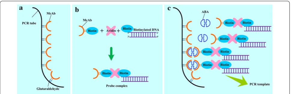
Binding affinity and specificity between analyte and its recognition factor are the key features for the accurate determination in IPCR. To obtain better binding speci-ficity and higher sensitivity, ABA monoclonal antibody (McAb) was used in this study as the recognition factor. Additionally, we prepared the optimized probe complex solution containing biotinylated ABA McAb and probe DNA, and their crosslinking agent avidin beforehand,thus PCR mix in 96-well plate was rapidly prepared. The oper-ational scheme of qIPCR was shown in Fig. 1: (1) through crosslinking reaction, ABA McAb was immobilized on the inner surface of PCR plate which was pretreated with glutaraldehyde (Fig. 1a). (2) Pre-preparing probe com-plex through avidin linking biotinylated ABA McAb and biotinylated probe DNA (Fig. 1b). (3) Adding probe com-plex and proper ABA sample into antibody coated tube for immuno-reaction, then washing the tube to remove excessive probe complex (Fig. 1c). (4) Making PCR mix and running the RT-PCR program.
Optimized ABA antibody fixation
PCR is an extreme signal enhancing method for DNA through exponential growth of products. In quantita-tive real time PCR, the amount of PCR products directly corresponds to the number of initial DNA template. For qIPCR, the consistency and stability of solidified probe DNA (that is DNA template) on the surface of PCR tube is a decisive factor for accurate quantification of analyte before performing PCR program. Therefore, high-homo-geneity PCR plate, high-performance crosslinking agent and proper wash buffer were seriously considered before
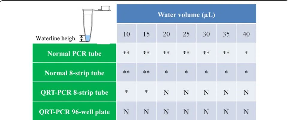
ABA McAb coating. To confirm high-homogeneity of the IPCR tube, we added certain volume of water in different type of polypropylene (PP) PCR tube and observed the waterline height to verify the homogeneity of PCR tube (Fig. 2). As shown in Fig. 2, ordinary PCR tube including normal PCR tube and 8-strip tube should be rejected to apply in IPCR because of higher inner surface deviation. Fortunately, most commercial 96/384-well QRT-PCR plates are homogeneous enough for high throughput quantification of analytes by qIPCR.
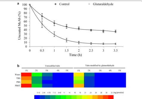
Glutaraldehyde is currently used as an effective cross-linker in generating chemically, biologically and thermally stable cross-links with hydroxyl organics. Additionally, the superfluous glutaraldehyde can be easily removed by rinsing with normal mild saline (such as PBS, TBS, PBST and even hyperpure water) or quenching with organic solvent (such as methanol, chloroform and ether) since it shows higher solubility in both organic and aque-ous phases [37, 38]. Moreover, glutaraldehyde can be employed in pretreating polyethylene or polypropylene tube to enhance binding characteristics with hydroxyl organics [30, 39]. Frequently-used PCR tube is made of polyethylene or polypropylene which owns lower binding capability and stability to proteins/antibodies comparing with polystyrene ELISA microplate. Since the PCR tube is an important restriction factor to affect sensitivity and repeatability of IPCR, we applied glutaraldehyde to pre-treat PCR tube and performed binding dynamics analysis through detecting concentration of uncoated antibod-ies in removed solution from PCR tube at different time intervals. The results showed that PCR tube modified by glutaraldehyde rapidly reached saturation stage in 2 h. The antibody binding efficiency of glutaraldehyde treated
Fig. 1 Operational scheme of qIPCR. a McAb is more efficiently coated on the inner surface of PCR tube after pretreating with glutaraldehyde;
tube was significantly higher than that of the untreated (Fig. 3a).
IPCR requires the antibody to stably bind to the solid phase surface. To test the binding stability of PCR tube and anti-ABA McAb, we detected the protein amount in washing buffer, including TBS (tris-buffered saline, 7.5), PBS buffered saline, 7.4), PBST (phosphate-buffered saline plus Tween, 7.5) and Milli Q water. The total protein content in whole volume washing buffer from antibody coated tube modified by glutaraldehyde was significantly lower than that from untreated tube, regardless the type of washing buffer used (Fig. 3b). This indicated that glutaraldehyde is an ideal stabilizer, thus pretreatment of PCR tube with glutaraldehyde before antibody coating was a recommended measure in qIPCR. Regarding the washing capability of different mild buff-ers, we found that more antibodies were detected in first two washing times using Milli Q water and PBST (Fig. 3b). We speculated that non-ionization water and Tween could elute part of bound McAb from coated PCR tube. TBS and PBS had no significant difference in wash-ing uncoated antibody (Fig. 3b).
Binding saturation analysis of fixed ABA McAb and antigen
In IPCR, unbounded analyte is removed along with washing buffer. Therefore, too much analyte in sample would lead to lower recovery and accuracy. The dose of analyte must not exceed the saturation and the upper limit of added ABA needs to be screened beforehand
in qIPCR system. We analyzed the binding saturation between ABA McAb (about 400 ng) and ABA. Firstly, the PCR tube coated with McAb was prepared. Then after, 2 ng ABA in 20 µL PBS was added into PCR tube and the mixture was incubated at 4 °C in dark. The standard ABA solution was removed at half hour interval and ABA con-centration in removed solution was determined by LC– MS/MS. The results showed that McAb quickly reacted with its antigen ABA within 2 h (Fig. 4).
Binding kinetics of biotinylated ABA McAb and biotinylated probe DNA
To improve the stability of probe complex, we employed the biotin–avidin system. ABA McAb and probe DNA were beforehand biotinylated and then avidin was used as their cross-linker. Avidin contains four duplicate sub-units and is a tetravalent binding glycoprotein for biotin to some extent. Competition exists between biotinylated ABA McAb and biotinylated probe DNA for binding site. To reveal the binding kinetics of biotin-McAb and bio-tin-DNA with avidin, we screened the molar mass ratio of biotin-McAb and biotin-DNA in the probe complex. In this test, the reaction solution contains one fmol avi-din and different amounts of McAb and biotin-DNA (Fig. 5). The ultrafiltration was applied to separate unbound biotin-DNA and the DNA amount in outflow was determined. The results suggested that fewer bio-tin-McAb and biotin-DNA were preferred for unsatu-rated and stable probe complex. The optimal molar mass
ratio was 1:1:1 for biotin-McAb, avidin and biotin-DNA (Fig. 5). Moreover, ultrafiltration was also employed to purify the probe complex.
Standard curve and recovery analysis
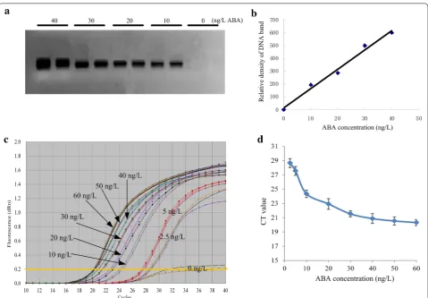
Firstly, we screened the specificity of IPCR. The PCR products were exhibited in 2% agarose gel and no non-specific band was found (Fig. 6a). Then, we calculated the relative densities of DNA bands through Image J software (https ://image j.nih.gov/ij/index .html). The concentra-tion of ABA vs DNA density showed better linear rela-tionship and the equation was y = 15.04x + 14.2 with the determinant coefficient (R2) of 0.9863 (Fig. 6b). Secondly,
the real-time IPCR was performed on QRT PCR instru-ment through SYBR green method. The amplification curves (Fig. 6c) showed that this method could be applied in ABA quantification. The CT (cycle threshold) value would increase along with the decrease of the concen-tration of ABA (Fig. 6c). Subsequently, a standard curve
Fig. 3 Effect of pretreatment with glutaraldehyde on the coating efficiency of ABA McAb. a Uncoated ABA McAb in wash buffer along with the coated time. Data represent the means and SD of three independent biological replicates (n = 5). b The binding stability analysis of ABA McAb coated (400 ng) on PCR tube in TBS (tris-buffered saline, 7.5), PBS (phosphate-buffered saline, 7.4), PBST (phosphate-buffered saline plus Tween, 7.5) and Milli Q water. X represents the number of wash times. The color meaning is the protein amount (ng) in washing buffer. Data represent the means of three independent biological replicates (n = 3)
was obtained using the relationship between the con-centration of ABA standard solution and the average CT value in which ABA was linear in the range of 10 ng/L and 40 ng/L (Fig. 6d). The linear regression equation was y =− 0.118x + 25.365 with the determinant coefficient (R2) of 0.9763. The limit of detection (LOD) was 10 ng/L,
which was converted as 2.5 fg ABA in one PCR reac-tion when utilizing 2.5 ng/L standard ABA solureac-tion. The repeatability and sensitivity of qIPCR method was very close to LC–MS/MS. In addition, the average recovery of ABA in the qIPCR method was 96.9%, similar to that of LC–MS/MS method of 98.1%.
High throughput analysis of ABA in plant samples
qIPCR offers a rapid and high throughput method to quantify analyte but is prone to interference in a num-ber of ways. Thus, it is advisable to validate for a par-ticular tissue by comparing to the qIPCR data with those obtained by a main stream technique with proven
Fig. 5 Binding kinetics of biotin-ABA McAb and biotin-DNA with avidin (1 fmol). The color meaning is percentage of unbound biotin-DNA in effluent after ultrafiltration through 100 kD ultra-filter tube (EMD Millipore UFC910024). Data represent the means of three independent biological replicates (n = 8)
Fig. 6 Amplification analysis of a serial concentrations of ABA by qIPCR. a qIPCR was performed in QRT-PCR tubes. The PCR products were exhibited in agarose gel; b the density values of DNA bands in agarose gel were estimated by Image J software (https ://image j.nih.gov/ij/index .html); c
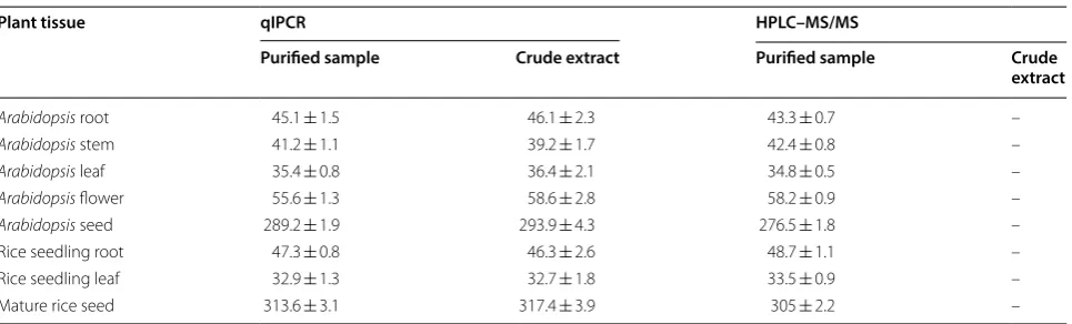
veracity. For this purpose, the qIPCR was validated in comparison with LC–MS/MS using deuterium-labelled ABA as an internal standard in this paper. To further test the repeatability of qIPCR, we determined ABA con-tent in eight different tissues from Arabidopsis and rice by 96-well plates. Comparing the ABA contents in plant tissues determined by qIPCR and LC–MS/MS, the dif-ferences between two methods for both the detected ABA concentration and repeatability were not significant (Table 1).
To further analyze the anti-interference capability, we tried to detect ABA content in the purified sample and the crude extract from different plant tissues (Table 1). The results indicated that qIPCR method showed high specificity for ABA and can be employed to accurately analyze the ABA in crude extracts. Of course, for higher accuracy, the salt content in sample should be limited in acceptable range because excessive ions would affect the amplification rate of PCR.
Discussion
The immuno-PCR (IPCR) method has the potential to quantify analytes at sub-femto gram levels through the efficient amplification of PCR. Theoretically, IPCR can be applied to detect all molecules, but in previous pub-lications, it was intensively employed in the quantifica-tion of bio-macromolecules, such as protein, nucleic acid and antibody [40–44]. Additionally, past efforts and practices were mostly focused on developing and screen-ing the interlinkscreen-ing methods between small organic mol-ecules and macromolmol-ecules. Consequently, IPCR has never been applied in phytohormonal quantification up to now although phytohormonal research is a hotspot in the plant biology field. This study is the first approach of IPCR application in phytohormonal quantification. In
the ABA qIPCR, the biotin–avidin system was employed to be the linkage of ABA and DNA. In detail, the McAb against ABA and probe DNA were biotinylated, and then cross-linked as the probe complex through streptavidin in a certain mixture ratio (Figs. 1, 5). This strategy solved the problem that phytohormones are difficult to directly react with bio-macromolecules in vitro. The pre-prep-aration of the probe complex was very helpful for the standard and high throughput operations in the followed QRT-PCR.
For IPCR, the key factors affecting the sensitivity and efficiency are the affinity and binding specificity between the support substrate surface and the analytes, or between different molecules. Nonspecific binding will lead to a certain level of DNA-tag amplification in samples (background level). In practice, the main aim of optimization of the IPCR method is to quickly obtain high level of amplification in the samples containing ana-lyte comparing to the negative controls [43]. In detail, the choice of support substrate, including binding char-acteristics and conjugation methods, should be seri-ously considered. In this study, we confirmed the type of PCR tubes and the pretreatment method in order to obtain high antibody/protein affinity and amplification efficiency. 96-well microplates show higher affinity for proteins/antibodies and are widely used for ELISA. How-ever, microplates are not suitable for running PCR pro-gram because of heat-instability in a PCR device. If using microplates, an additional step is that probe DNA needs to be detached from the antigen–antibody complex and to be transferred into another PCR tubes for amplifica-tion. Polypropylene PCR tubes were used for IPCR, but did not provide the needed binding characteristic for protein/antibody [45, 46]. Polycarbonate tube have improved protein-binding capacity and heat-stability, but
Table 1 ABA content in different tissues of Arabidopsis and rice (ng/g, fresh weight)
The purified sample was collected through standard extraction and purification processes. The crude extract was dissolved in acetonitrile after extracting with 80% acetone, removing solid impurity and drying [36]. “–” Represented no data because crude extract was not allowed in HPLC–MS/MS. Data represent the means and SD of three independent biological replicates (n = 5)
Plant tissue qIPCR HPLC–MS/MS
Purified sample Crude extract Purified sample Crude
extract
Arabidopsis root 45.1 ± 1.5 46.1 ± 2.3 43.3 ± 0.7 –
Arabidopsis stem 41.2 ± 1.1 39.2 ± 1.7 42.4 ± 0.8 –
Arabidopsis leaf 35.4 ± 0.8 36.4 ± 2.1 34.8 ± 0.5 –
Arabidopsis flower 55.6 ± 1.3 58.6 ± 2.8 58.2 ± 0.9 –
Arabidopsis seed 289.2 ± 1.9 293.9 ± 4.3 276.5 ± 1.8 –
Rice seedling root 47.3 ± 0.8 46.3 ± 2.6 48.7 ± 1.1 –
Rice seedling leaf 32.9 ± 1.3 32.7 ± 1.8 33.5 ± 0.9 –
experiments carried out in polycarbonate tube showed low amplification efficiency and detection sensitivity because of non-uniform distribution of heat in wells dur-ing PCR [47, 48] although the problem was partly solved through increasing denaturation temperature and elon-gation time [49, 50]. In this study, we screened high qual-ity polypropylene PCR tube and pretreated with 0.8% glutaraldehyde. The treated polypropylene PCR tube acquired significant improvement on the antibody-coat-ing capability (Fig. 3a).
Plant extracts are extremely complex, but phytohor-mones are present at trace amounts in plants, usually at the level of 0.1–50 ng/g fresh weight. An ideal analyti-cal method needs to be highly selective and sensitive in quantifying phytohormones. In the past decades, mass spectrometry (MS) has undergone spectacular develop-ment and become the main stream method in phyto-hormonal analysis for its extra sensitivity. Among them, LC–MS/MS is a powerful tool, but involves extra high costs, skilled operation and time consuming multi-step sample preparation. In this study, an easy-to-follow quan-tification method with extra sensitivity was developed to quantify ABA. In the ABA qIPCR, the limit of detection (LOD) reached 2.5 pg and it showed good reproducibil-ity at femtogram level of ABA. The sensitivreproducibil-ity and recov-ery were vrecov-ery close to that of LC–MS/MS. Moreover, the ABA qIPCR offered a sensitive and high throughput new method to quantify ABA in plant sample. Compar-ing with LC–MS/MS, an important improvement was that qIPCR can be used to analyze ABA in crude extract for plant samples. To fully exploit the advantages and potential of IPCR, further optimizations are still needed in protein coating, sampling and PCR condition. Under optimized conditions, qIPCR is expected to be applied in phytohormonal quantification at sub-femto gram level.
Conclusion
The ABA qIPCR is an easy-to-follow and extra sensitive method for phytohormonal quantification in trace plant sample. It can be applied in accurate detection of the ABA in crude extract for plant sample with high specific-ity and repeatabilspecific-ity. The sensitivspecific-ity and recovery of the qIPCR was very close to that of the widely recognized quantification method of LC–MS/MS.
Authors’ contributions
YS, WL and LX designed the research and developed the method. ZH, RW took part in data analysis. WL, QL and JT participated in part experiments. YS and LX prepared the manuscript. YS and WL are equal contribution in this study. All authors read and approved the final manuscript.
Author details
1 Hunan Provincial Key Laboratory of Phytohormones and Growth Develop-ment, Hunan Agricultural University, Changsha, China. 2 Southern Regional Collaborative Innovation Center for Grain and Oil Crops in China, Hunan
Agricultural University, Changsha, China. 3 Tea Research Institute, Hunan Acad-emy of Agriculture Science, Changsha 410125, China.
Acknowledgements
We thank Dr. Fen Xiang (Hunan Academy of Agricultural Science) for valuable suggestions and technical assistance.
Competing interests
The authors declare that they have no competing interests.
Availability of data and materials
The datasets used and/or analyzed during the current study are available from the corresponding author on reasonable request until they are made publicly available in a repository.
Consent for publication
Not applicable.
Ethics approval and consent to participate
Not applicable.
Funding
This work was financially supported by National Natural Science Foundation of China (Grants 91317312, 31570372 and 9141730003), National Key Research and Development Program-Seven major crops breeding Project (Grant 2016YFD0101803) and Scientific Research Fund of Hunan Provincial Education Department (Grants 13K065, 15K061).
Publisher’s Note
Springer Nature remains neutral with regard to jurisdictional claims in pub-lished maps and institutional affiliations.
Received: 24 June 2018 Accepted: 19 November 2018
References
1. Cutler SR, Rodriguez PL, Finkelstein RR, Abrams SR. Abscisic acid: emer-gence of a core signaling network. Annu Rev Plant Biol. 2010;61(1):651. 2. Li J, Wu Y, Xie Q, Gong Z. Abscisic acid. In: Li J, Li C, Smith SM, editors.
Hormone metabolism and signaling in plants. New York: Academic; 2017. p. 161–89.
3. Rajjou L, Duval M, Gallardo K, Catusse J, Bally J, Job C, Job D. Seed germi-nation and vigor. Annu Rev Plant Biol. 2012;63:507–33.
4. Tucker DJ, Mansfield TA. Effects of light quality on apical dominance in xanthium strumarium and the associated changes in endogenous levels of abscisic acid and cytokinins. Planta. 1971;102(2):140–51.
5. Tucker DJ, Mansfield TA. A simple bioassay for detecting “antitranspirant” activity of naturally occurring compounds such as abscisic acid. Planta. 1971;98(2):157–63.
6. Fuchs S, Fuchs Y. Immunological assay for plant hormones using specific antibodies to indoleacetic acid and gibberellic acid. Biochimica et Bio-physica. Acta (BBA) Gen Subj. 1969;192(3):528–30.
7. Boström EA, Tarkowski A, Bokarewa M. Resistin is stored in neutrophil granules being released upon challenge with inflammatory stimuli. Biochem Biophys Acta. 2009;1793(12):1894.
8. Guan J, Zhang ZY, Zhou ZQ, Li H, Tong DN, Zhou GW. Mesenchymal stem cell modulates T follicular helper cell to induce immunotolerance of islet allograft. Transpl Proc. 2015;47(6):2050–6.
9. Su YH, Yu XS, Liu YG, Zhang XS. Abscisic acid is required for somatic embryo initiation through mediating spatial auxin response in Arabidop-sis. Plant Growth Regul. 2013;69(2):167–76.
10. Wang ZY, Gehring C, Zhu J, Li FM, Zhu JK, Xiong L. The Arabidopsis vacu-olar sorting receptor1 is required for osmotic stress-induced abscisic acid biosynthesis. Plant Physiol. 2015;167(1):137–52.
12. Weiler EW, Ziegler H. Determination of phytohormones in phloem exu-date from tree species by radioimmunoassay. Planta. 1981;152(2):168–70. 13. Seeley SD, Powell LE. Electron capture gas chromatography for sensitive
assay of abscisic acid. Anal Biochem. 1970;35:530–3.
14. Horemans S, Van Onckelen HA, Rüdelsheim P, De Greef JA. Study of parameters involved in the determination of IAA and ABA in plant materi-als. J Exp Bot. 1984;35:1832–45.
15. Li X, Motte CEL, Stewart CR, Cloud NP, Wear-Bagnall S, Jiang CZ. Determi-nation of IAA and ABA in the same plant sample by a widely applicable method using GC–MS with selected ion monitoring. J Plant Growth Regul. 1992;11:55–65.
16. Hogge LR, Abrams GD, Abrams SR, Thibault P, Pleasance S. Characteriza-tion of abscisic acid and metabolites by combined liquid chromatog-raphy–mass spectrometry with ion-spray and plasma-spray ionization techniques. J Chromatogr A. 1992;623:255–63.
17. Chu J, Fang S, Xin P, Guo Z, Chen Y. Quantitative analysis of plant hor-mones based on. New York: LC–MS/MS; 2017.
18. Ding M. Simultaneous determination of gibberellic acid, indole-3-acetic acid and abscisic acid in wheat extracts by solid-phase extraction and liquid chromatography-electrospray tandem mass spectrometry. Talanta. 2008;76:798–802.
19. Hu X, Li N, Wu L, Li C, Li C, Zhang L, Liu T, Wang W. Quantitative iTRAQ-based proteomic analysis of phosphoproteins and ABA-regulated phos-phoproteins in maize leaves under osmotic stress. Sci Rep. 2015;5:15626. 20. Hernández L, Zapardiel A, Bermejo E, Pérez-López JA, Pérez-Fernández
JC. Electrochemical studies of ethamivan at glassy-carbon and platinum electrodes and its determination in urine by differential pulse voltamme-try. Anal Chim Acta. 1996;336(1):85–93.
21. Li R, Wang C, Hu Y, Zheng O, Guo L, Lin Z, Qiu B, Chen G. Electrochemilu-minescence biosensor for folate receptor based on terminal protection of small-molecule-linked DNA. Biosens Bioelectron. 2014;58(6):226–31. 22. Christmann A, Hoffmann T, Teplova I, Grill E, Müller A. Generation of
active pools of abscisic acid revealed by in vivo imaging of water-stressed arabidopsis. Plant Physiol. 2005;137(1):209–19.
23. Duan L, Dietrich D, Ng CH, Chan PM, Bhalerao R, Bennett MJ, Dinneny JR. Endodermal ABA signaling promotes lateral root quiescence during salt stress in Arabidopsis seedlings. Plant Cell. 2013;25(1):324–41.
24. Sano T, Smith CL, Cantor CR. Immuno-PCR: very sensitive antigen detection by means of specific antibody-DNA conjugates. Science. 1992;258(5079):120–2.
25. Lind K, Norbeck J. Immuno-qPCR detection of the tandem affinity puri-fication (TAP)-tag as a sensitive and accurate tool suitable for large-scale protein quantification. Proteomics. 2010;7(24):4414–23.
26. Li H, Cui X, Arnheim N. Direct electrophoretic detection of the allelic state of single DNA molecules in human sperm by using the polymerase chain reaction. Proc Natl Acad Sci USA. 1990;87(12):4580–4.
27. Chen H-Y, Zhuang H-S. A real-time immuno-PCR method for detecting 3,3′,4,4′-tetrachlorobiphenyl. Microchim Acta. 2011;172(1–2):233–9. 28. Dong J, Hasan S, Fujioka Y, Ueda H. Detection of small molecule
diagnos-tic markers with phage-based open-sandwich immuno-PCR. J Immunol Methods. 2012;377(1–2):1–7.
29. Fischer A, Von EC, Kuczius T, Omoe K, Peters G, Becker K. A quantitative real-time immuno-PCR approach for detection of staphylococcal entero-toxins. J Mol Med. 2007;85(5):461–9.
30. Meng XY, Li YS, Zhou Y, Zhang YY, Qiao B, Sun Y, Yang L, Hu P, Lu SY, Ren HL. Real-time immuno-PCR for ultrasensitive detection of pyrene and other homologous PAHs. Biosens Bioelectron. 2015;70:42.
31. Niemeyer CM, Adler M, Wacker R. Immuno-PCR: high sensitivity detection of proteins by nucleic acid amplification. Trends Biotechnol. 2005;23(4):208.
32. Barletta F, Ochoa TJ, Ecker L, Gil AI, Lanata CF, Cleary TG. Validation of five-colony pool analysis using multiplex real-time PCR for detection of diarrheagenic Escherichia coli. J Clin Microbiol. 2009;47(6):1915–7. 33. Gofflot S, Elmoualij B, Zorzi D, Melenlamalle L, Roels S, Quatpers D, Grassi
J, Vanopdenbosch E, Heinen E, Zorzi W. Immuno PCR quantitative pour la détection et la quantification de la protéine prion. Acta Physiol (Oxf ). 2004;7(6):142–9.
34. Li YW, Xia K, Wang RZ, Jiang JH, Xiao LT. An impedance immunosensor for the detection of the phytohormone abscisic acid. Anal Bioanal Chem. 2008;391(8):2869–74.
35. Murashige T, Skoog F. A revised medium for the rapid growth and bioas-say with tobacco tissue cultures. Physiol Plant. 1962;15(3):473–97. 36. Zhou LJ, Xiao LT, Xue HW. Dynamic cytology and transcriptional
regula-tion of rice lamina joint development. Plant Physiol. 2017;174(3):1728. 37. Gendler E, Gendler S, Nimni ME. Toxic reactions evoked by
glutaralde-hyde-fixed pericardium and cardiac valve tissue bioprosthesis. J Biomed Mater Res. 1984;18(7):727–36.
38. Migneault I, Dartiguenave C, Bertrand MJ, Waldron KC. Glutaraldehyde: behavior in aqueous solution, reaction with proteins, and application to enzyme crosslinking. Biotechniques. 2004;37(5):790–802.
39. Ji Y, He Q, Xu Y, Tu Z, Yang H, Qiu Y, Wang X, Liu Y. Phage displayed anti-idiotypic nanobody mediated immuno-PCR for sensitive and environ-mentally friendly detection of mycotoxin ochratoxin A. Anal Methods. 2016;8(43):7824–31.
40. Barletta J, Bartolome A, Constantine NT. Immunomagnetic quantitative immuno-PCR for detection of less than one HIV-1 virion. J Virol Methods. 2009;157(2):122–32.
41. Kuczius T, Becker K, Fischer A, Zhang W. Simultaneous detection of three CNS indicator proteins in complex suspensions using a single immuno-PCR protocol. Anal Biochem. 2012;431(1):4–10.
42. Niemeyer CM, Adler M, Wacker R. Detecting antigens by quantitative immuno-PCR. Nat Protoc. 2007;2(8):1918.
43. Ryazantsev DY, Voronina DV, Zavriev SK. Immuno-PCR: achievements and perspectives. Biochemistry. 2016;81(13):1754.
44. Tao X, He Z, Cao X, Shen J, Li H. Development of a highly sensitive real time immuno-PCR for the measurement of chloramphenicol in milk based on magnetic beads capturing. Anal Methods. 2014;6(23):9340–7. 45. Case MC, Burt AD, Hughes J, Palmer JM, Collier JD, Bassendine MF,
Yea-man SJ, Hughes MA, Major GN. Enhanced ultrasensitive detection of structurally diverse antigens using a single immuno-PCR assay protocol. J Immunol Methods. 1999;223(1):93–106.
46. Numata Y, Matsumoto Y. Rapid detection of alpha-human atrial natriu-retic peptide in plasma by a sensitive immuno-PCR sandwich assay. Clin Chim Acta. 1997;259(1–2):169.
47. Adler M, Langer M, Witthohn K, Eck J, Blohm D, Niemeyer CM. Detection of rViscumin in plasma samples by immuno-PCR. Biochem Biophys Res Commun. 2003;300(3):757–63.
48. Niemeyer CM, Adler M, Pignataro B, Lenhert S, Gao S, Chi L, Fuchs H, Blohm D. Self-assembly of DNA-streptavidin nanostructures and their use as reagents in immuno-PCR. Nucl Acids Res. 1999;27(23):4553–61. 49. Barletta J. Applications of real-time immuno-polymerase chain reaction
(qIPCR) for the rapid diagnoses of viral antigens and pathologic proteins. Mol Asp Med. 2006;27(2–3):224.