Development and validation of RP – HPLC method for the estimation of Tylosin tartrate in pure and pharmaceutical formulation
8
0
0
Full text
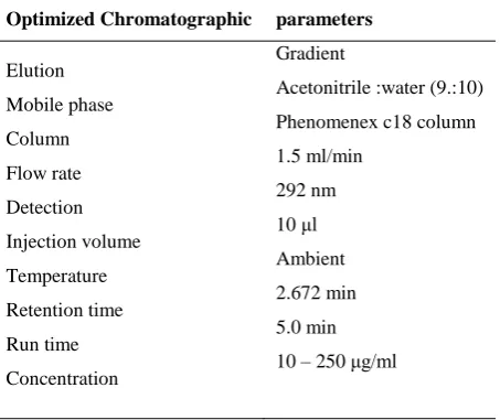
(2) Sailaja Kotha et al / Int. J. of Pharmacy and Analytical Research Vol-3(2) 2014 [214-221]. develop reverse phase high performance liquid chromatography method for the estimation of tylosin tartrate in pure and pharmaceutical dosage form without any derivatization and having short retention time. This method was found to be linear, precise, accurate, sensitive, specific, and robust, and therefore suitable for routine analysis.. was filtered through What man filter paper. The standard chromatogram for tylosin tartrate (100μg/ml) was shown in figure 2. Preparation of working standard solution Working standard solutions of tylosin tartrate were prepared by accurately transferring the (0.1, 0.5, 1.0, 1.5, 2.0 and 2.5 ml) aliquots of the standard stock solution into a series of six 10 ml volumetric flasks. The volume was made up to mark with mobile phase to obtain concentration range of 10 – 250 µg/ml.. MATERIALS AND METHOD Chemicals and Reagents Tylosin tartrate was obtained as a gift from vet India, Hyderabad. HPLC grade acetonitrile and, water was obtained from SD Fine Chemicals Ltd, Mumbai.. Preparation of sample solutions 0.5 ml of tylosin tartrate injection was taken into 100 mL volumetric flask and then the sample was diluted to 100 ml with mobile phase to get concentration of 100 µg/ml and used for analysis.. HPLC Instrumentation and Chromatographic conditions The analytical separations were carried out on a waters 2487 HPLC system equipped with UV detector. The output of signal was monitored and integrated using LC – solutions 2000 software. The analytical column was phenomenex C18 (150 × 4.6 mm, 5 µ). Mobile phase consisted Acetonitrile and water in the ratio of 90:10. Mobile phase was mixed, filtered through 0.45 µ membrane filter and degassed under ultrasonication. The mobile phase was used as diluent. The flow rate was 1.5 ml/min and runtime was 5 minute. The column was maintained at ambient temperature. UV detection was measured at 292 nm and the volume of sample injected was 10 μl.. RESULTS AND DISCUSSION HPLC method development and optimization To optimize the chromatographic conditions, different columns, mobile phases, flow rates etc., were tested. Acetonitrile and water in the ratio of 90:10 was preferred as mobile phase because it resulted in a greater response to tylosin tartrate after several preliminary investigatory runs compared with the different mobile phase combinations. The effect of the flow rate was studied in the range 0.9 to 2.0 ml/min and 1.5 ml/min was preferred to be effective. Under these conditions, the analytic peak obtained was welldefined and free from tailing. The retention time (RT) was found to be 2.672 min. The optimized chromatographic parameters were listed in table 1.. Preparation of standard stock solution 50 mg of tylosin tartrate was weighed accurately and dissolved in 50 ml of mobile phase to get the concentration of 1000 µg/ml. Resultant solution. Table 1: Optimized chromatographic parameters Optimized Chromatographic Elution Mobile phase Column Flow rate Detection Injection volume Temperature Retention time Run time Concentration. parameters Gradient Acetonitrile :water (9.:10) Phenomenex c18 column 1.5 ml/min 292 nm 10 μl Ambient 2.672 min 5.0 min 10 – 250 μg/ml. 215.
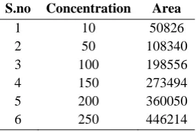
(3) Sailaja Kotha et al / Int. J. of Pharmacy and Analytical Research Vol-3(2) 2014 [214-221]. Graph 2. Optimized chromatogram. 2.672. 0.050. 0.045. 0.040. 0.035. AU. 0.030. 0.025. 0.020. 0.015. 4.578. 0.005. 3.434. 2.461. 0.010. 0.000 0.50. 1.00. 1.50. 2.00. 2.50 Minutes. 3.00. 3.50. 4.00. 4.50. 5.00. system. Retention time (RT), tailing factor (T), and peak asymmetry (AS), resolution (RS) were evaluated. The system suitability test was performed using five replicate injections of standards before analysis of samples. The system suitability method acceptance criteria set in each validation run were: capacity factor > 2.0, tailing factor ≤ 2.0 and theoretical plates > 2000. In all cases, the relative standard deviation (R.S.D) for the analytic peak area for two consecutive injections was <2.0%. System suitability parameters were shown in table 2.. Validation of the method When method development and optimization are complete, it is necessary to accomplish method validation. The validation studies include linear range (correlation coefficient), method precision (RSD, %), method accuracy (% recovery and RSD, %), sensitivity studies (LOD & LOQ), and robustness.. System suitability studies System-suitability tests are an integral part of method development and are used to ensure adequate performance of the chromatographic. Table 2: System suitability parameters. Parameters. Values. Retention time. 2.672min. system. Excellent correlation between tylosin tartrate peak area and concentration was observed with R2 = 0.999 (Figure.3). The regression equation was found to be Y = 1652x +30311. Statistical data are presented in table 3 and the calibration curve was shown in figure 3.. Linearity The linearity of the method was evaluated by preparing six series of standard solutions of tylosin tartrate in the range of 10 – 250 µg/ml in mobile phase and injecting the solutions into the HPLC. Table 3: Linearity results for tylosin tartrate S.no 1 2 3 4 5 6. Concentration 10 50 100 150 200 250. 216. Area 50826 108340 198556 273494 360050 446214.
(4) Sailaja Kotha et al / Int. J. of Pharmacy and Analytical Research Vol-3(2) 2014 [214-221]. 500000 y = 1652.1x + 30311 R² = 0.9993. 400000 300000 200000 100000 0 0. 50. 100. 150. 200. 250. 300. Figure 3: Calibration curve of tylosin tartrate analyzed using the proposed method. The percent relative standard deviation (% RSD) for peak responses was calculated. Results of system To study precision, five replicate standard solutions precision studies were shown in table 4. of tylosin tartrate (100µg/ml) were prepared and. Precision System precision: (Repeatability). Table 4: Results of system precision for tylosin tartrate S.no. Retention time(min). Area (mv.Sec). 1. 2.164. 108564. 2. 2.183. 1902567. 3. 2.207. 2036987. 4. 2.241. 2326987. 5. 2.173. 2569874. 6. 2.244. 2698745. Mean. 2.202. 1940620.667. Standard deviation. 0.03451377. 47620.365. % RSD. 1.56738262. 1.453872919. Method precision: (Reproducibility). and on different days for concentration of sample solutions of 100µg/ml. The result was reported in terms of relative standard deviation (% RSD). Results of method precision studies were shown in table 5. Table 5: Results of Method precision for tylosin tartrate. The intraday and inter-day precision of the proposed method was determined by analyzing the corresponding responses 6 times on the same day. S.no. Peak area. %labelled claim. 1 2 3 4 5 6 Mean SD % RSD. 105896 123698 165987 206598 215694 223654 173587.833 49996.3476 28.8017579. 90.236 101.235 102.325 106.325 123.36 125.365 108.141 9.36985456 0.4569854. 217.
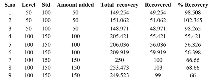
(5) Sailaja Kotha et al / Int. J. of Pharmacy and Analytical Research Vol-3(2) 2014 [214-221]. concentration of sample solutions 100 µg/ml. The percent relative standard deviation (% RSD) for peak responses was calculated. The results for intermediate precision were shown in table 6.. Intermediate precision The intermediate precision of the proposed method was determined by performing the method by two analysts (Analyst 1 and Analyst 2) for. S.NO. Table 6: Results of Intermediate precision for tylosin tartrate ANALYST 1 ANALYST 2 RT (MIN) AREA (MV.SEC) RT (MIN) AREA(MV.SEC). 1. 2.193. 1389879. 2.241. 1385965. 2. 2.736. 1397256. 2.661. 1377715. 3. 2.601. 1372689. 2.594. 1366121. 4. 2.606. 1377661. 2.693. 1345688. 5. 2.643. 1388821. 2.619. 1366127. 6. 2.493. 1401127. 2.514. 1399910. Mean. 2.54533333. 1387905.5. 2.553667. 1373587.667. SD. 0.18949899. 10986.0312. 0.165023. 17092.40841. % RSD. 7.44495764. 0.791554699. 6.462207. 1.244362397. standard drug solution (100 µg/ml) was added to a pre analyzed sample solution (100, 150, 200 µg/ml) and percentage drug content was measured. The closeness of obtained value to the true value indicates that the proposed method is accurate. Recovery studies were shown in table 7.. ACCURACY Accuracy of the method was confirmed by the standard addition method, which was carried out by performing recovery studies at 3 different concentrations 100%, 150% and 200% of these expected, in accordance with ICH guidelines, by replicate analysis (n=3). Known amount of. % Recovery = [(Ct –Cpa)/ Cs] × 100.. Where, Ct = Total concentration of analyte Cpa = Concentration of pre-analysed sample Cs = Concentration of standard added to pre-analysed sample.. Table 7: Results of recovery studies for tylosin tartrate S.no 1 2 3 4 5 6 7 8 9. Level 50 50 50 100 100 100 100 100 100. Std 100 100 100 150 150 150 150 150 150. Amount added 50 50 50 100 100 100 150 150 150. Total recovery 149.254 151.062 148.971 205.421 206.036 209.919 250 253.473 249.523 . Robustness The robustness study was performed to evaluate the influence of small but deliberate variation in the chromatographic condition. The robustness was checked by changing parameters like flow rate of mobile phase and detection wavelength. . 218. Recovered 49.254 51.062 48.971 55.421 56.036 59.919 100 103 99. % Recovery 98.508 102.365 98.265 55.421 56.326 56.398 66.66 68.66 66. Change in the detection wavelength by ± 2nm (294nm and 290nm) Change in flow rate by ± 0.1 ml/minute (1.6 ml/min and 1.4 ml/minute).
(6) Sailaja Kotha et al / Int. J. of Pharmacy and Analytical Research Vol-3(2) 2014 [214-221]. After each change, sample solution was injected Robustness values were and % assay with system suitability parameters were checked. Table 8: Results of Robustness for tylosin tartrate Parameter Flow rate(ml/min)1.7 1.3 Wavelength(nm). Rt(min) 2.229 2.569 2.204 2.18. given. in. table. 8. Area(mvsec) 389654 386954 1896.369 1852.369. Limit of Detection and Quantitation Detection and Quantitation limit were calculated by slope of the calibration plot, using the formula. the method based on the standard deviation () and Limit of Detection × 3.3/S Limit of Quantitation × 10/S Where = The standard deviation of the response. S = The slope of the calibration curve (of the analyte). Results of LOD & LOQ were shown in table 9. Table 9: Results of LOD, LOQ for tylosin tartrate S.No LOD LOQ 1 0.099 0.301 be present in the sample matrix. Chromatograms of Specificity standard and sample solutions were compared in Specificity of an analytical method is its ability to order to provide an indication of specificity of the measure the analyte accurately and specifically in method. the presence of component that may be expected to pharmaceutical dosage form And the % Assay results were shown in table 10.. Assay of pharmaceutical formulation The proposed validated method was successfully applied to determine tylosin tartrate in their. Table 10: Results of % assay S.No Amount Found %Assay 1 197.876 98.39 2 198.044 99.022 3 191.501 95.75 was found to be precise and accurate. The low detection and quantification limits achieved indicate the method is very sensitive. The robustness data gathered during method validation showed that the method is not susceptible to small changes in chromatographic conditions. The proposed RP-HPLC method developed by the author is suitable for routine analysis and quality assessment of tylosin tartrate in pharmaceutical products.. CONCLUSION A simple, rapid, accurate, and precise RP-HPLC method for the analysis of tylosin tartrate in pure and in pharmaceutical dosage forms had been developed and validated in accordance with ICH guidelines. The RP-HPLC method developed is cost-effective due to short retention time which enabled analysis of tylosin tartrate samples with a small amount of mobile phase. From the % RSD values of precision and recovery studies the method. 219.
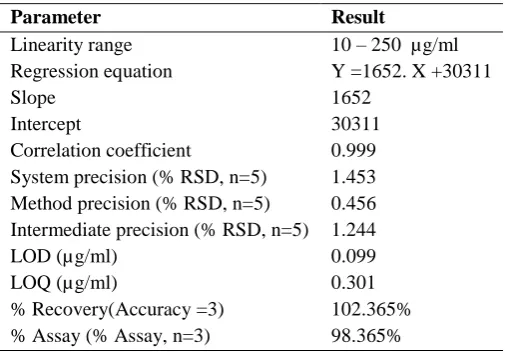
(7) Sailaja Kotha et al / Int. J. of Pharmacy and Analytical Research Vol-3(2) 2014 [214-221]. Table 12: Summary of validated parameters for proposed method Parameter Linearity range Regression equation Slope Intercept Correlation coefficient System precision (% RSD, n=5) Method precision (% RSD, n=5) Intermediate precision (% RSD, n=5) LOD (µg/ml) LOQ (µg/ml) % Recovery(Accuracy =3) % Assay (% Assay, n=3). Result 10 – 250 µg/ml Y =1652. X +30311 1652 30311 0.999 1.453 0.456 1.244 0.099 0.301 102.365% 98.365%. REFERENCES [1]. Parsons CG, Danysz W, Quack G. Memantine is a clinically well Tolerated N‐methyl‐D‐aspartate (NMDA) receptor antagonist‐a review of preclinical data. Neuropharmacology. 38; 1999: 735‐767. [2]. Sonkusare SK, Kaul CL, Ramarao P. Dementia of Alzheimer's disease and other neurodegenerative disorders‐memantine. Pharmacol Res. 51; 2005: 1‐17. [3]. Erickson CA, Posey DJ, Stigler KA, Mullett J, Katschke AR, Mc Dougle CJ. A retrospective study of memantine in children and adolescents with pervasive developmental disorders. Psychopharmacology. 191; 2007: 141‐147. [4]. Zdanys K, Tampi RR. A systematic review of off‐label uses of memantine for psychiatric disorders.Prog Neuropsychopharmacol Biol Psychiatry. 32; 2008: 1362‐1374. [5]. R F Suckow, M F Zhang, E D Collins, M W Fischman, T B Cooper. Sensitive and selective liquid chromatographic assay of memantine in plasma with fluorescence detection after pre-column derivatization. J Chromatogr B Biomed Sci Appl. 729 (1-2); 1999: 217-224 [6]. Afshin Zarghi, Alireza Shafaati, Seyed Mohsen Foroutan,Arash Khoddam, and Babak Madadian. Sensitive and Rapid HPLC Method for Determination of Memantine in Human Plasma Using OPA Derivatization and Fluorescence Detection. Application to Pharmacokinetic Studies. Sci Pharma. 78(4); 2010: 847–856. [7]. Belen Puente, Esther Hernandez, Susana Perez, Luis Pablo, Esther Prieto, Maria Angeles Garcia, And Miguel Angel Bregante. Determination of memantine in plasma and vitreous humour by HPLC with precolumn derivatization and fluorescence detection. Journal of Chromatographic Science. 49; 2011: 745-752 [8]. Leis HJ, Fauler G, Windischhofer W. Quantitative analysis of memantine in human plasma by gas chromatography/negative‐ion chemical ionization/mass spectrometry. J Mass Spectrom 37; 2002: 477‐480. [9]. K. Siddappa, Metre Mallikarjun, Tambe Mahesh, Kote Mallikarjun, Reddy Chandrakanth. Development and validation of a gas chromatographic method for the assay of memantine hydrochloride in pure and tablet dosage forms. Physics, Chemistry and Technology. 9(1); 2011: 1 - 8 [10]. Koeberle MJ, Hughes PM, Wilson, Skellern GG. Development of a liquid chromatography‐mass spectrometric method for measuring the binding of memantine to different melanins. J Chromatogr B Analyt Technol Biomed Life Sci. 787; 2003: 313‐322. [11]. Almeida AA, Campos DR, Bernasconi G, Calafatti S, Barros FAP,Eberlin MN. Determination of memantine in human plasma by liquid chromatography‐electrospray tandem mass spectrometry. J Chromatogr B Analyt Technol Biomed Life Sci. 848; 2007: 311‐316.. 220.
(8) Sailaja Kotha et al / Int. J. of Pharmacy and Analytical Research Vol-3(2) 2014 [214-221]. [12]. Liu MY, Meng SN, Wu HZ, Wang S, Wei MJ. Pharmacokinetics of single‐dose and multiple‐dose memantine in healthy Chinese volunteers using an analytical method of liquid chromatography‐tandem mass spectrometry. Clin Ther. 30; 2008: 641‐653. [13]. Pan RN, Chian TY, Kuo B, Pao LH. Determination of memantine in human plasma by LC–MS–MS. Application to a pharmacokinetic study. Chromatographia. 70; 2009: 783‐788. [14]. Sarita Karthikeyan, Anju Aji , Sarabjit Singh And Shivanand P. Puthli . An LC-MS/MS method for the quantification of Memantine in human plasma: Development, validation and application to a pharmacokinetic study. International Journal of Pharmacy and Biological Sciences. 3(2); 2013: 343354 [15]. Jagathi V, Anupama B, Praveen PS, Rao GD. Developed two simple, sensitive and reproducible colorimetric methods for the estimation of Memantine (MEM) in bulk and in pharmaceutical formulations. Int J Current Pharma Res. 2; 2010: 17‐18. [16]. Praveen PS, Jagathi V, Rao GD, Aparna A . Developed two simple and sensitive extractive spectrophotometric methods for the estimation of Memantine (MEM) in pure and pharmaceutical dosage forms. Res J Pharma Biological Chemical Sci. 1; 2010: 222‐225. [17]. Karim Michail, Hoda Daabees, Youssef Beltagy, Magdi Abdelkhalek, Mona Khamis*. Spectrophotometric And Spectrofluorimetric Determination of Memantine Hydrochloride in Bulk and Pharmaceutical Preparations. International Journal of Pharmacy and Pharmaceutical Sciences. 3(3); 2011:180-185 [18]. Bhavil Narola, A.S. Singh, P. Rita Santhakumar, and T.G. Chandrashekhar. A Validated Stabilityindicating Reverse Phase HPLC Assay Method for The Determination of Memantine Hydrochloride Drug Substance With UV-detection Using Pre column Derivatization Technique. Anal Chem Insights. 5; 2010: 37–45 [19]. kishore kumar Hotha, Satti Phani Kumar Reddy, V. Kishore Raju, L.K. Ravindranath., Forced degradation studies: Practical approach-overview of regulatory guidance and literature for the drug products and drug substances. Int. Res. J. Pharm. 4(5); 2013: 78-85. *******************************. 221.
(9)
Figure


+4

Related documents
Background: A systematic review assessing autologous versus alloplastic bone for secondary alveolar bone grafting in patients with cleft lip and palate was published in 2011
However, the nanosilver-coated orthodontic bracket can be utilized during fixed orthodontic treatment with caution, because the foreign particles which are brown-black granules
The results shows that Impact strength decreases after continues exposure to weathering and. sometimes it rapidly
The company creates also a classification on the amount of sale in the table of facts. According to the process of modeling, the table of information is selected in the first stage
The objectives of the study were to promote healthy lifestyle, by implementing school based intervention program (physical activity and nutrition education) for healthy body
This procedure is recommended over closed biopsy when the magnitude of the patient's illness and degree of pulmonary function do not allow acceptance of the risks
Hitch Hiker classifies LooCI bindings as high-priority and low-priority, and this classifica- tion allows Hitch Hiker to support data aggregation by appending low-priority data in
The pupils in the experimental school improved their nutrition knowledge, iron rich food intake which could have positively influenced their hemoglobin levels.. Deworming may also
