Electronic Thesis and Dissertation Repository
7-3-2014 12:00 AM
Sox9 conditional knockdown reduces chondroitin sulphate
Sox9 conditional knockdown reduces chondroitin sulphate
proteoglycan expression, increases neuroplasticity, and improves
proteoglycan expression, increases neuroplasticity, and improves
motor function in a mouse model of spinal cord injury
motor function in a mouse model of spinal cord injury
William M. McKillop
The University of Western Ontario
Supervisor Dr. Arthur Brown
The University of Western Ontario
Graduate Program in Anatomy and Cell Biology
A thesis submitted in partial fulfillment of the requirements for the degree in Doctor of Philosophy
© William M. McKillop 2014
Follow this and additional works at: https://ir.lib.uwo.ca/etd
Part of the Molecular and Cellular Neuroscience Commons
Recommended Citation Recommended Citation
McKillop, William M., "Sox9 conditional knockdown reduces chondroitin sulphate proteoglycan expression, increases neuroplasticity, and improves motor function in a mouse model of spinal cord injury" (2014). Electronic Thesis and Dissertation Repository. 2153.
https://ir.lib.uwo.ca/etd/2153
This Dissertation/Thesis is brought to you for free and open access by Scholarship@Western. It has been accepted for inclusion in Electronic Thesis and Dissertation Repository by an authorized administrator of
SOX9 CONDITIONAL KNOCKDOWN REDUCES CHONDROITIN SULFATE PROTEOGLYCAN EXPRESSION, INCREASES NEUROPLASTICITY, AND IMPROVES
MOTOR FUNCTION IN A MOUSE MODEL OF SPINAL CORD INJURY
Title Page
(Thesis format: Integrated-Article)
by
William M. McKillop
Graduate Program in Anatomy and Cell Biology
A thesis submitted in partial fulfillment
of the requirements for the degree of
Doctor of Philosophy
The School of Graduate and Postdoctoral Studies
The University of Western Ontario
London, Ontario, Canada
Abstract
This thesis investigates the effect of Sox9 knockdown on anti-regenerative scar gene
expression, neuroplasticity, and hind limb functional recovery following mouse spinal cord
injury. We hypothesized that Sox9 knockdown would reduce expression of anti-regenerative
chondroitin sulfate proteoglycans both at the lesion site and at sites distant to the injury, thus
providing an avenue for increased neuroplasticity and locomotor recovery after spinal cord
injury. The first chapter provides a general introduction to the biological problem of spinal cord
injury. The development of the glial scar and expression of the anti-regenerative chondroitin
sulfate proteoglycan (CSPG) extracellular matrix is introduced, and Sox9 is identified as a
transcription factor that may control expression of these anti-regenerative genes. The second
chapter is a manuscript that describes the molecular changes and improved locomotor function
seen when Sox9 knockdown is carried out just prior to spinal cord injury. The third chapter is
more clinically relevant as it is a manuscript detailing the effects of Sox9 knockdown after spinal
cord injury on the recovery of hind limb motor function. The fourth chapter is a manuscript
investigating the neuro-anatomical mechanism of the improved functional recovery seen in Sox9
knockdown mice after spinal cord injury. The fifth chapter reflects on the findings presented
herein, and suggests possible future plans of study. This dissertation demonstrates that inhibition
of Sox9 leads to reduced CSPG expression, improved hind limb function, and increased total
locomotion. It further provides compelling evidence that increased neuroplasticity as evidenced
by increased reactive sprouting and increased expression of the presynaptic markers
synaptophysin and VGLUT1 caudal to the injury site underlies the improved neurological
recovery observed in spinal cord injured Sox9 conditional knockdown mice.
Key words: spinal cord injury, Sox9, chondroitin sulfate proteoglycans, neuroplasticity, reactive
Co-authorship
Chapter 2 of this dissertation was co-authored for publication by myself, Magda Dragan,
Dr. Andreas Schedl, and Dr. Arthur Brown. Magda Dragan was instrumental in developing
astrocyte cell culture techniques. Chapter 3 of this dissertation has been submitted for publication
co-authored by myself, Eli York, and Dr. Arthur Brown. Eli York assisted in the day to day care
of the mice used for the delayed knockdown experiment, the evaluation of their locomotor
activity, and the spinal tissue cryosectioning as well as GFAP and CSPG immunostaining of
sectioned spinal cords. Chapter 4 of this dissertation has been prepared for publication
co-authored by myself, Dr. Todd Hryciw, Dr. Kathy Xu, Dr. Nicole Geremia, and Dr. Arthur
Brown. Dr. Todd Hryciw performed the BDA injections into the mouse primary motor cortex.
Dr. Kathy Xu helped to develop the BDA and fluorogold quantification techniques. Dr. Nicole
Geremia helped to develop the fluorogold administration technique. In each of these studies, I
planned and executed the experiments and completed the analysis of results with the
aforementioned assistance. Dr. Andreas Schedl contributed the Sox9flox/flox mice for our usage.
Dr. Arthur Brown conceptualized much of the experimental design, provided appropriate
guidance, assisted with technical problems, and edited manuscripts. Dr. Arthur Brown also
Acknowledgements
Thank you to my supervisor and mentor Dr. Arthur Brown and to the members of my
advisory committee, Dr. Vania Prado, Dr. Nagalingam Rajakumar, and Dr. Lique Coolen for
their guidance and assistance. I would also like to thank Dr. Greg Dekaban for all of his
assistance over the years, and Dr. Lynne Weaver for help with manuscript preparation.
I must extend a special thank you to Anna Pniak, Eli York, Dr. Kathy Xu, Dr. Todd
Hryciw, and Dr. Nicole Geremia who each directly affected this project. Thank you to the Brown
laboratory personnel; Stephen McDonald, Bethany Bass, Trina Rosenzweig, Vanessa Omaña,
John Pierce, Daniel Bottner, and Luc Rubinger, Dekaban laboratory personnel; Dr. Kemi
Adeyanju, Christy Willert, Sonali de Chickera, Ryan Buensuceso, Bryan Au, Robarts Research
Institute students; Arthur Lau, Cynthia Tang, and Peter Mitsopoulos, and finally my friends from
outside Western; Susan Sheng, Arash Behravan, Kevin Tsin, and Howard Fung. Each of these
people contributed something important to this project along the way.
Finally I would like to thank my parents Brian and Renee McKillop for their guidance,
love, and support.
This work could not have been completed without funding from the National Science and
Education Research Council of Canada, the Ontario Graduate Scholarship program, the
Dedication
Table of Contents
Title Page ... i
Abstract ... ii
Key words ... ii
Co-authorship ... iii
Acknowledgements ... iv
Dedication ... v
Table of Contents ... vi
List of Figures ... xi
List of Abbreviations ... xiii
Chapter 1: Introduction ... 1
1.0 Spinal cord injury ... 1
1.1 Nervous system plasticity... 3
1.2 Chondroitin Sulfate Proteoglycans (CSPGs) ... 5
1.3 Regenerative failure post-SCI – CSPGs in the glial scar ... 7
1.4 The perineuronal network ... 9
1.5 Anti-CSPG strategies show promise for the treatment of SCI ... 10
1.6 Existing therapies for SCI ... 11
1.7 Sox9 was identified as a potential regulator of CSPG biosynthesis ... 13
1.8 Transcription factor Sox9 ... 14
1.9 The role of Sox9 in cartilage formation ... 15
1.10 Sox9 modulates enzymes essential for CSPG production ... 17
1.11 The dorsal contusion spinal cord injury model used in these studies ... 18
1.12 Conditional Sox9 knockdown mouse breeding strategy ... 19
1.13 Specific goals ... 20
1.14 Summary ... 22
1.15 References ... 23
Chapter 2: Conditional SOX9 ablation reduces chondroitin sulfate proteoglycan expression and improves motor function following spinal cord injury ... 33
2.0 Abstract ... 34
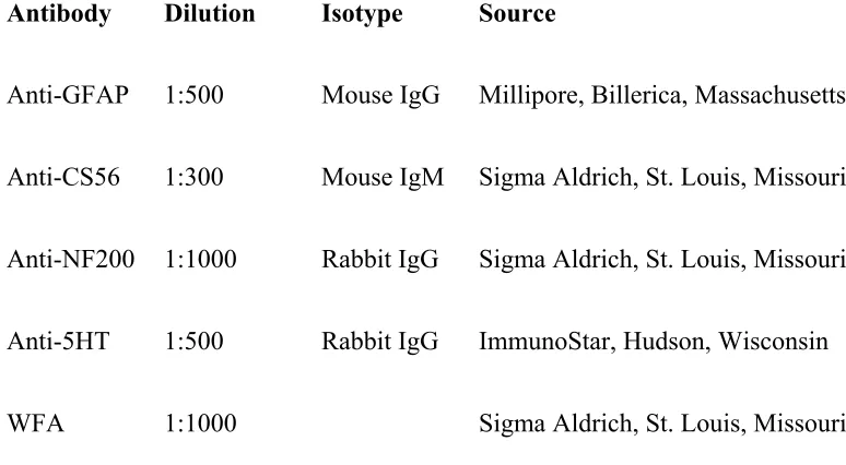
2.2 Materials and Methods ... 36
2.3 Results ... 47
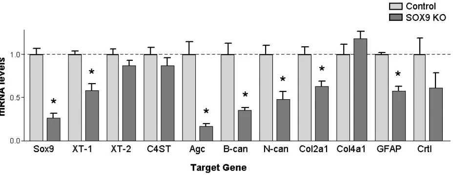
Figure 1. Astrocytes from Sox9 conditional knockdown mice demonstrate reduced glial scar gene expression compared to control mice. ... 49
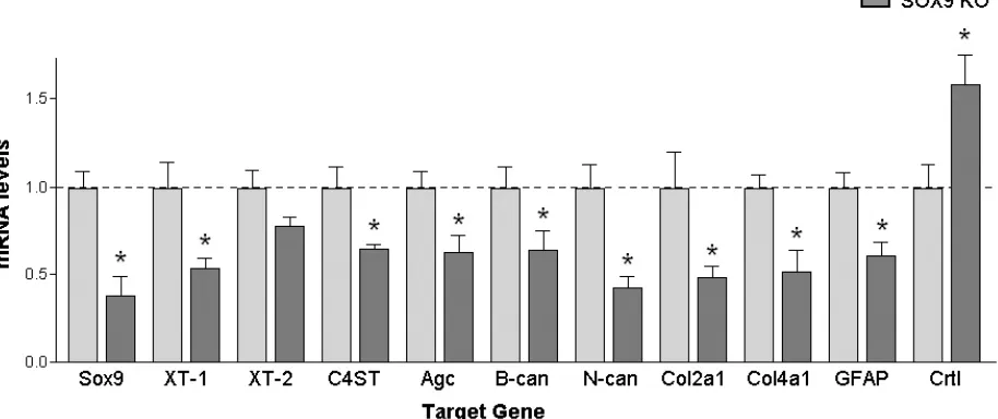
Figure 2. Sox9 conditional knockdown mice demonstrate reduced glial scar gene expression compared to control mice. ... 52
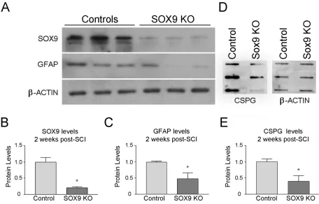
Figure 3. Sox9 conditional knockdown mice demonstrate reduced SOX9, GFAP, and CSPG protein 2 weeks post-SCI. ... 54
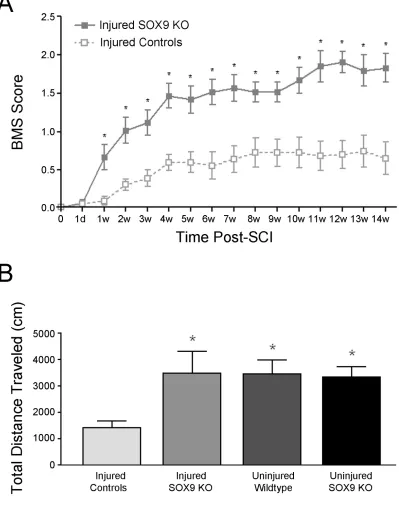
Figure 4. Sox9 conditional knockdown mice demonstrate improved locomotor recovery after SCI. ... 57
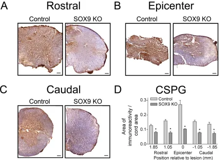
Figure 5. Sox9 conditional knockdown mice display reduced CSPG expression 14 weeks post-SCI. ... 60
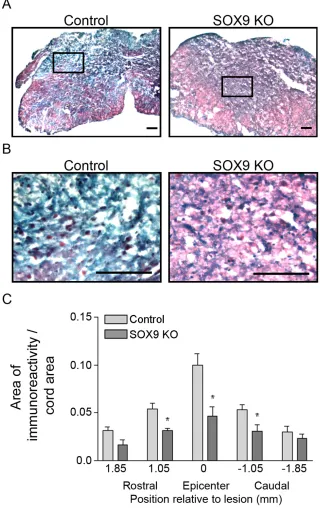
Figure 6. Sox9 conditional knockdown mice demonstrate reduced collagen at the lesion epicenter 14 weeks post-SCI. ... 62
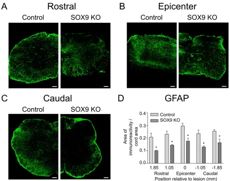
Figure 7. Sox9 conditional knockdown mice demonstrate reduced GFAP expression 14 weeks post-SCI. ... 64
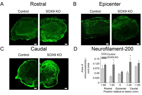
Figure 8. Sox9 conditional knockdown mice demonstrate increased neurofilament immunoreactivity rostral and caudal to their lesion epicenters 14 weeks post-SCI. ... 67
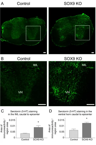
Figure 9. Sox9 conditional knockdown mice display increased 5-HT immunoreactivity caudal to the lesion. ... 69
Figure 10. Perineuronal net matrix is reduced in Sox9 conditional knockdown mice caudal to lesion 14 weeks post-SCI. ... 72
2.4 Discussion ... 73
2.5 Acknowledgments ... 79
2.6 Author Disclosure Statement ... 79
2.7 Supplementary Table 1. Two Way ANOVA Summary Table ... 80
2.8 References ... 80
Chapter 3: Conditional ablation of Sox9 after spinal cord injury reduces chondroitin sulfate proteoglycan expression and improves locomotor recovery ... 85
3.0 Abstract ... 86
3.1 Introduction ... 87
3.2 Materials and Methods ... 88
Figure 1. Experimental timeline. ... 92
Figure 2. Tamoxifen administration 1 week post-SCI requires 6 days to achieve significant
Sox9 knockdown. ... 101
Figure 3. Sox9, GFAP, neurocan and aggrecan mRNA and protein expression levels are reduced 6 weeks post-SCI following Sox9 ablation initiated at 1 week after injury. ... 104
Figure 4. Sox9 knockdown mice demonstrate improved locomotor recovery compared to control mice. ... 107
Figure 5. Reduced CSPG expression levels in Sox9 knockdown mice 14 weeks post-SCI. ... 110
Figure 6. Reduced GFAP expression levels in Sox9 knockdown mice 14 weeks post-SCI. ... 112
Figure 7. Reduced WFA staining in Sox9 knockdown mice 14 weeks post-SCI. ... 115
Figure 8. Increased 5-HT immunoreactivity caudal to the lesion in Sox9 knockdown mice 14 weeks post-SCI. ... 117
3.4 Discussion ... 118
3.5 Acknowledgments ... 123
3.6 Author Disclosure Statement ... 123
3.7 Supplementary Table 1. Two Way ANOVA Summary Table ... 123
3.8 References ... 124
Chapter 4: Sox9 knockdown promotes neuroplasticity after spinal cord injury ... 127
4.0 Abstract ... 128
4.1 Introduction ... 129
4.2 Materials and Methods ... 131
Figure 1. Experimental Timeline. ... 134
4.3 Results ... 140
Figure 2. WFA staining is reduced both just caudal to the injury site, as well as in the lumbar enlargement in Sox9 KO mice 10 weeks post-SCI. ... 143
Figure 3. Sox9 KO mice do not display increased axonal sparing or long range axonal regeneration post-SCI. ... 146
Figure 4. Sox9 KO mice display increased synaptophysin immunoreactivity in the ventral horn of the lumbar enlargement 10 weeks post-SCI. ... 148
Figure 5. Sox9 KO mice display increased VGLUT1 immunoreactivity in the ventral horn of the lumbar enlargement 10 weeks post-SCI. ... 150
Figure 7. Sox9 KO mice display increased pre-synaptic terminal synaptophysin positive
boutons in layer IX of ventral horn at the lumbar enlargement 10 weeks post-SCI. ... 155
Figure 8. Sox9 KO mice display increased pre-synaptic terminal VGLUT1 positive boutons in layer IX of ventral horn at the lumbar enlargement 10 weeks post-SCI. ... 157
Figure 9. Control and Sox9 KO mice display similar pre-synaptic terminal VGAT positive boutons in layer IX of ventral horn at the lumbar enlargement 10 weeks post-SCI. ... 159
Figure 10. High magnification images display increased BDA and VGLUT1 positive puncta around ventral horn motor neurons in the lumbar enlargement in Sox9 KO mice. ... 161
Figure 11. Sox9 KO mice display increased serotonin immunoreactivity both just caudal to the injury site and in the ventral horn of the lumbar enlargement 10 weeks post-SCI. ... 165
4.4 Discussion ... 166
Supplementary Figure 1. Sox9 KO mice also do not display increased sparing or long range axonal regeneration in propriospinal interneurons, the rubrospinal tract or the vestibular spinal tract post-SCI. ... 173
Supplementary Figure 2. Control and Sox9 KO mice display similar numbers of BDA labeled fibers in the cervical enlargement in both uninjured mice as well as 10 weeks post-SCI. ... 175
Supplementary Figure 3. High magnification images display similar numbers of BDA and VGLUT1 positive puncta around ventral horn motor neurons in the cervical enlargement in Sox9 KO mice. ... 177
4.5 Acknowledgments ... 178
4.6 Author Disclosure Statement ... 178
4.7 References ... 178
Chapter 5: Discussion ... 183
5.0 Sox9 knockdown, a potential pro-regenerative treatment for SCI ... 183
5.1 Obstacles to regeneration, activation of SOX9 post-SCI ... 184
5.2 Sox9 knockdown results in improved hind limb motor function post-SCI ... 185
5.3 Sox9 knockdown reduces anti-regenerative CSPG expression post-SCI ... 186
5.4 Investigating neuroplasticity post-SCI; axonal regeneration, reactive sprouting, and synapse plasticity ... 187
5.5 Sox9 knockdown does not promote axonal sparing post-SCI ... 188
5.6 Sox9 knockdown does not promote long range axonal regeneration post-SCI ... 189
5.8 Combination therapies may be required to produce maximal beneficial effect post-SCI 194 5.9 Complete Sox9 knockdown is unlikely to be optimal for recovery post-SCI; beneficial role
of astrocytes post-SCI ... 195
5.10 Complete Sox9 knockdown is unlikely to be optimal for recovery post-SCI; beneficial role of CSPGs post-SCI ... 196
5.11 Why do we our Sox9 knockdown mice only display ~65% Sox9 reduction? ... 197
5.12 Alternative competing strategies, chondroitinase ABC ... 198
5.13 Alternative competing strategies, decorin ... 199
5.14 Alternative competing strategies, inhibition of N-acetylgalactosaminyltransferase-1 ... 199
5.15 Alternative competing strategies, inhibition of Nogo-A ... 200
5.16 Alternative competing strategies, inhibition of Rho ... 201
5.17 Alternative explanations for functional recovery post Sox9 knockdown ... 202
5.18 Alternative explanations for functional recovery post Sox9 knockdown; the effect of Sox9 knockdown on inflammation ... 203
5.19 Alternative explanations for functional recovery post Sox9 knockdown; the effect of Sox9 knockdown on neural stem cells ... 205
5.20 Conclusion ... 207
5.21 References ... 208
Ethics Approval for Animal Usage on Protocol 2007-009-02... 215
JOHN WILEY AND SONS LICENSE ... 216
List of Figures
Chapter 2: Conditional SOX9 ablation reduces chondroitin sulfate proteoglycan expression and improves motor function following spinal cord injury
Figure 1. Astrocytes from Sox9 conditional knockdown mice demonstrate reduced glial scar gene expression compared to control mice. ... 49 Figure 2. Sox9 conditional knockdown mice demonstrate reduced glial scar gene expression compared to control mice. ... 52 Figure 3. Sox9 conditional knockdown mice demonstrate reduced SOX9, GFAP, and CSPG protein 2 weeks post-SCI. ... 54 Figure 4. Sox9 conditional knockdown mice demonstrate improved locomotor recovery after SCI. ... 57 Figure 5. Sox9 conditional knockdown mice display reduced CSPG expression 14 weeks post-SCI. ... 60 Figure 6. Sox9 conditional knockdown mice demonstrate reduced collagen at the lesion epicenter 14 weeks post-SCI. ... 62 Figure 7. Sox9 conditional knockdown mice demonstrate reduced GFAP expression 14 weeks post-SCI. ... 64 Figure 8. Sox9 conditional knockdown mice demonstrate increased neurofilament
immunoreactivity rostral and caudal to their lesion epicenters 14 weeks post-SCI. ... 67 Figure 9. Sox9 conditional knockdown mice display increased 5-HT immunoreactivity caudal to the lesion. ... 69 Figure 10. Perineuronal net matrix is reduced in Sox9 conditional knockdown mice caudal to lesion 14 weeks post-SCI. ... 72
Chapter 3: Conditional ablation of Sox9 after spinal cord injury reduces chondroitin sulfate proteoglycan expression and improves locomotor recovery
Figure 1. Experimental timeline. ... 92 Figure 2. Tamoxifen administration 1 week post-SCI requires 6 days to achieve significant
Chapter 4: Sox9 knockdown promotes neuroplasticity after spinal cord injury
Figure 1. Experimental Timeline. ... 134 Figure 2. WFA staining is reduced both just caudal to the injury site, as well as in the
lumbar enlargement in Sox9 KO mice 10 weeks post-SCI. ... 143 Figure 3. Sox9 KO mice do not display increased axonal sparing or long range axonal
regeneration post-SCI. ... 146 Figure 4. Sox9 KO mice display increased synaptophysin immunoreactivity in the ventral horn of the lumbar enlargement 10 weeks post-SCI. ... 148 Figure 5. Sox9 KO mice display increased VGLUT1 immunoreactivity in the ventral horn of the lumbar enlargement 10 weeks post-SCI. ... 150 Figure 6. Control and Sox9 KO mice display similar VGAT immunoreactivity in the ventral horn of the lumbar enlargement 10 weeks post-SCI. ... 152 Figure 7. Sox9 KO mice display increased pre-synaptic terminal synaptophysin positive boutons in layer IX of ventral horn at the lumbar enlargement 10 weeks post-SCI. ... 155 Figure 8. Sox9 KO mice display increased pre-synaptic terminal VGLUT1 positive boutons in layer IX of ventral horn at the lumbar enlargement 10 weeks post-SCI. ... 157 Figure 9. Control and Sox9 KO mice display similar pre-synaptic terminal VGAT positive boutons in layer IX of ventral horn at the lumbar enlargement 10 weeks post-SCI. ... 159 Figure 10. High magnification images display increased BDA and VGLUT1 positive puncta around ventral horn motor neurons in the lumbar enlargement in Sox9 KO mice. ... 161 Figure 11. Sox9 KO mice display increased serotonin immunoreactivity both just caudal to the injury site and in the ventral horn of the lumbar enlargement 10 weeks post-SCI. ... 165 Supplementary Figure 1. Sox9 KO mice also do not display increased sparing or long range axonal regeneration in propriospinal interneurons, the rubrospinal tract or the vestibular spinal tract post-SCI. ... 173 Supplementary Figure 2. Control and Sox9 KO mice display similar numbers of BDA labeled fibers in the cervical enlargement in both uninjured mice as well as 10 weeks post-SCI. ... 175 Supplementary Figure 3. High magnification images display similar numbers of BDA and VGLUT1 positive puncta around ventral horn motor neurons in the cervical enlargement in
Sox9 KO mice. ... 177
List of Abbreviations
AANS/CNS – American Association of Neurological Surgeons/Congress of Neurological Surgeons
ANOVA – analysis of variance
BBB – blood brain barrier
BMS – Basso Mouse Scale
BDNF – brain derived neurotrophic factor
C4ST – Chondroitin-4 sulfotransferase
CNS – central nervous system
CPG – central pattern generator
CSPG – chondroitin sulfate proteoglycans
DRG – dorsal root ganglion
ECM – extracellular matrix
GAG – glycosaminoglycans
GalNAc – N-acetylgalactosamine
GFAP – glial fibrillary acidic protein
GFP – green fluorescent protein
HMG – high-mobility-group
HRP – horse radish peroxidase
HSPG – heparin sulfate proteoglycans
IL – interleukin
LAR - leukocyte common antigen-related phosphatase receptor
MAG – myelin associated glycoprotein
MBP – myelin basic protein
MP – methylprednisolone
NASCIS – National Acute Spinal Cord Injury Study
PDGF – platelet derived growth factor
PGC-1alpha – Peroxisome proliferator-activated receptor gamma co-activator 1alpha
PNN – perineuronal net
PNS – peripheral nervous system
RPTPσ − receptor protein tyrosine phosphatase sigma
RT-PCR – real time PCR
SCI – spinal cord injury
SDS-PAGE – Sodium dodecyl sulphate polyacrylamide gel electrophoresis
TGF – transforming growth factor
VGAT – vesicular GABA transporter
VGLUT – vesicular glutamate transporter
XT – xylosyltransferase
Chapter 1: Introduction
1.0 Spinal cord injury
The spinal cord serves as the communication relay between the brain and the periphery. It
conveys messages from the brain to control movement, breathing, and other bodily functions,
and messages from sensory organs to the brain to allow us to interpret and act on those
sensations. These messages are transmitted by long extensions of neurons called axons. The
spinal cord is surrounded by vertebrae that protect it. If these bones are damaged, the spinal cord
and the axons therein may be injured and vital information that needs to be transmitted to, or
from, the brain does not reach its destination.
Spinal cord injury (SCI) is a devastating event resulting in immediate life-altering
consequences for not only the affected individual but their family and friends. SCI can result in
motor, sensory, and autonomic dysfunctions [1]. Depending on the severity of injury, SCI may
leave patients with lifetime disability [1]. Although paralysis is the symptom most people
associate with SCI, those who suffer a SCI may also develop numerous other debilitating
symptoms including chronic pain [2], muscle spasticity [3], poor control of bladder and bowel
function [4], and decreased sexual function [5, 6]. SCI resulting in permanent paralysis or
significant neurological deficit occurs with an annual incidence between 25 and 93 cases per
million North Americans [7]; in Canada this translates to over 1,000 new cases a year. SCI often
has a profound socio-economic impact on people as they may be forced to leave their jobs,
costs [8]. This socio-economic burden is exacerbated by the fact that these injures are most
common in young adults [7].
SCI occurs in a two-stage process. The initial stage is known as the primary SCI which
occurs at the time of injury due to physical trauma to the spinal cord [9]. Examples of primary
injury include hyperextension of the cord, destruction due to sheering or twisting of the cord,
vertebral fracture and contact with the side of the cord, and laceration due to a direct cut of the
cord by sharp object, or projectile [10]. Most often these injuries also result in reduction of blood
flow to the spinal cord resulting in ischemia [11] that contributes significantly to the destruction
of the grey matter which requires considerable oxygen supply [10]. Damage to the spinal cord is
not limited to this initial physical injury. A secondary phase of SCI begins in the hours following
the initial trauma to the cord. This secondary SCI is characterized by the body’s activation of
inter-related processes that lead to an expansion of the lesion and contribute to neuronal loss and
increased functional defect [12]. Such secondary processes contributing to SCI include
ischemia-reperfusion [13], edema [14], excitotoxicity [15], oxidative damage [16], inflammation [17], and
glial scarring [18].
The debilitating neurological damage that results as a consequence of SCI has long been
considered to be irreversible. Currently, there are no effective pharmacological treatments for
SCI [19]. Thus, improved therapeutic strategies for the treatment of SCI need to be developed. A
beneficial treatment for SCI will need to limit neuronal damage and subsequent functional
degeneration, or promote axonal regeneration culminating in significant functional recovery. The
1.1 Nervous system plasticity
Embryonic central nervous system (CNS) neurons display significant plasticity, however
in the adult CNS this intrinsic ability to grow becomes suppressed to maintain proper synaptic
organization [20]. Unlike embryonic CNS neurons, mature CNS neurons display minimal neurite
outgrowth, do not respond readily to neurotrophic stimuli, are significantly inhibited by
myelin-associated inhibitors, and form few growth cones [21, 22]. Much like CNS neurons, peripheral
neurons undergo similar reduction in their growth properties over time, but their intrinsic growth
capabilities can be reactivated by peripheral nerve injury that may result in increased expression
of pro-regeneration transcription factors and growth associated proteins thus allowing for
considerable peripheral nerve regeneration [20, 23, 24]. This type of reactivation of
pro-regenerative transcription factors is not seen in response to CNS injuries, and thus CNS neurons
do not regenerate to the same extent as peripheral nervous system (PNS) neurons post-injury.
However, this is not the sole reason why CNS neurons do not regenerate to the same extent as
PNS neurons. The PNS contains Schwann cells, and axons from both PNS and CNS neurons
grow well in a Schwann cell rich environment [25-27]. The CNS, however, contains astrocytes
and oligodendrocytes rather than Schwann cells, and culture experiments have shown that both
PNS and CNS axons grow poorly across astrocyte and oligodendrocyte rich environments
[28-30]. Following CNS damage, axonal and glial debris remains for a significant period of time,
often months, and has been suggested to act as a source of axonal growth inhibition. In
comparison, post-injury debris is cleared much faster in the PNS [31, 32]. Perhaps the most
significant impediment to axonal growth in the CNS is the response of cells located at the local
site of injury. Unlike in the periphery [33], the adult CNS experiences the activation of
disruption of the blood–brain barrier, which allows neutrophils and macrophage to extravasate
from the blood to assist endogenous microglia with antigen recognition and phagocytic
functions. Fibroblasts from the meninges converge with the inflammatory cells to the injury site
and release various cytokines and chemokines [34, 35]. These small molecules activate reactive
astrocytes to induce further gliosis and glial scar formation. This glial scar consists of a number
of growth inhibiting molecules that contribute to the failure of axon regeneration [18, 36-42].
Many of the molecules (semaphorins, ephrins, netrins and slit) that are expressed after injury in
the glial scar induce axonal growth cone collapse. Studies investigating the inhibition of these
molecules have demonstrated some promise for the functional recovery after CNS injuries
including SCI [43, 44]. There are also many derived inhibitors such as Nogo,
myelin-associated glycoprotein, repulsive guidance molecule, and oligodendrocyte myelin glycoprotein
each of which inhibit neurite outgrowth in vitro [45]. However, their contribution in vivo is still
under investigation as Nogo, myelin-associated glycoprotein, and oligodendrocyte myelin
glycoprotein triple knockout mice do not display improved recovery post-SCI [46, 47]. In 1990
Silver et al. found that a major component of the glial scar, chondroitin sulfate proteoglycans
(CSPGs) that are up-regulated post-neurotrauma [48], prevent axonal growth as cultured axons
from the E9 chick dorsal ganglia would not grow across a strip of CSPGs coated on
nitrocellulose [49]. CSPG digestion abolished this inhibition and allowed the chick axons to
grow across the plate. Thus, CSPGs became a focus for the investigation of axonal growth
1.2 Chondroitin Sulfate Proteoglycans (CSPGs)
CSPGs are a class of extracellular matrix macromolecules that share a common structure
consisting of a central core protein with a number of chondroitin sulfate side chains attached
[50]. More than 10 enzymes participate in the complex synthesis of a completed CSPG. First, the
CSPG core protein is produced. Next a tetrasaccharide linker is synthesized. The rate limiting
step in CSPG synthesis is the attachment of this tetrasaccharide linker to the CSPG core protein
by way of a serine-xyline interaction catalyzed by the enzyme xylosyltransferase (XT) isoforms I
and II (XT-I, XT-II) [51]. Chondroitin sulfate N-acetylgalactosaminyltransferase adds N
-acetylgalactosamine (GalNAc) to the tetrasaccharide linker [52], and chondroitin sulfate
synthase, as well as chondroitin polymerizing factor, add glucuronic acid (GlcA) to the available
N-acetylgalactosamine [53, 54]. This polymerization process repeats with the addition of
subsequent (GalNAc-GlcA) disaccharides one after another to form a long carbohydrate chain.
Finally, these side chains are sulfated by chondroitin 4-sulfotransferase (C4ST) [55] to make a
complete CSPG.
There are many different types of CSPGs, each defined by their core protein. There are
large aggregating proteoglycans including aggrecan [56] and versican [57] localized in most
smooth muscle tissues and in fibrous and elastic cartilage as well as the CNS, and CNS-specific
proteoglycans neurocan [58], brevican[59], NG2 [60], and phosphacan [61]. These CSPGs exist
abundantly within the CNS and interact with a variety of other ECM components including
laminin, fibronectin, tenascin, hyaluronic acid and several collagens by way of interaction
CSPG expression is tightly controlled in the embryonic brain, with expression restricted
during development to highly specialized and organized regions such as neural crest pathways
and the spinal cord where they act as axon guidance molecules [66, 67]. The removal of these
CSPGs results in abnormally formed axonal pathways [68]. In the adult CNS, the normal
expression patterns of CSPGs become more diffuse. Neurocan (produced by astrocytes and
oligodendrocyte precursor cells) is generally found in the white matter of the adult CNS [69].
NG2 is distributed throughout the adult brain existing on oligodendrocyte precursor cells [70], on
blood vessels and meningeal cells [50]; and is often cleaved and secreted into the ECM [71].
Phosphacan is present throughout the CNS and at particularly high levels in the cerebellum [72].
Versican is produced by oligodendrocyte precursor cells and is found in high concentration in the
white matter, [73]. Brevican is distributed throughout the CNS [74] and is produced
predominantly by astrocytes [75]. CSPGs also amass in extracellular matrix structures which
surround synapses on the neuronal surface known as perineuronal networks (PNNs) [76]. This
CSPG recruitment into PNN structures coincides with the end of the critical period of plasticity
which occurs in the transition to adulthood [76]. These CSPGs exist normally within the adult
CNS [77], but following SCI, their expression levels markedly increase [37, 78]. CSPGs become
up-regulated to play a beneficial role post-injury as they surround the lesion site and disrupted
blood-brain barrier to seal off the damaged tissue, limit the secondary inflammatory response,
and prevent increased cavitation [79]. However, as previously mentioned, CSPGs both in the
1.3 Regenerative failure post-SCI – CSPGs in the glial scar
Following SCI, axonal regeneration in the adult CNS is poor. However, axons do have an
intrinsic ability to grow in both the CNS tissue and degenerating white matter. Davies et al. [42]
demonstrated that adult dorsal root ganglion (DRG) sensory neurons transplanted into the white
matter tracts of adult rats could grow in the CNS. If the transplantation procedure caused
minimal damage to the local transplantation site, considerable numbers of regenerating adult
axons rapidly extended through the white matter and eventually invaded the grey matter. If the
surgical procedure caused considerable damage to the local area, failure in long range
regeneration was noted. Increased CSPG expression was seen within the extracellular matrix at
the transplant interface in those animals that displayed abortive regeneration, and minimal CSPG
expression was noted in animals which displayed successfully regenerating transplants [42]. In a
follow up study Davies et al. [36] transplanted DRG sensory neurons into degenerating white
matter of the adult rat spinal cord several millimeters rostral to a severe lesion of the dorsal
columns. DRG axonal growth away from the transplantation site, in both the rostral and caudal
directions, was noted. Upon reaching the lesion site the rapidly extending axons stopped their
growth, experienced growth cone collapse, and became dystrophic. High concentrations of
CSPGs were found at the site of cessation of growth in the reactive glial matrix. These results
suggested that the major impediment to axonal regeneration in the adult CNS was the molecular
barrier found in the glial scar that forms at the lesion site, and that inhibition of axon growth
correlates directly with elevated CSPG expression.
In vitro studies have shown that explanted glial tissue expressing CSPGs do not permit
expressing CSPGs [80]. Specific CSPGs have also been shown to inhibit neurite outgrowth
including: aggrecan [81], neurocan [82], phosphocan [83], brevican [75], versican [84], and NG2
[85]. Neurocan in particular has been identified as a major impediment to recovery post-SCI
[86]. Importantly, following mid-thoracic SCI neurocan expression is increased throughout the
lesion site both in the cervical dorsal columns and in the lumbar ventral horn much longer than
other CSPGs [86]. The long lasting increase of neurocan in gray matter regions at distal levels of
the spinal cord may contribute to the restriction of plasticity in the chronic phase after SCI.
The mechanism behind CSPG inhibition of axonal growth is believed to be due to both
the physical and molecular barrier that CSPGs impose at the glial scar. The negatively charged
sulfates present on the sugar side chains are repellent to axon fibers [87] and CSPGs sterically
inhibit access to substrate adhesion molecules [88]. Recently, the transmembrane receptor
protein tyrosine phosphatase sigma (RPTPσ) has been identified as an axonal high affinity
receptor for CNS CSPGs [89]. Following interaction with CSPGs, RPTPσ signals growth cone
collapse and cessation of axonal growth [90-92]. In vitro work demonstrated that cerebellar
granule neurons from RPTPσ knockdown mice display reduced sensitivity to CSPG growth
inhibition [93]. In vivo, corticospinal tract axons were found to regenerate through the injury site
and extend for long distances after a dorsal hemisection or contusion injury of the thoracic spinal
cord in RPTPσ deficient mice [93]. Transmembrane leukocyte common antigen-related
phosphatase receptor (LAR), another member of the RPTP family, has also been identified as a
receptor for CSPGs [94]. Functional blockade of LAR reversed neurite growth inhibition
induced by CSPGs and induced significant descending axonal growth and locomotor functional
CSPG up-regulation in the glial scar at the local injury site is certainly an obstacle to
regeneration post-SCI; however, CSPG inhibition of axonal regeneration does not only occur at
the glial scar. CSPGs also act as natural endogenous regulators of synaptic plasticity and
experience-dependent neural plasticity as the principle component of the perineuronal network at
sites distant to injury, and can thus restrict axonal plasticity throughout the CNS [95-97].
1.4 The perineuronal network
PNNs are composed of a highly condensed extracellular matrix that exists around the end
feet of astrocytes surrounding the cell bodies and dendrites of CNS neurons [98]. PNNs were
originally reported by Camillo Golgi in 1898 [99], and have since been confirmed to be produced
by both neurons [100] and glia [101, 102]. PNNs are believed to play a number of functions in
the adult brain; they maintain cellular positioning [103]; they modulate the chemotactic gradient
of growth factors around neurons [98]; and they generate a polyanionic ion-buffering
microenvironment to promote cell survival [104]. PNNs contain CSPGs, and tenascin-R [105],
both non-permissive strata for axonal growth or attachment [106, 107]. This suggests that PNNs
impede the formation of new synaptic contacts, and restrict axonal plasticity. In fact, PNNs play
a crucial role in regulation of what is referred to as the critical period of synaptic plasticity in the
brain.
As the juvenile brain develops, particular systems display critical periods of plasticity
where the neuronal circuitry controlling these systems shows significantly increased sensitivity
to experience [108-110]. Experience during these critical periods is believed to have a significant
and behaviors including interpretation of one’s senses and emotional processing [110, 111]. This
critical period comes to an end with a dramatic up-regulation of PNNs around neuronal cell
bodies and dendritic synapses bringing to a close this period of increased plasticity [98],
finalizing the adaptations to the neuronal network acquired during the critical period [112-114].
The PNNs contain CSPGs that stabilize those synapses formed by critical period experience,
preventing the formation of inappropriate synapses in the future by stunting axonal sprouting
onto incorrect targets after appropriate connections have been made [97, 101, 115-117]. This
mechanism appears to function as a method to retain proper understanding of sensation and
optimized motor behaviors without need for continual maintenance, as constantly plastic
synapses might leave individuals unable to use that circuitry optimally, and thus would be unable
to interact optimally with their environment. Multiple studies have shown that reducing CSPG
levels at the lesion site and/or in PNNs results in increased structural neuroplasticity [118-122].
Thus, therapies targeting a reduction in CSPGs both near and far from the lesion epicenter may
present an interesting strategy for improved recovery post-SCI.
1.5 Anti-CSPG strategies show promise for the treatment of SCI
Thus far, three strategies have been devised to have broad effects on CSPGs and have
been shown to improve axonal regeneration after SCI. The first strategy makes use of enzymatic
digestion of the chondroitin sulfate side chains found on all CSPGs using the enzyme
chondroitinase ABC. Chondroitinase treatment renders glia permissive to neurite outgrowth in
vitro [118], and intrathecal chondroitinase treatment in the rat resulted in minor improvements in
following chondroitinase treatment of the injured spinal cord was modest. The lack of extensive
improvement was attributed to the carbohydrate stubs left on the CSPG core proteins after
chondroitinase treatment [123]. Another strategy was devised to block glycosylation of
proteoglycan core proteins using an anti-XT-I ribozyme administered after SCI, and resulted in
improved axonal regeneration [124]. A third approach investigated mice carrying a gene
knockdown for Chondroitin sulfate N-acetylgalactosaminyltransferase-1, a key enzyme in CSPG
biosynthesis. N-acetylgalactosaminyltransferase-1 knockdown mice displayed reduced CSPG
expression, and recovered more completely from SCI than both wild-type mice or chondroitinase
treated mice [125]. Thus, strategies targeting CSPG expression are particularly interesting as
potential therapeutics for the treatment of SCI.
1.6 Existing therapies for SCI
Currently, there are remarkably few treatments for SCI. Spinal cord decompression is
widely practiced as a surgical intervention designed to relieve the physical pressure applied to a
compressed spine and to reduce secondary damage. Spinal decompression leads to improved
neurological function, reduced hospital stay, fewer secondary complications, earlier
mobilization, and quicker transfer to rehabilitation centers [126-130]. However, the
decompression surgery needs to occur immediately after SCI as studies have demonstrated that
no significant neurologic benefit was seen when spinal cord decompression was performed just
over 24 hours post-injury (mean 1.8 days) or during a more chronic time point (mean, 16.8 days)
A pharmacological treatment for SCI also exists; methylprednisolone (MP) has been used
as an approved anti-inflammatory clinical treatment for SCI due to its promising
immunosuppressive effects [132] and profound anti-oxidant properties [133] in pre-clinical
models. However, MP treatment is not without its detractors. In 1979 a multicenter, randomized,
double-blinded clinical trial referred to as National Acute Spinal Cord Injury Study (NASCIS)
analyzed 330 patients treated with MP or placebo and found no beneficial effect of MP treatment
on neurological recovery, motor function, or sensory function, at 6 weeks or 6 months after
injury [134]. However, animal studies suggested that the MP dose used in NASCIS I was not
sufficient to confer neuroprotection [135, 136]. Thus, a second multicenter trial (NASCIS II) was
initiated in 1985 using a higher MP dose, and 487 participants. The administration of a higher
dose of MP within 8 hours after injury was associated with a statistically significant
improvement in motor and sensory function at the 6-month follow up compared with patients
receiving placebo or MP at later time-points [137]. However, there were no functional outcome
measures designed to assess whether the statistical improvements noted with MP treatment were
indeed clinically relevant. As a whole, this trial was criticized for methodological, scientific, and
statistical design issues [138-141]. These criticisms resulted in the development of a third
NASCIS trial. The study began in 1991 and followed 499 patients, this time also assessing
self-care, mobility, locomotion, sphincter control, communication and social cognition in acute SCI
patients. Any benefit of MP treatment was only observed at the 6 week and 6 month follow-ups
and no positive effect was noted beyond 1 year [142, 143]. Importantly, all NASCIS trials
reported a statistically significant increase in adverse side effects, including: infections,
gastrointestinal hemorrhages, sepsis, pulmonary embolism, severe pneumonia, and death [140,
In 2002, the American Association of Neurological Surgeons/Congress of Neurological
Surgeons (AANS/CNS) released a statement indicating that the harmful side effects of MP
treatment outweighed the potential for clinical benefit [144]. In Canada, administration of MP
was common practice following NASCISII (administered in 76% of acute SCI cases) and,
following the AANS/CNS recommendations, Canadian clinicians dramatically reduced MP
usage for treatment of SCI (administered in only 24% of acute SCI) [145]. As MP is losing favor
among clinicians treating SCI, and spinal decompression intervention must occur in the very
acute stages after injury, there is clearly a need to develop new efficacious treatments for SCI.
1.7 Sox9 was identified as a potential regulator of CSPG biosynthesis
Given that CSPGs are such a potent obstacle to regeneration post-SCI the regulation of
XT-I, XT-II and C4ST, the enzymes that synthesize the chondroitin sulfate side chains on
CSPGs, was investigated. Following SCI, XT-I, XT-II and C4ST all show similar temporal
patterns of up-regulated gene expression [146], and are followed by the detection of increased
CSPG levels [146]. As the formation of the glial scar is a process that does not occur in the
undamaged adult spinal cord, it seemed reasonable to presume that a genetic program for the
elaboration of the glial scar would be activated post-SCI to up-regulate the expression of CSPGs
and other relevant genes. Thus, researchers in the Brown laboratory conducted an in silico
phylogenetic footprinting analyses [147] to identify transcription factors that could potentially
activate XT-I, XT-II, and C4ST expression. Genomatix software identified five transcription
factors which have putative binding sites within the promoters of human, rat, and mouse, XT-I,
development [148, 149] and positively regulates the expression of proteoglycans during
chondrogenesis in the periphery [150-152].
1.8 Transcription factor Sox9
SOX (SRY-type HMG box) transcription factors are part of a DNA-binding protein
subfamily with a high-mobility-group (HMG) domain [153]. Individual members of the SOX
family show greater than 50% identity in their HMG domain to SRY, the testes-determining
factor, and thus it is not surprising that SOX transcription factors play roles in sex determination
[153, 154]. However, SOX transcription factors are involved in a wide range of developmental
processes beyond sex determination, including neurogenesis, hematopoiesis, lens development,
and skeleton formation [153, 155-161].
Sox9 has two essential functions: it regulates the activity of genes required for testes
development, and it regulates essential genes required for cartilage and bone development.
Mutations to SOX9 have significant consequences for those afflicted. Disrupting Sox9 results in
sex reversal, in which a genetically male (XY) individual will appear phenotypically female as
Sox9 is unable to carry out its normal role in testes development [162, 163]. Mutations to Sox9
also result in a genetically dominant skeletal condition known as campomelic dysplasia that
presents alongside sex reversal [162, 163]. It is characterized by bone and cartilage abnormalities
causing short arms and legs, short digits, bowing of the legs, dislocated hips, feet that are
abnormally rotated, and small chest size. The condition is life threatening during the newborn
period, with death often resulting from breathing problems as a result of underdeveloped chest or
age they develop further orthopedic problems and hearing loss [165, 166]. Mutations to the
regions near the SOX9 locus may disrupt enhancer elements that normally regulate the activity
of Sox9. If this leads to reduced Sox9 activity an inability to properly control the development of
facial structures known as isolated Pierre Robin sequence may result [167]. These skeletal
abnormalities result from the individual’s inability to form proper cartilaginous structures due to
Sox9 mutation. Sox9’s role in cartilage development is particularly important to this study as
proteoglycans are required for proper cartilage formation.
1.9 The role of Sox9 in cartilage formation
Cartilage formation is an essential process during development. Cartilages do not only
constitute permanent skeletal structures in the respiratory tract, articular joints, and other organs,
but are also essential templates for the formation of endochondral bones. Cartilage is created by
chondrocytes secreting specific extracellular matrix components such as various collagens and
proteoglycans. Sox9 is expressed in all chondroprogenitors and differentiated active
chondrocytes [168]. In vitro studies have shown that Sox9 binds and activates
chondrocyte-specific enhancer elements for the Col2a1, Col11a2, Aggrecan, and CD-RAP genes in vitro,
demonstrating that these genes are targets of Sox9 [169-172]. Sox9 can also directly activate type
2 collagen expression when ectopically expressed in some non-cartilaginous sites in transgenic
mice [173].
Studies using mouse embryo chimeras derived from Sox9knockdown embryonic stem
(ES) cells demonstrated that Sox9 null mutant cells were excluded from chondrogenic
expression or aggrecan proteoglycan expression [174]. To elucidate further the role of Sox9 in
chondrogenesis and chondrocytic differentiation, Akiyama et al. [175] developed a Sox9
conditional knockdown line using the Cre recombinase system. Embryos, in which Sox9 was
deleted after mesenchymal condensations, exhibited a severe generalized chondrodysplasia.
Most cells were arrested as condensed mesenchymal cells and did not undergo overt
differentiation into chondrocytes. Furthermore, chondrocyte proliferation was severely inhibited
and joint formation was defective. Embryos missing Sox9 from undifferentiated mesenchymal
cells of limb buds displayed the complete absence of both cartilage and bone, and developed
chondrodysplasia. This was associated with dramatically reduced expression of Runx2, a
transcription factor needed for osteoblast differentiation, as well as an absence of Sox5/Sox6
expression [175]. Sox5 and Sox6 work alongside Sox9 to play a crucial role in chondrocytic
differentiation [176]. Sox5 and Sox6 are co-expressed with Sox9 in all chondroprogenitors and
all differentiated chondrocytes, and cooperate with Sox9 to activate the Col2a1 enhancer and the
Col2a1 gene [177]. Sox9’s involvement in the control of so many genes that contribute to
expression of the extracellular matrix, including collagen and proteoglycan genes, suggested that
Sox9 may be a master regulator of many genes required for scar formation. Thus, Sox9 was
chosen as the first transcription factor to be investigated in search of a putative CSPG modulator
1.10 Sox9 modulates enzymes essential for CSPG production
Researchers in the Brown laboratory have carried out chromatin immunoprecipitation
(ChIP) assays to determine whether Sox9 directly binds XT-I, XT-II and C4ST. Mouse genomic
DNA from developing testes that express Sox9 was immunoprecipitated with an anti-SOX9
antibody. The Sox9-immunoprecipitated DNA was purified and subsequently amplified by PCR
using primers that flank the Genomatix predicted Sox9 binding sites in the XT-I, XT-II, and
C4ST promoters. The ChIP experiments demonstrate in vivo binding of Sox9 to the XT-I and
C4ST promoters. Experiments are ongoing to identify Sox9 binding sites in the XT-II promoter
region.
To investigate the effects of Sox9 on astrocyte-produced XT-I, XT-II, and C4ST, a
primary rat astrocyte tissue culture model was used. Over-expression of Sox9 was accomplished
by transient transfection of a Sox9 expression cassette and demonstrated increased astrocyte
XT-I, XT-IXT-I, and C4ST expression. Sox9 knockdown in primary rat astrocyte cultures, accomplished
by treatment with anti-Sox9 siRNA, demonstrated decreased astrocyte XT-I, XT-II, and C4ST
expression along with increased laminin and fibronectin expression [146]. These findings
suggested that Sox9 plays a role in up-regulating anti-regenerative CSPG production as well as
down-regulating pro-regenerative laminin and fibronectin expression [146].
A second, seemingly unrelated, experimental strategy to promote recovery post-SCI, is
the intravenous administration of anti-CD11d monoclonal antibody. This anti-inflammatory
treatment that blocks leukocytes from infiltrating into the damaged spinal cord has been shown
to decrease lesion size, improve neurological recovery after rodent SCI, and indirectly result in
accompanied by a reduction in XT-I, XT-II, and C4ST expression as well as an increase in
laminin and fibronectin expression [179]. Consistent with these mRNA changes, CSPGs were
reduced by approximately one half in the lesions of anti-CD11d-treated rats, and laminin was
increased. These results are in keeping with our previous observation that pro-inflammatory
cytokines (IL-6, TGF-β2 and PDGF) up-regulate the expression of SOX9 [146]. We conjectured
that these changes in Sox9, CSPG, and laminin/fibronectin expression in the scars of anti-CD11d
mAb-treated rats greatly contributed to the increase in axonal growth and sprouting as well as
functional recovery observed [178]. We were thus interested in investigating CSPG expression,
recovery of hind limb motor function, and neuroplasticity in Sox9 knockdown mice.
1.11 The dorsal contusion spinal cord injury model used in these studies
The SCI model used in this dissertation is a thoracic spinal cord dorsal contusion. Mice
are anesthetized, restrained in a mechanical device which stabilizes their spinal cord, and a
laminectomy is performed to expose the 9th thoracic spinal segment. A computer controlled
injury is induced by the Infinite Horizons Impactor mechanically delivering a blunt contusion to
the exposed spinal cord (without disruption of the dura) [180]. The device records precise force
delivered and displacement of spinal tissue, thus allowing for consistent and reproducible
injuries. This contusion model of SCI produces primary mechanical trauma to the cord as well as
significant secondary damage [181]. The resulting injury generates motor and behavioral deficits
similar in morphology and pathology to common human SCI [182, 183]. Following SCI, motor
function is evaluated using the Basso Mouse Scale (BMS) for scoring hind limb function [184]
1.12 Conditional Sox9 knockdown mouse breeding strategy
Conventional Sox9 knockdown embryos have been generated but are unsuitable for
studies of SCI as Sox9 knockdown embryos do not survive to birth [174, 185]. Thus, to study the
effect of Sox9 knockdown in adult mice which develop with normal Sox9 activity, we bred two
existing mouse strains together. The first mouse strain used was Sox9flox/flox, a transgenic strain
carrying floxed Sox9 alleles (exons 2 and 3 of Sox9 surrounded by loxP sites) which has been
used successfully for the conditional knockdown of Sox9 in various cell types [148, 175]. The
second mouse strain used was CAGGCre-ER, a transgenic line that ubiquitously expresses Cre
recombinase fused to the mutated ligand binding domain of the mouse estrogen receptor under
the control of the chicken beta actin promoter/enhancer coupled to the Cytomegalovirus (CMV)
immediate early enhancer [186]. The mutated estrogen receptor ligand binding domain does not
bind endogenous estradiol but rather binds to exogenously administered tamoxifen. Cre
recombinase expressed in these animals is trapped outside the nucleus by the mutated estrogen
receptor until tamoxifen administration releases Cre allowing for its transport to the nucleus
where it excises floxed regions of DNA. By breeding the Sox9flox/flox mice to the CAGGCre-ER
mice we generate Sox9flox/flox;CAGGCre-ER offspring (Sox9flox/flox;Cre). The F1 pups of
Sox9flox/flox;Cre genotype were backcrossed with the Sox9flox/flox mice for more than 5 generations
to attain the Sox9flox/flox;Cre and their Sox9flox/flox littermates described herein. In these
Sox9flox/flox;Cre mice, tamoxifen administration allows us to examine the molecular, cellular, and
neurological responses to SCI in the presence of greatly reduced expression levels of Sox9. In all
of our studies Sox9flox/flox littermates that do not carry the CAGGCre-ER allele serve as controls
conditional knockdown (Sox9flox/flox;Cre) and control mice expressing normal levels of Sox9
(Sox9flox/flox) were used to evaluate the following specific goals for this thesis.
1.13 Specific goals
This thesis had three major goals: 1) to determine if the transcription factor Sox9 controls
chondroitin sulfate proteoglycan gene expression, 2) to determine if Sox9 conditional knockdown
mice display improved hind limb functional recovery post-SCI, and 3) to determine if Sox9
conditional knockdown mice display increased neuroplasticity post-SCI.
I set out the following seven objectives to investigate these goals:
1) To breed Sox9 conditional knockdown mice for use in studying the effect of Sox9
knockdown on recovery post-SCI in the mouse.
2) To determine if Sox9 conditional knockdown mice display decreased CSPG expression in
vitro.
3) To determine if Sox9 conditional knockdown mice display decreased CSPG expression in
vivo.
4) To determine if Sox9 conditional knockdown mice display increased hind limb function
post-SCI.
5) To determine if Sox9 conditional knockdown mice display increased locomotion
6) To determine if Sox9 conditional knockdown mice display increased axonal regeneration
post-SCI.
7) To determine if Sox9 conditional knockdown mice display increased synapse plasticity
post-SCI.
The first objective was accomplished by breeding Sox9flox/flox mice to CAGGCre-ER mice
to generate Sox9flox/flox;Cre and their Sox9flox/flox littermates. On tamoxifen administration the
Sox9flox/flox;Cre mice lose expression of Sox9 while the Sox9flox/flox littermates will still express
wild type levels of Sox9flox/flox. By comparing Sox9flox/flox;Cre and Sox9flox/flox littermates we can
ascertain the effect of Sox9 knockdown on mouse physiology post-SCI. The second objective
was accomplished by using real time PCR to monitor mRNA changes in Sox9 knockdown and
control primary astrocyte cultures. The third objective was accomplished by using western blot
and immunohistochemistry to monitor protein changes in Sox9 knockdown and control mice.
The fourth objective evaluated hind limb functional recovery in both a proof of principle model,
as well as a more clinically relevant delayed knockdown model. Hind limb functional recovery
from a T9 dorsal contusion SCI was evaluated in both models by way of Basso mouse scale hind
limb function scoring. The fifth objective was accomplished by use of rodent activity box
locomotion testing performed on Sox9 knockdown and control mice in both a proof of principle
model, as well as a more clinically relevant delayed knockdown model. The sixth objective was
accomplished by neuronal labeling techniques including retrograde labeling to assess for sparing
and long range regeneration of axons through the injury site, and anterograde labeling to assess
for axonal sprouting caudal to the injury site. The seventh and final objective was investigated
1.14 Summary
The failure of CNS axons to undergo significant regeneration following injury is
considered to be the main reason why most animals do not display significant functional
recovery post-neurotrauma. One of the most detrimental causes of this failure of CNS axons to
undergo significant regeneration is the expression of CSPG extracellular matrix post-injury. We
have previously identified Sox9 as a transcription factor which may up-regulate expression of
this anti-regenerative CSPG extracellular matrix. This thesis will attempt to determine if Sox9
ablation will inhibit CSPG extracellular matrix production both in vitro and in vivo, result in
increased neuroplasticity and axonal regeneration, and lead to improved hind limb motor
function post-SCI. Chapter 2 of this thesis contains our proof of principle study in which we
show that, following SCI, Sox9 knockdown mice display reduced CSPG expression and
improved hind limb functional recovery. Chapter 3 of this thesis details a more clinically
relevant injury model in which Sox9 is knocked down approximately 2 weeks post-injury, and
still results in reduced CSPG expression and improved hind limb functional recovery. Chapter 4
of this thesis investigates the neuronal mechanism behind this improved hind limb functional
recovery and finds evidence for increased neuroplasticity, revealed as reactive sprouting and
1.15 References
1. Fehlings, M.G. and D.C. Baptiste, Current status of clinical trials for acute spinal cord injury. Injury, 2005. 36 Suppl 2: p. B113-22.
2. Siddall, P.J., D. Taylor, and M.J. Cousins, Pain associated with spinal cord injury. Curr Opin Neurol, 1995. 8(6): p. 447-50.
3. Krause, J.S., Factors associated with risk for subsequent injuries after traumatic spinal cord injury. Arch Phys Med Rehabil, 2004. 85(9): p. 1503-8.
4. Lynch, A.C., et al., Bowel dysfunction following spinal cord injury. Spinal Cord, 2001.
39(4): p. 193-203.
5. Dahlberg, A., et al., Sexual activity and satisfaction in men with traumatic spinal cord lesion. J Rehabil Med, 2007. 39(2): p. 152-5.
6. Allard, J., et al., Spinal cord control of ejaculation. World J Urol, 2005. 23(2): p. 119-26. 7. Pickett, G.E., et al., Epidemiology of traumatic spinal cord injury in Canada. Spine,
2006. 31(7): p. 799-805.
8. Krueger, H., et al., The economic burden of traumatic spinal cord injury in Canada.
Chronic diseases and injuries in Canada, 2013. 33(3): p. 113-22.
9. Taoka, Y. and K. Okajima, Spinal cord injury in the rat. Prog Neurobiol, 1998. 56(3): p. 341-58.
10. Dumont, R.J., et al., Acute spinal cord injury, part I: pathophysiologic mechanisms.
Clinical neuropharmacology, 2001. 24(5): p. 254-64.
11. Taoka, Y. and K. Okajima, Spinal cord injury in the rat. Progress in neurobiology, 1998.
56(3): p. 341-58.
12. Carlson, S.L., et al., Acute inflammatory response in spinal cord following impact injury.
Exp Neurol, 1998. 151(1): p. 77-88.
13. Reece, T.B., et al., The evolution of ischemic spinal cord injury in function, cytoarchitecture, and inflammation and the effects of adenosine A2A receptor activation.
J Thorac Cardiovasc Surg, 2004. 128(6): p. 925-32.
14. Joseph, G., et al., Spinal cord infarction due to a self-inflicted needle stick injury. Spinal Cord, 2004. 42(11): p. 655-8.
15. Agrawal, S.K. and M.G. Fehlings, Role of NMDA and non-NMDA ionotropic glutamate receptors in traumatic spinal cord axonal injury. J Neurosci, 1997. 17(3): p. 1055-63. 16. Demopoulos, H.B., et al., The free radical pathology and the microcirculation in the
major central nervous system disorders. Acta Physiol Scand Suppl, 1980. 492: p. 91-119. 17. Popovich, P.G., P. Wei, and B.T. Stokes, Cellular inflammatory response after spinal
cord injury in Sprague-Dawley and Lewis rats. J Comp Neurol, 1997. 377(3): p. 443-64. 18. Fawcett, J.W. and R.A. Asher, The glial scar and central nervous system repair. Brain
Res Bull, 1999. 49(6): p. 377-91.
19. Conti, A., et al., Role of inflammation in the secondary injury following experimental spinal cord trauma. J Neurosurg Sci, 2003. 47(2): p. 89-94.
20. Abe, N. and V. Cavalli, Nerve injury signaling. Current opinion in neurobiology, 2008.
18(3): p. 276-83.
21. Sun, F. and Z. He, Neuronal intrinsic barriers for axon regeneration in the adult CNS.
22. Yang, P. and Z. Yang, Enhancing intrinsic growth capacity promotes adult CNS regeneration. Journal of the neurological sciences, 2012. 312(1-2): p. 1-6.
23. Smith, D.S. and J.H. Skene, A transcription-dependent switch controls competence of adult neurons for distinct modes of axon growth. J Neurosci, 1997. 17(2): p. 646-58. 24. Stam, F.J., et al., Identification of candidate transcriptional modulators involved in
successful regeneration after nerve injury. Eur J Neurosci, 2007. 25(12): p. 3629-37. 25. Richardson, P.M., V.M. Issa, and A.J. Aguayo, Regeneration of long spinal axons in the
rat. Journal of neurocytology, 1984. 13(1): p. 165-82.
26. Richardson, P.M., U.M. McGuinness, and A.J. Aguayo, Axons from CNS neurons regenerate into PNS grafts. Nature, 1980. 284(5753): p. 264-5.
27. David, S. and A.J. Aguayo, Axonal elongation into peripheral nervous system "bridges" after central nervous system injury in adult rats. Science, 1981. 214(4523): p. 931-3. 28. Fawcett, J.W., et al., The growth of axons in three-dimensional astrocyte cultures. Dev
Biol, 1989. 135(2): p. 449-58.
29. Fawcett, J.W., J. Rokos, and I. Bakst, Oligodendrocytes repel axons and cause axonal growth cone collapse. Journal of cell science, 1989. 92 ( Pt 1): p. 93-100.
30. Bandtlow, C., T. Zachleder, and M.E. Schwab, Oligodendrocytes arrest neurite growth by contact inhibition. J Neurosci, 1990. 10(12): p. 3837-48.
31. Perry, V.H., M.C. Brown, and S. Gordon, The macrophage response to central and peripheral nerve injury. A possible role for macrophages in regeneration. The Journal of experimental medicine, 1987. 165(4): p. 1218-23.
32. Stoll, G., B.D. Trapp, and J.W. Griffin, Macrophage function during Wallerian degeneration of rat optic nerve: clearance of degenerating myelin and Ia expression. J Neurosci, 1989. 9(7): p. 2327-35.
33. Berry, M., et al., Deposition of scar tissue in the central nervous system. Acta neurochirurgica. Supplementum, 1983. 32: p. 31-53.
34. Silver, J. and J.H. Miller, Regeneration beyond the glial scar. Nat Rev Neurosci, 2004.
5(2): p. 146-56.
35. Brouty-Boye, D., et al., Chemokines and CD40 expression in human fibroblasts.
European journal of immunology, 2000. 30(3): p. 914-9.
36. Davies, S.J., et al., Robust regeneration of adult sensory axons in degenerating white matter of the adult rat spinal cord. J Neurosci, 1999. 19(14): p. 5810-22.
37. McKeon, R.J., et al., Reduction of neurite outgrowth in a model of glial scarring following CNS injury is correlated with the expression of inhibitory molecules on reactive astrocytes. J Neurosci, 1991. 11(11): p. 3398-411.
38. Reier, P.J. and J.D. Houle, The glial scar: its bearing on axonal elongation and transplantation approaches to CNS repair. Adv Neurol, 1988. 47: p. 87-138.
39. Bahr, M., C. Przyrembel, and M. Bastmeyer, Astrocytes from adult rat optic nerves are nonpermissive for regenerating retinal ganglion cell axons. Exp Neurol, 1995. 131(2): p. 211-20.
40. Jones, L.L., et al., NG2 is a major chondroitin sulfate proteoglycan produced after spinal cord injury and is expressed by macrophages and oligodendrocyte progenitors. J Neurosci, 2002. 22(7): p. 2792-803.