Original citation:
Bowman, Andrew, Koide, Akiko, Goodman, Jay S., Colling, Meaghan E., Zinne, Daria, Koide,
Shohei and Ladurner, Andreas G.. (2017) sNASP and ASF1A function through both
competitive and compatible modes of histone binding. Nucleic Acids Research, 45 (2). pp.
643-656.
Permanent WRAP URL:
http://wrap.warwick.ac.uk/86270
Copyright and reuse:
The Warwick Research Archive Portal (WRAP) makes this work of researchers of the
University of Warwick available open access under the following conditions.
This article is made available under the Creative Commons Attribution 4.0 International
license (CC BY 4.0) and may be reused according to the conditions of the license. For more
details see:
http://creativecommons.org/licenses/by/4.0/
A note on versions:
The version presented in WRAP is the published version, or, version of record, and may be
cited as it appears here.
sNASP and ASF1A function through both competitive
and compatible modes of histone binding
Andrew Bowman
1,*, Akiko Koide
2,3, Jay S. Goodman
2, Meaghan E. Colling
1, Daria Zinne
1,
Shohei Koide
2,3and Andreas G. Ladurner
1,4,5,*1Biomedical Center Munich, Physiological Chemistry, Faculty of Medicine, Ludwig-Maximilians-Universit ¨at M ¨unchen,
Großhaderner Str. 9, 82152 Planegg-Martinsried, Germany,2Department of Biochemistry and Molecular Biology,
University of Chicago, Chicago, IL 60637, USA,3Perlmutter Cancer Center, New York University Langone Medical
Center, New York, NY 10016, USA,4Center for Integrated Protein Science Munich (CIPSM),
Ludwig-Maximilians-Universit ¨at M ¨unchen, Butenandt Str. 5–13, 81377 Munich, Germany and5Munich Cluster for
Systems Neurology (SyNergy), Ludwig-Maximilians-Universit ¨at M ¨unchen, Feodor Lynen Str. 17, 81377 Munich, Germany
Received June 28, 2016; Revised September 19, 2016; Accepted October 04, 2016
ABSTRACT
Histone chaperones are proteins that interact with hi-stones to regulate the thermodynamic process of nu-cleosome assembly. sNASP and ASF1 are conserved histone chaperones that interact with histones H3 and H4 and are found in a multi-chaperoning com-plex in vivo. Previously we identified a short pep-tide motif within H3 that binds to the TPR domain of sNASP with nanomolar affinity. Interestingly, this peptide motif is sequestered within the known ASF1– H3–H4 interface, raising the question of how these two proteins are found in complex together with his-tones when they share the same binding site. Here, we show that sNASP contains at least two additional histone interaction sites that, unlike the TPR–H3 pep-tide interaction, are compatible with ASF1A binding. These surfaces allow ASF1A to form a quaternary complex with both sNASP and H3–H4. Furthermore, we demonstrate that sNASP makes a specific com-plex with H3 on its own in vitro, but not with H4, suggesting that it could work upstream of ASF1A. Further, we show that sNASP and ASF1A are capa-ble of folding an H3–H4 dimer in vitro under native conditions. These findings reveal a network of bind-ing events that may promote the entry of histones H3 and H4 into the nucleosome assembly pathway.
INTRODUCTION
Histone chaperones are defined as proteins that contribute to the thermodynamic assembly of nucleosomes without
be-ing part of the final product. Histone chaperones adopt nu-merous proteins folds, often containing multiple domains and can exist in multi-subunit complexes (1). Many of these interactions have been elucidated at the structural and bio-chemical level, revealing a highly diverse repertoire of hi-stone interaction modules (2,3). Understanding how these interactions function together in multi-protein complexes is crucial for a better understanding of the process of nucleo-some assembly.
With regards to histone chaperones specific to H3–H4, a series of crystal structures has demonstrated how this speci-ficity is achieved, supporting the concept of ‘histone hand-off’ between chaperoning complexes proposed from previ-ous biochemical analysis of histone chaperone interactions (2,4). For example, the unique specificity of the HIRA com-plex for histone H3.3 is mediated through the recognition of three residues unique to H3.3 by UBN1 (5). The binding site occupied by UBN1 is not compatible with the structure of
the chaperone DAXX in complex with H3.3–H4 (6), in
sup-port of previous findings showing that the DAXX-ATRX
and HIRA form two discrete complexes in the cell (7,8).
Similarly, the histone chaperone ASF1 was shown through biochemical analysis to bind to histones H3–H4
concomi-tantly with the MCM helicase complex (9–11), a finding
supported by recent crystal structures of the ASF1–H3–
H4–MCM2 co-chaperoning complex (12,13). These
ter-tiary and quaternary complexes are thought to represent snapshots of dynamic ‘histone hand-off’ mechanisms dur-ing histone folddur-ing and chromatin maturation.
The human chaperone NASP represents a unique fam-ily of TPR motif containing proteins that interact
specif-ically with histones H3–H4 (14–16). In the cell, NASP
is found in a multi-subunit complex containing the
co-*To whom correspondence should be addressed. Tel: +44 24 761 50220; Email: [email protected]
Correspondence may also be addressed to Andreas G. Ladurner. Email: [email protected]
Present address: Andrew Bowman, Division of Biomedical Cell Biology, Warwick Medical School, University of Warwick, Coventry CV4 7AJ, UK.
C
The Author(s) 2016. Published by Oxford University Press on behalf of Nucleic Acids Research.
chaperones RbAp46 and ASF1A and the histone acetyl-transferase HAT1, amongst other components (8,10,11,17– 22). This interaction network is highly conserved, occurring in evolutionary distant organisms such as the budding yeast Saccharomyces cerevisiae(17,23) and the ciliated protozoan Tetrahymena thermophila (24). We recently demonstrated that the TPR domain of sNASP (the somatic splice isoform of human NASP) binds to a discrete H3 peptide motif found within the globular core of the H3–H4 dimer with nanomo-lar affinity (25). Interestingly, whilst the NASP epitope is
distinct from the interaction site of RbAp46 and HAT1 (26–
29), the interaction site overlaps significantly with that of
ASF1 (30–32). This raised the question of how two histone
chaperones are found in complex with each other when they share the same binding site for their histone substrate.
In order to reconcile these findings, we undertook a comprehensive interaction analysis between sNASP and
ASF1A and their histone cargo. Using in vitro
biochem-ical reconstitution assays we confirmed that sNASP and ASF1A do indeed compete for the C-terminal epitope of H3, with sNASP outcompeting ASF1A, and also discov-ered that sNASP forms a stable complex with full length H3, but not with H4. Interaction analysis with an H3–H4 dimer revealed that both sNASP and ASF1A can interact with the same dimer at the same time, and that sNASP contributes to the solubility of the hetero-tetrameric complex under phys-iological conditions. Importantly, using a cellular interac-tion assay we show that ASF1A outcompetes sNASP at the C-terminus of H3 when bound to the H3–H4 dimer, as is
suggested by the crystal structure of ASF1 (30), and
pro-pose that additional interactions between sNASP and H3– H4 must exist to retain sNASP within the sNASP–H3–H4– ASF1A complex. To investigate this further we generated two site-specific monobodies against sNASP that revealed additional interaction sites involving the central acidic do-main that interrupts the TPR2 motif, and one other site on the TPR domain that lies outside of the central H3 peptide-binding channel. Interestingly, these additional interaction sites were occupied both when sNASP was in complex with H3 alone and when sNASP was in complex with an H3–H4 dimer, suggesting that sNASP may hold H3 in a conforma-tion that is conducive to folding with H4. To test this hy-pothesis, we carried outin vitrofolding reactions and found that sNASP and ASF1A are capable of producing a folded H3–H4 dimer from monomeric subunits under physiolog-ical conditions, and that precomplexation of sNASP with histones before the addition of ASF1A was necessary for efficient folding to occur. Our findings reveal a dynamic interplay between two conserved histone chaperones, and suggests an intricate network of histone binding events that contribute to efficient H3–H4 folding and entry into the hi-stone deposition pathway.
MATERIALS AND METHODS
Protein expression and purification
sNASP was expressed in bacteria as an N-terminal (His)6
fusion construct using Ni-NTA affinity resin (GE Health-care) and was further purified by ion-exchange and gel
fil-tration chromatography after cleavage of the (His)6fusion
by TEV protease, as described previously (25). sNASP
mu-tants, truncations and the budding yeast homolog Hif1 were expressed and purified using the same method. N-terminal tagged GST-ASF1A was expressed in bacteria and puri-fied over a Glutathione Sepharose resin (GE Healthcare), the GST tagged removed with Precision Protease cleav-age, and further purified using anion exchange and gel
fil-tration chromatography. Mb13 was expressed as a (His)6,
Avitag fusion in bacteria, and after cleavage of the (His)6
fusion, was further purified by ion exchange and size
ex-clusion chromatography. Full lengthXenopushistones H3
and H4 were purified and refolded as described previously to form the H3–H4 dimer/tetramer (33). For reconstitution of monomeric histones with chaperones, H3 and H4 were dialyzed to water and then added directly to the chaperone. MBP–H3 (116–135) and MBP-mb1 were expressed in the same way as sNASP and purified over Dextrin Sepharose (GE Healthcare) in 20 mM HEPES–KOH pH 7.5 and 0.5 M sodium chloride, eluted in the same buffer supplemented with 10 mM maltose. Maltose was removed by dialysis prior to storage at –80◦C.
Analytical gel filtration
Analytical gel filtration was carried out using either a
Su-perdex S200 10/300 GL column, or later a Superdex 200
Increase 10/300 GL column (GE Healthcare), in 20 mM
HEPES–KOH pH 7.5 and 200 mM sodium chloride, unless otherwise stated in the text. Samples were made up in the same buffer containing 5 mM dithiothreitol prior to
sep-aration. Typically, 0.5l fractions were collected
encom-passing the void and bed volumes of the column. Fractions were separated by SDS-PAGE and stained with Coomassie Brilliant Blue (SERVA). Proteins and complexes were
re-constituted at a concentration of 20 M, unless
other-wise stated in the text, before separating out by gel filtra-tion chromatography. For sNASP N330 and sNASP cTPR, the sNASP mutants were added to equimolar amounts of ASF1A–H3–H4 in 20 mM HEPES–KOH pH 7.5 and 500 mM NaCl. The higher salt concentration was used to pre-vent potential precipitation of the ASF1A–H3–H4 complex in the absence of sNASP binding. Gel filtration was then carried out as above in 20 mM HEPES–KOH pH 7.5 and 200 mM sodium chloride.
Solubilization of ASF1A-H3–H4 through salt titration, or by sNASP
For the salt titration, ASF1A was mixed with H3–H4
dimers at 20 M each in a volume of 50 l containing
20 mM HEPES–KOH pH 7.5 and 0.2 M sodium chlo-ride, resulting in near complete precipitation of the com-plex. Sodium chloride was then titrated to the
concentra-tions shown in Figure2A. Samples were incubated for 30
min at 30◦C, before separating soluble and precipitated ma-terial by centrifugation. The precipitate was lightly washed
with 50l of the same buffer to remove residual soluble
protein. Insoluble proteins were solubilized by addition of
2×SDS-PAGE loading buffer and equal volumes of
insolu-ble and soluinsolu-ble material were separated by SDS-PAGE. For
made up as before, but increasing concentrations of sNASP were titrated into the precipitate whilst keeping the sodium chloride concentration constant.
Fluorescence-2-hybrid assay
For analysis of protein–protein interactions using the F2H assay we used a human U2OS cell line harbouring the
sta-bly integrated LacO (256×) array, as has been described
earlier (34). F2H assays were essentially performed as pre-viously described in (35,36) and (25,34). U2OS cells were cultured in DMEM containing 10% FBS (GIBCO), 2 mM
L-glutamine (Sigma), 100 U ml−1 penicillin, 100g ml−1 streptomycin (Sigma) in a humidified atmosphere with 5% CO2 at 37◦C. Transient transfections of cells seeded onto
an eight-well -Slide (ibidi) were carried out using Xfect
(Clonetech) according to manufacturers instructions. Imag-ing was carried out on a Zeiss AxioObserver Z1 confo-cal spinning disk microscope equipped with an AxioCam HRm CCD camera (Zeiss) through a Zeiss C-Apochromat 40×/1.2 water immersion objective lens. Image analysis was manually performed with ImageJ image analysis software. Fluorescence intensity at the LacI array was calculated as a percentage increase over the nucleoplasm. Briefly, a region of interest (ROI) was drawn around the LacO array as de-marcated by the LacI-mCherry fusion protein. The average intensity of the fluorescence from the ROI was then com-pared to the average intensity from a same size ROI taken from an adjacent region of the nucleus. The Wilcoxen Rank Sum Test was used as a normal distribution could not be assumed. Only cells in which a clearly discernable array was present were counted. As with previous experiments (25), a total of 20 cells were counted as not to inflate the calculated P-value.
Selection of monobodies and affinity measurements
A phage-display library of monobodies, dubbed ‘side
libarry’ (37) was used for the selection of the
sNASP-binding monobodies using previously described methods (37–39). A total of four rounds of phage-display library selection were performed using the target concentrations of 100, 100, 50 and 50 nM. The enriched pools of mono-body clones were converted into yeast libraries after per-forming gene shuffling among them, as described previously (37), and two rounds of library sorting using fluorescence-activated cell sorters were performed using the target con-centrations of 500 and 250 nM. Affinity of the monobodies was measured using the yeast display method as described
previously (39). We have validated using numerous
mono-bodies that theKDvalues measured in this manner are
con-sistent with those determined from biophysical methods
us-ing purified monobodies (39–41). Cell lysate of HEK293T
cells was prepared in the high salt buffer (10 mM Tris–HCl pH 8, 420 mM NaCl, 0.1% NP-40) and the lysate concen-tration was adjusted so that its OD280nmin the binding assay
was 3.0.
Analysis of monobody–sNASP interactions
Analytical gel filtration of sNASP complexes with mono-bodies was carried out under 200 mM NaCl, 20 mM
HEPES–KOH pH 7.5 and 1 mM EDTA on a Superdex 200
10/300 GL column (GE Healthcare). 30 uM of each
com-ponent was made up in the same buffer with 2 mM DTT, with monobodies being added first followed by sNASP, hi-stones and then ASF1A. Peaks from the elution profiles were separated out on SDS-PAGE gel and stained with Coomassie Brilliant Blue (SERVA).
H3–H4 folding assay
Purified sNASP and ASF1A were dialyzed to 200 mM sodium chloride, 20 mM HEPES–KOH pH 7.5,
concen-trated to 200M and supplemented with 2 mM
dithiothre-itol. Lyophilized H3 and H4 were dissolved in water at a
concentration of 200 M and supplemented with 2 mM
dithiothreitol. 5l of each component was added to 30l
of buffer containing 200 mM sodium chloride and 20 mM HEPES–KOH pH 7.5, in all possible orders of addition,
to give a final volume of 50l and final concentration of
20M. Samples were incubated at 30◦C for 30 min before
isolating soluble and insoluble material by centrifugation. Proteins were separated by SDS-PAGE and stained with Coomassie Brilliant Blue (SERVA). Total relative
precipi-tation from each lane was quantified using ImageJ (http:
//imagej.net/Fiji). The maximum and minimum values were used to normalise values from three independent experi-ments.
H3–H4 deposition assay
Deposition of H3–H4 onto DNA was carried out in a sim-ilar fashion as has been previously reported (42–45). A 91 base pair DNA corresponding to the central portion of the Widom 601 DNA sequence (46,47) was mixed at a final
con-centration of 500 nM with 1, 2 or 4M of prefolded H3–
H4 dimers, unfolded H3 and H4, sNASP–H3–H4–ASF1A complex made from prefolded histones, or the same com-plex made from unfolded histones, in a final volume of 20
l. The buffer composition was 20 mM HEPES–KOH pH
7.5 and 200 mM NaCl. Samples were incubated at 25◦C for
1 h, precipitates removed by centrifugation, 1l of soluble
material was added to 10l of 4% sucrose and resolved on
a 7% poly-acrylamide gel in 0.5% TBE buffer at 4◦C. The
gel was visualized by staining with ethidium bromide.
RESULTS
sNASP and ASF1A bind competitively to both an H3 C-terminal peptide and to full length H3
In a screen looking for the peptide binding epitope of the TPR domain of sNASP, we previously identified a short motif close to the C-terminus of H3 that bound with high affinity and specificity (25). Interestingly, residues crucial to this interaction are sequestered in the interface between H3
and the co-chaperone ASF1 (Figure1A and B) (30–32). We
therefore wanted to determine if sNASP and ASF1A bind to this region competitively. Incubation of ASF1A with an H3 peptide (incorporating residues 116–135) fused to MBP resulted in co-elution of the two proteins in a single com-plex as seen by coomassie staining of fractions from gel
fil-tration chromatography (Figure1C and D), as was
Figure 1. sNASP and ASF1A bind competitively to histone H3. (A) Crystal structure of ASF1A bound to an H3–H4 dimer (PDB code: 2IO5) with residues from the previously mapped sNASP binding site (25) shown as yellow spheres. (B) Detailed view of H3 residues involved in binding the TPR domain of sNASP in complex with ASF1A. (C) Gel filtration elution profile of free MBP H3 (116–135). (D) Gel filtration elution profile of ASF1A bound to MBP H3 116–135 (ASF1A was kept at a molar excess over the MBP H3 (116–135) peptide to visualise both free and bound ASF1A). (E) Elution profile of equimolar amounts of sNASP, ASF1A and MBP H3 116–135, revealing that sNASP can effectively outcompete ASF1A for binding to the H3 C-terminal peptide. (F) Elution profile of sNASP complexed with full-length histone H3. (G) Elution profile of sNASP, ASF1A and full-length H3. ASF1A is unable to bind to H3 whilst it is associated with sNASP, eluting in its unbound fraction.
by the shifting of the MBP–H3 116–135 and ASF1A peak to earlier eluting fractions when compared to their elution
alone (for ASF1A elution see Figure 1E and G). We next
wanted to know whether sNASP effectively competed with ASF1A for H3 peptide binding. Mixing equimolar concen-trations of MBP–H3 (116–135), ASF1A and sNASP and separating out the complexes by gel filtration chromatog-raphy, we found that the MBP–H3 (116–135) peptide
co-eluted with sNASP rather than ASF1A (Figure 1E),
sug-gesting that with respect to the very C-terminus of H3, sNASP and ASF1A bind competitively, with sNASP out-competing ASF1A.
Next, we wanted to know whether sNASP and ASF1A binding are mutually exclusive with regard to the full-length H3 protein. Histones are notoriously aggregation-prone in
the absence of their folding partner. We found, however, that sNASP formed a stable, soluble complex with H3 in the absence of H4 (Figure1F). Interestingly, whilst soluble on their own, H3 and ASF1A formed a precipitate when mixed in stoichiometric amounts (Supplementary Figure S1A). Crucially, addition of sNASP to this precipitate resulted in solubilization of both proteins, with sNASP and H3 coelut-ing in a scoelut-ingle complex and ASF1A elutcoelut-ing on its own when
analyzed by gel filtration chromatography (Figure1G). In
for binding to the C-terminal region of H3, in agreement with previous structural and biochemical analysis of these
chaperones (25,30), and that sNASP forms a soluble
com-plex with full length H3 that excludes ASF1A binding.
sNASP and ASF1A bind compatibly to an H3–H4 dimer
Although a complex containing sNASP and ASF1A has been extensively studied previously, the majority of these studies have focused on chaperoning complexes isolated from cultured cells, where a number of other components
were present (8,11,15–21). To determine if sNASP and
ASF1A can form a complex with H3–H4 in the absence of other factors, we attempted anin vitroreconstitution using purified chaperones and an H3–H4 dimer. As the complex formed between ASF1A and H3–H4 is prone to precipita-tion under physiological salt concentraprecipita-tions (30,32,48) (Fig-ure2A), we initially attempted to reconstitute complexes in high salt buffers (0.6 M sodium chloride or higher). This at-tempt failed, as sNASP did not associate with ASF1–H3– H4 under the conditions needed for ASF1–H3–H4 solubil-ity. Interestingly, however, we noticed that titrating sNASP into a precipitated ASF1–H3–H4 complex resulted in the same solubilizing effect as raising the ionic strength (Figure
2B), similar to the effect seen with ASF1A and H3
(Sup-plementary Figure S1A). However, whilst ASF1A was
ex-cluded from the sNASP–H3 dimer (Figure1G), upon
sepa-ration of the solubilized precipitate by gel filtsepa-ration, we ob-served co-elution of all four proteins, forming a sNASP–
H3–H4–ASF1A tetramer (Figure2C). We could not detect
any interaction between sNASP and ASF1A in the absence
of H3 and H4 under the same conditions (Figure2D),
sug-gesting that the interaction between sNASP and ASF1A is mediated through their histone substrate.
Previously, we were unable to detect an interaction be-tween the budding yeast homolog of sNASP, Hif1 and an
H3 116–135 peptide of H3 (25), even though the
interac-tion was detectable in a more evoluinterac-tionary distant plant ho-molog. We therefore wondered whether Hif1 was capable of interacting with H3–H4 whilst in complex with ASF1, as has been shown previously (23). To test this, we recon-stituted the Hif1–H3–H4–ASF1A complex, and separated out the components using gel filtration chromatography. As the globular domain of human ASF1A and yeast Asf1, the main interaction site between the histone chaperone and H3–H4, is highly conserved we used the human ASF1A in these experiments. However, it should be noted that the yeast Asf1 has a longer acidic tail region compared to hu-man ASF1A, that may carry functional importance. Inter-estingly, Hif1 coeluted with H3, H4 and ASF1A (Figure
2E), suggesting that although Hif1 is unable to interact with the H3 C-terminal peptide with any significant affinity (25), it has retained its ability to interact with an H3–H4 dimer in the presence of ASF1A, as has recently been suggested (49). These findings reveal that sNASP plays a crucial role in pre-venting aggregation of the H3–H4–ASF1A complex, and
recapitulates previousin vivofindings showing that sNASP
forms a stable complex with ASF1 and histones through in-teractions that are evolutionary conserved from yeast to hu-mans.
ASF1A H3 H4
25
18 14
-ASF1A H3 H4
25
18 14
-MW (kDa)
[NaCl]
In
soluble
Soluble
0.2 M0.4 M0.6 M 0.8 M 1.0 M
66
35
18 14
-sNASP
ASF1A H3 H4 MW (kDa)
66
35
18 14 116
116
-sNASP
ASF1A H3 H4
Insoluble
S
oluble
0.2 M NaCl
[image:6.612.331.564.71.459.2]0.0 0.2 0.4 0.6 0.8 1.0 1.2 [sNASP] [Asf1-H3-H4]
66 45
25
18
-14 - H3
sNASP
ASF1A
H4
8 10 12 14 16 18
sNASP-H3 -H4-ASF1A
ASF1A
Elution volume (mL)
M (kDa)
0.2 M NaCl
18 66 45
-25 - ASF1A
sNASP
8 10 12 14 16 18
sNASP ASF1A
Elution volume (mL) M
(kDa) A
D
B
C
0.2 M NaCl
E
H3 Hif1
ASF1A
H4
Hif1-H3 H4-ASF1A
ASF1A
0.2 M NaCl
Elution volume (mL)
8 10 12 14 16 18
18 66
25
-M (kDa)
14
-Figure 2.sNASP solubilises H3–H4–ASF1A by forming a stable sNASP–
H3–H4–ASF1A complex.(A) Salt titration showing the solubility of the H3–H4–ASF1A complex is dependent on ionic strength. Soluble and in-soluble material were separated by centrifugation before analysis by SDS-PAGE SDS-PAGE and coomassie staining. (B) The precipitate formed at lower ionic strength conditions in (A) (200 mM sodium chloride) can be solubi-lized through titration of sNASP. At an equimolar ratio of sNASP to H3– H4–ASF1A near complete solubilization is observed. (C) Gel filtration elution profile of sNASP bound to the H3–H4–ASF1A complex showing co-elution of all four proteins as visualized by SDS-PAGE and coomassie staining. A molar excess of ASF1A over all other components was used to gauge the stoichiometry of the complex. (D) Gel filtration elution pro-file of sNASP and ASF1A showing that the two chaperones elute in sepa-rate fractions, and therefore do not interact in the absence of their histone cargo. (E) Elution profile of the yeast homolog of sNASP, Hif1, showing that the complex formed between sNASP family of histone chaperones and H3–H4–ASF1A is evolutionary conserved.
The TPR–H3 peptide interaction is not required for sNASP– H3–H4–ASF1A complex formation
de-scribed. To determine which chaperone may contain the pu-tative secondary mode of binding, we used previously
de-fined point mutants of ASF1A (V94R) (31) and sNASP
(E246A/Y249S/L253S, termed EYL>ASS) (25) that dis-rupt their known binding sites to histones: ASF1A V94R mutates a key hydrophobic patch that interacts with the
H3 C-terminal region, whereas sNASP EYL>ASS mutates
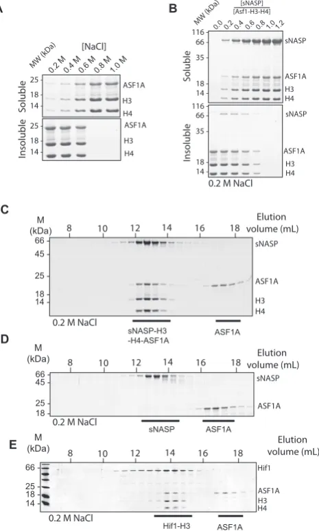
residues within the H3 peptide-binding pocket formed by the TPR repeat motifs. We hypothesized that a retained in-teraction in the presence of a mutation would be indicative of a secondary mode of binding. As the sub-components of the sNASP–H3–H4–ASF1A complex are prone to aggre-gationin vitro(Figure2A) we initially employed the Fluo-rescent 2-Hybrid (F2H) assay to probe the complex in live cells. In the F2H assay a protein–protein interaction can be observed by assessing the ability of a bait protein, tethered to an integrated LacO array, to recruit a soluble prey pro-tein fused to a fluorescent propro-tein (Figure3A) (35,36). Us-ing this assay we found that an mCherry-LacI-sNASP bait construct efficiently recruited a soluble mEGFP-ASF1A fu-sion construct to the LacO array, but that mEGFP-ASF1A was not recruited to the empty mCherry-LacI fusion
(Fig-ure3A–C). As sNASP and ASF1A do not interact directly
in vitro (Figure2D), this interaction is most likely medi-ated through endogenous H3–H4 present in the cell (as
il-lustrated in Figure3A). Using the ASF1A V94R mutation
to disrupt histone binding, we abolished the recruitment of ASF1A V94R to the sNASP-tethered array, whereas muta-tion of the sNASP TPR–H3 peptide binding interface had
little effect on recruitment (Figure3B and C), suggesting
that a secondary mode of binding exists between sNASP and the H3–H4 dimer. These results are in agreement with a previous study in which the V94R mutation of ASF1A failed to pull down NASP from whole cell extracts (18). To further test the idea of a secondary mode of interaction be-tween sNASP and histones, and to exclude possible effects from extraneous cellular components, we reconstituted the
sNASP–H3–H4–ASF1A complexin vitrousing the sNASP
EYL>ASS mutant and found that it forms a complex with
H3–H4–ASF1A similar to wild-type sNASP (Figure3D).
Taken together, these findings suggest that it is the sNASP component of the sNASP–H3–H4–ASF1A complex which contains a secondary, as yet uncharacterized, mode of his-tone interaction, and that, unlike the TPR–H3 peptide
in-teraction (Figure 1E and F), this mode of interaction is
compatible with co-binding of ASF1A.
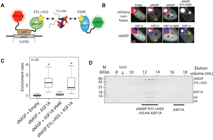
Generation of monobodies specific to sNASP
As a means to investigate the secondary mode of sNASP’s interaction with H3–H4 further, we attempted to isolate
monobody binders to sNASP (Figure 4A–C).
Monobod-ies are synthetic, monoclonal binding proteins constructed with the fibronectin type III scaffold that can be isolated against specific antigens using various display technolo-gies (37,50,51). They function as exquisite molecular probes in studying protein–protein interactions (40,41,52,53). In designing the sNASP antigen for monobody generation, we removed the unstructured C-terminal region of sNASP (residues 331–449), but retained the central acidic domain that interrupts its TPR2 motif so as not to overtly
con-strain the folded core of the TPR domain (Figure4A and
B), because we were interested in obtaining monobodies directed to the structured regions of sNASP. The result-ing sNASP N-330 construct retained its interaction with the H3–H4–ASF1A complex (Supplementary Figure S2A), and was used to screen a large monobody library to isolate
sNASP-specific binders (Figure4C).
We identified two sNASP-specific monobodies termed Mb(sNASP 1) and mbsNASP 13 (for brevity referred to as mb1 and mb13; see Supplementary Figure S2B for sequence information) that interacted with sNASP with nanomo-lar affinity (Figure4D), as measured by yeast display (37). Binding of mb1 and mb13 to sNASP was further tested us-ing full-length sNASP and monobodies expressed and puri-fied from bacteria. Upon recombinant expression, mb1 had limited solubility on its own, and so was expressed as a mal-tose binding protein (MBP) fusion, which also aided in re-solving the two monobodies during SDS-PAGE. As mb1 and mb13 were isolated separately against a truncated form of sNASP, we first addressed whether the two monobod-ies interacted with full-length sNASP, and whether their binding was mutually exclusive (representing overlapping binding sites) or not (representing non-overlapping binding sites). Mixing equimolar concentrations of sNASP, MBP-mb1 and MBP-mb13 we observed that all three proteins eluted in a single peak during gel filtration chromatography
(Fig-ure 4E, peak 1), confirming their interaction with
full-length sNASP, and demonstrating that they bind to non-overlapping surfaces of sNASP. Next, we wanted to know if we could further define the interaction sites of the mono-bodies on the surface of sNASP. Previously, we demon-strated that the central acidic domain of sNASP could be removed, leaving a contiguous TPR (cTPR) construct that retained H3-peptide binding ability (25). To test whether the acidic domain was required for either mb1 or mb13 binding, we repeated the binding experiment using the cTPR mutant. Interestingly, whilst mb13 binding was not affected,
co-eluting with sNASP (Figure4F, peak 1), MBP-mb1 eluted
on its own in a separate fraction (Figure4F, peak 2). This suggests that the acidic domain of sNASP comprises at least part of the binding epitope of mb1, whereas mb13 binds to another site on sNASP’s TPR domain. Therefore, these monobodies represent unique probes for investigating the involvement of different surfaces of sNASP in histone recognition.
Surface mapping of sNASP reveal two additional histone in-teraction sites required for sNASP–H3–H4–ASF1A complex formation
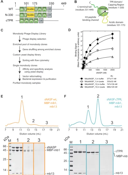
As monobodies bind to discrete surface epitopes on their target protein, we wanted to address whether the interac-tion sites of mb1 and mb13 overlapped with any of the hi-stone binding interfaces of sNASP. We first tested compat-ibility of monobody binding with the H3 C-terminal
pep-tide (Figure5A). Whilst mb13 remained largely associated
to sNASP in the presence of MBP–H3 (116–135), a sig-nificant proportion of MBP-mb1 was displaced, eluting in
a separate peak (Figure 5A, peak 2). This suggests that
D A
sNAS P + ASF1A
sNASP + ASF1A
V94R
sNASP + Empt
y
n=20
Enrichment ratio
C
sNASP EY L>ASS +
ASF1A
sNASP ASF1
H3-H4
LacO LacI
mCherry
mEGFP
(x256)
B
66 45 25 18
-14 - H3
sNASP EYL>ASS
ASF1A
H4
8 10 12 14 16 18
M
(kDa) volume (mL)Elution
sNASP EYL>ASS -H3-H4-ASF1A 4
3
2
1
ASF1A EYL>ASS V94R
[image:8.612.130.494.70.309.2]* * Void
Figure 3. The effect of sNASP and ASF1A mutants on sNASP–H3–H4–ASF1A complex formation. (A) Diagrammatic representation of the F2H
exper-iment. sNASP is tethered to a LacO array through an mCherry-LacI fusion, whilst ASF1A is expressed as a soluble mEGFP fusion. As the chaperones do not interact directly, interaction between the two chaperones is likely mediated through endogenous cellular H3–H4. Yellow stars represent mutations that disrupt the known histone binding surfaces of the two chaperones. (B) mEGFP-ASF1A does not recruit to the empty mCherry-LacI construct, but does recruit to the sNASP fusion. Disruption of the H3 binding interface of ASF1A by the V94R mutation abrogates recruitment of ASF1A, however, disruption of the sNASP TPR-H3 peptide interaction through the EYL>ASS triple mutation has little effect of ASF1A recruitment. (C) Quantification of images shown in (B). Asterisks represent aPvalue of<0.001 as determined by the Wilcoxon rank sum test. (D) Reconstitution of the sNASP EYL> ASS-H3–H4–ASF1Ain vitrodemonstrates that disruption of the TPR–H3 peptide interaction has little effect on the sNASP’s ability to form a complex with H3–H4–ASF1A.
domain/capping region (Figure4F), but outside of the cen-tral H3 peptide-binding channel. We next tested whether binding of mb1 and mb13 is compatible with full-length H3 interaction (Figure5B). Surprisingly, although the epi-tope of mb13 was outside of the H3 peptide-binding region, mb13 was displaced from sNASP upon interaction with H3. Mb1 was also displaced as expected, with the majority of both monobodies eluting in their own fractions (peaks 2 and 3), whilst sNASP and H3 eluted as a single
com-plex (Peak 3) (Figure 5B). Finally, we addressed whether
mb1 and mb13 remain associated to sNASP whilst it is in
complex with H3–H4 and ASF1A (Figure5C). Similar to
full-length H3, mb1 and mb13 were displaced upon bind-ing of the H3–H4–ASF1A complex, elutbind-ing in their indi-vidual fractions (peaks 2 and 3) when analyzed by gel fil-tration chromatography, whereas sNASP eluted as a
com-plex with H3–H4–ASF1A (Peak 1) (Figure 5C).
Consid-ering the mapping of the monobody binding sites to two
discrete epitopes on the surface of sNASP (Figure4E and
F), these findings suggest that sNASP makes extensive in-teractions with both full-length H3 on its own, and with an H3–H4 dimer in the context of the H3–H4–ASF1A complex. This secondary mode of interaction involves at least two additional interactions sites on sNASP: the acidic domain that interrupts the two helices of the TPR2 mo-tif, and the surface of the TPR domain or capping region outside of the central peptide-binding cavity. In summary, and in support of biochemical analysis of the sNASP–H3– H4–ASF1A complex, surface mapping of sNASP using
our monobodies mb1 and mb13 has revealed that sNASP makes an extensive interaction interface with both full-length H3 and with the H3–H4–ASF1A complex, and that
the outer surface of TPR domain/capping region and the
central acidic domain constitute at least part of this inter-face (for a summary of the monobody interactions see Fig-ure5D).
The acidic domain of sNASP is crucial for sNASP–H3–H4– ASF1A complex formation
Next, we wanted to independently confirm the importance of the secondary histone binding sites of sNASP identi-fied by monobody surface mapping. Attempts to gain high-resolution structural information on the mb1 and mb13-sNASP complexes through co-crystalization have thus far not succeeded, making the exact location of monobody binding difficult to determine. However, as the mb1 binding site can be disrupted by removing the acidic domain to form
a contiguous TPR domain (sNASP cTPR) (Figure4F) (25),
A
D
C
1
2
3
sNASP wt, MBP-mb1, mb13
sNASP cTPR, MBP-mb1, mb13
1
2
3
1
2
3
- mb13 - MBP-mb1 - sNASP
- cTPR
- mb13 - MBP-mb1
14 25 45 66
-kDaM
M
B
449
330
1
101
175
N-330 TPR1 Acidic TPR3 TPR4 CR
WT TPR1 Acidic TPR3 TPR4 CR
C
sNASP
H3 peptide binding channel
Acidic domain (residues 101-175) C-terminal tail
(residues 331-449)
TPR domain/ Capping Region (residues 1-330)
7 9 11 13 15 17 19 21 7 9 11 13 15 17 19 21
Elution volume (mL) Elution volume (mL)
1
2
3
cTPR TPR
1
TPR 3
TPR 4 CR TPR
2
F
E
14 25 45 66
-kDa
Monobody Phage-Display LibraryPhage display selection
Enriched pool of monobody clones
Custom yeast display library
Sorting with flow cytometry
Vector reformatting
Bacterial expression & purification Affinity and specificity analysis using yeast display Single monobody clones
Purified monobody samples
Gene shuffling among enriched clones
0
0 500 1000 1500 2000
Binding Signal
(Mean Fluorescence in arbitrary units)
sNASP (nM)
Mb(sNASP_1) in lysate 333±32 nM
Mb(sNASP_13) in lysate 686±129 nM Mb(sNASP_13) in buffer 747±139 nM 1
2 3 4 5 6 7 8
[image:9.612.91.501.71.631.2]Mb(sNASP_1) in buffer 377±17 nM KD
Figure 4. Isolation and characterization of two sNASP specific monobodies. (A) Domain diagrams of wild-type (WT) sNASP and truncation mutants
- mb13 - MBP-mb1
- MBP-H3 116-135 - sNASP
1
2
3
sNASP, MBP-H3 116-135, MBP-mb1, mb13
M
- mb13 - MBP-mb1 - sNASP
- H3 - H4 - ASF1A sNASP+ H3-H4+ ASF1A+ MBP-mb1+ mb13
1
2
3
M
14 25 45 66 -kDa
7 9 11 13 15 17 19 21
1
2
3
7 9 11 13 15 17 19 21
1 2
3
- mb13 - MBP-mb1 - sNASP
- H3
1
2
3
sNASP, H3, MBP-mb1, mb13
7 9 11 13 15 17 19 21
1 2
3
A
D C
B
13 1
MBP C
sNASP
ASF1
H3H4
13 1
MBP C
sNASP MBP 13
1
MBP
C
mb13 mb1
MBP
C
sNASP
H3
MBP
H3
ASF1
H3H4
MBP-H3 116-135
H3 peptide binding channel Acidic
domain C-terminal tail TPR domain/
Capping Region sNASP-mb1-mb13
complex
sNASP
14 25 45 66 -kDa
[image:10.612.109.513.65.525.2]14 25 45 66 -kDa
Figure 5.Probing the secondary modes of sNASP interaction using monobodies mb1 and mb13. (A) Gel filtration profile of equimolar amounts of sNASP,
MBP–H3 116–135, mb1 and mb13. Whilst mb13 remains predominantly associated with the sNASP-MBP–H3 116–135 complex (peak 1), MBP-mb1 is partially displaced, suggesting that it undergoes binding site competition with the H3 peptide (free MBP–H3 116–135 and MBP-MBP-mb1 have partially overlapping elution profile represented by peak 2). (B) Gel filtration profile of sNASP, MBP-mb1, H3 full-length and mb13, showing that MBP-mb1 and mb13 are largely excluded from the sNASP–H3 complex (peak 1), eluting in their separate fractions (peaks 2 and 3, respectively). (C) Elution profile of sNASP, MBP-mb1, mb13, H3–H4 and ASF1A, showing that MBP-mb1 (peak 2) and mb13 (peak 3) are largely displaced from the sNASP–H3–H4– ASF1A complex (peak 1). (D) Diagrammatic representation of the complexes formed between sNASP, its various histone substrates and the monobodies mb1 and mb13. Whilst mb13 retains its binding to sNASP in the presence of the H3 116–135 peptide, it is displaced upon either H3 full-length or H3–H4– ASF1A complex binding. MBP-mb1 is partially displaced by the H3 116–135 peptide, suggesting binding site competition, and is fully displaced upon H3 full-length or H3–H4–ASF1A complex binding.
mixed the cTPR mutant with H3–H4 and ASF1A at high salt concentration (0.6 M sodium chloride) to prevent
pre-cipitation of the components (Figure2A and B) and
sepa-rated out the formed complexes by gel filtration
chromatog-raphy (Figure 6A–C: ASF1A was kept at a molar excess
over the other components to judge the stoichiometry of the complex and to serve as an internal control in marking the elution point of free ASF1A). Interestingly, the sNASP
cTPR mutant failed to co-migrate with the H3–H4–ASF1A complex, whereas the H3–H4–ASF1A complex eluted in
the bed volume of the column (Figure6B and C). As the
H3–H4–ASF1A complex is aggregation-prone under phys-iologically relevant salt concentrations used in the
exper-iment (Figure 2A), elution in the bed volume most likely
A
B
449 330
1 101 175
WT TPR1 Acidic TPR3 TPR4 CR
cTPR TPR
1 TPR
3 TPR
4 CR TPR
2
C
H3 sNASP cTPR
ASF1A H4
sNASP cTPR 66
45
25 18 14
-MW (kDa)8 10 12 14 16 18 20 22 Elution volume (mL)
sNASP cTPR ASF1A ASF1A H3-H4
Bed volume
6 8 10 12 14 16 18 20 22 Elution volume (mL)
24
5 45 95
Absorbance (280 nm) 25
30 35 40 Conduc
tivit
y
(mS/cm)
Void volume
Bed volume
[image:11.612.48.286.66.326.2]sNASP cTPR
Figure 6. The central acidic domain of sNASP is necessary for H3–H4–
ASF1A binding. (A) Domain diagrams of the contiguous (cTPR) trun-cation mutant compared to wild-type sNASP. (B) Elution profile of the sNASP cTPR mutant and H3–H4–ASF1A separated out by gel filtration chromatography. The conductivity spike representing the higher salt from sample preparation eluting in the bed volume of the column is indicated. (C) Fractions from the elution separated by SDS-PAGE reveal that the cTPR mutant, although retaining its ability to interact with the H3 116– 135 peptide, is unable to bind to the H3–H4–ASF1A complex.
be noted that the same conditions were used for success-fully separating the entire sNASP–H3–H4–ASF1A com-plex containing the sNASP N-330 truncation mutant (Sup-plementary Figure S2A), showing that under these condi-tion of high salt loading the sNASP–H3–H4–ASF1A com-plex is still able to form normally. This finding suggests that the acidic domain, although dispensable for binding the H3 C-terminal peptide of H3 (25), is necessary for the sec-ondary mode of interaction between sNASP and the H3– H4 dimer whilst it is in complex with ASF1A, support-ing our view that sNASP utilizes secondary histone-bindsupport-ing sites.
sNASP and ASF1 cooperate to fold an H3–H4 dimer under native conditionsin vitro
As sNASP forms a stable complex with H3 in the absence
of H4 (Figure1F), sNASP may work upstream of ASF1A
in the histone chaperoning pathway, scaffolding free H3 and aiding in its folding with H4 to form an sNASP–H3– H4–ASF1A complex. If this were the case, we wondered whether the sNASP–H3–H4–ASF1A complex could be re-constituted using histone monomers as substrates, bypass-ing the requirement for usbypass-ing pre-folded H3–H4 dimers
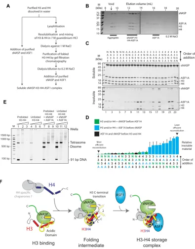
(Figure7A). To test this, we mixed sNASP with monomeric
H3 and H4 (dissolved in water) and ASF1A, and analyzed the resulting mixture by gel filtration chromatography
(Fig-ure 7B). Remarkably, we found that sNASP, H3, H4 and
ASF1A all eluted in a stable complex, comparable to that seen when using pre-folded H3–H4 dimers to reconstitute the complex (Figure2C), in addition to a small quantity of aggregates eluting in the void volume of the column (Figure
7B).
Whilst reconstituting the sNASP–H3–H4–ASF1A from unfolded histones, we noticed that the order in which the histones and chaperones were combined, greatly affected the efficiency of reconstitution, with some orders of addi-tion producing marked precipitaaddi-tion over others. We rea-soned that some interactions may need to occur before oth-ers in order to drive complex assembly. We investigated this possibility by systematically combining the four com-ponents (sNASP, H3, H4 and ASF1A) in all possible or-ders of addition, and measured the reconstitution efficiency by quantifying the precipitate formed (Figure7C). Interest-ingly, when we ranked the orders of addition according to total relative precipitate, we see that in the top quartile of most efficient reconstitutions, sNASP is always added be-fore ASF1A. Conversely, we see that in the bottom quartile (the least efficient reconstitutions), ASF1A is always added before sNASP. This reveals a general rule in which efficient
reconstitution of the tetrameric complexin vitro requires
sNASP to be added before ASF1A. These results support the idea that sNASP functions upstream of ASF1A in the histone chaperoning pathway.
To test if the H3 and H4 are folded correctly with the reconstituted sNASP–H3–H4–ASF1A complex, we carried out a tetrasome reconstitution assay (42,45), with the view that a correctly folded H3–H4 dimer should behave iden-tically to a prefolded H3–H4 dimer in both its efficiency of tetrasome reconstitution and its migration pattern dur-ing native PAGE. In this assay, the ability of a chaperone to mitigate aggregation and promote correct folding of a tetrasome particle is assessed under increasing histone to DNA ratios. At lower ratios, disomes are formed (a single H3–H4 dimer associated with DNA), leading to tetrasome formation (two H3–H4 dimers associated with DNA) and then aggregation when histones are in a large excess, as has been observed previously (42,44). Similarly, we see that both sNASP and ASF1A can individually aid in depositing H3– H4 onto DNA, with sNASP preferentially forming tetra-somes and ASF1A preferentially forming ditetra-somes, whereas traditional salt dialysis results in tetrasome formation (Sup-plementary Figure S4). Furthermore, when sNASP and ASF1A are combined, both disomes and tetrasomes are formed with disomes predominating (Supplementary Fig-ure S4).
Direct addition of pfolded H3–H4 dimers to DNA re-sults in poor assembly of tetrasomes, with increasing
hi-stone to DNA ratios resulting in aggregation (Figure7E,
lanes 1–3). The addition of unfolded H3 and H4 results in even poorer tetrasome reconstitution, as would be expected
(Figure7E, lanes 4–6). However, when H3–H4 dimers are
first assembled in an sNASP–H3–H4–ASF1A complex be-fore adding DNA, reconstitution is greatly enhanced
(Fig-ure 7E, lanes 7–9), as may be expected from the
ei-dissolved in water
sNASP and ASF1
Soluble sNASP-H3-H4-ASF1 complex Lyophilisation
Resolubilisation and mixing of H3 & H4 in 7 M guanidinium-HCl
Dialysis against 1 M NaCl
chromatography
Dialysis/dilution to 0.2 M NaCl
sNASP and ASF1
1000 bp 500 bp
100 bp
-- Disome - Tetrasome
- 91 bp DNA
1500 bp
-- Wells Prefolded H3-H4 Unfolded H3-H4 Unfolded H3-H4 + sNASP + ASF1A Prefolded H3-H4 + sNASP + ASF1A
M 1 2 3 4 5 6 7 8 9 10 11 12
E A B C D Soluble Insoluble 66 45 35 25 18 14 66 45 35 25 18 14 -- sNASP - ASF1A - H4 - H3 - sNASP - ASF1A - H4 - H3
1 3 5 7 9 11 13 15 17 19 21 23
N A 3 4 N A 4 3 N 4 3 A N 4 A 3 N 3 4 A N 3 A 4 A N 3 4 A N 4 3 A 3 N 4 A 3 4 N A 4 N 3 A 4 3 N 3 N A 4 3 N 4 A 3 A N 4 3 A 4 N 3 4 N A 3 4 A N 4 N A 3 4 N 3 A 4 A N 3 4 A 3 N 4 3 N A 4 3 A N Order of addition M (kDa) H3 ASF1A sNASP 10
-8 10 12 14 16 18 20
0.2 M NaCl 15 20 25 37 50 75 -sNASP-H3 -H4-ASF1A ASF1A Aggregates H4 M (kDa)
Elution volume (mL) Void C C sNASP ASF1 ASF1 Acidic Domain sNASP
H3
H3H4sNASP
H3H4
C
Folding intermediate
H3 binding H3-H4 storagecomplex
[image:12.612.115.511.65.571.2]chaperones ? H3 C-terminal transition C C N N N CC N C N N CC F
H4
Order of addition Relative insoluble material Least reconstitution Most reconstitution N A 3 4 N A 4 3 N 3 A 4 A N 3 4 A N 4 3 A 3 4 N 3 N A 4 3 4 N A 4 3 A N N 4 3 A N 4 A 3 4 N A 3 4 N 3 A N 3 4 A 4 3 N A 3 N 4 A 3 A N 4 3 4 A N 4 A 3 N 4 A N 3 A 4 N 3 A 4 3 N 3 A 4 N A 3 N 4 H3 and/or H4 + sNASP before ASF1A H3 and/or H4 + ASF1A before sNASP ASF1A and sNASP before H3 and H4Figure 7. sNASP and ASF1A are capable of folding an H3–H4 dimerin vitro. (A) Flow chart showing the two different strategies for reconstituting the
ther the efficiency of tetrasome reconstitution or the migra-tion pattern of histone–DNA complexes when comparing sNASP–H3–H4–ASF1A complex formed from pre-folded
H3–H4 dimers (Figure7E, lanes 7–9) and sNASP–H3–H4|
ASF1A complex formed from monomeric H3 and H4
(Fig-ure7E, lanes 10–12). This suggests that sNASP and ASF1A
are fully capable of efficiently folding an H3–H4 dimer from
unfolded monomeric substrates in vitroand supports the
notion that sNASP functions upstream of ASF1A in the histone chaperoning pathway.
DISCUSSION
sNASP represents a family of TPR motif containing chap-erones that specifically interact with histones H3 and H4. In vivosNASP has been isolated in complex with other
co-chaperones, including ASF1A/B, RbAp46 and the histone
acetyl-transferase HAT1, which represents the major solu-ble source of H3–H4 within the cell. Previously, we iden-tified that the H3 binding site of sNASP overlaps signifi-cantly with that of ASF1A (25), suggesting that (1) binding site competition may be important in H3–H4 maturation,
and (2), as ASF1A and sNASP exist in complex with each
other, either sNASP or ASF1A contain secondary interac-tion sites with their histone substrate.
In this current study, we have presented a detailed bio-chemical investigation into how sNASP and ASF1A co-chaperone histones H3 and H4, and present a model in which sNASP and ASF1A cooperate through both compet-itive and compatible interactions to fold and retain an H3–
H4 dimer in an aggregation-resistant state (Figure7F). In
this model, formation of a sNASP–H3 complex is upstream of the sNASP–H3–H4–ASF1A complex. Importantly, in a cellular context this is most likely also associated with the HAT1-complex and other accessory factors (10,11,19,20). Interaction between sNASP and H3 in the sNASP–H3 com-plex is predominantly mediated by the high affinity
TPR-peptide interaction (25). However, displacement of both
mb1 and mb13 monobody probes also suggested that sec-ondary modes of interaction between sNASP and H3 are present, involving both the acidic domain and the outer
sur-face of the TPR domain/capping region (Figure 6B). In
contrast to sNASP, ASF1A mediates its interaction pre-dominantly with the folded surface of the H3–H4 dimer (30,32), agreeing with our observation that ASF1A is un-able to compete with sNASP for binding of H3 in the
ab-sence of H4 (Figure1C–G). Upon H3 folding with H4,
se-questration of residues important in mediating the TPR–H3 peptide interaction (Ala127, Arg131) (Figure1A) within the histone fold may act to reduce the grip of sNASP on this re-gion and result in a transition from sNASP being bound at the C-terminus of H3 to ASF1A being bound as the H3– H4 dimer forms. sNASP, however, is still retained within the ASF1–H3–H4 complex through its secondary interac-tion sites involving the central acidic domain and the outer
surface of the TPR/capping region (Figures5and6). Due
to their ability to solubilise the otherwise aggregation prone ASF1–H3–H4 complex, these secondary modes of interac-tion between sNASP and H3–H4 most likely aid in pre-venting non-specific interactions within a cellular context, ensuring that a soluble pool of histones is maintained at
all times. In this regard, the molecular model we propose is consistent with findings showing that NASP contributes to the fine-tuning of a soluble reservoir of H3–H4 through
inhibition of chaperone-mediated autophagy (CMA) (21).
Furthermore, as sNASP and ASF1A are sufficient for
fold-ing of an H3–H4 dimer in vitro, the isolation of protein
folding chaperones associated with histone H3 (17) may re-late to a role of the these proteins in CMA/quality control, rather than in generating an H3–H4 dimer.
Most biochemical analysis to date has involved individ-ual analysis of histone chaperone function in isolation, this being especially true for NASP (15,16,54,55). However, H3 and H4 are found in multi-chaperone complexes in the cell, and so the individual function of each chaperone is likely only to play an important but small role in the histone de-position process. We have shown that sNASP has synergis-tic functions with ASF1 towards H3 and H3–H4. Interest-ingly, H4 has also been shown to have specific chaperones, binding RbAp46 and HAT1, which form a stable dimeric HAT1 complex, important in acetylation of lysine 5 and 12 of H4 (26,56,57). A recent crystal structure of the Saccha-romyces cerevisiae homolog of the RbAp46–HAT1 dimer bound to H4 and H3 peptides revealed that nearly half of the H4 molecule is sequestered within the binding pockets of the HAT1 complex (27), including the majority of the
N-terminal tail and the␣1 helix of the histone core domain.
Interestingly, the␣1 helix of H4 and the␣3 helix of H3 re-side in close proximity to each other on the same re-side of the histone fold dimer. Binding and releasing of these regions within the core histone fold domain may therefore aid in guiding H3 and H4 down a productive folding pathway.
The role that sNASP plays in the folding, maturation and storage of histones is complex and most likely involves a number of other chaperones in addition to ASF1. In or-der to further unor-derstand the process, high-resolution struc-tures of the stable components of the pathway, namely the sNASP–H3 dimer and the sNASP–H3–H4–ASF1A tetramer, need to be solved and the dynamic intermediates probed through complementary, biophysical means. The role of additional factors, such as the HAT1 holo-enyme, and how they function in concert with sNASP and ASF1 also has to be addressed. Importantly, novel approaches will need to be developed in order to observe the rapid process
of ‘histone hand-off’in vivo and to validate such
mecha-nisms, as we and others have proposed (17,21). In this re-port, we have biochemically characterized the sNASP–H3 and sNASP–H3–H4–ASF1A complexes and demonstrated that multiple interaction interfaces exist between sNASP and histones. We have shown how these contend and coop-erate with the chaperone ASF1A to form a folded H3–H4 dimer, establishing a mechanistic platform for the further investigation of these dynamic histone chaperoning pro-cesses.
SUPPLEMENTARY DATA
Supplementary Data are available at NAR Online.
ACKNOWLEDGEMENTS
Koide groups for helpful discussions and Gyula Timinszky for technical advice and discussions.
Author contributions: A.B. conceived of the project in dis-cussion with A.G.L. and carried out the majority of the experiments. A.K., J.S.G. and S.K. isolated and carried out initial characterization of the monobodies. D.Z. con-tributed to biochemical analysis of monobody interactions. M.E.C. contributed to biochemical analysis of sNASP mu-tants and their interaction with the ASF1–H3–H4 complex. A.B. and A.G.L. wrote the manuscript with contributions from S.K.
FUNDING
EMBO Long-term Fellowship [ALTF 887-2011 to A.B.]; Marie Curie Intra-European Fellowship for career devel-opment [331726 FP7-PEOPLE-2012-IEF]; Wellcome Insti-tutional Strategy Support Fund [WT105627MA]; Research in the Ladurner laboratory is supported by the Bavar-ian Research Network for Molecular Biosystems (BioSys-Net); Deutsche Forschungsgemeinschaft; EU FP7 Network of Excellence ‘EpiGeneSys’; National Institutes of Health [R01-DA036887]; University of Chicago Comprehensive Cancer Center, NIH grant [P30-CA014599]. Funding for open access charge: Deutsche Forschungsgemeinschaft. Conflict of interest statement.A.K. and S.K. are listed as in-ventors on a patent application filed by The University of Chicago that covers a design of monobody libraries (United
States Patent application 13/813,409). The other authors
declare no competing financial interests.
REFERENCES
1. Gurard-Levin,Z.A., Quivy,J.P. and Almouzni,G. (2014) Histone chaperones: assisting histone traffic and nucleosome dynamics.Annu. Rev. Biochem.,83, 487–517.
2. Liu,W.H. and Churchill,M.E. (2012) Histone transfer among chaperones.Biochem. Soc. Trans.,40, 357–363.
3. Das,C., Tyler,J.K. and Churchill,M.E.A. (2010) The histone shuffle: histone chaperones in an energetic dance.Trends Biochem. Sci.,35, 476–489.
4. Burgess,R.J. and Zhang,Z. (2013) Histone chaperones in nucleosome assembly and human disease.Nat. Struct. Mol. Biol.,20, 14–22. 5. Daniel Ricketts,M., Frederick,B., Hoff,H., Tang,Y., Schultz,D.C.,
Singh Rai,T., Grazia Vizioli,M., Adams,P.D. and Marmorstein,R. (2015) Ubinuclein-1 confers histone H3.3-specific-binding by the HIRA histone chaperone complex.Nat. Commun.,6, 7711. 6. Elsasser,S.J., Huang,H., Lewis,P.W., Chin,J.W., Allis,C.D. and
Patel,D.J. (2012) DAXX envelops an H3.3-H4 dimer for H3.3-specific recognition.Nature,491, 560–565.
7. Goldberg,A.D., Banaszynski,L.A., Noh,K.M., Lewis,P.W., Elsaesser,S.J., Stadler,S., Dewell,S., Law,M., Guo,X., Li,X.et al.
(2010) Distinct factors control histone variant H3.3 localization at specific genomic regions.Cell,140, 678–691.
8. Drane,P., Ouararhni,K., Depaux,A., Shuaib,M. and Hamiche,A. (2010) The death-associated protein DAXX is a novel histone chaperone involved in the replication-independent deposition of H3.3.Genes Dev.,24, 1253–1265.
9. Foltman,M., Evrin,C., De Piccoli,G., Jones,R.C., Edmondson,R.D., Katou,Y., Nakato,R., Shirahige,K. and Labib,K. (2013) Eukaryotic replisome components cooperate to process histones during chromosome replication.Cell Rep.,3, 892–904.
10. Ask,K., Jasencakova,Z., Menard,P., Feng,Y., Almouzni,G. and Groth,A. (2012) Codanin-1, mutated in the anaemic disease CDAI, regulates Asf1 function in S-phase histone supply.EMBO J.,31, 2013–2023.
11. Groth,A., Corpet,A., Cook,A.J., Roche,D., Bartek,J., Lukas,J. and Almouzni,G. (2007) Regulation of replication fork progression through histone supply and demand.Science,318, 1928–1931. 12. Richet,N., Liu,D., Legrand,P., Velours,C., Corpet,A., Gaubert,A.,
Bakail,M., Moal-Raisin,G., Guerois,R., Compper,C.et al.(2015) Structural insight into how the human helicase subunit MCM2 may act as a histone chaperone together with ASF1 at the replication fork.
Nucleic Acids Res.,43, 1905–1917.
13. Huang,H., Stromme,C.B., Saredi,G., Hodl,M., Strandsby,A., Gonzalez-Aguilera,C., Chen,S., Groth,A. and Patel,D.J. (2015) A unique binding mode enables MCM2 to chaperone histones H3–H4 at replication forks.Nat. Struct. Mol. Biol.,22, 618–626.
14. Nabeel-Shah,S., Ashraf,K., Pearlman,R.E. and Fillingham,J. (2014) Molecular evolution of NASP and conserved histone H3/H4 transport pathway.BMC Evol. Biol.,14, 139.
15. Wang,H., Ge,Z., Walsh,S.T. and Parthun,M.R. (2012) The human histone chaperone sNASP interacts with linker and core histones through distinct mechanisms.Nucleic Acids Res.,40, 660–669. 16. Wang,H., Walsh,S.T. and Parthun,M.R. (2008) Expanded binding
specificity of the human histone chaperone NASP.Nucleic Acids Res.,
36, 5763–5772.
17. Campos,E.I., Fillingham,J., Li,G., Zheng,H., Voigt,P., Kuo,W.H., Seepany,H., Gao,Z., Day,L.A., Greenblatt,J.F.et al.(2010) The program for processing newly synthesized histones H3.1 and H4.Nat. Struct. Mol. Biol.,17, 1343–1351.
18. Jasencakova,Z., Scharf,A.N., Ask,K., Corpet,A., Imhof,A., Almouzni,G. and Groth,A. (2010) Replication stress interferes with histone recycling and predeposition marking of new histones.Mol. Cell,37, 736–743.
19. Groth,A., Ray-Gallet,D., Quivy,J.P., Lukas,J., Bartek,J. and Almouzni,G. (2005) Human Asf1 regulates the flow of S phase histones during replicational stress.Mol. Cell,17, 301–311. 20. Tagami,H., Ray-Gallet,D., Almouzni,G. and Nakatani,Y. (2004)
Histone H3.1 and H3.3 complexes mediate nucleosome assembly pathways dependent or independent of DNA synthesis.Cell,116, 51–61.
21. Cook,A.J., Gurard-Levin,Z.A., Vassias,I. and Almouzni,G. (2011) A specific function for the histone chaperone NASP to fine-tune a reservoir of soluble H3–H4 in the histone supply chain.Mol. Cell,44, 918–927.
22. Campos,E.I., Smits,A.H., Kang,Y.H., Landry,S., Escobar,T.M., Nayak,S., Ueberheide,B.M., Durocher,D., Vermeulen,M., Hurwitz,J.
et al.(2015) Analysis of the histone H3.1 interactome: a suitable chaperone for the right event.Mol. Cell,60, 697–709.
23. Fillingham,J., Recht,J., Silva,A.C., Suter,B., Emili,A., Stagljar,I., Krogan,N.J., Allis,C.D., Keogh,M.C. and Greenblatt,J.F. (2008) Chaperone control of the activity and specificity of the histone H3 acetyltransferase Rtt109.Mol. Cell. Biol.,28, 4342–4353. 24. Garg,J., Lambert,J.P., Karsou,A., Marquez,S., Nabeel-Shah,S.,
Bertucci,V., Retnasothie,D.V., Radovani,E., Pawson,T., Gingras,A.C.
et al.(2013) Conserved Asf1-importin beta physical interaction in growth and sexual development in the ciliate Tetrahymena thermophila.J. Proteomics,94, 311–326.
25. Bowman,A., Lercher,L., Singh,H.R., Zinne,D., Timinszky,G., Carlomagno,T. and Ladurner,A.G. (2016) The histone chaperone sNASP binds a conserved peptide motif within the globular core of histone H3 through its TPR repeats.Nucleic Acids Res.,44, 3105–3117.
26. Murzina,N.V., Pei,X.Y., Zhang,W., Sparkes,M., Vicente-Garcia,J., Pratap,J.V., McLaughlin,S.H., Ben-Shahar,T.R., Verreault,A., Luisi,B.F.et al.(2008) Structural basis for the recognition of histone H4 by the histone-chaperone RbAp46.Structure,16, 1077–1085. 27. Li,Y., Zhang,L., Liu,T., Chai,C., Fang,Q., Wu,H., Agudelo
Garcia,P.A., Han,Z., Zong,S., Yu,Y.et al.(2014) Hat2p recognizes the histone H3 tail to specify the acetylation of the newly synthesized H3/H4 heterodimer by the Hat1p/Hat2p complex.Genes Dev.,28, 1217–1227.
28. Song,J.J., Garlick,J.D. and Kingston,R.E. (2008) Structural basis of histone H4 recognition by p55.Genes Dev.,22, 1313–1318. 29. Nowak,A.J., Alfieri,C., Stirnimann,C.U., Rybin,V., Baudin,F.,
Ly-Hartig,N., Lindner,D. and Muller,C.W. (2011)
Su(z)12 through partially overlapping binding sites.J. Biol. Chem.,
286, 23388–23396.
30. English,C.M., Adkins,M.W., Carson,J.J., Churchill,M.E.A. and Tyler,J.K. (2006) Structural basis for the histone chaperone activity of Asf1.Cell,127, 495–508.
31. Mousson,F., Lautrette,A., Thuret,J.Y., Agez,M., Courbeyrette,R., Amigues,B., Becker,E., Neumann,J.M., Guerois,R., Mann,C.et al.
(2005) Structural basis for the interaction of Asf1 with histone H3 and its functional implications.Proc. Natl. Acad. Sci. U.S.A.,102, 5975–5980.
32. Natsume,R., Eitoku,M., Akai,Y., Sano,N., Horikoshi,M. and Senda,T. (2007) Structure and function of the histone chaperone CIA/ASF1 complexed with histones H3 and H4.Nature,446, 338–341.
33. Luger,K., Rechsteiner,T.J., Flaus,A.J., Waye,M.M.Y. and
Richmond,T.J. (1997) Characterization of nucleosome core particles containing histone proteins made in bacteria.J. Mol. Biol.,272, 301–311.
34. Czarna,A., Berndt,A., Singh,H.R., Grudziecki,A., Ladurner,A.G., Timinszky,G., Kramer,A. and Wolf,E. (2013) Structures of Drosophila cryptochrome and mouse cryptochrome1 provide insight into circadian function.Cell,153, 1394–1405.
35. Zolghadr,K., Mortusewicz,O., Rothbauer,U., Kleinhans,R., Goehler,H., Wanker,E.E., Cardoso,M.C. and Leonhardt,H. (2008) A fluorescent two-hybrid assay for direct visualization of protein interactions in living cells.Mol. Cell. Proteomics : MCP,7, 2279–2287.
36. Zolghadr,K., Rothbauer,U. and Leonhardt,H. (2012) The fluorescent two-hybrid (F2H) assay for direct analysis of protein–protein interactions in living cells.Methods Mol. Biol.,812, 275–282. 37. Koide,A., Wojcik,J., Gilbreth,R.N., Hoey,R.J. and Koide,S. (2012)
Teaching an old scaffold new tricks: monobodies constructed using alternative surfaces of the FN3 scaffold.J. Mol. Biol.,415, 393–405. 38. Koide,A., Wojcik,J., Gilbreth,R.N., Reichel,A., Piehler,J. and
Koide,S. (2009) Accelerating phage-display library selection by reversible and site-specific biotinylation.Protein Eng. Des. Sel.: PEDS,22, 685–690.
39. Koide,S., Koide,A. and Lipovsek,D. (2012) Target-binding proteins based on the 10th human fibronectin type III domain ((1)(0)Fn3).
Methods Enzymol.,503, 135–156.
40. Stockbridge,R.B., Koide,A., Miller,C. and Koide,S. (2014) Proof of dual-topology architecture of Fluc F- channels with monobody blockers.Nat. Commun.,5, 5120.
41. Tanaka,S., Takahashi,T., Koide,A., Ishihara,S., Koikeda,S. and Koide,S. (2015) Monobody-mediated alteration of enzyme specificity.
Nat. Chem. Biol.,11, 762–764.
42. Scorgie,J.K., Donham,D.C. 3rd and Churchill,M.E. (2012) Analysis of histone chaperone antisilencing function 1 interactions.Methods Enzymol.,512, 223–241.
43. Liu,W.H., Roemer,S.C., Port,A.M. and Churchill,M.E. (2012) CAF-1-induced oligomerization of histones H3/H4 and mutually exclusive interactions with Asf1 guide H3/H4 transitions among histone chaperones and DNA.Nucleic Acids Res.,40, 11229–11239.
44. Donham,D.C. 2nd, Scorgie,J.K. and Churchill,M.E. (2011) The activity of the histone chaperone yeast Asf1 in the assembly and disassembly of histone H3/H4-DNA complexes.Nucleic Acids Res.,
39, 5449–5458.
45. Bowman,A., Ward,R., Wiechens,N., Singh,V., El-Mkami,H., Norman,D.G. and Owen-Hughes,T. (2011) The histone chaperones Nap1 and Vps75 bind histones H3 and H4 in a tetrameric conformation.Mol. Cell,41, 398–408.
46. Thastrom,A., Lowary,P.T., Widlund,H.R., Cao,H., Kubista,M. and Widom,J. (1999) Sequence motifs and free energies of selected natural and non-natural nucleosome positioning DNA sequences.J. Mol. Biol.,288, 213–229.
47. Lowary,P.T. and Widom,J. (1998) New DNA sequence rules for high affinity binding to histone octamer and sequence-directed
nucleosome positioning.J. Mol. Biol.,276, 19–42.
48. English,C.M., Maluf,N.K., Tripet,B., Churchill,M.E.A. and Tyler,J.K. (2005) ASF1 binds to a heterodimer of histones H3 and H4: A two-step mechanism for the assembly of the H3–H4 heterotetramer on DNA.Biochemistry,44, 13673–13682. 49. Haigney,A., Ricketts,M.D. and Marmorstein,R. (2015) Dissecting
the molecular roles of histone chaperones in histone acetylation by type B histone acetyltransferases (HAT-B).J. Biol. Chem.,290, 30648–30657.
50. Richards,J., Miller,M., Abend,J., Koide,A., Koide,S. and Dewhurst,S. (2003) Engineered fibronectin type III domain with a RGDWXE sequence binds with enhanced affinity and specificity to human alphavbeta3 integrin.J. Mol. Biol.,326, 1475–1488.
51. Koide,A., Bailey,C.W., Huang,X. and Koide,S. (1998) The
fibronectin type III domain as a scaffold for novel binding proteins.J. Mol. Biol.,284, 1141–1151.
52. Sha,F., Gencer,E.B., Georgeon,S., Koide,A., Yasui,N., Koide,S. and Hantschel,O. (2013) Dissection of the BCR-ABL signaling network using highly specific monobody inhibitors to the SHP2 SH2 domains.
Proc. Natl. Acad. Sci. U.S.A.,110, 14924–14929.
53. Gilbreth,R.N., Truong,K., Madu,I., Koide,A., Wojcik,J.B., Li,N.S., Piccirilli,J.A., Chen,Y. and Koide,S. (2011) Isoform-specific monobody inhibitors of small ubiquitin-related modifiers engineered using structure-guided library design.Proc. Natl. Acad. Sci. U.S.A.,
108, 7751–7756.
54. Kato,D., Osakabe,A., Tachiwana,H., Tanaka,H. and Kurumizaka,H. (2015) Human tNASP promotes in vitro nucleosome assembly with histone H3.3.Biochemistry,54, 1171–1179.
55. Finn,R.M., Browne,K., Hodgson,K.C. and Ausio,J. (2008) sNASP, a histone H1-specific eukaryotic chaperone dimer that facilitates chromatin assembly.Biophys J.,95, 1314–1325.
56. Verreault,A., Kaufman,P.D., Kobayashi,R. and Stillman,B. (1998) Nucleosomal DNA regulates the core-histone-binding subunit of the human Hat1 acetyltransferase.Curr. Biol.,8, 96–108.