inorganic papers
i140
Roh and Hong Ba5(VO4)3Cl doi:10.1107/S1600536805018854 Acta Cryst.(2005). E61, i140–i142 Acta Crystallographica Section E
Structure Reports Online
ISSN 1600-5368
Apatite-type Ba
5(VO
4)
3Cl
Yun-Ho Roh and Seung-Tae Hong*
LG Chem Research Park, Daejeon 305-380, Korea
Correspondence e-mail: [email protected]
Key indicators
Single-crystal X-ray study
T= 298 K
Mean(V–O) = 0.004 A˚
Rfactor = 0.015
wRfactor = 0.034
Data-to-parameter ratio = 14.2
For details of how these key indicators were automatically derived from the article, see http://journals.iucr.org/e.
#2005 International Union of Crystallography Printed in Great Britain – all rights reserved
The title compound, pentabarium tris(vanadate) chloride, crystallizes in the well known apatite structure type, with space groupP63/mandZ= 2. The crystal structure contains isolated
(VO4) 3
tetrahedra that are bridged by Ba2+ ions. The intermediate Cl anions, situated on positions with 3 symmetry, are octahedrally surrounded by Ba2+cations.
Comment
Single crystals of the title compound were first obtained as an impurity during our attempt to explore new compounds in the Ba–V–Ru–O system, with BaCl22H2O used as an excess flux
material. Once its composition and preliminary crystal struc-ture were identified by elemental and X-ray diffraction analyses, it was possible to prepare single-phase material of Ba5(VO4)3Cl as both powder and single crystals. To our
knowledge, only the cell dimensions and the powder pattern of Ba5(VO4)3Cl have been reported to date (Powder Diffraction
File No. 19-0099; ICDD, 2001), without any further details of the crystal structure. Here, we report the synthesis of the compound and its crystal structure, as determined from single-crystal data.
Ba5(VO4)3Cl crystallizes in the well known apatite structure
type, like the related compound Sr5(PO4)3Br and its Eu 2+
doped analogues, which find application as photoluminescent materials (No¨tzold & Wulff, 2000). The structure consists of VO4
3
tetrahedra bridged by Ba2+ions, and intermediate Cl
[image:1.610.204.458.498.676.2]Received 12 May 2005 Accepted 14 June 2005 Online 6 July 2005
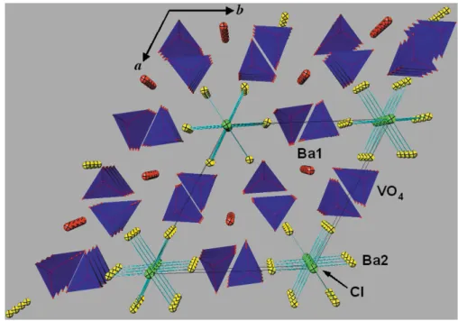
Figure 1
A polyhedral representation of the crystal structure of Ba5(VO4)3Cl,
projected on (001). The unit cell is outlined. The V ions are at the centre of blue VO4tetrahedra. Ba1, Ba2 and Cl ions are drawn as red, yellow
anions. A projection of the crystal structure down thecaxis is shown in Fig. 1.
Two crystallographically distinct Ba2+cations are present in the structure. Atom Ba1 is located on a threefold axis and is coordinated to nine O atoms, as shown in Fig. 2. It is connected to six VO4
3
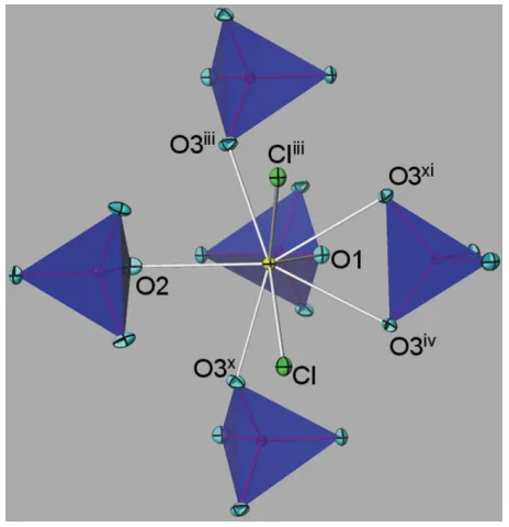
tetrahedra, three of which chelate to the Ba1 cation. Atom Ba2 is coordinated by six O atoms and two Cl atoms. It bridges five VO4
3
tetrahedra, one of which has also chelating character (Fig. 3). The Clanion has site symmetry 3 and is coordinated by six Ba2 atoms. The resultant [ClBa6]
octahedron shows a slight angular distortion.
The empirical expression for bond valence, which has been widely adopted to estimate valences in inorganic solids (Brown, 2002), was used to check the Ba5(VO4)3Cl crystal
structure. The bond-valence sums (Brown & Altermatt, 1985; Brese & O0Keeffe, 1991) for atoms Ba1, Ba2, V, Cl, O1, O2 and O3 are 2.15, 2.08, 5.09, 1.18, 2.24, 1.98 and 1.99 v.u., respectively, and these match the expected charges of the ions reasonably well. All interatomic distances (Table 1) are within the expected ranges.
Experimental
Ba5(VO4)3Cl powder was prepared from a mixture of high-purity
BaCO3, V2O5and BaCl22H2O in the stoichiometric ratio of 9:3:1.
The powder was mixed in an agate mortar, pressed into pellets and then placed in an alumina boat. It was heated twice at 1173 K for 12 h with intermittent mixing and pressing, and finally cooled to room temperature. The heating and cooling rates were 200 K h1. No other
phases were observed in the powder X-ray diffraction pattern of the light-yellowish product. Single crystals were obtained from a mixture of Ba5(VO4)3Cl powder and BaCl22H2O with a weight ratio of 1:2,
heated at 1373 K for 12 h in an alumina crucible, cooled slowly to 1073 K at a rate of 6.25 K h1, and finally cooled to room
tempera-ture at 200 K h1. The remaining BaCl2was washed out from the
resultant product with distilled water, yielding transparent single crystals of Ba5(VO4)3Cl.
Crystal data
Ba5(VO4)3Cl
Mr= 1066.92
Hexagonal,P63=m a= 10.5565 (1) A˚ c= 7.7584 (1) A˚ V= 748.76 (1) A˚3 Z= 2
Dx= 4.732 Mg m
3
MoKradiation Cell parameters from 526
reflections = 7.5–70
= 14.94 mm1
T= 298 K
Polyhedron, colourless 0.150.120.10 mm
Data collection
Bruker SMART APEX-2 diffractometer !scans
Absorption correction: multi-scan (SADABS; Bruker, 2002) Tmin= 0.17,Tmax= 0.22
3252 measured reflections
661 independent reflections 567 reflections withI> 2(I) Rint= 0.04
max= 28.2
h=13!10 k=13!9 l=10!5
Refinement
Refinement onF2 R[F2> 2(F2)] = 0.015
wR(F2) = 0.034 S= 0.46 567 reflections 40 parameters
Method of quasi-unit weights, w= [1/(2Fo)]2
(/)max= 0.001 max= 0.68 e A˚
3 min=0.52 e A˚ 3
Extinction correction: Larson (1970)
Extinction coefficient: 76.8 (16)
inorganic papers
Acta Cryst.(2005). E61, i140–i142 Roh and Hong Ba
[image:2.610.54.285.71.317.2]5(VO4)3Cl
i141
Figure 2The local environment around Ba1 (red), surrounded by six VO4
tetrahedra (blue), and coordinated by nine O ions (light blue). All atoms are drawn with 50% probability displacement ellipsoids. [Symmetry codes: (i)y, 1x+y,z(vi)yx, 1x,z; (vii) 1y, 1 +xy,z; (viii) 1x, 1y,z; (ix)xy,x,z1
2.]
Figure 3
The local environment around Ba2 (yellow), coordinated by six O and two Cl atoms. The representation is as in Fig. 2. [Symmetry codes: (iii)y,
yx,1
[image:2.610.323.555.72.312.2]Table 1
Selected geometric parameters (A˚ ,).
Ba1—O1 2.751 (3)
Ba1—O2i
2.733 (3)
Ba1—O3 3.045 (3)
Ba2—O1ii
2.603 (4) Ba2—O3iii
2.699 (3)
Ba2—O3iv 2.838 (3)
Ba2—O2 2.974 (5)
Ba2—Cl1 3.2878 (3)
V1—O3 1.698 (3)
V1—O2 1.722 (4)
V1—O1 1.723 (4)
O3—V1—O3v
106.2 (2)
O3—V1—O2 111.95 (14)
O3—V1—O1 106.78 (14)
O2—V1—O1 112.8 (2)
Symmetry codes: (i) y;1xþy;z; (ii) 1y;xy;z; (iii) y;yx;zþ1 2; (iv) yx;x;z; (v)x;y;1
2z.
Cell parameters were refined from powder data collected between 15 and 140in 2
[526 data points; Cu K1radiation (1.5405 A˚ )]. The
LeBail fit method was applied using theTOPASprogram (Bruker, 2002).
Data collection:SMART(Bruker, 2002); cell refinement:TOPAS
(Bruker, 2002); data reduction:SAINT(Bruker, 2002); program(s) used to solve structure: SHELXS97 (Sheldrick, 1997); program(s) used to refine structure:CRYSTALS(Betteridgeet al., 2003);
mole-cular graphics: ATOMS (Dowty, 2000); software used to prepare material for publication:CRYSTALS.
The authors thank Seung Jae Lee at Chungnam National University (South Korea) for kind help in the single-crystal data collection.
References
Betteridge, P. W., Carruthers, J. R., Cooper, R. I., Prout, C. K. & Watkin, D. J. (2003).J. Appl. Cryst.36, 1487.
Brese, N. E. & O’Keeffe, M. (1991).Acta Cryst.B47, 192–197. Brown, I. D. & Altermatt, D. (1985).Acta Cryst.B41, 244–247.
Brown, I. D. (2002). The Chemical Bond in Inorganic Chemistry. Oxford University Press.
Bruker (2002). SMART (Version 5.611), SAINT-Plus (Version 7.12), SADABS(Version 2.03) and TOPAS(Version 2.1). Bruker AXS Inc., Madison, Wisconsin, USA.
Dowty, E. (2000).ATOMS for Windows. Version 5.1. Shape Software, 521 Hidden Valley Road, Kingsport, TN 37663, USA.
ICDD (2001). The Powder Diffraction File. International Centre for Diffraction Data, 12 Campus Boulevard, Newtown Square, PA 19073-3273, USA.
Larson, A. C. (1970).Crystallographic Computing, edited by F. R. Ahmed, S. R. Hall & C. P. Huber, pp. 291–294. Copenhagen: Munksgaard.
No¨tzold, D. & Wulff, H. (2000).Phys. Status Solidi A,177, 281–292. Sheldrick, G. M. (1997).SHELXS97. University of Go¨ttingen, Germany.
inorganic papers
i142
Roh and Hong Basupporting information
sup-1 Acta Cryst. (2005). E61, i140–i142
supporting information
Acta Cryst. (2005). E61, i140–i142 [https://doi.org/10.1107/S1600536805018854]
Apatite-type Ba
5(VO
4)
3Cl
Yun-Ho Roh and Seung-Tae Hong
Pentabarium tris(vanadate) chloride
Crystal data Ba5(VO4)3Cl
Mr = 1066.92
Hexagonal, P63/m Hall symbol: -P 6c a = 10.5565 (1) Å c = 7.7584 (1) Å V = 748.76 (1) Å3
Z = 2 F(000) = 924
Dx = 4.732 Mg m−3
Mo Kα radiation, λ = 0.71073 Å Cell parameters from 526 reflections θ = 7.5–70°
µ = 14.94 mm−1
T = 298 K
Polyhedron, colourless 0.15 × 0.12 × 0.10 mm
Data collection
Bruker SMART APEX-2 diffractometer
Graphite monochromator ω scans
Absorption correction: multi-scan (SADABS; Bruker, 2002) Tmin = 0.17, Tmax = 0.22 3252 measured reflections
661 independent reflections 567 reflections with I > 2σ(I) Rint = 0.04
θmax = 28.2°, θmin = 3.4°
h = −13→10 k = −13→9 l = −10→5
Refinement Refinement on F2 Least-squares matrix: full R[F2 > 2σ(F2)] = 0.015
wR(F2) = 0.034
S = 0.46 567 reflections 40 parameters 0 restraints
Primary atom site location: structure-invariant direct methods
Method of quasi-unit weights, w = [1/(2*Fobs)]2 (Δ/σ)max = 0.001
Δρmax = 0.68 e Å−3 Δρmin = −0.52 e Å−3
Extinction correction: Larson (1970) Extinction coefficient: 76.8 (16)
Fractional atomic coordinates and isotropic or equivalent isotropic displacement parameters (Å2)
x y z Uiso*/Ueq
Ba1 0.3333 0.6667 −0.00077 (6) 0.0099
Ba2 0.25686 (4) 0.01113 (4) 0.2500 0.0095 V1 0.37108 (11) 0.40508 (10) 0.2500 0.0069
Cl1 0.0000 0.0000 0.0000 0.0142
supporting information
sup-2 Acta Cryst. (2005). E61, i140–i142
O3 0.2597 (3) 0.3541 (4) 0.0750 (4) 0.0153
Atomic displacement parameters (Å2)
U11 U22 U33 U12 U13 U23 Ba1 0.01142 (15) 0.01142 (15) 0.00687 (19) 0.00571 (8) 0.000 0.0000 Ba2 0.01043 (19) 0.00921 (18) 0.00862 (17) 0.00475 (14) 0.000 0.0000 V1 0.0069 (4) 0.0078 (4) 0.0065 (4) 0.0040 (3) 0.0000 0.0000 Cl1 0.0128 (6) 0.0128 (6) 0.0169 (11) 0.0064 (3) 0.0000 0.0000 O1 0.011 (2) 0.009 (2) 0.016 (2) 0.0026 (16) 0.0000 0.0000 O2 0.019 (2) 0.023 (2) 0.015 (2) 0.017 (2) 0.0000 0.0000 O3 0.0120 (13) 0.0238 (16) 0.0106 (14) 0.0094 (13) −0.0030 (12) −0.0055 (13)
Geometric parameters (Å, º)
Ba1—O1 2.751 (3) Ba2—O2 2.974 (5)
Ba1—O2i 2.733 (3) Ba2—Cl1 3.2878 (3)
Ba1—O3 3.045 (3) V1—O3 1.698 (3)
Ba2—O1ii 2.603 (4) V1—O2 1.722 (4)
Ba2—O3iii 2.699 (3) V1—O1 1.723 (4)
Ba2—O3iv 2.838 (3)
O3—V1—O3v 106.2 (2) O3—V1—O1 106.78 (14)
O3—V1—O2 111.95 (14) O2—V1—O1 112.8 (2)