http://www.sciencepublishinggroup.com/j/ijacm doi: 10.11648/j.ijacm.20190702.11
ISSN: 2376-7766 (Print); ISSN: 2376-7774(Online)
Airway Block for Interventional Rigid Bronchoscopy
..
Pros and Cons
Walid Youssef Youssef Kamel
*, Amr Mohammed Hilal Abdou, Eman Mohammed Kamal Aboseif
Department of Anesthesia, ICU and Pain Management, Faculty of Medicine, Ain Shams University, Cairo, Egypt
Email address:
*
Corresponding author
To cite this article:
Walid Youssef Youssef Kamel, Amr Mohammed Hilal Abdou, Eman Mohammed Kamal Aboseif. Airway Block for Interventional Rigid Bronchoscopy….. Pros and Cons. International Journal of Anesthesia and Clinical Medicine. Vol. 7, No. 2, 2019, pp. 31-36.
doi: 10.11648/j.ijacm.20190702.11
Received: July 29, 2019; Accepted: September 3, 2019; Published: September 16, 2019
Abstract:
Introduction: The patients presented for airway surgery either an elderly patients with coexisting disease or young children for foreign body removal. Multiple challenges for safe anaesthesia in that patient population are available. Block of the superior laryngeal nerves bilaterally, along with translaryngeal injection of local anaesthetic, provides anaesthesia of the airway from the infraglottic area to the epiglottis. Additional topical application of local anesthetic to the oral, along with appropriate sedation, by fentayl and propofol provides satisfactory analgesia for endoscopic procedures The glossopharyngeal nerve (i.e., cranial nerve IX) supplies sensation to the posterior third of the tongue, the pharynx, and the superior surface of the epiglottism. Methods: The level of sedation was recorded using Ramsay sedation score. The time of the block, duration of the procedure were also recorded. Arterial blood gases (ABG) was withdrawn after the beginning of the procedure and by the end of the procedure and recorded, event of desaturation as well, The operator and the patients’ satisfaction were also assessed. After the end of the procedure the patients were admitted to recovery room till gag and cough reflexes regained and by the time the patients were reporting that they became no more feeling numbness, clear fluid were started. Results: The mean Duration of procedure, Time of block and Amount of sedation was 44.7±24.06 min, 15.2±3.05 min, and 18.9±10.2 ml respectively. tracheal biopsy was the most common procedure done (40%). In all the patients the Ventilation were Spontaneous and Stable all through the procedure. Arterial blood gases were normal in all the cases. no statistical significance between the depth of the sedation and patients satisfaction but there was statistical significance between the level of the sedation and the operator satisfaction. The mean recovery time from the end of the procedures till the patients fully awake, and hospital stay was 3.5±2.6 min, and 2.2±0.42 hr respectively. No Postoperative complications were recorded. 90% of the Patients were satisfied, while operator satisfaction was 60%. Conclusion: Airway block with sedation is a safe and reliable practice for high risks patients scheduled for interventional bronchoscopic procedures on a day case basis.Keywords:
Airway, Local, Block, Complications, Rigid, Bronchoscopy1. Introduction
Airway manipulation is challenging. It requires high communication skills between the operators and the anesthetists. The anesthetists role doesn’t only facilitate the surgery but also to carry oxygen and anesthesia, eliminate carbon dioxide and guarantee a rapid return of consciousness and airway reflexes after surgery [1].
The patients presented for airway surgery either an elderly patients with coexisting disease and malignant lesion with complications of its treatment or young children which may
be with abnormal mentation for foreign body removal. Those patients or their relative usually come with psychosocial burdens because of fear of choking, death and many return for multiple procedures.
which strategy is selected [2].
Postoperative care for those patients is crucial as there are multiple risks including laryngeal spasm, aspiration and airway obstruction due to oedema or haematoma formation, those risks necessitate that the equipment for emergency airway access should be available and anaesthetic as well as surgical teams must remain nearby until the patient is ready for discharge.
Difficult airway trolleys should be readily available and it is recommended that one be present in the recovery area and that all the staff be familiar with its contents [1].
Rigid bronchoscopy is almost performed under deep sedation or general anesthesia and requires a standard preoperative assessment; particular attention should be paid not only to the oral cavity, jaw, and neck mobility but to the location, size, extent, mobility of the lesion, the effects on laryngeal function and airway patency. [1]. Basic laboratory tests such as complete blood count, metabolic profile, chest imaging and electrocardiogram should be done before the procedure. Additional testing may be necessary, depending on the patient’s medical history [3].
A variety of options are available for ventilating the patient, and selection of an approach should be determined by local expertise, equipment availability, and patient factors that may make one approach more advantageous over another [2].
Several options are available for ventilating the patients
including Apneic oxygenation, Spontaneous assisted
ventilation, Controlled ventilation (closed system), Manual jet ventilation and high-frequency jet ventilation (HFJV). The selection of the approach should be dependent on the anaesthetist experience, equipment availability and patient factors as well [2].
Supraglotti airway device insertion, direct laryngoscopy and intubation, rigid indirect optical devices. Intubation, tracheotomy and cricothyrotomy are all techniques that can be performed under topical anaesthesia in awake patients [4].
Block of the superior laryngeal nerves bilaterally, along with translaryngeal injection of local anaesthetic, provides anaesthesia of the airway from the infraglottic area to the epiglottis. Additional topical application of local anesthetic to the oral and nasal mucosa, along with appropriate sedation, provides satisfactory analgesia for endoscopic procedures [4].
Nerve blocks produce more profound and longer-lasting anaesthesia than topical anaesthesia. superior laryngeal nerve block created by injection through the thyrohyoid membrane is the least invasive and block the area between the vocal cords and the epiglottis [5].
A translaryngeal block is simple to perform and results in anaesthesia of the trachea below the vocal cords [6]. However, local anaesthetic usually stimulates the cough reflex, and should be avoided when coughing is undesirable [7].
The glossopharyngeal nerve (i.e., cranial nerve IX) supplies sensation to the posterior third of the tongue, the pharynx, and the superior surface of the epiglottis. Careful
aspiration because of proximity of the carotid artery [8].
2. Aim of the Work
This study is to evaluate the efficacy of the airway block as a safe modality for anaesthesia for rigid bronchoscopy as well as the effect on patients discharge.
Sample size and statistics
The study is a Single arm intervention descriptive study. Sample size was calculated using PASS® version 11 program, setting the type-1 error (α) at 0.05, power (1-β) at 0.8 and confidence width level at 0.1. Results from a previous pilot study showed that the complication rate among cases was 0%. Calculation according to these values produced a minimal sample size of 43 cases.
3. Patient and Methods
The study is a single armed clinical trial descriptive study that was conducted in Ain shams university hospital on 50 patients that were scheduled for rigid bronchoscopy after approval of the ethical committee and informed written consent from the patients in the period from April 2013 to November 2018.
3.1. Inclusion Criteria
1.Age 30- 70
2.BW 70- 100
3.Height 160 – 190 cm
4.ASA II- IV
3.2. Exclusion Criteria
1.Patient refusal to participate in the study 2.Patient refusing the blocks
3.Known hypersensitivity to lidocaine 4.Failure of the block
During the Preoperative visit, the patients were clinically evaluated, laboratory finding was requested, ECG were revised as well as the imaging studies. The Laboratory works were including CBC, liver function, renal function and coagulation profiles. Imaging studies include CXR, CT neck/chest and echocardiography.
The procedure was explained to the patients, the consent was signed and intravenous access was secured.
ASA standard monitors were attached to the patients including 5 leads ECG, NIBP and pulse oximetry.
The patients gargled 5 ml of lidocaine 10% (Rx Debocaine 10%) diluted in 5 ml of distilled water to a total volume of 10 ml for 3 min., after then they spilt out the lidocaine and lay down in a supine position. A 25-gauge, spinal needle bent 1 cm from the distal end is directed laterally into the submucosa along the caudal aspect of the PTP (palatopharyngeal fold). After careful aspiration for blood and cerebrospinal fluid, 2 mL of lidocaine was injected [9].
side, the hyoid bone displaced laterally toward the other side, and a 25-gauge, 2.5-cm needle is walked off the greater cornu of the hyoid bone inferiorly and advanced 2 to 3 mm. As the needle passes through the thyrohyoid membrane and where a slight loss of resistance was felt, 3 mL of local anesthetic solution (Rx Debocaine 10%) is injected superficial and deep to this structure by this way Superior laryngeal nerve were blocked The block was then repeated on the opposite side. After then 5 ml of lidocaine 2% (Rx Debocaine 10%) were injected through cricothyroid membrane through a 20 G cannula.
With the patient in the supine position, the cricothyroid membrane is located, and a 20-gauge cannula was introduced in the midline. The trocar was withdrawn with the plastic catheter held firmly in place; aspiration of air confirms correct catheter placement. Between 3 and 5 mL of a 5% lidocaine solution is injected rapidly and usually results in a vigorous cough, which aids in spread of the solution within the trachea
The level of sedation was recorded using Ramsay sedation score Table 1. The time of the block, duration of the procedure were also recorded.
Table 1. Ramsay sedation scale.
Response
1 Anxious or restless or both 2 Cooperative, orientated and tranquil 3 Responding to commands 4 Brisk response to stimulus 5 Sluggish response to stimulus 6 No response to stimulus
Arterial blood gases (ABG) was checked 10 min. after the beginning of the procedure and by the end of the procedure, PH, PaCO2, PaO2 were all recorded, event of desaturation as well; The operator satisfaction was assessed by Yes/ No questions.
The oxygen saturation was graded in 3 grades; grade I: 94- 100%, grade II: 85- <94%, grade III: <85 and recorded as well
After the procedure, the patients were discharged to the
recovery room till gag and cough reflexes regained. humidified oxygen was administered to all patients to reduce the dryness of the oral mucosa and the inspissations of secretions.
During this period any mishaps were reported including but not limited to aspiration, laryngospasm, airway oedema, local anesthetic toxicity, respiratory distress and bleeding.
4. Results
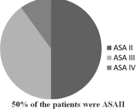
The mean age was 49.4±14.73 with a range of 32-72 year, 50% were males (table 3). 50% of cases were ASA II and 40% were ASA III (figure 1).
Figure 1. Description of the ASA distribution among the patients population.
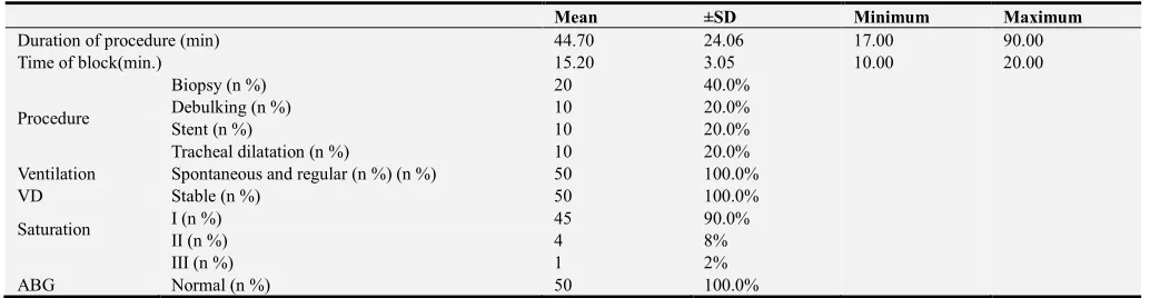
The mean duration of procedure, time of block was 44.7±24.06 min, 15.2±3.05 min, respectively as in table 4. 40% of the cases were tracheal biopsy, others were tumor debulking, tracheal stent insertion and tracheal dilatation (20% for each).
The Ventilation was Spontaneous and Stable with a normal arterial blood gases in all of the cases. The oxygen saturation was grade I in 90% of the patients, 8% of the patients were grade II and 2% were grade III.
There was no significant statistical difference between the depth of the sedation and patients satisfaction, but as the depth of the sedation increased the operator satisfaction does (table 2).
Table 2. The relation between the level of sedation and the satisfaction level regarding the patients as well as the operator.
RSS
P Sig
RSS 4 RSS 5 RSS 6
N % N % N %
Patient satisfaction No 0 .0% 0 .0% 5 14.3% 0.598** NS
Yes 5 100.0% 10 100.0% 30 85.7%
Operator satisfaction No 0 .0% 0 .0% 20 57.1% 0.001* HS
Yes 5 100.0% 10 100.0% 15 42.9%
The mean recovery time (from the end of the procedures till the patients became fully awake), and hospital stay was 3.5±2.6 min, and 2.2±0.42 hr respectively (table 3).
No Postoperative complications were recorded. 90% of the Patients were satisfied, while operator satisfaction was 60%.
Table 3. Description of the post-operative data among cases.
Mean ±SD Minimum Maximum
Recovery time (min.) 3.50 2.64 1.00 10.00
Hospital stay (hr) 2.20 .42 2.00 3.00
Postoperative complication No (n%) 50 100.0%
Mean ±SD Minimum Maximum
Not satisfied (n%) 5 10.0%
Operator satisfaction Satisfied (n%) 30 60.0%
Not satisfied (n%) 20 40.0%
Cause of operator dissatisfaction (n=15) Frequent coughing (n%) 10 20.0%
Upward movement of bronchus (n%) 5 10.0%
Table 4. Description of operative data among cases.
Mean ±SD Minimum Maximum
Duration of procedure (min) 44.70 24.06 17.00 90.00
Time of block(min.) 15.20 3.05 10.00 20.00
Procedure
Biopsy (n %) 20 40.0%
Debulking (n %) 10 20.0%
Stent (n %) 10 20.0%
Tracheal dilatation (n %) 10 20.0%
Ventilation Spontaneous and regular (n %) (n %) 50 100.0%
VD Stable (n %) 50 100.0%
Saturation I (n %) 45 90.0%
II (n %) 4 8%
III (n %) 1 2%
ABG Normal (n %) 50 100.0%
5. Discussion
Airway block with sedation is a common practice for flexible fiberoptic bronchoscopy by variable anesthetic agent with a constant oxygen delivery.
This was to ensure a safer practice than the traditional approach by using general anesthesia and the associated airway management that is considered as a true challenge for the anesthetist, especially with the type of the patients scheduled for such a procedure.
A further step toward the patient safety, airway block was examined in our study for rigid bronchoscopy aiming to decrease the ventilatory management as well as airway management.
The study was extended beyond the patient safety to the team safety by decreasing the room pollution that was reported by Paul & Jeremi on using inhalational anesthestic.
The current study could increase the patient satisfaction by decreasing the use of TIVA and the reported increase in the patient awareness during anesthesia with this technique [9].
In positive pressure ventilation, the bronchoscopist has to cover the proximal end each time the anesthetist generate positive pressure with hand ventilation, this impair the ability to go through the procedure and necessitate frequent interruption of the ventilation and subsequent apnea with the associated hypercapnia that couldn’t be detected even with the use of end-tidal CO2 due to excessive leak [10].
Jet ventilation if ever available with the sophisticated setting and the especial need for connection like luer lok connection “to avoid too much slippage” and sampling catheter to frequently measure airway pressure or alternatively use of manual jet ventilation with the high risk
for barotraumas and associated pneumothorax or
pneumomediastinum and hemodynamic collapse [9, 10]. Painless, comfortable, and peaceful procedures are common targets for both the patients and pulmonologist, several studies are held with a conclusion that sedation
results in less problems compared to procedures that run without sedation [12, 14, 16, 17].
Conscious sedation markedly improves the tolerability of bronchoscopy, but, it does not totally abort cough, choking, pain, and anxiety [15, 18, 19]. In addition, it does not improve the patient cooperation [13, 14], the corresponding association with hypoxic events and bronchoscopy-related mortality (50%) [12, 14]. Positive-pressure ventilation was used to overcome hypoxic event in bronchoscopy [22] as this event of de-saturation, in combination with tachycardia, may result in myocardial ischemia, especially in lengthy procedures [23].
This study was conducted to test the efficiency of airway block with deep sedation as a safe alternative for patients undergoing interventional rigid bronchoscopy. Perrin and colleagues [24] explained their experiment on 124 rigid bronchoscopies in which ventilation was done by using the spontaneous assisted technique. After 3 minutes duration of preoxygenation by mask, anaesthesia was induced by intravenous administration of propofol, phenoperidine, and diazepam or midazolam. Afterwards, ventilation was done manually using high-flow oxygen (FIO2, 0.6–1.0) through the ventilation port of the bronchoscopy. Anaesthesia was
maintained by repeated injections of intravenous
anaesthetics, and ventilation was assisted manually in case of prolonged apnea or de-saturation. Perrin and colleagues reported complications in 22 procedures and included severe
pre-operative as well as postoperative hypoxemia,
bronchospasm, and laryngospasm [24]. In comparison to our study, we found that the arterial blood gases were not changed in almost all of the cases and the oxygen saturation was maintained in 90% of the patients.
the comparison of midazolam and propofol for sedation during rigid bronchoscopy. Crawford et al, reported that propofol is more vulnerable to exceed the desired level of moderate sedation [26], Moreover, midazolam was found to induce reduction in the mean blood pressure, the respiratory rate, and O2 saturation especially in elderly patients [27, 28]. This de-saturation event is associated with myocardial insult [22]. Propofol maintain O2 saturation in elderly patients above 70 years of age [23].
The present study revealed that, the ventilation and the oxygenation were almost unchanged and the patients’ satisfaction was 90% in the selected population. The presented approach could provide a safe alternative especially when alternate apnea technique could present a true risk for the patients, as the presented approach was examined on patients ASA II (50%), III (40%) and IV (10%). On the other side, the presented study showed a lower satisfaction index for operators that is estimated as 60%, that was mainly attributed to the frequent coughing of the patients especially on examining the left main bronchus as well as its tributaries, which could be explained by the poor spread of the local anesthetic in the left bronchus.
The efficiency of the presented approach is extended to the postoperative periods as well, whereas the mean recovery time where 3.5 min. whereas the mean time of the patients’ discharge was 2.20 hr with no complication in the population of the patients selected. These results go with the approach as a day case surgery. This in contrast to the study by Ayers & Beamis who found that the Patients requiring high levels of supplemental O2 and those with baseline hypercarbia and hemodynamic instability are at increased risk for intra- and post procedural complications, and they concluded that risk and benefit of the procedure should be weighed carefully
6. Conclusion
Airway blocks are simple procedure, have a high success rate, and offer significant potential advantages to patients with a high satisfaction. Airway block for rigid bronchoscopy are associated with a low rate of complications. The likelihood of significant long-term morbidity is low.
Airway block with sedation is a safe and reliable practice for high risks patients scheduled for interventional bronchoscopic procedures on a day case basis.
References
[1] J. English, A. Norris and N. Bedforth. Anaesthesia for airway surgery. Continuing Education in Anaesthesia, The Board of Management and Trustees of the British Journal of Anaesthesia Critical Care & Pain 2006; 6: 1.
[2] V. Pathak, I. Welsby, K. Mahmood, M. Wahidi, N. MacIntyre, and Shofer S. Ventilation and Anesthetic Approaches for Rigid Bronchoscopy Annals ATS 2014; 11: 4.
[3] J. Beamis. Rigid bronchoscopy. In: J. Beamis, P. Mathur, editors. Interventional pulmonology. New York, NY:
McGraw-Hill; 1999: 17–28.
[4] J. Henderson. Airway Management in the Adult. in: RD. Miller, LI. Eriksson, LA. Fleisher, JP. Wiener-Kronish, WL. Young. Miller’s anesthesia 7th edition chuchil livingstone el sevier; 2010: 50: 1573-1611.
[5] P. Kundra, S. Kutralam, M. Ravishankar: Local anaesthesia for awake fibreoptic nasotracheal intubation. Acta Anaesthesiol Scand 2000; 44: 511-516.
[6] T. Shawn. Simmons, Schleich: Airway Regional Anesthesia for Awake Fiberoptic Intubation Regional Anesthesia and Pain Medicine 2002; 27.
[7] Saunders. Translaryngeal block. In Brown D. Atlas of Regional Anesthesia 2nd edition: Philadelphia; 1999: 215-216.
[8] Nehthorn RW, Amayem A, Ganta R. Which method for intraoral glossopharyngeal nerve block is better? Letters to the Editor. Anesth Analg. 1995; 81: 1114.
[9] Paul H Alfille, jeremi Mountjoy, Anesthesia for adult bronchoscopy, uptodate 2018.
[10] de Lima A, Kheir F, Majid A, Pawlowski J. Anesthesia for interventional pulmonology procedures: a review of advanced diagnostic and therapeutic bronchoscopy. Can J Anaesth 2018; 65: 822.
[11] AR Selzer, M Murrell, E Shostak. New trends in interventional pulmonology. Curr Opin Anaesthesiol 2017; 30: 17.
[12] F. Maltais, F. Laberge, M. Laviolette. A randomized, double-blind, placebo controlled study of lorazepam as premedication for bronchoscopy. Chest 1996; 109: 1195-1198.
[13] SJ Pearce. Fibreoptic bronchoscopy: is sedation necessary? BMJ 1980; 281: 779-780.
[14] MQF Hatton, MB Allen, AS Vathenen. Does sedation help in fibreoptic bronchoscopy? BMJ 1994; 309: 1206-1207.
[15] GL Samsoon, JR Young. Difficult tracheal intubation: a retrospective study. Anaesthesia 1987; 42: 487-490.
[16] S. Putinati, L. Ballerin, L. Corbetta, L. Trevisani, A. Potena. Patient satisfaction with conscious sedation for bronchoscopy. Chest 1999; 115: 1437-1440.
[17] Ni YL, Lo YL, Lin TY, Fang YF, Kuo HP. Conscious sedation reduces patient discomfort and improves satisfaction in flexible bronchoscopy. Chang Gung Med J 2010; 33: 443452.
[18] HJ Lim, YJ. Cho, JS. Park, H. Yoon, Lee J-H, CT. Lee, et al. Predictors of hypoxemia developed during fiberoptic bronchoscopy under monitored anesthesia care. Chest 2012; 142: 915.
[19] G. Maguire, AR. Rubinfeld. Patients prefer sedation for fibreoptic bronchoscopy. Respirology 1998; 3: 81-85.
[20] HM. Hadzri, SMS. Azarisman, ARM. Fauzi, H. Roslan, AM. Roslina, ATN. Adina, et al. Can a bronchoscopist reliably assess a patient's experience of bronchoscopy? JRSM Short Rep 2010; 1: 35-42.
[22] B. Maître, S. Jaber, SM. Maggiore, E. Bergot, JC. Richard, H. Bakthiari, et al. Continuous positive airway pressure during fiberoptic bronchoscopy in hypoxemic patients. A randomized double-blind study using a new device. Am J Respir Crit Care Med 2000; 162: 1063-1067.
[23] I. Matot, MR. Kramer, L. Glantz, B. Drenger, S. Cotev. Myocardial ischemia in sedated patients undergoing fiberoptic bronchoscopy. Chest 1997; 112: 1454-1458.
[24] G. Perrin, H. Colt, C Martin, M Mak, J Dumon, F. Gouin. Safety of interventional rigid bronchoscopy using intravenous anesthesia and spontaneous assisted ventilation: a prospective study. Chest 1992; 102: 1526–1530.
[25] M. Ayers, J. Beamis. Rigid bronchoscopy in the twenty-first century. Clin Chest Med 2001; 22: 355–364.
[26] M. Crawford, J. Pollock, K. Anderson, RJ. Glavin, D. Macintyre, D. Vernon. Comparison of midazolam with propofol for sedation in outpatient bronchoscopy. Br J Anaesth 1993; 70: 419-422.
[27] GC. Sun, MC. Hsu, YY. Chia, PY. Chen, FZ. Shaw. Effects of age and gender on intravenous midazolam premedication: a randomized double-blind study. Br J Anaesth 2008; 101: 632-639.