2807712971
THE ONTOG ENY OF INTESTINAL IRON ABSORPTION
IN THE GUINEA PIG
by
GEORGE FRANCIS EMMANUEL CHOW RIM OO TOO
Thesis submitted for the degree of Doctor of Philosophy
in the University of London
HAMPSTEAW
ROYAL FREE HOSPITAL SCHOOL OF M EDICINE
ProQuest Number: U549515
All rights reserved
INFORMATION TO ALL USERS
The quality of this reproduction is dependent upon the quality of the copy submitted. In the unlikely event that the author did not send a complete manuscript and there are missing pages, these will be noted. Also, if material had to be removed,
a note will indicate the deletion.
uest.
ProQuest U549515
Published by ProQuest LLC(2016). Copyright of the Dissertation is held by the Author. All rights reserved.
This work is protected against unauthorized copying under Title 17, United States Code. Microform Edition © ProQuest LLC.
ProQuest LLC
789 East Eisenhower Parkway P.O. Box 1346
A B S T R A C T
Homeostasis of body iron content is regulated by intestinal iron absorption.
In neonatal animals however, absorption of iron is enhanced despite having
substantial iron stores. In order to examine the cellular basis for this effect,
intestinal iron absorption has been compared in adult and neonatal guinea
pigs using a variety of techniques.
Iron measurement has shown the neonates are not iron deficient compared
to the adult. Studies carried out in vivo using radiolabelled iron confirmed
previous studies that the rate of duodenal iron transfer in the adult was an
order of magnitude greater than that of the terminal 20 cm of ileum. In the
neonate however, the rate of transfer was similar in both duodenum and
ileum. Transition to the adult duodenal/ileal ratio occurs after the first 16
days of postnatal life. This localisation change may be relevant to the
enhanced iron absorption in neonatal mammals.
Several approaches, including autoradiography of everted duodenal and ileal
segments and uptake of iron by duodenal and ileal brush border membrane
vesicles, confirmed that the regulatory site for iron absorption is not at the
brush border membrane.
Chromatographic and immunoprécipitation studies of enterocyte cytosolic
was associated with ferritin. Some was associated with a protein of low
molecular weight of about 10-12 kD. In contrast, in newborn animals and
those up to 21 days old, 80% of newly absorbed radioactive iron was found
to be associated with this low molecular weight protein. These results were
confirmed by parallel immunoblotting studies which showed the levels of
ferritin to be low in newborn but increased with age. Similarly the levels of
the low molecular weight protein was shown to be high at birth and decrease
during development.
Studies of transferrin receptor number using isolated duodenal and ileal
enterocytes indicated that changes in transferrin binding was not responsible
for the developmental changes in intestinal iron absorption.
The results presented in this thesis throw further light on the mechanisms
regulating intestinal iron absorption during development.
It is concluded that neither brush border membrane transport nor events
involving transferrin binding to enterocytes account for the enhanced iron
absorption in the neonate. Changes in iron absorption during development
may be explained by segmental or enterocyte maturation differences,
intracellular events involving iron binding proteins or differences in iron
ACKNOWLEDGEMENTS
I wish to thank Professor N McIntyre, Professor M Spyer and Professor D
Chapman for allowing me to study in their respective departments. I would
like to express my sincere gratitude to Drs ES Debnam, SKS Srai and O
Epstein for their constant encouragement and supervision throughout the
period of my studies. I also wish to thank Dr M Gillett, Mr D Brown, Mrs M
Kozma, Mr A McKay, Ms D Elliott, Mr J Hadden and all of those who have
freely given their advice and technical assistance during these studies.
Finally I wish to thank my family, whose encouragement and patience
Ab b r e v ia t io n
LIST OF ABBREVIATIONS
BBM Brush border membrane
BSA Bovine serum albumin
EDTA Ethylenediaminetetra-acetate
FADHg Flavin adenine dinucleotide (reduced)
FMNH. Flavin mononucleotide (reduced)
IREBP Iron responsive element binding protein
Mol. wt. Molecular weight
NAD"
NADH
Nicotinamide adenine dinucleotide
(oxidised)
Nicotinamide adenine dinucleotide
(reduced)
RNA Ribonucleic acid
mRNA Messenger RNA
SDS-PAGE Sodium deodecyl sulphate polyacrylamide gel
C O N T E N T S
A
b s tr a c tACKNOWLEDGED ENTS
A
b b r e v ia t io n sS
e c t io nP
a g eL
is t o fF
ig u r e s20
L
is t o f t a b l e s27
1.0
Introduction to iron, its metabolism, physiology and
pathology
28
P
a g e1.1.2 Iron chemistry
29
1.2
Iron transport and storage proteins
32
1.2.1 Transferrin
32
1.2.1.1
Transferrin synthesis
34
1.2.1.2
Transferrin gene location and
regulation
35
1.2.1.3
Transferrin receptors
35
1.2.1.4
Cellular uptake of iron from
transferrin
36
1.2.2 Ferritin
38
1.2.2.1
Isoferritins
38
1.2.2.2
Ferritin function
40
1.2.2.3
Iron binding and release from
P
a g e1.2.2.4
Ferritin gene regulation
42
1.2.2.5
Ferritin receptors
43
1.2.3 Lactoferrin
44
1.2.3.1
Function of lactoferrin
45
1.2.3.2
Lactoferrin receptors
47
1.2.4 Iron responsive element binding
proteins
47
1.3
Homeostasis of body iron in mammals
48
1.4
Development of the intestinal tract
49
1.5
Iron absorption
53
1.5.1 Intraluminal factors affecting iron
P
a g e1.5.2 Coporeal factors affecting iron
absorption
55
1.5.3 Mucosal factors affecting iron
absorption
57
1.5.4 Mechanism of iron absorption
58
1.5.5 Mechanisms involved in iron
absorption
60
1.5.6 Proteins involved in iron absorption
64
1.5.7 Absorption of Haem iron
65
1.6
Iron transport and utilisation
66
1.7
Disturbances associated with iron
67
1.7.1 Iron deficiency
67
1.7.2 Iron overload
69
P
a g e1.9
Choice of animal
71
2.0
Materials and Methods
72
2.1
Animals and anaesthesia
72
2.2
Enterocyte isolation
72
2.3
Gel filtration
73
2.4
Electrophoresis
74
2.5
Staining of gels
74
2.6
Drying of gels
75
P
a g e2.8
Protein assay
75
2.9
ECL
76
3.0
Iron status of the adult and developing guinea pig
77
3.1
Introduction
77
3.2
Materials and method
78
3.2.1 Materials
78
3.2.2 Sample collection
78
3.2.3 Chemical measurement of serum iron 78
3.2.4 Chemical measurement of liver iron
79
3.3
Results and Discussion
82
P
a g e4.0
Autoradiographic localisation of iron uptake by
the small intestine of the developing guinea pig
during ontogeny
85
4.1
Introduction
85
4.2
Materials and Methods
86
4.2.1 Autoradiography and histology
86
4.3
Results
88
4.4
Discussion
91
P
a g e5.0
Regional localisation of iron absorption by the guinea
pig small intestine during ontogeny
94
5.1
Introduction
94
5.2
Materials and Methods
94
5.2.1 Regional localisation of iron transfer to
blood
94
5.3
Results
95
5.3.1 The appearance of absorbed ®^Fe in the
blood
95
5.4
Discussion
99
P
a g e6.0
Iron uptake and binding across the brush border
membrane during ontogeny
104
6.1
Introduction
104
6.2
Materials and Methods
105
6.2.1 Brush border membrane vesicles
105
6.2.2 Iron solution
106
6.2.3 Uptake
106
6.2.4 Binding studies
107
6.2.5 Duodenal iron binding protein
107
6.2.6 Alkaline phosphatase assay
108
6.3
Results
109
P
a g e6.3.3 Uptake studies
110
6.3.4 Binding studies
110
6.3.5 Duodenal iron binding proteins in adult 110
6.4
Discussion
122
6.5
Conclusion
128
7.0
Iron binding proteins in enterocyte cytosol during
ontogeny
129
7.1
Introduction
129
7.2
Materials and Methods
129
7.2.1
In Vivo
iron absorption
129
P
a g e7.2.3 Characterisation of radiolabelled peaks 130
7.2.4 Western blotting for ferritin
131
7.2.5 Characterisation of radiolabelled
peak IV
131
7.2.6 Antibody to peak IV protein
132
7.2.7 Partial purification of the low molecular
weight iron binding proteins
132
7.3
Results
133
7.3.1 Profiles of iron binding in enterocyte
cytosol
133
7.3.2 Immunoprécipitation for ferritin
133
7.3.3 Western blotting for ferritin
134
7.3.4 SDS PAGE of enterocyte soluble
supernatant
134
7.3.5 Characterisation of peak IV
134
7.3.6 Partial purification of peak IV low
P
a g e7.4
Discussion
146
7.5
Conclusion
152
8.0
Iron transferrin binding to isolated guinea pig
enterocytes during ontogeny
153
8.1
Introduction
153
8.2
Materials and methods
154
8.2.1 Saturation of transferrin with iron
154
8.2.2 Radiolabelling of transferrin with ^^1
154
8.2.3 Binding of transferrin to isolated
enterocytes
155
P
a g e8.3.1 Characterisation of differic-transferrin
binding by isolated intestinal cells
156
8.4
Discussion
161
8.5
Conclusion
166
9.0
General Discussion and Conclusion
167
10.0 List of Publications
175
LIST OF FIGURES
F
ig u r eP
a g e1.1
Iron complexes in solution
31
1.2
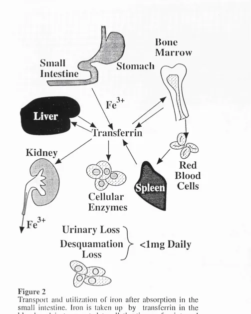
Transport and utilisation of iron after
absorption in the small intestine.
68
3.1
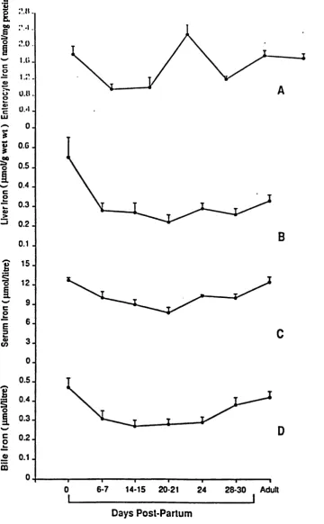
Level of iron in enterocytes, liver, serum and
bile during ontogeny.
81
4.1
Apparatus used for autoradiographic uptake of
F
ig u r eP
a g e4.2
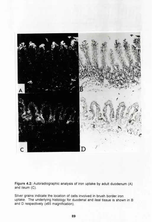
Autoradiographic analysis of iron uptake by
adult duodenum and ileum.
89
4.3
Autoradiographic analysis of iron uptake by
neonatal duodenum and ileum.
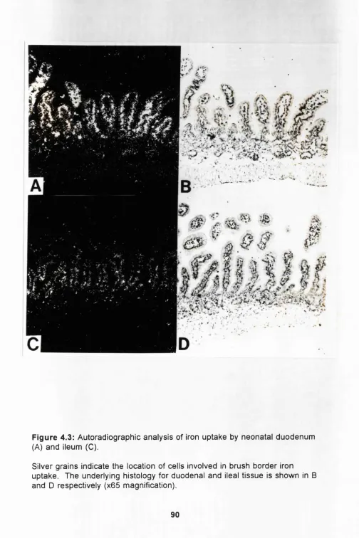
90
5.1
Time dependence for the appearance of ®®Fe
in adult peripheral blood following exposure
of duodenal mucosa to ®®Fe-ascorbate.
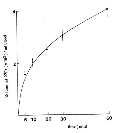
97
5.2
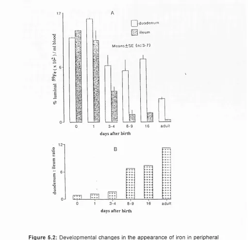
Developmental changes in the appearance of iron
in peripheral blood following exposure of
F
ig u r eP
a g e6.1
Time dependence of iron uptake by adult brush
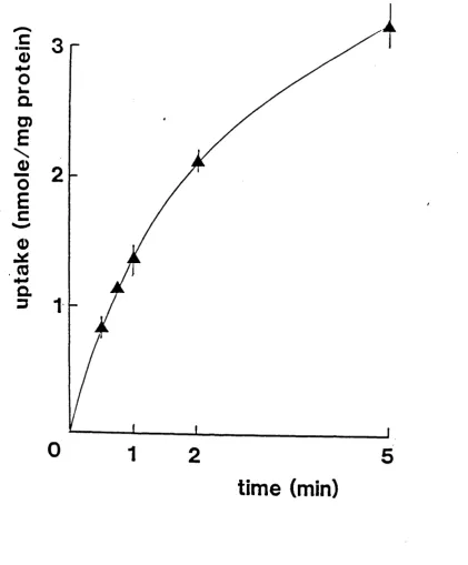
border membrane vesicles prepared from ileal
mucosa.
113
6.2
Concentration dependence of Fe^"" uptake by adult
and neonate brush border prepared from duodenal
mucosa.
114
6.3
Concentration dependence of Fe^^ uptake by adult
and neonate brush border membrane vesicles
prepared from duodenal mucosa.
115
6.4
Concentration dependence of Fe^"^ uptake by adult
and neonate brush border membrane vesicles
F
ig u r eP
a g e6.6
Binding of
to duodenal brush border
membrane as a function of time
117
6.6
Binding of Fe^" to duodenal brush border
membrane as a function of concentration
118
6.7
®®Fe profile of solubilised duodenal brush
border membrane proteins fractionated on
sepharose CI-6B.
120
6.8
SDS-PA G E and identification of iron binding
protein by autoradiography in adult brush
F
ig u r eP
a g e7.1
Absorption spectrum of enterocyte cytosol.
Protein fractions eluted from Sepharose CL-6B
column.
136
7.2
®^Fe elution profile of soluble supernatant of
adult and neonate enterocyte, fractionated
on sepharose CI-6B.
137
7.3
Relative distribution of ®®Fe associated with
peak I and IV of fractionated enterocyte cytosol
during ontogeny.
138
7.4
Immunoprécipitation of fractionated enterocyte
soluble supernatant ^®Fe peak I and IV for
F
ig u r eP
a g e7.5
Changes in the level of ferritin in enterocyte
cytosolic supernatant by immunoblotting.
140
7.6
Changes in the level of ferritin in liver
cystolic supernatant by immunoblotting.
141
7.7
SDS-PAGE of enterocyte soluble-cytosolic
supernatant of enterocyte during ontogeny.
142
F
ig u r eP
a g e7.9
Semi-quantification of the low molecular weight iron
binding protein during ontogeny by
immunoblotting.
144
7.10
SDS-PAGE of partial purification of the low
molecular weight iron binding protein from the
soluble supernatant of neonates enterocyte.
145
8.1
Binding of ^^^l-labelled guinea pig transferrin
to duodenal enterocyte.
158
8.2
Binding of ^^^l-labelled guinea pig transferrin
to duodenal enterocytes as a function of
LIST OF TABLES
T
a b l eP
a g e6.1
Purity of brush border membrane measured by
alkaline phosphatase activity.
112
6.2
Kinetic parameters for high affinity (specific) binding
of Fe^'" binding to adult and neonatal BBM
vesicles.
119
8.1
Kinetic parameters for high affinity (specific)
binding of ^^l-transferrin to duodenal and ileal
enterocytes from adult and neonatal guinea
1.0
INTRODUCTION TO IRON, ITS METABOLISM, PHYSIOLOGY AND
PATHOLOGY
1.1
I
ronIron is a ubiquitous metal and because of its abundance in the earth's crust
and subtle chemical properties, it has been selected in molecular evolution
to perform a large number of biological functions. After the creation of the
earth, iron existed largely in its reduced form (ferrous) due to the reducing
atmosphere (Frieden 1974). As atmospheric oxygen increased, iron
oxidised to its ferric form (Whittemore 1979) which is less available for
acquisition by biological systems. In response to this change, life forms
adjusted to it by developing special devices to acquire iron. For example
bacteria synthesize and secrete high affinity chelators that extract iron from
the environment (Neilands 1980). Certain roots of plant can secrete
substances that facilitate iron absorption (Brown 1979).
In higher animals iron is used in many ways. Because it readily undergoes
oxidation and reduction it was ideally selected in the molecular evolution of
many biological systems involved in electron transport such as the
cytochromes and succinic dehydrogenase. The enzymes catalase and
peroxidase utilise iron because of their capacity to decompose hydrogen
have the ability to bind large amounts of oxygen in a readily reversible way.
In higher animals haemoglobin binds oxygen in the lungs and delivers it to
tissues at the same time transporting carbon dioxide from the tissues to the
lungs. Iron also serves as an efficient catalyst in biological systems
(Bothwell 1979). It is also known that certain enzymes, although not
containing iron, appear to require iron for their function, these include the
enzyme aconitase (Bothwell 1979) which is involved in the isomerization of
citrate to isocitrate in the citric acid cycle. In addition because free iron is
toxic to cells and the propensity of it to hydrolyse in aqueous solution,
special molecules are needed for its transport to tissues and storage for
when it is required. These are provided in the form of transferrin and ferritin
respectively (Aisen and Listowsky 1980). Therefore body iron may be
divided into the following pools which include storage, haemoglobin,
myoglobin, transport, enzymes and labile iron.
I.1.2
Iron Chemistry
Although iron can be found in oxidation states as high as VI and as low as
-II, the most common oxidation states are II and III. In biological systems
only the iron II and iron III oxidation states are found, although there is
speculation that in the haem moiety of the enzyme horseradish peroxidase,
iron can be found in states as high as IV and V (Peisach et al, 1967). The
most stable oxidation state in both acid and alkaline solution is the ferrous
state. However, even mild oxidants such as oxygen are capable of oxidizing
both oxygen and water is the ferric form.
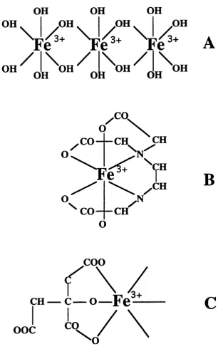
As shown in Figure 1.1 iron has six co-ordinating bonds and in aqueous
solution it forms macromolecules with hydroxyl groups which eventually
precipitate out of solution as conditions become more alkaline. Some
chelators are required for iron absorption in biosystems. Chelators such as
EDTA can bind to all the co-ordinating bonds thus making the iron
unavailable for absorption. On the other hand chelators such as citrate and
ascorbate bind only a few of the co-ordinating bonds and increase
absorption by increasing the solubility of iron and preventing the formation of
hydroxyl groups (Conrad 1987). Iron within the haem moiety has only two of
its co-ordinate bonds available for complex formation and is not affected by
OH
OH
OH
OH
-OH
-OH
OH
OH ^oL'^OH
^ o t^ O H
OH
CO
CH
CO
CH
CH
CH
CO
CH
B
coo
CH — c — o — F e
3+
ooc
CO
\
o
Figure 1.1. Iron complexes in aqueous solution
Like other transition metals iron has a 3-d orbital that contains some
unpaired electrons. These allow iron to form weak co-ordinate bonds with
other ligands. This makes iron suitable to perform various functions in the
body. For example, in mammals, the iron containing protein haemoglobin
can form weak bonds with oxygen in the lung, it can then be transported to
cells where oxygen is released and can be utilised in cellular respiration.
Similarly after releasing oxygen haemoglobin is then free to bind carbon
dioxide, a waste product of cells, and transport it to the lung where it can be
expelled from the body.
1.2
I
ron transport and storage proteinsThe iron transport and storage proteins include transferrin, ferritin and
lactoferrin. These proteins may have an important role in the regulation of
iron absorption in mammals. These proteins are discussed in detail here.
1.2.1
Transferrin
Following iron absorption from the environment effective distribution within
the organism is needed since free iron is insoluble.
In invertebrates several means of transporting iron are present (Aisen and
transferrin. This protein was first isolated in 1947 by Laurel! and Ingelman
and since then much information has been found out about its structure and
function.
Transferrin is a glycoprotein of molecular mass of about 76-90,000 KDa
depending on the species (Bezkorovainy 1964; Charlwood 1963). It consists
of a single polypeptide chain of 679 amino acids (MacGillivray et al 1985)
and two N-linked glycan chains (Forland et al 1977; Karlsson et al 1978).
The transferrin molecule can be divided into two homologous domains, the
N-terminal domain residues 1-336 and the C terminal domain residues
(337-679) with the carbohydrate moieties in the C-terminal domain at position 413
and 611 (De Jong et al 1990). Although iron is the most important metal
that binds transferrin in vivo, it can also bind other metal ions such as
Ca^*, Mn^", Cr^*, Cd^\ Zn^* and Co^"' (De Jong et al 1990). The amount of
carbohydrate in transferrin also varies with species ranging from 2-12%
(Graham and William 1975). The predominant form of human serum
transferrin has two doubly branched glycans, joining the protein by
N-asparaginyl linkage and each branch terminates in a sialic acid (Spik et al
1982). But small amounts of triple and quadruple branched glycans have
also been identified in human (Marz et al 1982). In contrast, both rat and
rabbit transferrins have been shown to have only one glycan chain per
molecule (Evans et al 1988; I rie et al 1988). The carbohydrate chains do
not appear to be important in transferrin-cell interactions and their function
1.2.1.1
Transferrin Synthesis
The principal site of transferrin synthesis is the liver. The hepatocytes being
the main cell type involved in the synthesis of transferrin (Aisen 1984).
Other cells that have been shown to produce transferrin include, the sertoli
cells o fth e testis (Skinner and Grisworld 1983), the brain cells (Bloch et al
1985; Dickson et al 1985) and the lactating mammary gland where
production can exceed that of the liver (Jordan and Morgan 1969). It has
also been shown that transferrin production in certain tissues changes with
development after birth (Morgan 1981). Transferrin synthesis in the spleen,
muscle and kidney decreases during ontogeny whereas synthesis in the
brain increases significantly after birth. Transferrin synthesis also is
dependent on body size and metabolism (Munro 1969). Transferrin is
synthesised as a precursor with an additional 19 to 20 amino acids and
these are removed by proteases before secretion. Inhibition of this event
prevents the secretion of transferrin. This extra peptide may be the signal
necessary for translocation of mRNA-ribosome complex from the cytoplasm
to the rough endoplasmic reticulum (de Jong et al 1990). However inhibition
of the glycosylation process does not prevent secretion (de Jong et al 1990).
The newly synthesized transferrin in the hepatocyte is found on ribosomes
and membrane of the rough endoplasmic reticulum with small amounts
within smooth endoplasmic reticulum and Golgi apparatus. From this
observation it has been suggested that the protein is first assembled on the
ribosomes of the rough endoplasmic reticulum, passed through the
protein Is completed and finally taken up by exocytotic vesicles, transported
to the plasma membrane and released into the portal blood (Morgan 1981;
de Jong et al 1990).
1.2.1.2
Transferrin Gene Location and Regulation
The transferrin gene location has been identified to be on chromosome 3 (Le
Beau et al 1985; Yang et al 1984). On the same chromosome a
pseudogene has also been identified (Schaeffer et al 1987).The complete
structure of the human transferrin gene has been described by Le Beau et al
(1985). The gene is about 33.5 kilobase pairs (kb) in length containing 17
exons separated by 16 introns. The gene appears to be derived from a
precursor gene by internal duplication (Park et al 1985; William 1982). The
mechanisms involved in the transcriptional and translational regulation of
transferrin synthesis have not been fully elucidated, but studies with iron
deficient animals reveal that enhancement of transferrin synthesis by iron
deficiency reflects transcriptional rather than translational regulatory
mechanism (Idzerda et al 1986; McKnight 1980).
1.2.1.3
Transferrin Receptors
The existence of membrane receptors for transferrin on reticulocytes have
been postulated since 1960 (Jandl and Katz 1963). Subsequent to this
there have been great advances in our knowledge of this protein. It is a
transmembrane glycoprotein made up of a dimer of subunits of molecular
Each subunit contains a transferrin binding site, two N-asparagine linked
glycan and a complex carbohydrate chain. The transferrin receptors have
very high affinity for diferric transferrin with an estimate Ka of 2-7 x 10*®M
(Newman et al 1984; Testa 1985; Trowbridge et al 1984). Thus, very low
amounts of transferrin can be bound to the receptor. The gene for
transferrin receptor has also been located on chromosome 3 (Le Beau et al
1985). It is calculated to contain 31 kb pairs and at least 12 exons.
Synthesis of the receptor is highly regulated by iron (Ward 1987). Like
transferrin, iron deficiency induced expression of transferrin receptors on the
cell surface and iron overload reduced their appearance (Mattia et al 1984).
With regards to cellular iron uptake, the transferrin receptor may be more
important than transferrin since its gene expression has been shown to be
more sensitive to intracellular iron concentration (Owen and Kuhn 1987; Rao
et al 1985, 1986; Ward et al 1984).
Some of the tissues where transferrin receptors have been demonstrated
include placenta, liver, testis, epidermis, pancreas, brain, bone marrow and
intestine (Banerjee et al 1986; Huebers and Finch 1987). Apart from cellular
surfaces, the receptors have also been identified in intracellular
compartments using labelled transferrin or monoclonal antibodies (Nikinmaa
et al 1984; Trowbridge and Newman 1984; Willingham and Pastan 1985).
1.2.1.4
Cellular Uptake of Iron from Transferrin
first binds to transferrin receptors on cell membranes in an energy and
temperature dependent process (Zaman et al 1980). The complex is then
internalised by endosomes and proton pumps within the endosomal wall,
rapidly reduce the internal pH to a value of 5.5. This drop in pH promotes
the release of iron from transferrin and the released iron is moved to its
destination within the cell by an unknown mechanism (Egyed 1982; Konopka
and Romslo 1981; Patteson et al 1984; Veldman et al 1984; Thorstensen
and Romslo 1990). The apotransferrin receptor complex, which binds tightly
at the low internal pH of the endosome, is then cycled to the cell surface,
where it dissociates at the higher extacellular pH (Dautry-Varsat et al 1983;
Karin and Mintz 1981). This recycling process relies on the high affinity of
holo transferrin for the receptor at neutral pH and the high affinity of apo
transferrin for the receptor at acidic pH (Klausner et al 1984; Morgan 1983),
and low affinity of apo transferrin for the receptor at neutral pH.
Recent studies have revealed another pathway involving NADH reductase
activity. Binding of differic transferrin to the transferrin receptor of rat
hepatocytes followed by reduction of ferric ions to the ferrous state with
conversion of NADH to NAD*. The Fe II released from this reaction is
transported across the plasma membrane (Low et al 1985; Thorstensen
1989; Thorstensen and Romslo 1988). Reticulocytes display little NADH
reductase activity and transferrin uptake occurs predominantly by
receptor-mediated endocytosis. Hepatocytes on the other hand display
1.2.2
Ferritin
Iron is an essential requirement for ceils and this need for iron poses a
problem for the cells. This is because at physiological pH, free ferric iron is
insoluble and also ferrous ions can generate hydroxyl radicals from
hydrogen peroxide with lethal effects on cell function and viability. This
demands the storage of iron in a sequestered and accessible form. Many
organisms respond to this by producing the protein apoferritin which can
store up to 4,500 atoms of iron (Mann et al 1986). X-ray and electron
microscopic studies have revealed the protein to consist of a spherical shell
with an external diameter of 120-130Â and a central cavity of about 75Â
(Aisen 1980). The iron is encapsulated in the central cavity which has eight
shallow pockets (Harrison 1986), so the variable iron content does not
appreciably alter the surface charge of the protein and has no direct effect
on electrophoretic mobility, immunology and other properties depending on
the protein surface (Harisson 1964). The protein coat is formed from 24
subunits of approximately 175 amino acids in length surrounding the variable
iron-oxide phosphate complex (Heusterspreute and Crichton 1981).
1.2.2.1
Isoferritins
The subunits of ferritin are of two types, designated H (heavy) and L (light).
These have molecular weights of about 21,000 and 19,000 respectively
(Arosio et al 1978). Molecules containing different proportions of the two
metabolicaiiy and possibly functionally. They have different electrophoretic
and chromatographic properties (Drysdale et al 1977; Urushizaki et al 1971).
L-rich ferritins predominate in tissues that store large amounts of iron such
as liver, spleen and bone marrow. The H-rich ferritins are found mainly in
organs with no major iron storage function. Such as heart, kidney, pancreas
and rapidly proliferating tissues (Alpert et al 1973; Bomford,
Conlon-Hollingstead and Munro 1981; Kohgo et al 1980; Powell et al 1974). It is
also know that during development and under certain pathological conditions
such as iron overload and cancer there are changes in ferritin phenotype
(Alpert et al 1973; Kohgo et al 1980; Powell et al 1974). Most of these
differences appear to be the consequence of variation in production of H
type ferritins (Vezzoni et al 1986).
Although it is generally agreed that most species have at least the two
subunit types H and L, it has been proposed that the subunit classes may
be subjected to further heterogeneity. For example, serum ferritin and a
ferritin-like protein in milk binds to concanavalin A which indicate that they
may be glycosylated (Samtambrogio et al 1987). Serum ferritin was found
to react with antibody to the L ferritin and not to H type and therefore is
thought to be related to L type (Cragg et al 1981; Santambrogio et al 1987).
On the other hand, three types of ferritin mRNAs that are expressed in a
tissue specific manner have been found in tadpoles. Two are H type and
1.2.2.2
Ferritin Function
Ferritin is found in most cell types of humans, other vertebrates,
invertebrates, higher plants, fungi and bacteria. All ferritin functions in iron
storage but have a different role according to the cell type, its metabolic
activities and physiological function (Theil 1990). It has been reported that
the difference between ferritin and ferritin messenger RNA, either total,
relative abundance of subunits (or both) alters with the changing role of iron
in the cell (Beaumont et al 1987; Chou et al 1986; Dickey et al 1987; White
and Munro 1988). The role of ferritin in different cell types includes (a)
specialised function which stores iron to be used by other cells as in
recycling iron in macrophages, short and long term storage in red cells of
embryos and in hepatocytes which store iron for long periods as an
emergency reserve to replenish iron lost through bleeding or iron deficiency
(Theil 1990) and (b) intracellular housekeeping function, in which iron is
stored in a cell for its own use as required for the cytochromes of the
respiratory chain and ribonucleotide reductase and possibly detoxification if
excess iron enters the cell. The amount and rate of iron used varies with
cell type and therefore may reflect the variations observed in ferritin structure
and iron content (Aisen and Listowsky 1980).
1.2.2.3
Iron Binding and Release from Ferritin
The physiological processes and the mechanisms involved In the binding
and release of iron is unclear. It is known that for iron to be released from
have shown that Iron can be mobilised from ferritin by reduction with agents
such as dithionite, thioglycolate, or other thiols (Crichton 1973) and the
removal can be made easier by chelators. It is also known that reduced
flavin such as FADH, FMNHj, riboflavin or coupled NADH-FMN systems can
effectively reduce ferritin iron under anaerobic conditions (Sirivech et al
1974). The Fe^"^ ions can then be easily removed by chelating agents.
In a previous study it was found that two equivalents of Fell I were reduced
per equivalent of flavin in a sequential one electron transfer reaction (Aisen
1980). Reaction rates were fast and compatible with a possible role for
flavins in iron release.
It is also known that iron release and rate of reduction on ferritin can be
influenced by certain conditions including buffers (Watt et al 1985); the age
of the protein (lhara et al 1984) and disorder of the iron core (Yang et al
1986).
For binding of iron by ferritin it has been suggested that iron is gradually
incorporated into the apoferritin, and in the event of low level iron which is
below the requirement for induction of protein synthesis, iron is taken up by
pre-existing ferritin molecules which are partly filled (Aisen 1980). Iron is
taken up more readily in the partly filled ferritin than apoferritin. This
variability can be due to the possible shift in their intracellular location as
existing iron micelles for deposition of additional iron (Harrison et al 1977).
Among the models which have been proposed for the mechanisms of iron
uptake by ferritin includes the formation of an iron aggregate first which then
promotes assembly of protein subunits around themselves (Pape et al
1968). The other model proposed is that the protein is first formed into a
hollow sphere through which iron is channelled through to polymerize in the
central cavity (Aisen 1980). This model is consistent with the observation
that labelled amino acids are first incorporated initially into apoferritin
during iron induced ferritin synthesis (Aisen and Listowsky 1980; Harrison
and Gregory 1968).
The above is further supported by other studies which found that apoferritin
accelerates the oxidation of Fe(ll) and Felll in vitro which results in an
aggregate of ferric oxide formed in the central cavity of the protein
(Aisen and Listowsky 1981). It has been suggested that oxygen may be the
oxidising agent in vitro (Crichton and Roman 1978).
1.2.2.4
Ferritin gene and Regulation
Much information is available concerning the ferritin gene. In human the
expressed H-gene subunit is located on chromosome 11 (Cragg et al 1985)
and the expressed L-subunit gene is on chromosome 19 (Gatti et al 1987).
Regulation of ferritin synthesis is maintained at both transcriptional and
demonstrated that the initial response of ferritin synthesis to iron is
predominantly at the translational control (Drysdale and Munro 1986).
However because of the different functions of ferritin both as a
housekeeping protein and as a specialised cell protein, mechanisms of
regulation could vary depending on the cell type. The translational control
mechanism provides the necessity to respond quickly to iron load (Theil
1987). There appears to be an identical response for both L and H subunits
of ferritin synthesis at translational control (White and Munro 1988).
However there is a difference at the transcriptional level. An increase in L to
H subunit ratio in the total cellular RNA is observed upon iron induction
(Bomford et al 1981; Kohgo et al 1980) which result from an increase in the
rate of transcription of the L-subunit gene. In the situation of high iron
stress, it is more advantageous for cells to produce L-rich ferritins since
these proteins retain iron more firmly and turnover more slowly than H-rich
ferritins (Bomford et al 1981; Mann et al 1986; Wagstaff et al 1978).
Tissues that serve as major iron stores such as liver and spleen have
ferritins which are rich in L-subunit, and tissue which does not serve as
storage organ such as heart produce ferritins which are H-rich (Bomford et al
1981; Linder-Harowitz et al 1970).
1.2.2.5
Ferritin Receptors
Despite the fact that ferritin is produced in all tissues, membrane ferritin
receptors appear to be present. Injection of labelled ferritin in rats results in
30 minutes (Unger and Hershko 1974). Binding studies have shown high
affinity, saturable binding of ferritin to human placenta membrane (Takami et
al 1986), isolated rat hepatocytes (Mack et al 1985), guinea pig reticulocytes
(Pollack and Campana 1981) and Hela cells (Bretscher and Thomson 1983)
suggesting the presence of a ferritin receptor. Ferritin receptors have been
detected on isolated rat hepatocytes and number some 30,000 ferritin
binding sites per cell with Ka 1 x 10® molar (Mack et al 1983).
A human hepatic ferritin receptor has been proposed (Adams et al 1988).
An affinity constant of 6.0 x 10® was calculated for the receptor and its
molecular weight was estimated to be 53 KD by gel electrophoresis. The
precise physiological function of the ferritin receptor is unknown. But it is
thought that ferritin is secreted by Kupffer cells (Aisen 1987) and could be
taken up by hepatocytes via the receptors.
1.2.3
Lactoferrin
The protein lactoferrin was first reported by Sorensen and Sorensen in 1939.
They observed a red protein in human milk. It was first isolated from human
(Johansson 1960) and bovine milk (Groves 1960). Lactoferrin is closely
related to the serum transferrin. It consists of a single polypeptide chain of
molecular weight 75-90 kD (Querinjeum et al 1971; Weiner 1975) and it
binds two ferric ions together with the binding of two bicarbonate ions
(Massons and Herman 1968). It differs from the serum transferrin by being
of lactoferrin is 9.0 (Malamud and Drysdale 1978), and as a result in the
presence of relatively low affinity iron chelator lactoferrin retains its iron
below pH 4.0, whereas that of transferrin is lost as the pH becomes lower
than pH 6.0 (Johansson 1960).
Human milk is known to contain high concentrations of lactoferrin ranging
from 7 mg/ml in colostrum to 1 mg/ml in mature milk (Masson and Herman
1971). Bovine colostrum contains 5 mg/ml of lactoferrin and this level drops
rapidly to 20-200 pg/ml in mature milk (Reiter 1978). Lactoferrin has also
been found in other exocrine secretions including vaginal, nasal, bronchial
and intestinal (Masson et al 1966). It has also been identified in pancreatic
juice and neutrophils (Masson et al 1966; Masson et al 1968; Masson et al
1969). Studies have shown that apolactoferrin in bovine milk is rapidly
degraded by proteolytic enzymes whereas the saturated lactoferrin is
relatively resistant to prolonged tryptic digestion and gives rise to only large
fragments which retain their binding capacity for iron (Brock et al 1978).
1.2.3.1
Function of Lactoferrin
Although the precise role of lactoferrin is unclear, there are several possible
functions. Iron in human milk is absorbed by about 50% compared to cows
milk or infant formula which is as low as 5-20% (Sarinen et al 1977). It is
thought that this is due to the higher lactoferrin concentration in human milk.
Studies with human biopsies have indicated that lactoferrin has the ability to
in the rabbit small intestine have been described (Mazurier et al 1985).
Using brush border membrane vesicles prepared from the monkey small
intestine, binding studies have demonstrated the presence of specific
lactoferrin membrane receptors with Ka of 9 x lO'^M (Davidson and
Lonnerdal 1988).
It was concluded that lactoferrin in milk may function in iron absorption by
interaction with a small intestinal receptor and that fucosylates glycans on
the carbohydrate chain of lactoferrin are necessary for binding.
Other work however, on the basis of an inverse relationship between iron
status and lactoferrin concentration in duodenal fluids of adults concluded
that lactoferrin may inhibit iron absorption (De Vet and Van Gool 1974).
However the capacity to digest lactoferrin is greater in adult than the
newborn.
Another possible role for lactoferrin is as a bacteriostatic. This is based on
observations that the protein inhibits a variety of potential pathogenic
bacterial strains including Escherichia coli by depriving them of iron required
for growth (Bullen et al 1972). A third possible role for lactoferrin is as a
mitogenic agent due to its ability to bind to monocytes, leucocytes and
macrophages functioning as a growth promoting factor (Birgen et al 1983;
1.2.3.2
Lactoferrin Receptors
Several observations indicate the presence of a specific lactoferrin receptor
in the mammalian small intestine. The existence of lactoferrin receptors was
described in brush border membrane of the rabbit small intestine using
ligand blotting (Mazurier et al 1985). It was demonstrated that human and
monkey lactoferrin bound to monkey brush border membrane vesicle where
as bovine lactoferrin does not (Davidson and Lonnerdal 1988). Lactoferrin
receptors have been isolated and partially characterised in mouse intestinal
brush border (Hu et al 1990). The latter study showed the protein to be of
molecular weight about 130,000 daltons and consisting of a single
polypeptide chain. The protein is glycosylated by bi and triantennary
glycans and one molecule of receptor appears to bind one molecule of
lactotransferrin.
The cellular mechanism by which the lactotransferrin receptor allows for the
intake of lactotransferrin bound iron is still not known but it could constitute
one pathway for iron uptake by mucosal cells.
1.2.4
Iron responsive element binding protein
An iron responsive element with a highly conserved sequence of 28
nucleotides have been reported in the 5' untranslated region of ferritin mRNA
of amphibian, birds and human (Aziz and Munro 1987), and 3' untranslated
region of transferrin receptor mRNA (Casey et al 1988). These play an
regulation by cells of the transferrin receptors (iron uptake) and ferritin (iron
storage). A cytosolic protein which in the absence of iron binds to the iron
responsive element to suppresses translation of ferritin mRNA (Leibold and
Munro 1988) and to increase translation of transferrin receptor mRNA by
increasing its stability (Mullner et al 1989). Conversely when iron is
abundant, the iron responsive element binding protein (IRE-BP) dissociates
from the iron responsive element so that ferritin mRNA translation proceeds
and iron is stored in the newly synthesised ferritin. At the same time levels
of transferrin receptor mRNA decrease, transferrin receptor becomes less
abundant and iron uptake by the cell is decreased. The gene for this protein
has been located on chromosome 9 (Klausner and Harford 1989) but its role
in the regulation of iron absorption remain unclear. Since the gene for
hereditary haemochromatosis is on chromosome 6, IRE-BP is unlikely to be
directly involved in the pathogenesis of haemochromatosis.
1.3
H
omeostasis ofB
odyI
ron inM
ammalsIn human and other mammals body iron is maintained at a relatively
constant level. This is achieved by establishing an equilibrium between
requirement and absorption of iron by the small intestine (McCance and
Widdowson 1937). Requirements include the needs for growth and
replacement of physiological loss (Green et al 1968; Dallman 1980). It is
suggested that iron supply poses a limiting factor for iron balance. Only a
minor proportion of iron is absorbed from the diet depending on its
Most of the iron enters a common pool within the intestinal lumen as either
haem or non-haem (Bothwell et al 1979; Cook and Finch 1975). Although
the availability of haem iron for absorption is greater, most dietary iron exists
as non-haem iron and is in itself of low availability (Hallberg 1981).
Most cells contain iron in the same proportion as the amount in body stores
and the daily loss of cells from the skin, gut and secretion such as bile and
sweat provides a limited but selective loss of body iron (Conrad et al 1964;
Dubach et al 1955; Finch and Loden 1959; Fletcher and Sutler 1969). The
villous structure also has a role in the control of iron balance. The epithelial
cells generated from the intestinal crypts migrate up the shaft of the villus
under the pressure of newly generating cells behind them. On reaching the
villus tip they are shed into the lumen carrying away iron associated with
them. The life span of the cells is 2-3 days in rats and 3-4 days in the
human (Trier 1968). This continual cycle of the mucosal epithelium is
important as cells differ in their avidity for iron. In iron deficient animals, the
mucosal cells absorb iron more easily whilst in iron overload, there is a
reduced absorption of iron (Conrad and Crosby 1964). It has also been
suggested that macrophages in the lamina propria of the villi are also
involved in controlling the turnover of iron. The macrophages may prevent
absorption of iron and return it to the lumen (Crosby et al 1963).
1.4
D e v e lo p m e n t o f t h e I n t e s t i n a l T r a c tnewborn mammal to enteral nutrition. Those changes are at birth and
weaning (W eaver and Carrick 1989). Most of the information obtained on
the ontogeny of absorptive function of the intestine are obtained from rodent
models.
Rats and mice have a gastrointestinal tract that is functionally immature at
birth and for the first 2 weeks of life. Changes of digestive activity start at
about the third postnatal week and are complete by the fourth week
(Henning 1987). The newborn rat and mouse are born after a short
gestation and during the suckling period they require milk high in protein,
rich in immunoglobulin and containing trophic factors which may contribute
to perinatal gastrointestinal adaptation (W eaver 1986). Major changes in the
gastrointestinal mucosal morphology and function occur during weaning
(Henning 1987, Clark and Hardy 1969). Weaning is the period of transition
from milk to solid food. Neonate milk is the only source of nutrients before
solids are introduced to the diet. Milk differs from solid food in the
proportion of nutrients and the nature of carbohydrates. The major
carbohydrate is lactose (Jenness et al 1964) and high activities of lactase
are found in the intestinal mucosa of the suckling animal (Deren 1968;
Kretchmer 1971). Solid food contains mainly sucrose and starch, and in
many species including humans, there is a lower lactase activity in the adult
than the newborn (Deren 1968). Therefore changes in dietary composition
necessitate changes in digestive function. Maltese has low activity during
in these enzymes (Reddy and Wostmann 1966; Rubino et al 1964). The
enzyme for sucrose cannot be detected during the first two weeks in the
newborn rat and is then increased rapidly to the adult level (Reddy and
Woosnam 1966; Rubuino et al 1964). These developmental changes in
enzyme activities of the intestine allow the young rat to make dietary
changes from lactose to maltose and sucrose after weaning (Henning 1981).
In neonatal rats, intact macromolecules including protein are absorbed by
the small intestine by pinocytosis (Henning 1981). In the neonate, haem as
well as nutritional protein and immunoglobulin is transferred intact to the
circulation from the jejunum where pinocytosis is specific for these molecules
(Jones 1978; Morris and Morris 1977; Rodewald 1970). In the ileum
however pinocytosis is non-specific and macromolecules are delivered to the
lysosomes (Gonnella and Neura 1984; Wilson et al 1982). In suckling rats
following ileal absorption, proteins are digested by lysosomal cathepsins and
cytosolic peptidase (Jones 1978; Morris and Morris 1977). Macromolecular
absorption also occurs in premature and mature human infants (Eastham et
al 1978) and it has been reported that a small amount of intact protein and
other macromolecules can cross the healthy adult small intestine (Gardner
1983).
Apart from absorption of intact protein, the intestine of the suckling rat
displays increased absorptive capacities for several nutritional metals such
intestine displays elevated absorption of iron (Oski 1981). The enhanced
absorptive capacity in rats declines to adult levels during the third and fourth
1.5
I r o n A b s o r p t i o nIn the adult iron absorption occurs mainly in the duodenum. The cellular
mechanism of intestinal iron absorption remains unknown. However it is
suggested that the transfer of iron from the lumen of the intestine to the
portal blood stream could involve at least two steps (Manis and Schachter
1962), the initial passage of iron across the brush border into the mucosal
cell and the subsequent transfer of some of this iron across the basolateral
membrane of the enterocyte into the blood.
The study of the regulation of iron absorption has concentrated on three
main areas: the lumen of the gut, the enterocyte transport processes and
corporeal factors.
1.5.1
Intraluminal Factors Affecting Iron Absorption
The composition of the diet is one of the factors affecting iron absorption.
For its absorption dietary iron must be exposed to the intestinal absorptive
surface for a sufficient length of time and in an absorbable form (Conrad
1987). There are two main sources of dietary iron. Haem iron which when
supplied as haemoglobin or with meat or soy protein is well absorbed
(Matseshe et al 1980; Callender et al 1957). These proteins prevent
formation of poorly absorbed macromolecular haem polymers (Conrad et al
1967). Haem absorption is generally not affected by the composition of the
diet, but haem constitutes only 10-15% of total daily iron intake in food. The
percentage of total daily iron intake (Bezkorovainy 1989; Finch and Huebers
1982). The physicochemical form of inorganic iron affects its absorption.
Dietary constituents that solubilize iron may enhance absorption whereas
compounds which can cause precipitation or aggregation of iron decrease
absorption (Benjamin et al 1967; Bothwell 1989). It is established that
ferrous iron is absorbed much more rapidly than is ferric iron, especially if it
is stabilised by ascorbate (Bezrovainy 1989; Marx and Stiekema 1982).
Ascorbate is an antioxidant and can also increase ferric absorption because
of its reducing powers. Meat diet also increases inorganic iron absorption
due to its cysteine and glutathione contents which work via the redox
capabilities (Taylor et al 1986). Enhancers of iron absorption in the diet
include ascorbic acid, malic acid and inhibitors include polyphenols and
phytates which are derived from plants, calcium and phosphate (Bothwell et
al 1989). Iron absorption is also affected by pH. Ferric iron is poorly
absorbed because it is insoluble in aqueous solutions with a pH above 3
unless it is reduced or chelated whereas most ferrous iron remains soluble
at pH 8 (Conrad 1987). Gastrointestinal secretion also plays a role in iron
absorption. The acidic contents of the stomach stabilise ferric iron to make
it available to chelators that enhance iron absorption in the more alkaline
environment of the small intestine (Benjamin et al 1967; Conrad 1987). Bile
also enhances iron absorption due to its high ascorbic acid content (Jacobs
and Miles 1970). Pancreatic bicarbonate is also known to reduce iron
absorption whereas certain enzymes that catalyse the breakdown of protein