Article
1
Trans-Kingdom Conjugation Within Solid Media
2
from Escherichia coli to Saccharomyces cerevisiae
3
Maximillian P.M. Soltysiak 1, Rebecca S. Meaney 2,3,†, Samir Hamadache 1,3,†, Preetam Janakirama
4
2, David R. Edgell 3 and Bogumil J. Karas 2,3,*
5
1 Department of Biology, The University of Western Ontario, London, N6A 5C1, Ontario, Canada;
6
[email protected], [email protected]
7
2 Designer Microbes Inc., London, Canada; [email protected], [email protected],
8
9
3 Department of Biochemistry, Schulich School of Medicine and Dentistry, University of Western Ontario,
10
London, N6A 5C1, Ontario, Canada; [email protected], [email protected], [email protected],
11
12
* Correspondence: [email protected]
13
14
Abstract: Conjugation is a bacterial mechanism for DNA transfer from a donor cell to a wide range
15
of recipients, including both prokaryotic and eukaryotic cells. In contrast to conventional DNA
16
delivery techniques, such as electroporation and chemical transformation, conjugation eliminates
17
the need for DNA extraction, thereby preventing DNA damage during isolation. While most
18
established conjugation protocols allow for DNA transfer in liquid media or on a solid surface, we
19
developed a procedure for conjugation within solid media. Such a protocol may expand conjugation
20
as a tool for DNA transfer to species that require semi-solid or solid media for growth. Conjugation
21
within solid media could also provide a more stable microenvironment in which the conjugative
22
pilus can establish and maintain contact with recipient cells for the successful delivery of plasmid
23
DNA. Furthermore, transfer in solid media may enhance the ability to transfer plasmids and
24
chromosomes greater than 100 kbp. Using our optimized method, plasmids of varying sizes were
25
tested for transfer from E. coli to S. cerevisiae. We demonstrated that there was no substantial
26
decrease in conjugation frequency as plasmid size increased—up to 138.5 kbp in length. Finally, we
27
established an efficient PCR-based synthesis protocol to generate custom conjugative plasmids.
28
Keywords: conjugation; solid media; Saccharomyces cerevisiae; Trans-Kingdom; Escherichia coli;
pTA-29
Mob; yeast assembly
30
31
1. Introduction
32
Conjugation is a widespread bacterial mechanism for DNA transfer and a major contributor to
33
the spread of antibiotic resistance and virulence factors [1]. Through the advent of recombinant DNA
34
technology, conjugation has been adapted for extensive use in biotechnology as a simple alternative
35
for DNA transfer to a broad range of recipient species. Although initially described as a prokaryotic
36
phenomenon, conjugal transfer is not limited strictly to bacterial recipients. Trans-kingdom
37
conjugation is observed in nature in the form of T-DNA transfer from Agrobacterium species to plants
38
[2,3]. For bioengineering purposes, various bacterial donor species have been used to deliver DNA
39
to eukaryotic recipients such as the yeast Saccharomyces cerevisiae [4–8], algal diatoms Phaeodactylum
40
tricornutum and Thalassiosira pseudonana [8–12], and mammalian cells [13–16].
41
Conventional transformation techniques, such as electroporation and chemical transformation
42
[17], have been developed for many species, yet suffer from some drawbacks [18]. For one, these
43
methods require pure, intact DNA molecules, which can be challenging since large molecules (>100
44
kbp) are prone to damage from shear forces during purification and handling [19]. Conjugation
45
provides an alternative approach to achieve delivery of either self-transferring (cis) or mobilizable
46
(trans) plasmids into a wide range of recipient species. For in situ applications, conjugation is
47
especially useful when conventional transformation techniques can be difficult or even impossible to
48
use, such as in DNA delivery to soil rhizospheres [20,21] or gut microbiomes [22]. Although easy to
49
use when transferring DNA between prokaryotic cells, optimal conditions for conjugal transfer to
50
eukaryotes remain poorly explored. Furthermore, while it has been previously demonstrated that
51
plasmids up to 875 kbp in size can be transferred to prokaryotic recipients, the upper size limit of
52
conjugal transfer to eukaryotes has yet to be determined [23].
53
The majority of conjugation systems use a subfamily of the type IV secretion system (T4SS) to
54
export DNA to recipient cells; however, the composition and structure of T4SS complexes differ
55
among the identified conjugative plasmid groups [24,25]. Some conjugation systems, such as IncF,
56
IncH, and IncI plasmids, transfer DNA more efficiently in liquid media, while others, including the
57
IncN, IncM, IncP, and IncW plasmids, achieve higher DNA transfer frequencies on solid media [26].
58
It is suspected that the ability to transfer DNA in different environmental conditions is related to
59
variation in pilus formation, structure, and stability of cells during the conjugation process. If
60
conjugation would occur within solid media—as opposed to on the surface or in liquid—the
61
environment may be more stable. The additional stability could prolong the time a donor bacterium
62
is attached to the recipient cell, and therefore, may be more conducive to transfer of large plasmids
63
or even whole chromosomes. A protocol where conjugal transfer occurs within solid media may also
64
expand the use of conjugation as a tool for DNA transfer to species that require semi-solid or solid
65
media for growth [27–29]. Furthermore, such a protocol may permit more accurate enumeration of
66
transconjugants by avoiding the use of a spreader while plating cells on selective media [30].
67
We developed and optimized a simple protocol for cis and trans conjugal transfer from E. coli to
68
S. cerevisiae within solid media. Using the newly developed solid media conjugation protocol, we
69
transferred plasmids to S. cerevisiae ranging in size from 18.1 kbp to 138.5 kbp. Notably, we showed
70
that there was no substantial decrease in conjugation frequency as the size of the plasmids increased.
71
We also established an efficient and reproducible PCR-based synthesis pipeline to generate the
72
conjugative plasmid pTA-Mob 2.0, a derivative of the IncP plasmid pTA-Mob [31]. Both tools
73
improve how we build and deliver DNA via conjugation from prokaryotic to eukaryotic cells.
74
2. Materials and Methods
75
2.1. Strains and growth conditions
76
NEB 5-alpha Electrocompetent Escherichia coli (New England Biolabs Ltd., #C2987) and
77
Transformax Epi300 Electrocompetent E. coli (Lucigen, #EC300110) were grown in Luria-Bertani (LB)
78
media supplemented with the appropriate antibiotic(s): chloramphenicol (30 µ g/mL) and/or
79
gentamicin (40 µ g/mL). Saccharomyces cerevisiae VL6-48 (ATCC no. MYA-3666) was grown in 2x yeast
80
extract/peptone/dextrose media (YPD) supplemented with 200 µ g/mL adenine hemisulfate
(Sigma-81
Aldrich, #A2545)(YPDA) and 100 µ g/mL ampicillin. For yeast spheroplast transformation/plasmid
82
assembly, complete minimal (CM) glucose media lacking histidine and uracil (Teknova, #C7221)
83
supplemented with adenine hemisulfate (100 µ g/mL) and 1 M D-sorbitol. For yeast conjugation
84
plates, either CM glucose media lacking histidine supplemented with 60 µ g/mL adenine (Teknova,
85
#C7112) or CM glucose media lacking histidine and uracil supplemented with adenine hemisulfate
86
(100 µ g/mL) was used when appropriate.
87
88
2.2. Construction of pTA-Mob 2.0 plasmid
89
The first iteration the pTA-Mob 2.0 plasmid was generated by PCR amplification of pTA-Mob
90
[31]in nine overlapping fragments (D501F/R – D509F/R); amplification of HIS3-CEN6-ARS4
91
(D510F/R) and URA3 (D512F/R)from a Designer Microbes Inc. plasmid (pDMI-1.0, unpublished); and
the RK2/RP4 origin of transfer sequence (oriT) (D511F/R) from p0521s [9]. In the final optimized
93
protocol, the pTA-Mob 2.0 plasmid was generated by amplifying the vector as ten overlapping
94
fragments using primers (Fragment_1_F/R – Fragment_10_F/R) listed in Supplementary Table 1.
95
Each fragment was individually amplified in a 50 µ L PCR reaction using PrimeSTAR GXL
96
polymerase (Takara Bio Inc., #R050A), 1 µ L of template DNA (see specific concentrations below), and
97
the respective forward and reverse primers in a final concentration of 0.2 µ M. For the final optimized
98
protocol, 1 µ L of 10 ng/µ L template plasmid was used for fragments 1–6 and 8–10, while 1 µ L of 50
99
ng/µ L template plasmid was used for fragment 7 due to poor initial amplification. (Note: For the
100
initial 12 fragment assembly each fragment was amplified using 1–2 ng/µ L of template DNA.) The
101
PCR programming for the optimized protocol was as follows: 25 cycles of 98°C for 10 seconds, 61°C
102
for 15 seconds, and 68°C for 70 seconds, followed by 1 cycle of 68°C for 60 seconds, ending with an
103
infinite hold at 12°C. Amplification was confirmed using agarose gel electrophoresis by runnning 1
104
µ L of PCR product on a 1.4% agarose (w/v) gel.
105
To eliminate the template DNA from the PCR products, each reaction was treated with 10 units
106
(0.5 µ L) of DpnI restriction endonuclease (New England Biolabs Ltd., #R0176), incubated at 37°C for
107
30 minutes, and deactivated for 20 minutes at 80°C. Fragments were then purified from each solution
108
using the EZ-10 Spin Column PCR Products Purification Kit (BioBasic Inc., #BS363) and diluted to
109
approximately 60 ng/µ L in nuclease-free water. Equimolar quantities of each of the ten purified
110
fragments were mixed into a single 1.5-mL microcentrifuge tube (with a total volume of 20.4 µ L). For
111
negative controls, partial assembly mixes containing either fragment 1, fragment 2, or fragment 3
112
were used.
113
The purified fragments were assembled using yeast spheroplast transformation as described in
114
Karas et al. [32] with the exception that the bacterial culture was replaced by mixtures of DNA
115
fragments were used. Following the polyethylene glycol treatment and recovery, 100 µ L of
116
transformed spheroplasts were added to 8 mL of molten CM glucose media lacking histidine and
117
uracil supplemented with adenine hemisulfate and with 1 M D-sorbitol and 2% agar. After mixing
118
the cells by inversion, the media was poured directly into a Petri dish. Plates were incubated at 30°C
119
for 24 hours prior to the addition of 8 mL of liquid CM glucose media lacking histidine and uracil to
120
pool the transformed S. cerevisiae colonies. After an additional incubation for three days at 30°C, the
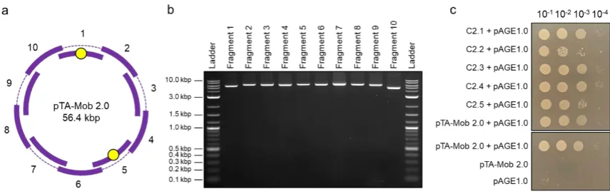
121
liquid layer was transferred to a 15 mL centrifuge tube. Plasmid isolation was then carried out
122
according to Karas et al. [9] and pelleted DNA was resuspended in 50 µ L of elution buffer (Qiagen,
123
#19086).
124
For the initial 12-fragment assembly, 1 µ L of isolated pooled yeast DNA was added to 30 µ L of
125
NEB 5-alpha electrocompetent E. coli cells in a 1.5 mL microcentrifuge tube on ice. The mixture was
126
transferred to a cold 1 mm electroporation cuvette and electroporated at 1.8 kV using the BioRad
127
GenePulser. For the final optimized protocol, 25 µ L of TransforMax Epi300 Electrocompetent E. coli
128
cells were mixed with 1 µ L of isolated pooled yeast DNA on ice. The mixture was transferred to a
129
cold 2 mm electroporation cuvette and electroporated at 2.5 kV. Cells were recovered in 1 mL of
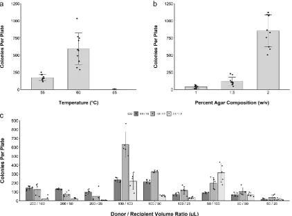
130
Super Optimal broth with Catabolite repression [33] at 37°C, shaking at 225 rpm for 1 hour. Following
131
recovery, 100 µ L of the transformed cell mixture was plated on 1.5% agar (w/v) LB media plates
132
containing gentamicin (40 µ g/mL) and incubated at 37°C overnight.
133
To screen for correctly assembled plasmids, individual E. coli colonies were tested as follows:
134
For the initial 12-fragment assembly, 50 E. coli pTA-Mob 2.0 colonies were streaked onto a fresh
135
LB agar plate containing gentamicin (40 µ g/mL) and incubated at 37°C overnight. A second LB agar
136
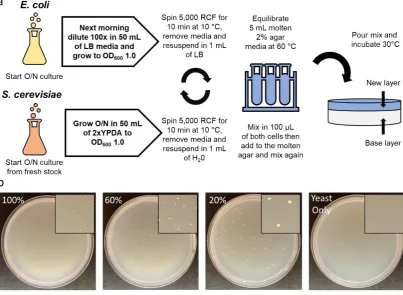
plate supplemented with chloramphenicol (30 µ g/mL) was streaked with Epi300 E. coli containing
137
the pAGE2.0 plasmid and incubated at 37°C overnight [8]. The next morning, each E. coli pTA-Mob
138
2.0 colony was streaked on top of E. coli pAGE2.0 on non-selective LB agar plates and incubated for
3 hours at 37°C. Cells were scraped and resuspended in liquid LB media before performing 10-fold
140
serial dilutions from 10-1 to 10-4. Then, 5 µ L of the serial dilutions for each sample were spot-plated
141
on LB agar plates containing gentamicin (40 µ g/mL) and chloramphenicol (30 µ g/mL) and incubated
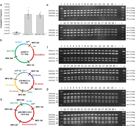
142
at 37°C overnight. The ability of each E. coli colony to conjugate was assessed by transconjugant
143
colony growth at each dilution the following day. One clone was selected, and the plasmid isolated
144
from this strain was named pTA-Mob 2.0. Plasmid DNA was isolated using the EZ-10 Spin Column
145
Plasmid DNA Miniprep Kit (BioBasic Inc., #BS413) and sent for Complete Plasmid Sequencing at the
146
Massachusetts General Hospital DNA Core.
147
For the final optimized plasmid assembly protocol, 30 colonies were tested. First, the colonies
148
were streaked on LB plates supplemented with gentamicin (40 µ g/mL) and incubated at 37°C
149
overnight. A second LB agar plate containing chloramphenicol (30 µ g/mL) was streaked with Epi300
150
E. coli containing the pAGE1.0 plasmid and incubated at 37°C overnight [8]. The next day, overnight
151
cultures of 3 mL LB supplemented with gentamicin (40 µ g/mL) were inoculated with one of the 30
152
newly assembled donor E. coli pTA-Mob 2.0 colonies. In addition, two overnight cultures of recipient
153
E. coli pAGE1.0 were grown in 3 mL LB containing chloramphenicol (30 µ g/mL). As a positive control
154
for conjugation, a 3 mL LB with gentamicin (40 µ g/mL) culture of the original E. coli pTA-Mob 2.0
155
was also inoculated. Cultures were incubated overnight at 37°C in a tube rotator. After 16 hours, 100
156
µ L of donor and 100 µ L of recipient cultures were mixed, spread on non-selective LB agar plates, and
157
incubated for 1 hour at 37°C. The following controls were also performed: i) original donor E. coli
158
pTA-Mob 2.0 with recipient E. coli pAGE1.0 (positive control); ii) original donor E. coli pTA-Mob 2.0
159
with water (negative control), and; iii) recipient E. coli pAGE1.0 with water (negative control).
160
Conjugation plates were then scraped with 1 mL LB media, and cells were transferred to a 1.5 mL
161
microcentrifuge tube. Each mixture was serially diluted 10-fold from 10-1 to 10-5 using LB media, and
162
a 5 µ L volume of each of the serial dilutions was spot-plated onto LB agar plates supplemented with
163
gentamicin (40 µ g/mL) and chloramphenicol (30 µ g/mL). Plates were incubated overnight at 37°C
164
and photographed the next day.
165
2.3. Conjugation within solid media
166
2.3.1. Preparation of E. coli
167
Cultures of 50 mL of LB media supplemented with the appropriate antibiotic (40 µ g/mL
168
gentamicin for E. coli pTA-Mob 2.0 or 40 µ g/mL gentamicin and 30 µ g/mL chloramphenicol for E. coli
169
pAGE1.0 [8] with pTA-Mob [31] and E. coli pBK-RBYV-25-2 [9,34] with pTA-Mob) were inoculated
170
with freshly grown E. coli and incubated overnight at 37°C and 225 rpm in 250 mL Erlenmeyer flasks.
171
The following morning, saturated bacterial cultures were diluted 100x to a total volume of 50 mL in
172
LB media with the appropriate antibiotic in a new flask. The diluted cultures were grown to an optical
173
density at 600 nm (OD600) of 0.5, 1.0, or 2.0. The cultures were then transferred to 50 mL centrifuge
174
tubes and pelleted for 10 minutes at 5,000 relative centrifugal force (rcf). After centrifugation, the
175
supernatants were decanted, and the pellets were resuspended in 1 mL LB media.
176
2.3.2. Preparation of S. cerevisiae VL6-48
177
A 50 mL overnight culture was inoculated with fresh yeast and grown at 30°C at 225 rpm in 2x
178
YPDA media supplemented with ampicillin (100 µ g/mL) in a 250 mL Erlenmeyer flask. The culture
179
was grown to an OD600 of 1.0. The culture was then transferred to a 50 mL centrifuge tube and pelleted
180
for 10 minutes at 5,000 rcf. After decanting the supernatant, the cell pellet was resuspended in 1 mL
181
sterile double deionized H2O (sddH2O).
182
183
2.3.3. Conjugation in solid media procedure
Mixtures of 25, 50, or 100 µ L of E. coli cell suspension with 50, 100, or 200 µ L of S. cerevisiae cell
185
suspension were brought to a final volume of 1 mL with sddH2O in a 1.5 mL microcentrifuge tube.
186
Centrifuge tubes containing 5 mL of either molten CM glucose media lacking histidine and uracil
187
with 1%, 1.5%, or 2% agar (w/v) or molten CM glucose media lacking histidine supplemented with
188
adenine with 2% agar were prepared and held in a water bath set to either 55°C, 60°C, or 65°C. The
189
tubes were removed from the water bath, the cell mixture was added to the molten media and
190
inverted three times to mix. Cells resuspended in agar were then poured onto the respective base
191
plate of 25 mL CM glucose media lacking histidine and uracil with 2% agar or CM glucose media
192
lacking histidine with 2% agar preincubated at 37°C. After a 20-minute drying period, conjugation
193
plates were incubated at 30°C. Successful transconjugant S. cerevisiae colonies were then counted
194
under 5x magnification using the Zeiss Discovery.V8 SteREO microscope at the Biotron Experimental
195
Climate Change Research Centre at the University of Western Ontario.
196
To quantify conjugation frequency, the colony-forming units for the recipient S. cerevisiae were
197
obtained by creating serial dilutions and plating 100 µ L of diluted cells onto YPDA 1% agar plates.
198
The S. cerevisiae plates were dried and then incubated for two days at 30°C prior to counting colonies.
199
2.3.4. Confirmation of successfully transferred plasmids
200
After the four-day incubation period, colony yields were counted. Single colonies were picked
201
from the agar and streaked onto CM glucose media lacking histidine with 2% agar plates containing
202
ampicillin (100 µ g/mL). A total of three successive passages were performed, streaking a small
203
portion of cells after a two-day incubation at 30°C each time. Following the second passage, a small
204
portion of cells were streaked onto LB plates containing gentamicin (40 µ g/mL) to test for surviving
205
donor E. coli. After the third passage, S. cerevisiae colony streaks were resuspended in 100 µ L TE buffer
206
(pH 8). Cell resuspensions were then lysed at 98°C for 15 minutes. Multiplex PCR was performed
207
using Qiagen Multiplex PCR Kit (Qiagen, #206143) using the cell lysate as template for primers
208
(Supplementary Table 1) that bind to pAGE1.0, pTA-Mob 2.0, and pBK-RBYV-25-2 in locations
209
distributed around each plasmid. The Multiplex PCR conditions were as follows: a 95°C hot start for
210
15 minutes, 35 cycles of 94°C for 30 seconds, 60°C for 90 seconds, and 72°C for 45 seconds, followed
211
by a final extension at 72°C for 10 minutes, and a 12°C infinite hold. Multiplex amplification was
212
confirmed using agarose gel electrophoresis with 2 µ L of each Multiplex PCR reaction run on a 2%
213
agarose (w/v) gel.
214
3. Results
215
3.1. PCR-based synthesis of conjugative plasmid pTA-Mob 2.0
216
In previous studies, the non-mobilizable helper plasmid pTA-Mob was used to transfer
217
destination plasmids from bacteria to recipient bacteria [31], algae [9,11,12], and yeast [8]. The
pTA-218
Mob plasmid encodes the machinery required for conjugal transfer of vectors that contain an origin
219
of transfer (oriT) [31]. For this study and future applications, we designed a method to build an
220
alternative version of pTA-Mob, named pTA-Mob 2.0, that can self-mobilize (cis) and replicate in
221
both E. coli and S. cerevisiae. To this end, we used PCR to initially amplify pTA-Mob as nine fragments
222
along with three additional fragments: the S. cerevisiae URA3 gene; a gene cassette containing the S.
223
cerevisiae HIS3 gene, yeast centromere (CEN6), and an autonomously replicating sequence (ARS4);
224
and an oriT cassette. After assembling the fragments into whole plasmids in yeast, we transferred the
225
DNA to E. coli and tested individual colonies for conjugation on top of agar to recipient E. coli. For
226
subsequent work we selected one colony harboring pTA-Mob 2.0 and isolated the plasmid for
227
optimization of a simplified PCR-based plasmid synthesis pipeline. The reason for this optimization
228
step was to simplify the assembly from twelve to ten fragments as well as to obtain cleaner PCR
229
products. This improved protocol functions to accelerate the creation of designer pTA-Mob 2.0
230
variants. We designed new primers to amplify the vector as ten approximately equal-sized
overlapping fragments (Figure 1a, b). Once again, following yeast assembly, we transferred DNA to
232
E. coli and tested for conjugation to a recipient E. coli strain. This initial E. coli to E. coli conjugation
233
analysis provided a rapid approach to evaluate successfully assembled plasmids before testing
234
conjugal transfer to eukaryotic cells. Of the colonies tested, 23 out of the 30 donor colonies conjugated
235
at least as well as the parental pTA-Mob 2.0 (Figure 1c, Supplementary Figure 1).
236
237
Figure 1. Optimized PCR-based synthesis of plasmid pTA-Mob 2.0: (a) Schematic of pTA-Mob 2.0
238
split into ten overlapping fragments. Yellow circles indicate positions of the yeast selection markers
239
HIS3 in fragment 1 and URA3 in fragment 5; (b) Gel electrophoresis of pTA-Mob 2.0 amplified as ten
240
overlapping fragments; (c) Partial conjugation results of newly assembled pTA-Mob 2.0 plasmids
241
(colonies C2.1 to C2.5) to recipient E. coli containing the pAGE1.0 plasmid.
242
3.2. Development and optimization of conjugation within solid media
243
The initial protocol for conjugation in solid media was inspired by the method for direct transfer
244
of DNA from bacteria to yeast, where a polyethylene glycol-treated mixture of bacteria and yeast
245
spheroplasts is suspended in molten agar media and then plated in a Petri dish [19,35]. For the
246
development of the conjugation protocol, we used intact yeast cells and did not treat the mixture with
247
polyethylene glycol. Early attempts performing the conjugation protocol resulted in inconsistent
248
results (data not shown) prompting us to develop an optimized protocol. Optimization parameters
249
included: i) molten agar media temperature, ii) molten agar media agar concentration, and iii)
250
volumes of E. coli and S. cerevisiae cell suspensions harvested at various optical densities (ODs). We
251
used the optimal value for each parameter in subsequent optimization experiments.
252
First, when testing the effect of molten agar media temperature, we found that a temperature of
253
60°C was optimal for transconjugant colony formation (Figure 2a). For the initial agar concentration,
254
2% agar (w/v) was the most conducive to colony formation (Figure 2b). Higher agar concentrations
255
were not practical due to the rapid solidification of the media. We then tested 27 combinations of
256
various volumes of donor and recipient cells harvested at different ODs and counted the number of
257
yeast colonies per plate (Figure 2c). Based on this experiment, mixing 100 µ L of donor and recipient
258
cell suspensions, harvested at an OD600 of 1.0, resulted in the highest number of transconjugant
259
colonies.
261
Figure 2. Optimization of the conjugation protocol in solid media. Colonies per plate yielded from
262
optimization experiments testing: (a) Molten agar media temperature; (b) Molten agar media agar
263
composition prior to the addition of cell mixture; (c) Volumes of E. coli and S. cerevisiae cell
264
suspensions harvested at various optical densities (OD600).
265
We summarized the optimization results in a final protocol for the conjugation in solid media
266
method, as illustrated in Figure 3a, Supplementary Note 1. Using this optimized protocol, the highest
267
number of transconjugants are obtained however the final colonies appear small (Figure 3b). Using
268
smaller volumes of the final mix (100 µ L/100 µ L donor and recipient cell) such as 60% or 20%, fewer
269
but larger colonies can be obtained (Figure 3b).
271
Figure 3. Optimized protocol for conjugation in solid media: (a) Schematic for the final protocol for
272
conjugation from donor E. coli to recipient S. cerevisiae within solid media; (b) Comparison of using
273
100%, 60%, and 20% of the optimized 100 µ L/100 µ L donor and recipient cell ratio.
274
3.3. Conjugation of IncP-based vectors of increasing size
275
Using the optimized protocol, we tested conjugation of plasmids of increasing size: pAGE1.0
276
(18.1 Kbp) [8], pTA-Mob 2.0 (56.4 Kbp), and pBK-RBYV-25-2 (138.5 Kbp) [9,34]. To transfer pAGE1.0
277
and pBK-RBYV-25-2, the donor E. coli also contained the pTA-Mob helper plasmid to allow for
278
mobilization the plasmids in trans [31]. Surprisingly, conjugation with the larger plasmids produced
279
more yeast colonies than with the smaller pAGE1.0 plasmid. Conjugation frequencies were then
280
calculated using the number of transconjugant colonies divided by the colony-forming units obtained
281
from the recipient S. cerevisiae serial dilutions. The conjugation frequencies for pAGE1.0, pTA-Mob
282
2.0, and pBK-RBYV-25-2 to S. cerevisiae were determined to be 1.9 x 10⁻⁶, 3.3 x 10⁻⁵, and 3.1 x 10⁻⁵,
283
respectively (Figure 4a).
285
Figure 4. Conjugal transfer of plasmids increasing in size: (a) Conjugation frequency with donor E.
286
coli containing either pTA-Mob 2.0 or pTA-Mob with either pAGE1.0 or pBK-RBYV-25-2 to recipient
287
S. cerevisiae; Plasmid map and Multiplex PCR primer locations for (b) pAGE1.0; (c) pTA-Mob 2.0; or
288
(d) pBK-RBYV-25-2; Diagnostic Multiplex PCR for 30 transconjugant S. cerevisiae colonies containing
289
(e) pAGE1.0; (f) pTA-Mob 2.0; or (g) pBK-RBYV-25-2. Note: A faint band can be seen around 400 bp
290
in the negative control of the Multiplex PCR for pBK-RBYV-25-2, which is most likely non-specific
291
amplification.
292
To verify that entire plasmids were successfully transferred to yeast, we consecutively
293
restreaked the yeast colonies three times on selective media containing ampicillin to remove any
294
leftover donor E. coli. After the second passage, a portion of each colony was streaked onto an LB
295
plate containing gentamicin and incubated at 37°C overnight. The LB plate did not yield any E. coli
296
colonies after incubation, confirming that any leftover donor E. coli was eliminated during the
297
passage of the yeast colonies. Plasmid DNA from the yeast transconjugants was then isolated after
298
the third passage by lysing the cells in TE buffer and used for diagnostic Multiplex PCR (Figure 4e, f,
299
g). For each plasmid, we tested 30 individual recipient colonies, and all 90 colonies yielded bands of
300
the expected sizes. The Multiplex PCR results indicate that complete conjugal transfer of each
301
plasmid was achieved for every colony tested and suggests that the frequency of conjugation to S.
302
cerevisiae—when using either the pTA-Mob (trans) or pTA-Mob 2.0 (cis) system—does not decrease
303
as plasmid size increases within the tested range using the solid media conjugation protocol.
304
4. Discussion
306
The development of engineered conjugative plasmids such as pTA-Mob [31], pLS20 [36],
307
pRK2013 [18], pRH210 [37], and RP4 [38] provide a simple alternative for DNA transfer to
308
prokaryotic and eukaryotic recipient cells. As recently demonstrated, the development of conjugation
309
methods for species such as the eukaryotic algae P. tricornutum and other species allow for rapid
310
advances in genetic engineering of these organisms [9,11,12]. In this study, we aimed to develop a
311
pipeline for building conjugative plasmids and subsequently demonstrate that such plasmids can be
312
efficiently delivered from a bacterial donor to a eukaryotic recipient within solid media. To achieve
313
this we used E. coli as a conjugative donor and the model yeast S. cerevisiae as the eukaryotic recipient.
314
Trans-kingdom conjugation to S. cerevisiae from E. coli had already demonstrated in liquid conditions
315
and on the surface of solid media, providing a basis for the creation of the conjugation in solid media
316
protocol [7,8].
317
First, we developed a PCR-based plasmid synthesis pipeline for building conjugative plasmids
318
that can propagate in E. coli and yeast. Using this method, we found that 23 out of 30 plasmid clones
319
tested were able to conjugate at least as well as the parental pTA-Mob 2.0 clone. After sequencing
320
three of these plasmids, an average of 6.33 mutations per 56.4 kbp plasmid was found. It has been
321
previously demonstrated that Takara PrimeSTAR GXL polymerase has an error rate of about 8.4 x
322
10-6 substitutions per base per PCR doubling [39]. If we consider each PCR cycle in the optimized
323
protocol as a doubling, the error rate found in our plasmids was determined to be lower than the
324
previously determined value, at around 4.5 x 10⁻⁶ mutations per base per PCR doubling,
325
corresponding to a mutation about every 8.9 kbp. Some of these mutations may have arisen outside
326
of the PCR conditions during plasmid assembly in yeast and or during propagation of plasmids in
327
yeast or E. coli. If desired, mutations could be eliminated by first cloning each correct fragment
328
flanked by preferred unique restriction sites in the plasmid of interest. Fragments could then be easily
329
released by restriction digest, followed by yeast assembly. Since this pipeline utilizes PCR fragments
330
for assembly, incorporating modular components and customizing vectors is rapid and simple.
331
Additional components can be incorporated into the assembly mixture if there is built-in homology
332
on the terminal ends of the fragment. Homology can be easily introduced during amplification by
333
designing primers that contain a “hook” with sequence complementarity to the flanking DNA
334
fragments. To delete components, such as an undesirable gene, a single fragment can be amplified as
335
two separate fragments flanking the unwanted DNA region, creating a seamless deletion of the
336
target. Not only can the pipeline generate functional conjugative plasmids—as indicated with the
337
creation and testing of pTA-Mob 2.0—the pipeline can be adapted for use with any desired
338
customizable vector.
339
During the optimization process, it was identified that the ideal holding temperature for the
340
molten agar media, and at the time the cell mixture was added, was 60°C. Previous studies using
341
different bacterial species have also demonstrated that a “heat shock” step during conjugation results
342
in improved conjugation efficiencies [40–42]. However, it remains unclear as to why the heat shock
343
at 60°C yields an increased number of transconjugant S. cerevisiae colonies. Next, we demonstrated
344
that the number of transconjugant colonies per plate increased with higher agar composition,
345
producing the best results with 2% agar prior to the addition of the cell mixture. As agar concentration
346
increases, the movement of cells is restricted and, therefore, may provide additional stability of the
347
pili during the formation of cell-to-cell attachments between bacteria and yeast. A molten media
348
composition of higher than 2% agar was difficult to pour and impacted the ability to ensure the top
349
layer was even for the conjugation plates. If needed, higher percentage compositions of the media
350
could be achieved through the use of low-melting point agarose, to avoid premature solidification of
351
the top layer.
352
Next, we demonstrated that cell density had a significant effect on yeast colony formation. After
353
optimization, the most consistent conditions for transconjugant colony yields was achieved when
both donor and recipient cultures were harvested at an OD600 of 1.0, and 100 µ L of each cell
355
resuspension was used (approximately 2.0 x 10⁷ yeast cells per mL of top agar based on
colony-356
forming unit counts). For experiments where faster growth of yeast colonies is beneficial or to obtain
357
larger transconjugant yeast colonies, decreasing the volume of cell resuspensions used can yield such
358
a result. During the early optimization process, it was found that the thickness in the top agar layer
359
had an impact on the formation of transconjugant yeast colonies (data not shown). Thus, it is essential
360
that the total top agar plus cell mixture volume is 6 mL.
361
Once the protocol for conjugation within solid media was optimized, three plasmids of
362
increasing size were then tested: pAGE1.0 (18.1 Kbp), pTA-Mob 2.0 (56.4 Kbp), and pBK-RBYV-25-2
363
(138.5 Kbp). For donor E. coli containing pAGE1.0 and pBK-RBYV-25-2, the vectors were mobilized
364
using pTA-Mob as a helper plasmid for trans conjugation. In comparison, pTA-Mob 2.0 is able to
self-365
transfer via cis conjugation. As both types of vectors are no longer mobilizable once in S. cerevisiae,
366
the difference in colony yields would not be confounded by re-conjugation of recipients in the cis
367
setup. It has been previously stated that conjugation efficiency decreases as vector size increases
368
[5,43]. In this study, however, there was found to be no noticeable drop when comparing the
369
transconjugant yields between the 56.4 kbp vector (pTA-Mob 2.0) and the 138.5 kbp vector
(pBK-370
RBYV-25-2). From the Multiplex PCR reactions, we were able to confirm that the complete transfer
371
of all three vectors occurred in every colony screened. With no substantial decrease in conjugation
372
frequency and complete transfer of each vector confirmed, the upper size limit of conjugal transfer to
373
eukaryotic cells has not yet been met. As for why pAGE1.0, a relatively small vector, did not conjugate
374
as well as the larger plasmids, additional investigation is required.
375
Prior to this study, conjugation within solid media to yeast had not been demonstrated. This
376
method allows for the transfer plasmids of at least 138.5 kbp; however, the upper size limit of conjugal
377
transfer to eukaryotes has still yet to be determined. The protocol may be adjusted for transfer to
378
other prokaryotic or eukaryotic organisms as well. For example, some culturing methods for
379
microanaerobic and anaerobic bacteria rely on coating or mixing cells with a semi-solid agar media
380
layer for growth and certain eukaryotic algae have been cultured on plates when allowed to grow
381
within the agar layer of the plate itself [27–29]. Species that require semi-solid or solid media to grow
382
within may be able to be engineered using an adapted version of this conjugation protocol.
383
Additionally, protocols for more accurate enumeration of colony-forming units rely on submerging
384
cells in agar and allowing an increased number of smaller colonies to grow and be counted per plate
385
[30]. By eliminating the need for plating with a spreader, a more accurate count of cells in a sample
386
can be made by removing the error arising from cells sticking to the spreader. Thus, with this
387
optimized conjugation protocol, a more accurate enumeration of transconjugants can be estimated
388
for experimental conditions. The result is a simple, accurate protocol for conjugal transfer to S.
389
cerevisiae that permits the transfer of large vectors of at least up to 138.5 kbp.
390
Supplementary Materials: Supplementary materials are available online.
391
Author Contributions: Conceptualization, M.P.M.S., B.J.K; methodology, M.P.M.S., R.S.M., S.H., P.J., B.J.K;
392
validation, M.P.M.S., R.S.M., S.H., P.J; investigation, M.P.M.S., R.S.M., S.H., P.J., B.J.K; formal analysis,
393
M.P.M.S.; resources, D.R.E., B.J.K; writing—original draft preparation, M.P.M.S., B.J.K; writing—review and
394
editing, M.P.M.S., R.S.M., S.H., P.J., D.R.E., B.J.K; visualization, M.P.M.S., R.S.M., S.H., P.J., D.R.E., B.J.K;
395
supervision, P.J., B.J.K; project administration, B.J.K; funding acquisition, B.J.K.
396
Funding: This research was funded by: Defense Advanced Research Projects Agency (DARPA), Agreement
397
Number: D18AC00035. The views, opinions and/or findings expressed are those of the author and should not
398
be interpreted as representing the official views or policies of the Department of Defense or the U.S. Government.
399
Research in the labs of D.R.E., and B.J.K. is also supported by Natural Sciences and Engineering Research Council
400
of Canada (NSERC), RGPIN-2015-04800, RGPIN-2018-06172 respectively. In addition, this study was supported
401
by Designer Microbes Inc (DMI).
Conflicts of Interest: B.J.K. is Chief Executive Officer of Designer Microbes Inc. B.J.K and P.J. hold Designer
403
Microbes Inc. stock.
404
References
405
1. Cabezón, E.; Ripoll-Rozada, J.; Peña, A.; de la Cruz, F.; Arechaga, I. Towards an integrated
406
model of bacterial conjugation. FEMS Microbiol. Rev. 2014, 39, 81–95.
407
2. Chilton, M.-D.; Drummond, M.H.; Merlo, D.J.; Sciaky, D.; Montoya, A.L.; Gordon, M.P.;
408
Nester, E.W. Stable incorporation of plasmid DNA into higher plant cells: the molecular basis
409
of crown gall tumorigenesis. Cell 1977, 11, 263–271.
410
3. Christie, P.J. Agrobacterium tumefaciens T-complex transport apparatus: a paradigm for a
411
new family of multifunctional transporters in eubacteria. J. Bacteriol. 1997, 179, 3085–3094.
412
4. Stachel, S.E.; Zambryski, P.C. Generic trans-kingdom sex? Nature 1989, 340, 190–191.
413
5. Heinemann, J.A.; Sprague, G.F. Bacterial conjugative plasmids mobilize DNA transfer
414
between bacteria and yeast. Nature 1989, 340, 205–209.
415
6. Bundock, P.; den Dulk-Ras, A.; Beijersbergen, A.; Hooykaas, P.J. Trans-kingdom T-DNA
416
transfer from Agrobacterium tumefaciens to Saccharomyces cerevisiae. EMBO J. 1995, 14,
417
3206–3214.
418
7. Moriguchi, K.; Yamamoto, S.; Ohmine, Y.; Suzuki, K. A Fast and Practical Yeast
419
Transformation Method Mediated by Escherichia coli Based on a Trans-Kingdom Conjugal
420
Transfer System: Just Mix Two Cultures and Wait One Hour. PLoS One 2016, 11, e0148989.
421
8. Brumwell, S.L.; MacLeod, M.R.; Huang, T.; Cochrane, R.R.; Meaney, R.S.; Zamani, M.;
422
Matysiakiewicz, O.; Dan, K.N.; Janakirama, P.; Edgell, D.R.; et al. Designer Sinorhizobium
423
meliloti strains and multi-functional vectors enable direct inter-kingdom DNA transfer. PLoS
424
One 2019, 14, e0206781.
425
9. Karas, B.J.; Diner, R.E.; Lefebvre, S.C.; McQuaid, J.; Phillips, A.P.R.; Noddings, C.M.; Brunson,
426
J.K.; Valas, R.E.; Deerinck, T.J.; Jablanovic, J.; et al. Designer diatom episomes delivered by
427
bacterial conjugation. Nat. Commun. 2015, 6, 6925.
428
10. Diner, R.E.; Bielinski, V.A.; Dupont, C.L.; Allen, A.E.; Weyman, P.D. Refinement of the Diatom
429
Episome Maintenance Sequence and Improvement of Conjugation-Based DNA Delivery
430
Methods. Front. Bioeng. Biotechnol. 2016, 4, 65.
431
11. Slattery, S.S.; Diamond, A.; Wang, H.; Therrien, J.A.; Lant, J.T.; Jazey, T.; Lee, K.; Klassen, Z.;
432
Desgagné-Penix, I.; Karas, B.J.; et al. An Expanded Plasmid-Based Genetic Toolbox Enables
433
Cas9 Genome Editing and Stable Maintenance of Synthetic Pathways in Phaeodactylum
434
tricornutum. ACS Synth. Biol. 2018, 7, 328–338.
435
12. Wang, H.; Slattery, S.; Karas, B.; Edgell, D. Delivery of the Cas9 or TevCas9 System into
436
Phaeodactylum tricornutum via Conjugation of Plasmids from a Bacterial Donor.
PROTOCOL 2018, 8.
438
13. Waters, V.L. Conjugation between bacterial and mammalian cells. Nat. Genet. 2001, 29, 375–
439
376.
440
14. Kunik, T.; Tzfira, T.; Kapulnik, Y.; Gafni, Y.; Dingwall, C.; Citovsky, V. Genetic transformation
441
of HeLa cells by Agrobacterium. Proc. Natl. Acad. Sci. 2001, 98, 1871–1876.
442
15. Fernández-González, E.; de Paz, H.D.; Alperi, A.; Agúndez, L.; Faustmann, M.; Sangari, F.J.;
443
Dehio, C.; Llosa, M. Transfer of R388 derivatives by a pathogenesis-associated type IV
444
secretion system into both bacteria and human cells. J. Bacteriol. 2011, 193, 6257–6265.
445
16. Schröder, G.; Schuelein, R.; Quebatte, M.; Dehio, C. Conjugative DNA transfer into human
446
cells by the VirB/VirD4 type IV secretion system of the bacterial pathogen Bartonella henselae.
447
Proc. Natl. Acad. Sci. U. S. A. 2011, 108, 14643–14648.
448
17. Aune, T.E.V.; Aachmann, F.L. Methodologies to increase the transformation efficiencies and
449
the range of bacteria that can be transformed. Appl. Microbiol. Biotechnol. 2010, 85, 1301–1313.
450
18. Heinze, S.; Kornberger, P.; Grätz, C.; Schwarz, W.H.; Zverlov, V. V.; Liebl, W. Transmating:
451
conjugative transfer of a new broad host range expression vector to various Bacillus species
452
using a single protocol. BMC Microbiol. 2018, 18, 56.
453
19. Karas, B.J.; Jablanovic, J.; Sun, L.; Ma, L.; Goldgof, G.M.; Stam, J.; Ramon, A.; Manary, M.J.;
454
Winzeler, E.A.; Venter, J.C.; et al. Direct transfer of whole genomes from bacteria to yeast. Nat.
455
Methods 2013, 10, 410–412.
456
20. Hill, K.E.; M. Top, E. Gene transfer in soil systems using microcosms. FEMS Microbiol. Ecol.
457
1998, 25, 319–329.
458
21. Brophy, J.A.N.; Triassi, A.J.; Adams, B.L.; Renberg, R.L.; Stratis-Cullum, D.N.; Grossman,
459
A.D.; Voigt, C.A. Engineered integrative and conjugative elements for efficient and inducible
460
DNA transfer to undomesticated bacteria. Nat. Microbiol. 2018, 3, 1043–1053.
461
22. Ronda, C.; Chen, S.P.; Cabral, V.; Yaung, S.J.; Wang, H.H. Metagenomic engineering of the
462
mammalian gut microbiome in situ. Nat. Methods 2019, 16, 167–170.
463
23. Itaya, M.; Sato, M.; Hasegawa, M.; Kono, N.; Tomita, M.; Kaneko, S. Far rapid synthesis of
464
giant DNA in the Bacillus subtilis genome by a conjugation transfer system. Sci. Rep. 2018, 8,
465
8792.
466
24. Zechner, E.L.; Moncalián, G.; de la Cruz, F. Relaxases and Plasmid Transfer in Gram-Negative
467
Bacteria. In Type IV Secretion in Gram-Negative and Gram-Positive Bacteria; Backert, S.,
468
Grohmann, E., Eds.; Springer, Cham, 2018; Vol. 413, pp. 93–113 ISBN 978-3-319-75241-9.
469
25. Christie, P.J. Type IV secretion: Intercellular transfer of macromolecules by systems
470
ancestrally related to conjugation machines. Mol. Microbiol. 2001, 40, 294–305.
26. Bradley, D.E.; Taylor, D.E.; Cohen, D.R. Specification of surface mating systems among
472
conjugative drug resistance plasmids in Escherichia coli K-12. J. Bacteriol. 1980, 143, 1466–1470.
473
27. Mason, J.H. Isolation of anaerobic bacteria by a modified shake method. J. Gen. Microbiol. 1953,
474
8, 263–264.
475
28. Nagasaki, K.; Imai, I. Solid-phase culture of marine phytoflagellates. Bull. Japanese Soc.
476
Mircobial Ecol. 1994, 9, 37–43.
477
29. Lakeman, M.B.; Cattolico, and R.A. Cryptic Diversity in Phytoplankton Cultures is Revealed
478
Using a Simple Plating Technique. J. Phycol. 2007, 43, 662–674.
479
30. Koch, A.L. Growth Measurement. In Methods for General and Molecular Biology; Gerhard, P.,
480
Murray, R.G.E., Wood, W.A., Krieg, N.R., Eds.; American Society for Microbiology Press:
481
Washington, D.C., 2007; pp. 172–199.
482
31. Strand, T.A.; Lale, R.; Degnes, K.F.; Lando, M.; Valla, S. A new and improved
host-483
independent plasmid system for RK2-based conjugal transfer. PLoS One 2014, 9, e90372.
484
32. Karas, B.J.; Jablanovic, J.; Irvine, E.; Sun, L.; Ma, L.; Weyman, P.D.; Gibson, D.G.; Glass, J.I.;
485
Venter, J.C.; Hutchison, C.A.; et al. Transferring whole genomes from bacteria to yeast
486
spheroplasts using entire bacterial cells to reduce DNA shearing. Nat. Protoc. 2014, 9, 743–750.
487
33. Hanahan, D. Studies on transformation of Escherichia coli with plasmids. J. Mol. Biol. 1983,
488
166, 557–580.
489
34. Diner, R.E.; Noddings, C.M.; Lian, N.C.; Kang, A.K.; McQuaid, J.B.; Jablanovic, J.; Espinoza,
490
J.L.; Nguyen, N.A.; Anzelmatti, M.A.; Jansson, J.; et al. Diatom centromeres suggest a
491
mechanism for nuclear DNA acquisition. Proc. Natl. Acad. Sci. U. S. A. 2017, 114, E6015–E6024.
492
35. Karas, B.J.; Moreau, N.G.; Deerinck, T.J.; Gibson, D.G.; Venter, J.C.; Smith, H.O.; Glass, J.I.
493
Direct Transfer of a Mycoplasma mycoides Genome to Yeast Is Enhanced by Removal of the
494
Mycoides Glycerol Uptake Factor Gene glpF. ACS Synth. Biol. 2019, 8, 239–244.
495
36. Miyano, M.; Tanaka, K.; Ishikawa, S.; Takenaka, S.; Miguel-Arribas, A.; Meijer, W.J.J.; Yoshida,
496
K. ichi Rapid conjugative mobilization of a 100kb segment of Bacillus subtilis chromosomal
497
DNA is mediated by a helper plasmid with no ability for self-transfer. Microb. Cell Fact. 2018,
498
17, 13.
499
37. Moriguchi, K.; Yamamoto, S.; Tanaka, K.; Kurata, N.; Suzuki, K. Trans-Kingdom Horizontal
500
DNA Transfer from Bacteria to Yeast Is Highly Plastic Due to Natural Polymorphisms in
501
Auxiliary Nonessential Recipient Genes. PLoS One 2013, 8, e74590.
502
38. Quandt, J.; Clark, R.G.; Venter, A.P.; Clark, S.R..; Twelker, S.; Hynes, M.F. Modified RP4 and
503
Tn5-Mob derivatives for facilitated manipulation of large plasmids in Gram-negative bacteria.
504
Plasmid 2004, 52, 1–12.
39. Potapov, V.; Ong, J.L. Examining Sources of Error in PCR by Single-Molecule Sequencing.
506
PLoS One 2017, 12, e0169774.
507
40. Zhang, S.; Chen, T.; Jia, J.; Guo, L.; Zhang, H.; Li, C.; Qiao, R. Establishment of a highly efficient
508
conjugation protocol for Streptomyces kanamyceticus ATCC12853. Microbiologyopen 2019, 8,
509
e00747.
510
41. Zeng, X.; Ardeshna, D.; Lin, J. Heat Shock-Enhanced Conjugation Efficiency in Standard
511
Campylobacter jejuni Strains. Appl. Environ. Microbiol. 2015, 81, 4546–4552.
512
42. Kirk, J.A.; Fagan, R.P. Heat shock increases conjugation efficiency in Clostridium difficile.
513
Anaerobe 2016, 42, 1–5.
514
43. Heinemann, J.A.; Sprague, G.F. Transmission of Plasmid DNA to Yeast by Conjugation with
515
Bacteria. Methods Enzymol. 1991, 194, 187–195.