Copyright © 2002, American Society for Microbiology. All Rights Reserved.
Development and Use of Multimeric Major Histocompatibility
Complex Molecules
Tim F. Greten
1and Jonathan P. Schneck
2*
Department of Gastroenterology and Hepatology, Medizinische Hochschule Hannover, 30655 Hannover, Germany,1and
Department of Pathology, Johns Hopkins University, Baltimore, Maryland 212052
Enumeration and analysis of antigen-specific T cells play a central role in cellular immunology. While many methodolo-gies have been established for the analysis of humoral re-sponses, until recently the direct analysis of antigen-specific T-cell responses was extremely difficult. Difficulty in analyzing T cells was partially due to the fact that T lymphocytes recog-nize cell surface peptide-major histocompatibility complex (MHC) complexes and also due to the intrinsic low affinity of T-cell receptors (TCR) for cognate ligand. In this review, we discuss some new methodologies for visualizing antigen-spe-cific T cells and how they have impacted our understanding of cellular immune responses.
In the past, several methods were used to quantitate antigen-specific T-cell responses. Traditionally, these assays were func-tional assays, dependent on the proliferation or lytic activity of antigen-specific T cells in vitro. To improve the sensitivity of these assays, it was necessary to expand the population of antigen-specific T cells in vitro before measuring effector func-tions, such as cytolysis, proliferative responses, or cytokine release. Serial limiting-dilution assays (LDA) were the old “gold standard” for quantitative analysis of antigen-specific cytotoxic T lymphocytes (CTL) (19). This assay depended on the ability of antigen-specific T cells to survive, function, and proliferate in vitro upon stimulation. Therefore, the number of antigen-specific CTL determined by LDA significantly under-estimated the true number of antigen-specific CTL and also lacked further detailed information about the characteristics of antigen-specific CD8⫹T cells in vivo.
Recently, new assays based on cytokine induction and/or release have been developed. These assays do not require in vitro proliferation of antigen-specific T cells but do depend on peptide-induced production of cytokines. Cytokine production can be detected in an ELISPOT assay (34), intracellularly (29, 31), or captured on the cell surface (8) and analyzed by flow cytometry. Analysis for cytokine-producing cells has influenced our estimate of antigen-specific T-cell precursor frequency. While these methods have been useful in visualizing cells on the basis of effector cytokine release, there is also a skewing that occurs depending on which cytokine is being assayed that is not present if one can visualize T cells solely based on TCR specificity. In addition, due to technical aspects of the assays, one cannot keep cells alive for additional studies after ELI-SPOT assay or intracellular cytokine staining.
Anticlonotypic TCR-specific antibodies have also been
use-ful in analyzing the antigen-specific T-cell response in vivo. However, different T cells can use a variety of TCR to recog-nize the same peptide-MHC complex; therefore, anticlono-typic TCR, by definition, underestimate the polyclonal nature of the T-cell response.
Recent advances have enabled the use soluble multivalent analogs of MHC complexes to study T-cell specificity. While it has been possible to analyze TCR-peptide-MHC interactions using recombinant soluble monovalent MHC molecules, these molecules do not bind T cells stably and allow flow cytometry-based visualization of antigen-specific cells. TCR have a low binding affinity (Kd) for peptide MHC complexes that is much
lower than that of antibodies for their antigen (39) and also a very short dwell time (11, 51), making it difficult to analyze the interaction of soluble monovalent MHC with cognate TCR by standard flow cytometry-based assays. Therefore, recombinant monomeric MHC molecules have not been used to detect antigen-specific T cells. However, multimerized MHC mole-cules have a significantly increased avidity for their TCR. Mul-tivalent MHC molecules bind with their different arms inde-pendently to the same cell, resulting in a stable binding even if individual TCR-peptide-MHC complex interactions are unsta-ble (Fig. 1).
There are two broad approaches for generating multivalent MHC complexes. Altman et al. (2) first described the use of HLA-peptide tetrameric complexes. Peptide-MHC tetrameric complexes depend on the production of soluble peptide-MHC tagged with a biotin residue and complexed through fluores-cent labeled strepavidin. The modified MHC heavy-chain ex-tracellular domain sequences are expressed inEscherichia coli
and refolded around a specific peptide and2m. These mol-ecules bind stably to T cells displaying the cognate TCR and have been successfully used to directly visualize antigen-spe-cific cells ex vivo by flow cytometry (2). Tetramer technology has recently been reviewed (5, 54).
Multimeric peptide-MHC complexes can also be formed by using immunoglobulin as a molecular scaffold (13). In this system, the extracellular domains of MHC molecules are fused with the constant region of an immunoglobulin heavy chain separated by a short amino acid linker. In contrast to tet-rameric MHC complexes, MHC-immunoglobulin G (MHC-Ig) fusion proteins are expressed in eukaryotic cells, making it unnecessary to refold the denatured MHC molecule. They can be easily loaded with the peptide of interest and are stable at 4°C. Peptide-loaded MHC-IgG dimers can be used for the identification of antigen-specific CTL as first described by Greten et al. in 1998 (22). A number of currently available techniques for monoclonal antibodies can be easily combined
* Corresponding author. Mailing address: Johns Hopkins Univer-sity, 720 Rutland Ave., Ross 664G, Baltimore, MD 21205. Phone: (410) 614-4589. Fax: (410) 614-3548. E-mail: [email protected].
216
on August 17, 2020 by guest
http://cvi.asm.org/
with these MHC-Ig fusion antibodies (such as binding antibod-ies to specific beads, etc.) to phenotypically characterize anti-gen-specific CTL populations.
Use of multimeric MHC complexes in analysis of the cellu-lar immune response.Some of the first insights into the robust nature of the CD8 portion of the immune response were found when multimeric MHC were used to analyze virus-specific CD8⫹T cells. Many studies have shown virus-specific T-cell
frequencies of between 0.1 and 15% of the CD8⫹ T cells.
T-cell responses to major viral infections, including human immunodeficiency virus, Epstein-Barr virus, human T-cell leu-kemia virus type 1 (HTLV-1), hepatitis B virus, and hepatitis C virus, have been evaluated. The fact that up to 10 to 15% of all CD8⫹cells are directed at one viral epitope raised the
ques-tion of how the T-cell memory repertoire can accommodate new infections. A recent analysis of this issue indicates plastic-ity in the repertoire of memory T cells that changes upon new infection (46). Upon infection with a new virus, the memory compartment changes and cross-reactive T-cell epitope re-sponses become more prominent. Thus, individuals accommo-date to new viral infections by changes in the TCR repertoire in memory T-cell population (46).
Multimeric MHC complexes have also been used to identify tumor-specific CTL in melanoma patients (4, 27, 35, 56). High frequencies of Melan-A specific CD8⫹ T cells can also be
found in healthy individuals (44). Others have investigated the T-cell response to different vaccination approaches in vivo (14, 26, 37). Experiments using animal tumor models are just be-ginning to shed some more light on the role of antigen-specific CTL in tumor immunology (28, 49).
Multimeric MHC class I complexes are also used to analyze the antigen-specific T-cell response to malaria (6), cytomega-lovirus (20, 47),Mycobacterium tuberculosis (50), and simian immunodeficiency virus (15, 32, 42). Others have used tet-rameric MHC molecules to analyze minor MHC-restricted CTL (41) and have used nonclassical MHC tetramers, which bind to NK cells (1, 7).
Multimeric MHC molecules allow not only for enumeration and characterization of the antigen-specific T-cell response in the peripheral blood or spleens of mice but also in local ana-tomic niches and sites of inflammatory pathology. This infor-mation can be valuable for the understanding of the pathology of immunology-related disorders. Greten et al. were able to demonstrate a threefold enrichment of HTLV-1-specific CTL cells in the cerebrospinal fluid of patients with HAM/TSP, a demyelinating disorder (22). Others have analyzed antigen-specific T cells in melanoma tumors (3), directly isolated from the livers of patients with hepatitis (25), and in the synovial fluid of patients with treatment-resistant Lyme arthritis (40). All these studies show that a higher frequency of the presump-tive pathogenic cells accumulate at the site of pathology and strongly implicate these cells in the disease process.
Multimeric MHC molecules can be used to analyze antigen-specific T cells from tissue in situ. Several groups have tried using multimeric MHC complexes in immunohistochemistry analysis. However, the model of how multimeric MHC mole-cules bind stably to antigen-specific T cells (see above) suggests that these reagents may only work on viable cell membranes. It is very difficult to imagine that different MHC complexes on one molecule find different TCR on a cell membrane in a fixed
FIG. 1. Multivalent molecules bind stably to T cells. Different arms from the same molecule bind independently to the same cell. Therefore, the avidity of these complexes is increased, inducing a stable binding of multimeric molecules to the TCR.
on August 17, 2020 by guest
http://cvi.asm.org/
tissue section. Therefore, initial experiments used a technique with viable 200- to 500-m tissue sections (23, 48). The ability to identify antigen-specific T cells directly in situ will provide important insights into the anatomic localization of antigen-specific T cells during both normal and pathological immune responses.
Recent studies have also highlighted the use of multimeric MHC in analysis of the maturation of immune responses. In-vestigators have shown, by using multimeric MHC, that with there is an avidity maturation of immune responses (9, 45). This has been interpreted as a focusing of affinity during the maturation of immune responses. Focusing of affinity was thought to primarily occur through selection of specific high-affinity TCR clones. However, recent work has shown that the state of activation of the T cell has a dramatic impact on its ability to bind multivalent MHC (21). This occurs by the pro-cess of TCR reorganization (21) and occurs even though TCR do not increase their sensitivity by undergoing a process of gene mutations that mature to become high-affinity receptors for antigen (reviewed in reference 38). This process is of avid-ity maturation can ensure that T cells, once activated, readily recognize the low density of antigen in the periphery even in the presence of a relatively modest intrinsic affinity of the TCR for a specific peptide-MHC complex. A recent follow-up study also showed that tetramer binding was sensitive to cholesterol levels (16). This may be reflected in different cholesterol levels associated with different states of activation that affect multi-meric MHC binding. Thus, alternate states of activation could also explain the difference seen in avidity during the matura-tion of the immune response in vivo.
Differences in effector function have been reported since not all multimer-positive cells express effector functions such as the lysis of target cells or the release of cytokines such as interferon gamma. This has been observed in multiple viral systems (10, 22, 55, 57) and was recently reviewed (52). The appearance of these dysfunctional CD8 cells may relate to magnitude of antigen presented to T cells and/or to the loss of CD4 population. It appears that the dysfunctional state can be reversed by the addition of exogenous cytokines. The physio-logic role of antigen-specific cells that do not produce “nor-mal” effector cytokines is obviously an area of great interest.
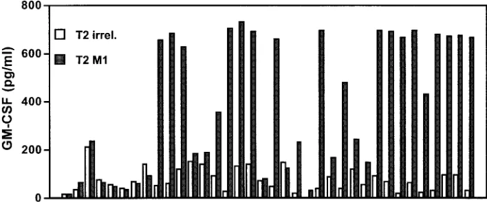
Potential therapeutic uses of multimeric MHC.While many investigators have used multimeric peptide-MHC complexes to enumerate T cells, these molecules are potentially valuable therapeutic tools for the generation of antigen-specific T cells. After one or two in vitro stimulations of peripheral blood mononuclear cells (PBMC) with a specific peptide, antigen-specific CTL cells can be identified by multimeric peptide-MHC complexes and directly isolated from mixed CTL bulk cultures by cell sorting (17, 18). This method clearly simplifies the generation of antigen-specific CTL and may facilitate adoptive T-cell therapies. More interestingly, investigators have been able to separate antigen-specific CTL with high-affinity TCR from those with low-high-affinity TCR by using pep-tide-MHC tetramers (53, 56). Sorted T cells can further be amplified in vitro and used therapeutically. A simpler tech-nique with MHC-immunoglobulin complexes does not even require a cell sorter instrument but instead combines the use of multimeric MHC complexes with magnetic beads specific for murine IgG molecules to enrich for antigen-specific cells. Us-ing this approach, we have been able to enrich influenza-specific T cells (Fig. 2). PBMC from an HLA-A2-positive do-nor were stimulated with the influenza M1 peptide for 8 days in the presence of recombinant interleukin-2 (IL-2). PBMC were incubated with M1 peptide-loaded HLA-A2–IgG. Excess MHC-IgG protein was washed off, and anti-IgG1-coated mag-netic beads were added for the isolation of M1-specific CD8⫹
T cells by using an MACS column. Purified M1-specific T cells were subcloned into 96-well plates in different dilutions and nonspecifically restimulated using phytohemagglutinin and IL-2. Analysis of T-cell clones by cytokine release with peptide-loaded T2 cells as targets revealed that a high percentage of these clones was specific for the influenza M1 peptide. This clearly demonstrates the power of this technique to isolate peptide-specific T cells from in vitro cultures to generate T-cell clones.
MHC class II tetramer and dimer.Similar to the analysis of CD8⫹CTL, a number of groups have begun to use class II
dimers and tetramers to analyze the antigen-specific CD4⫹
T-cell response. Multimeric MHC class II complexes differ from MHC class I constructs due to structural differences be-tween MHC class I and class II molecules. Whereas MHC class
FIG. 2. Using magnetically labeled anti-IgG antibodies, influenza M1-specific CTL were enriched from peripheral blood, restimulated in vitro and tested for specificity in a granulocyte-macrophage colony-stimulating factor secretion assay. A total of 36 independent clones were analyzed, and 22 of these demonstrated clear specificity for the influenza M1 peptide.
on August 17, 2020 by guest
http://cvi.asm.org/
I molecules consist of a single chain (␣1-␣3, transmembrane and intracellular domain), MHC class II molecules consist of two transmembrane glycoprotein chains, which are nonco-valently bound to each other. Currently, two general ap-proaches exist for the generation of multimeric class II MHC complexes (12, 24, 40). The two chains of the MHC molecule are expressed in different cell systems and are either fused to a biotinylation site to make tetravalent molecules or fused to the heavy and light chain of an immunoglobulin sequence for the generation of dimeric MHC-immunoglobulin molecules. In most multimeric MHC class II constructs, the antigenic pep-tide has to be covalently bound to the beta chain of the class II MHC complex. This affords additional stability to the complex. To facilitate chain pairing in these constructs, a rate-limiting step, Kalandadze et al. have included c-jun and c-fos zippers sites at the carboxy-terminal end of the alpha and beta chains to improve the stability of the MHC class II molecules (30).
Current data on MHC class II-specific T cells suggest that the number of antigen-specific CD4⫹T cells are very low and
might actually be under the detection limit of flow cytometry in many cases. In many systems examined so far, multimeric MHC class II constructs can only detect specific CD4⫹T cells
after one in vitro stimulation (33, 43). A number of studies with MHC class II tetramers indicate that the frequency of antigen-specific T lymphocytes, which is in the range of 0.2 to 0.6% of the T cells, is under the detection limit of flow cytometry. Combining the dimer and/or tetramer technology with the use of a fluorescent dye (carboxyfluorescein succinimide esther [CSFE]) (36) allows for the quantitative analysis after cell division. The CSFE technique is applicable to in vitro cell division, as well as to in vivo division of adoptively transferred cells, and can resolve multiple successive generations by using flow cytometry. Each time a cell divides, CSFE is apportioned equally among the two daughter cells, which contain half of the fluorescence. The number of cell divisions can be determined by comparing the resultant CSFE fluorescence to the original fluorescence of the parent population. Using this approach, the absolute number of precursor frequencies can be esti-mated, although this type of assay still involves a single in vitro stimulation, which may influence TCR repertoire usage and T-cell function.
In summary, recent findings in molecular and structural im-munology have led to the development of a new technique in cellular immunology. Multimeric MHC molecules have not only brought new insights into direct cellular T-cell responses in infectious disease but also into different aspects of tumor immunology. Apart from simple counting of antigen-specific CTL, these reagents can also be used for qualitative analysis, and recent findings suggest that they might even have an effect as an immunomodulating reagent.
ACKNOWLEDGMENTS
Support for this work was provided by NIH grants AI-29575 and AI-44129 and by NMSS (RG 2637 A2/1). Additional funding for this study was provided by Pharmingen. Under licensing agreements be-tween Pharmingen and Johns Hopkins University, J.P.S. and T.F.G. are entitled to a share of the royalty received by the university on sales of DimerX products. J.P.S. is also a paid consultant to Pharmingen. The terms of this agreement are managed by Johns Hopkins University in accordance with its conflict-of-interest policies. T.F.G. is supported by a grant from the Deutsche Forschungsgemeinschaft (Gr 1511 1-1
and 2-1) and the Wilhelm Sander Stiftung. Additional details about the Schneck lab are available online (http://pathology2.jhu.edu /schnecklab).
REFERENCES
1.Allan, D. S., M. Colonna, L. L. Lanier, T. D. Churakova, J. S. Abrams, S. A. Ellis, A. J. McMichael, and V. M. Braud.1999. Tetrameric complexes of human histocompatibility leukocyte antigen (HLA)-G bind to peripheral
blood myelomonocytic Cells. J. Exp. Med.189:1149–1156.
2.Altman, J. D., P. Moss, P. Goulder, D. H. Barouch, W. M. McHeyzer, J. I. Bell, A. J. McMichael, and M. M. Davis.1996. Phenotypic analysis of
anti-gen-specific T lymphocytes. Science274:94–96.
3.Anichini, A., A. Molla, R. Mortarini, G. Tragni, I. Bersani, M. Di Nicola, A. M. Gianni, S. Pilotti, R. Dunbar, V. Cerundolo, and G. Parmiani.1999. An expanded peripheral T cell population to a cytotoxic T lymphocyte (CTL)-defined, melanocyte-specific antigen in metastatic melanoma patients impacts on generation of peptide-specific CTLs but does not overcome tumor escape from immune surveillance in metastatic lesions. J. Exp. Med.
190:651–667.
4.Baurain, J. F., D. Colau, N. van Baren, C. Landry, V. Martelange, M. Vikkula, T. Boon, and P. G. Coulie.2000. High frequency of autologous anti-melanoma CTL directed against an antigen generated by a point
mu-tation in a new helicase gene. J. Immunol.164:6057–6066.
5.Bercovici, N., M. T. Duffour, S. Agrawal, M. Salcedo, and J. P. Abastado.
2000. New methods for assessing T-cell responses. Clin. Diagn. Lab.
Immu-nol.7:859–864.
6.Bonelo, A., D. Valmori, F. Triponez, J. M. Tiercy, G. Mentha, J. Oberholzer, P. Champagne, J. F. Romero, F. Esposito, I. Nebie, C. Barbey, P. Romero, S. Herrera, G. Corradin, and J. A. Lopez.2000. Generation and
characteriza-tion of malaria-specific human CD8⫹lymphocyte clones: effect of natural
polymorphism on T cell recognition and endogenous cognate antigen
pre-sentationby liver cells. Eur. J. Immunol.30:3079–3088.
7.Braud, V. M., D. S. Allan, C. A. O’Callaghan, K. Soderstrom, A. D’Andrea, G. S. Ogg, S. Lazetic, N. T. Young, J. I. Bell, J. H. Phillips, L. L. Lanier, and A. J. McMichael.1998. HLA-E binds to natural killer cell receptors CD94/
NKG2A, B and C. Nature391:795–799.
8.Brosterhus, H., S. Brings, H. Leyendeckers, R. A. Manz, S. Miltenyi, A. Radbruch, M. Assenmacher, and J. Schmitz.1999. Enrichment and
detec-tion of live antigen-specific CD4⫹and CD8⫹T cells based on cytokine
secretion. Eur. J. Immunol.29:4053–4059.
9.Busch, D. H., and E. G. Pamer.1999. T cell affinity maturation by selective
expansion during infection. J. Exp. Med.189:701–710.
10.Chang, J., A. Srikiatkhachorn, and T. J. Braciale.2001. Visualization and
characterization of respiratory syncytial virus F-specific CD8⫹T cells during
experimental virus infection. J. Immunol.167:4254–4260.
11.Corr, M., A. E. Slanetz, L. F. Boyd, M. T. Jelonek, S. Khilko, B. K. al-Ramadi, Y. S. Kim, S. E. Maher, A. L. Bothwell, and D. H. Margulies.1994. T cell receptor-MHC class I peptide interactions: affinity, kinetics, and
spec-ificity. Science265:946–949.
12.Crawford, F., H. Kozono, J. White, P. Philippa Marrack, and J. Kappler.
1998. Detection of antigen-specific T cells with multivalent soluble class II
MHC covalent peptide complexes. Immunity8:675–682.
13.Dal Porto, J., T. E. Johansen, B. Catipovic, D. J. Parfiit, D. Tuveson, U. Gether, S. Kozlowski, D. T. Fearon, and J. P. Schneck.1993. A soluble divalent class I major histocompatibility complex molecule inhibits
alloreac-tive T cells at nanomolar concentrations. Proc. Natl. Acad. Sci. USA90:
6671–6675.
14.Dhodapkar, M. V., R. M. Steinman, M. Sapp, H. Desai, C. Fossella, J. Krasovsky, S. M. Donahoe, P. R. Dunbar, V. Cerundolo, D. F. Nixon, and N. Bhardwaj.1999. Rapid generation of broad T-cell immunity in humans after
a single injection of mature dendritic cells. J. Clin. Investig.104:173–180.
15.Donahoe, S. M., W. J. Moretto, R. V. Samuel, P. A. Marx, T. Hanke, R. I. Connor, and D. F. Nixon.2000. Direct measurement of CD8⫹T cell re-sponses in macaques infected with simian immunodeficiency virus. Virology
272:347–356.
16.Drake, D. R. R., and T. J. Braciale.2001. Cutting edge: lipid raft integrity affects the efficiency of MHC class I tetramer binding and cell surface TCR
arrangement on CD8⫹T cells. J. Immunol.166:7009–7013.
17.Dunbar, P. R., J. L. Chen, D. Chao, N. Rust, H. Teisserenc, G. S. Ogg, P. Romero, P. Weynants, and V. Cerundolo.1999. Cutting edge: rapid cloning of tumor-specific CTL suitable for adoptive immunotherapy of melanoma.
J. Immunol.162:6959–6962.
18.Dunbar, P. R., G. S. Ogg, J. Chen, N. Rust, P. van der Bruggen, and V. Cerundolo.1998. Direct isolation, phenotyping and cloning of low-frequency antigen-specific cytotoxic T lymphocytes from peripheral blood. Curr. Biol.
8:413–416.
19.Eichmann, K., I. Falk I. Melchers, and M. M. Simon.1980. Quantitative studies on T cell diversity. I. Determination of the precursor frequencies for two types of streptococcus A-specific helper cells in nonimmune, polyclonally
activated splenic T cells. J. Exp. Med.152:477–492.
20.Engstrand, M., C. Tournay, M. A. Peyrat, B. M. Eriksson, J. Wadstrom, B. Z. Wirgart, F. Romagne, M. Bonneville, T. H. Totterman, and O.
on August 17, 2020 by guest
http://cvi.asm.org/
gren.2000. Characterization of CMVpp65-specific CD8⫹T lymphocytes
using MHC tetramers in kidney transplant patients and healthy participants.
Transplantation69:2243–2250.
21.Fahmy, T. M., J. G. Bieler, M. Edidin, and J. P. Schneck.2001. Increased TCR avidity after T cell activation: a mechanism for sensing low-density
antigen. Immunity14:135–143.
22.Greten, T. F., J. E. Slansky, R. Kubota, S. S. Soldan, E. M. Jaffee, T. P. Leist, D. M. Pardoll, S. Jacobson, and J. P. Schneck.1998. Direct visualization of
antigen-specific T cells: HTLV-1 Tax11–19-specific CD8⫹T cells are
acti-vated in peripheral blood and accumulate in cerebrospinal fluid from HAM/
TSP patients. Proc. Natl. Acad. Sci. USA95:7568–7573.
23.Haanen, J. B., M. G. van Oijen, F. Tirion, L. C. Oomen, A. M. Kruisbeek, F. A. Vyth-Dreese, and T. N. Schumacher.2000. In situ detection of
virus-and tumor-specific T-cell immunity. Nat. Med.6:1056–1060.
24.Hamad, A. R., S. M. O’Herrin, M. S. Lebowitz, A. Srikrishnan, J. Bieler, J. Schneck, and D. Pardoll.1998. Potent T cell activation with dimeric peptide-major histocompatibility complex class II ligand: the role of CD4 coreceptor.
J. Exp. Med.188:1633–1640.
25.He, X. S., B. Rehermann, F. X. Lopez-Labrador, J. Boisvert, R. Cheung, J. Mumm, H. Wedemeyer, M. Berenguer, T. L. Wright, M. M. Davis, and H. B. Greenberg.1999. Quantitative analysis of hepatitis C virus-specific CD8⫹T
cells in peripheral blood and liver using peptide-MHC tetramers. Proc. Natl.
Acad. Sci. USA96:5692–5697.
26.Jager, E., S. Gnjatic, Y. Nagata, E. Stockert, D. Jager, J. Karbach, A. Neumann, J. Rieckenberg, Y. T. Chen, G. Ritter, E. Hoffman, M. Arand, L. J. Old, and A. Knuth.2000. Induction of primary NY-ESO-1 immunity: CD8⫹
T lymphocyte and antibody responses in peptide-vaccinated patients with
NY-ESO-1⫹cancers. Proc. Natl. Acad. Sci. USA97:12198–203.
27.Jager, E., Y. Nagata, S. Gnjatic, H. Wada, E. Stockert, J. Karbach, P. R. Dunbar, S. Y. Lee, A. Jungbluth, D. Jager, M. Arand, G. Ritter, V. Cerun-dolo, B. Dupont, Y. T. Chen, L. J. Old, and A. Knuth.2000. Monitoring CD8 T cell responses to NY-ESO-1: correlation of humoral and cellular immune
responses. Proc. Natl. Acad. Sci. USA97:4760–4765.
28.Ju, D. W., Y. Yang, Q. Tao, W. G. Song, L. He, G. Chen, S. Gu, C. C. Ting, and X. Cao.2000. Interleukin-18 gene transfer increases antitumor effects of suicide gene therapy through efficient induction of antitumor immunity.
Gene Ther.7:1672–1679.
29.Jung, T., U. Schauer, C. Heusser, C. Neumann, and C. Rieger.1993.
Detec-tion of intracellular cytokines by flow cytometry. J. Immunol. Methods159:
197–207.
30.Kalandadze, A., M. Galleno, L. Foncerrada, J. L. Strominger, and K. W. Wucherpfennig.1996. Expression of recombinant HLA-DR2 molecules. Re-placement of the hydrophobic transmembrane region by a leucine zipper dimerization motif allows the assembly and secretion of soluble DR alpha
beta heterodimers. J. Biol. Chem.271:20156–20162.
31.Kern, F., I. P. Surel, C. Brock, B. Freistedt, H. Radtke, A. Scheffold, A. Blasczyk, P. Reinke, J. Schneider-Mergener, A. Radbruch, P. Walden, and H.-D. Volk.1998. T-cell epitope mapping by flow cytometry. Nat. Med.
4:975–978.
32.Kuroda, M. J., J. E. Schmitz, D. H. Barouch, A. Craiu, T. M. Allen, A. Sette, D. I. Watkins, M. A. Forman, and N. L. Letvin.1998. Analysis of Gag-specific cytotoxic T lymphocytes in simian immunodeficiency virus-infected rhesus monkeys by cell staining with a tetrameric major histocompatibility complex
class I-peptide complex. J. Exp. Med.187:1373–1381.
33.Kwok, W. W., A. W. Liu, E. J. Novak, J. A. Gebe, R. A. Ettinger, G. T. Nepom, S. N. Reymond, and D. M. Koelle. 2000. HLA-DQ tetramers identify epitope-specific T cells in peripheral blood of herpes simplex virus type 2-infected individuals: direct detection of immunodominant
antigen-respon-sive cells. J. Immunol.164:4244–4249.
34.Lalvani, A., R. Brookes, S. Hambleton, W. J. Britton, A. Hill, and A. J. McMichael.1997. Rapid effector function in CD8⫹memory T cells. J. Exp.
Med.186:859–865.
35.Lee, P. P., C. Yee, P. A. Savage, L. Fong, D. Brockstedt, J. S. Weber, D. Johnson, S. Swetter, J. Thompson, P. D. Greenberg, M. Roederer, and M. M. Davis.1999. Characterization of circulating T cells specific for
tumor-asso-ciated antigens in melanoma patients. Nat. Med.5:677–685.
36.Lyons, A. B., and C. R. Parish.1994. Determination of lymphocyte division
by flow cytometry. J. Immunol. Methods171:131–137.
37.Marchand, M., N. van Baren, P. Weynants, V. Brichard, B. Dreno, M. H. Tessier, E. Rankin, G. Parmiani, F. Arienti, Y. Humblet, A. Bourlond, R. Vanwijck, D. Lienard, M. Beauduin, P. Y. Dietrich, V. Russo, J. Kerger, G. Masucci, E. Jager, J. De Greve, J. Atzpodien, F. Brasseur, P. G. Coulie, P. van der Bruggen, and T. Boon.1999. Tumor regressions observed in patients
with metastatic melanoma treated with an antigenic peptide encoded by gene
MAGE-3 and presented by HLA-A1. Int. J. Cancer80:219–230.
38.Margulies, D. H.2001. TCR avidity: It’s not how strong you make it, it’s how
you make it strong. Nat. Immunol.2:669–670.
39.Matsui, K., J. J. Boniface, P. A. Reay, H. Schild, B. Fazekas de St. Groth, and M. M. Davis.1991. Low affinity interaction of peptide-MHC complexes with
T cell receptors. Science254:1788–1791.
40.Meyer, A. L., C. Trollmo, F. Crawford, P. Marrack, A. C. Steere, B. T. Huber, J. Kappler, and D. A. Hafler.2000. Direct enumeration of borrelia-reactive CD4 T cells ex vivo by using MHC class II tetramers. Proc. Natl. Acad. Sci.
USA97:11433–11438.
41.Mutis, T., G. Gillespie, E. Schrama, J. H. Falkenburg, P. Moss, and E. Goulmy. 1999. Tetrameric HLA class I-minor histocompatibility antigen peptide complexes demonstrate minor histocompatibility antigen-specific cy-totoxic T lymphocytes in patients with graft-versus-host disease. Nat. Med.
5:839–842.
42.Nixon, D. F., S. M. Donahoe, W. M. Kakimoto, R. V. Samuel, K. J. Metzner, A. Gettie, T. Hanke, P. A. Marx, and R. I. Connor.2000. Simian immuno-deficiency virus-specific cytotoxic T lymphocytes and protection against chal-lenge in rhesus macaques immunized with a live attenuated simian
immu-nodeficiency virus vaccine. Virology266:203–210.
43.Novak, E. J., A. W. Liu, G. T. Nepom, and W. W. Kwok.1999. MHC class II
tetramers identify peptide-specific human CD4⫹T cells proliferating in
response to influenza A antigen. J. Clin. Investig.104:R63–R67.
44.Pittet, M. J., D. Valmori, P. R. Dunbar, D. E. Speiser, D. Lienard, F. Lejeune, K. Fleischhauer, V. Cerundolo, J. C. Cerottini, and P. Romero.
1999. High frequencies of naive Melan-A/MART-1-specific CD8⫹T cells in
a large proportion of human histocompatibility leukocyte antigen (HLA)-A2
individuals. J. Exp. Med.190:705–715.
45.Savage, P. A., J. J. Boniface, and M. M. Davis.1999. A kinetic basis for T cell
receptor repertoire selection during an immune response. Immunity10:485–
492.
46.Selin, L. K., M. Y. Lin, K. A. Kraemer, D. M. Pardoll, J. P. Schneck, S. M. Varga, P. A. Santolucito, A. K. Pinto, and R. M. Welsh.1999. Attrition of T cell memory: selective loss of LCMV epitope-specific memory CD8 T cells
following infections with heterologous viruses. Immunity11:733–742.
47.Singhal, S., J. C. Shaw, J. Ainsworth, M. Hathaway, G. M. Gillespie, H. Paris, K. Ward, D. Pillay, P. A. Moss, and D. J. Mutimer.2000. Direct
visualization and quantitation of cytomegalovirus-specific CD8⫹cytotoxic
T-lymphocytes in liver transplant patients. Transplantation69:2251–2259.
48.Skinner, P. J., M. A. Daniels, C. S. Schmidt, S. C. Jameson, and A. T. Haase.
2000. Cutting edge: In situ tetramer staining of antigen-specific T cells in
tissues. J. Immunol.165:613–617.
49.Slansky, J. E., F. M. Rattis, L. F. Boyd, T. Fahmy, E. M. Jaffee, J. P. Schneck, D. H. Margulies, and D. M. Pardoll.2000. Enhanced antigen-specific anti-tumor immunity with altered peptide ligands that stabilize the
MHC-pep-tide-TCR complex. Immunity13:529–538.
50.Smith, S. M., R. Brookes, M. R. Klein, A. S. Malin, P. T. Lukey, A. S. King, G. S. Ogg, A. V. Hill, and H. M. Dockrell.2000. Human CD8⫹CTL specific
for the mycobacterial major secreted antigen 85A. J. Immunol.165:7088–
7095.
51.Sykulev, Y., A. Brunmark, M. Jackson, R. J. Cohen, P. A. Peterson, and H. N. Eisen.1994. Kinetics and affinity of reactions between an antigen-specific T
cell receptor and peptide-MHC complexes. Immunity1:15–22.
52.Welsh, R. M.2001. Assessing CD8 T cell number and dysfunction in the
presence of antigen. J. Exp. Med.193:F19–F22.
53.Whelan, J. A., P. R. Dunbar, D. A. Price, M. A. Purbhoo, F. Lechner, G. S. Ogg, G. Griffiths, R. E. Phillips, V. Cerundolo, and A. K. Sewell.1999. Specificity of CTL interactions with peptide-MHC class I tetrameric
com-plexes is temperature dependent. J. Immunol.163:4342–4348.
54.Whiteside, T. L.2000. Monitoring of antigen-specific cytolytic T lymphocytes in cancer patients receiving immunotherapy. Clin. Diagn. Lab. Immunol.
7:327–332.
55.Xiong, Y., M. A. Luscher, J. D. Altman, M. Hulsey, H. L. Robinson, M. Ostrowski, B. H. Barber, and K. S. MacDonald.2001. Simian
immunodefi-ciency virus (SIV) infection of a rhesus macaque induces SIV-specific CD8⫹
T cells with a defect in effector function that is reversible on extended
interleukin-2 incubation. J. Virol.75:3028–3033.
56.Yee, C., P. A. Savage, P. P. Lee, M. M. Davis, and P. D. Greenberg.1999. Isolation of high avidity melanoma-reactive CTL from heterogeneous
pop-ulations using peptide-MHC tetramers. J. Immunol.162:2227–2234.
57.Zajac, A. J., J. N. Blattman, K. Murali-Krishna, D. J. Sourdive, M. Suresh, J. D. Altman, and R. Ahmed.1998. Viral immune evasion due to persistence
of activated T cells without effector function. J. Exp. Med.188:2205–2213.