Automated Detection of New Vessels on the
Optic Disc using Retinal Photography
Neha R. Bherde
1, Sanjeevani K. Shah
2PG Student, Dept. of E & TC Engineering, STES’S Smt. Kashibai Navale College of Engineering, Pune, India1
HOD E&TC, Dept. of E & TC Engineering, STES’S Smt. Kashibai Navale College of Engineering, Pune, India2
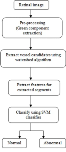
ABSTRACT: One of the rarest condition of severe visual impairment is diabetic retinopathy which can be observed by the development of unusual new retinal vessels. To identify these abnormal new vessels, an algorithm is designed which uses retinal photography method. Firstly, Watershed lines are used to detect new vessels which are candidate segments. Further different parameters like shape, position, color, brightness, contrast are evaluated for each segment. After that, support vector machine (SVM) is used to distinguish between normal and abnormal vessels depending upon parameters described. The accuracy provided by this method is close enough to satisfy the clinical needs for the detection of new vessels.
KEYWORDS: Watershed algorithm, Support vector machine(SVM), optic disc, retinal images, optic vessels.
I. INTRODUCTION
Diabetic retinopathy is the worst condition of retina in highly diabetic patients which results in improper functioning of retina, leads to blindness. It is the most leading disease in UK & US. Worldwide, one third of 285 million people with diabetes show the sign of DR. The direct damage to the vessels of retina is the main cause of blindness. The human brain can identify the conversion of light which produces visual images. Diabetic retinopathy damages the retinal vessels which often leads to leakage of fluid and distort vision. It is classified as non-proliferative diabetic retinopathy and proliferative diabetic retinopathy . NPDR is milder and mostly symptomless. PDR is the most advanced stage of DR which leads to formation of new and unusual vessels in retina. The most observed symptoms of DR are blurred vision, difficulty in seeing colours, fluid leakage, redness, irritation and even complete loss of vision. The leakage of newly formed vessel into vitreous gel that fills an eye, stopping light from reaching retina which ultimately leads to vitreous haemorrhage. A detached retina is the resultant of scar tissues which pulls retina back from the eye. The formation of new blood vessels leads to the blockage of normal flow of fluid which increases ocular pressure of an eye increasing the risk of optic nerve damage resulting in glaucoma. Macular edema is one of the mechanisms associated with DR. The swelling of macula leads to vision loss as macula holds tightly packed cones that provide sharp vision to human eye. A number of experiments are done to detect the formation of new blood vessels. In this paper, a method for detection of abnormalities on optic disc is described and implemented.
II. RELATED WORK
There are different aspects of image formation and image compression. Image formation, enhancement, masking, compression are very crucial while studying human eye. These factors help to distinguish between the different parts of human eye which needs very much clarity in order to detect any abnormality in the functioning of an eye.
The detection of exudates in [1] presented an effective method to improve the efficiency of diabetic retinopathy screening procedures. This study observed whether automated grading improves the detection of observable diabetic retinopathy. A novel approach for glaucoma detection using fractal analysis in [2] proposed a perimeter method using fractal algorithm for the fast detection of glaucoma. It uses preprocessing techniques followed by K-means clustering for identification of cup to disc ratio. The use of multiscale amplitude modulation and frequency modulation is proposed in [3] to identify normal and abnormal vessels on optic disc. This proposed system overall shows the efficient use of automated DR screening
.
Automatic detection of red lesions in digital color fundus photographs in [4] described red lesion detection method based on hybrid approach. The first approach is red lesion detection method using pixel classification and second approach is to add extensive features to classify each candidate segment separately. Fast detection of optic disc and fovea in color fundus photographs in [5] presented fast method to detect fovea and optic disc of retina in digital photographs. A deep study is investigated on near about 350 images using diabetic retinopathy screening services.
III.PROPOSEDMETHODOLOGY
A. Pre-processing
The system uses pre-processing to avoid false results. Pre-processing helps to exempt vessel area during detection of lesions. Green channel is used to distinguish as blood vessels images appear more contrasted as compared to blue and red channels in RGB format. Only green channel is used for pre processing suppressing other two channels.
B. Segmentation
The pre-processed images are then subjected to segmentation. The vessels are segmented to find normal and abnormal vessels on retina. Afterwards, Watershed algorithm is implemented. Watershed regions are calculated using Meyer’s. The calculation of dark ridges is given as,
𝐾 =𝐿𝑥 2 𝐿𝑦𝑦 + 𝐿2𝑦 𝐿𝑥𝑥 − 2𝐿𝑥 𝐿𝑦𝐿𝑥𝑦 (𝐿2𝑥 + 𝐿𝑦2)
3 2 ⁄
where L is the Gaussian filtered image. The subscripts denotes partial derivatives
C. Extract Features
Fifteen segment features are described which are partly based on observation of human to identify abnormal vessels. Segment length, gradient, gradient variations, density, direction, number segments, mean vessel gradient are the features extracted.
D. SVM classification
The extracted features are train to SVM classifier. Trained algorithm is used to detect new vessels. Different sets of images are tested for better results.
E. Classification of performance
IV.EXPERIMENTALRESULTS
All the experiments are conducted on the Matlab R2013a. Detection of new vessels can be observed by studying following figure 2.
Fig.2: Detection of new vessels
V. CONCLUSION
This paper focuses on the detection of new blood vessels on retina which are caused due to diabetic retinopathy. We have implemented the method for the detection of new blood vessels on retina which are caused due to diabetic retinopathy. Here, SVM classifier is used to extract features so that exact abnormalities can be detected. Further investigation is needed so that exact optic disc is located.
REFERENCES
[1] M. Mirmehdi, B. Thomas, and R. Markham, “Detection of diabetic retinal exudates in digital colour images,” vol. 97, pp. 1220–1223, 2019
[2] Xiong, Li, Huiqi Li, and Yan Zheng. “Automatic detection of glaucoma in retinal images,” in IEEE Industrial Electronics and Applications (ICIEA), 9th Conference, pp. 1016–1019, 2014.
[3] Multiscale AM-FM Methods for Diabetic Retinopathy Lesion Detection, Herbert Davis, Stephen Russell, Michael Abràmoff, Member, IEEE, and Peter Soliz, Member, IEEE, September 2015
[4] B. V. Ginneken, J. Staal, M. S. A. Suttorp-Schulten, and “Automatic detection of red lesions in digital color fundus photographs,”IEEE Trans. Med. Imag., vol. 24, no. 5, pp. 584–592, June 2010