International Journal of Nanomedicine
Dove
press
O r I g I N A L r e s e A r c h
open access to scientific and medical research
Open Access Full Text Article
New surface-modified solid lipid nanoparticles
using N-glutaryl phosphatidylethanolamine
as the outer shell
soheila Kashanian1
Abbas hemati Azandaryani1
Katayoun Derakhshandeh2,3
1school of chemistry, Nanoscience
and Nanotechnology research center and sensor and Biosensor research center, razi University, 2Department
of Pharmaceutics, Kermanshah University of Medical sciences,
3Nanoscience and Technology
research center school of Pharmacy, Kermanshah University of Medical sciences, Kermanshah, Iran
correspondence: Katayoun Derakhshandeh Department of Pharmaceutics, Faculty of Pharmacy, Kermanshah University of Medical sciences, Kermanshah 67145-1673, Iran
Tel +98 831 427 6485 Fax +98 831 427 6496
email [email protected]
Background: Solid lipid nanoparticles (SLNs) are colloidal carrier systems which provide controlled-release profiles for many substances. In this study, we prepared aqueous dispersions of lipid nanoparticles using a modified, pH-sensitive derivative of phosphatidylethanolamine.
Methods: SLNs were prepared using polysorbate 80 as the surfactant and tripalmitin glyceride and N-glutaryl phosphatidylethanolamine as the lipid components. Particle size, polydispersity index, and zeta potential were examined by photon correlation spectroscopy. Morphological evaluation was performed using scanning electron microscopy, atomic force microscopy, and differential scanning calorimetry.
Results: Photon correlation spectroscopy revealed a particle hydrodynamic diameter of
165.8 nm and zeta potential of −41.6.0 mV for the drug-loaded nanoparticles. Atomic force
microscopy investigation showed the nanoparticles to be 50–600 nm in length and 66.5 nm in height. Differential scanning calorimetry indicated that the majority of SLNs possessed less ordered arrangements of crystals compared with corresponding bulk lipids, which is favorable for improving drug-loading capacity. Drug-loading capacity and drug entrapment efficiency values for the SLNs were 25.32% and 94.32%, respectively.
Conclusion: The SLNs prepared in this study were able to control the release of triamcinolone acetonide under acidic conditions.
Keywords: solid lipid nanoparticles, high-shear homogenization, triamcinolone acetonide, tripalmitin, phosphatidylethanolamine
Introduction
Solid lipid nanoparticles (SLNs) were developed in the early 1990s as an alternative carrier system to traditional colloidal systems, including emulsions, liposomes, and polymeric microparticles and nanoparticles.1 Tremendous progress has been made
in the treatment of disorders using new drug delivery systems, including SLNs, which are made from solid lipids (ie, lipids solid at room temperature as well as body temperature) and stabilized by surfactant(s).2 They are generally composed of a solid
hydrophobic core with a monolayer of phospholipids or phospholipid-derived coating. The solid core contains the active drug dissolved or dispersed in a solid fat matrix with the hydrophobic ends of the phospholipid chains embedded in the fat matrix. Thus, they have the potential to carry lipophilic or hydrophilic drug(s) for diagnostic purposes.3–5
A clear advantage of SLNs over polymeric nanoparticles is the fact that the lipid matrix is made from physiologically tolerated lipid components, which decreases the potential for acute and chronic toxicity.5,6 SLNs have fewer storage and drug leakage
International Journal of Nanomedicine downloaded from https://www.dovepress.com/ by 118.70.13.36 on 23-Aug-2020
For personal use only.
Number of times this article has been viewed
This article was published in the following Dove Press journal: International Journal of Nanomedicine
Dovepress
Kashanian et al
problems compared with systems such as liposomes and also have good stability for 2–3 years.6–9
It is noteworthy that tumors and inflamed tissues are often associated with leaky vascular architecture as a result of the poorly regulated nature of tumor angiogenesis. Furthermore, the acidity of these regions differs from physiological pH. Therefore, the use of acid-sensitive drug carriers has been one of the most extensively studied active triggering strategies.10,11
Dong and Hoffman prepared and reported pH-sensitive hydrogels for drug delivery.12 Oh and Lee created a novel
vector for delivery of anticancer therapy to solid tumors using diafiltration to synthesize pH-sensitive polymeric micelles, and observed that release of doxorubicin as micelles was triggered at pH 6.8.13 Reddy and Low prepared a pH-sensitive
lipid formulation from dioleylphosphatidylethanolamine and citraconic anhydride, and used this to prepare liposomes. They observed that the resulting liposomes were stable at neutral pH but fusogenic at pH 5.11
The nature of tumor angiogenesis enables submicron-sized particulate matter, such as SLNs, to penetrate preferentially into tumor tissue and be retained there. Consequently, tumor-specific drug delivery is attracting considerable attention in cancer therapy.7,14–16 Moreover,
with advances in surface engineering technology, the biodistribution of SLNs can be further manipulated by modifying the surface physicochemical properties of SLNs to target them to the tissue of interest, particularly to tumors and inflamed tissue.7,17 So far, researchers have prepared
various derivatives of phospholipids and used them on the outer shells of SLNs.18
The aim of the present study was to investigate the feasibility of the inclusion of triamcinolone acetonide into surface-modified solid lipid nanoparticles. For this purpose, nanoparticles were covered with modif ied phosphatidylethanolamine as an outer shell. Triamcinolone acetonide is usually administered via the parenteral route to treat inflammatory disorders and cancer of the lymph nodes. It is a drug with poor water solubility and high lipophilicity, which makes it suitable for SLN encapsulation and in vitro characterization. SLN release has been studied using lyophilized nanoparticles in different media. Modified high shear homogenization and ultrasound techniques have been used to produce low particle sizes and high encapsulation eff iciency,19 while photon correlation spectroscopy,
differential scanning calorimetry, atomic force microscopy, and scanning electron microscopy have been used for their characterization.
Materials and methods
The following materials were obtained from the indicated sources and used in our study without further purification. Tripalmitin glyceride was purchased from Alfa Aesar (Germany), and phosphatidylethanolamine and triamcinolone acetonide from Sigma-Aldrich (St Louis, MO). Glutaric anhydride, polysorbate 80, and sucrose were obtained from Merck (Darmstadt, Germany). High-pressure liquid chromatography (HPLC) grade methanol and analytical grade chloroform and ethanol were also purchased from Merck. Other reagents were of analytical or HPLC grade. Double-distilled water was prepared in our laboratory.
Preparation of modified
phosphatidylethanolamine
For preparation of modified phosphatidylethanolamine, 5 mg of lipid was dissolved in 10 mL of chloroform, and an approximately five-fold molar excess (3.85 mg) of glutaric anhydride was then added in the presence of 7.5 µL pyridine, and the mixture was incubated at 20°C for 5 hours.20
Modified phosphatidylethanolamine was identified by iodine staining.21 N-glutaryl phosphatidylethanolamine was isolated
using plate chromatography and chloroform-methanol (65:35 v/v) as the solvent, with a yield of 90%:
1H NMR (200 MHz, CDCl
3) δ 0.88 (6H), 1.30 (32 H),
1.59 (4H), 1.99 (2H), 2.16 (8H), 2.27 (6H), 2.61 (2H), 3.48 (2H), 3.95 (2H), 4.13 and 4.38 (2H), 4.20 and 4.51 (2H), 4.71 (1H), 5.33 (4H), and 7.38 (1H) that peaked in 7.38 ppm indicated amidic hydrogen.
Preparation of aqueous sLNs and
lyophilization
Triamcinolone acetonide (0.75% w/v), tripalmitin glyceride (2% w/v), and N-glutaryl phosphatidylethanolamine (0.5% w/v) were dissolved in 10 mL of chloroform and methanol mixture (1:1). Organic solvents were completely removed using a rotoevaporator (Laborota 4003, Heidolph, Schwabach, Germany). The drug-embedded lipid layer was melted by heating at 70°C above the melting point of the lipid. An aqueous phase was prepared by dissolving polysorbate 80 (1% w/v) in double-distilled water and heated to the same temperature as the oil phase. The hot aqueous phase was added to the oil phase, and homogenization was achieved (at 10,000 rpm and 70°C) using a Diax 900 homogenizer (Heidolph) for 2 minutes. The resulting coarse hot oil in water emulsion was ultrasonicated using a Sonoplus ultrahomogenizer (Bandelin, Germany) for 15 minutes.
International Journal of Nanomedicine downloaded from https://www.dovepress.com/ by 118.70.13.36 on 23-Aug-2020
Dovepress N-glutaryl phosphatidylethanolamine lipid nanoparticles
Triamcinolone acetonide-SLNs were obtained by allowing the hot nanoemulsion to cool off at room temperature.
Sucrose was used in the freeze-drying process as a cryoprotector at a concentration of 3 wt%.22 The SLN
sus-pension was frozen in an aqueous sucrose solution at −70°C overnight, and the sample was subsequently transferred to the freeze-drier (ZiRBuS Technology VaCo 5, D-37539) at −50°C for 72 hours, and finally the SLN powder was col-lected for further experiments.
Determination of entrapment efficiency
and drug-loading capacity
The percentage of incorporated triamcinolone acetonide (entrapment efficiency) was determined by spectropho-tometric determination at 234 nm using an Agilent 8453-spectrophotometer, after ultracentrifugation of the aqueous dispersion (40,000 rpm for 45 minutes). The amount of free drug was detected in the supernatant and the amount of incor-porated drug was calculated as the initial drug minus the free drug. The drug entrapment efficiency and drug-loading in the SLNs were calculated using equations (1) and (2):
EE (%) = W W
W 100
a s
a
− ×
(1)
DL (%) = W W
W -W +W 100
a s
a s L
− ×
(2)
where EE is entrapment efficiency, DL is drug-loading, and Wa, Ws, and WL are the weight of drug added into the system, analyzed weight of drug in the supernatant, and weight of lipid added into the system, respectively.23
Particle diameter, polydispersity index,
and zeta potential
Measurement of hydrodynamic diameter, polydispersity index, and zeta potential of the nanoparticles was accom-plished using photon correlation spectroscopy (Nano ZS4700, Malvern Instruments, Worcestershire, UK). For size measurement, all formulations were diluted by deion-ized water to eliminate the effect of viscosity caused by the ingredients. Zeta potential measurements were conducted for samples prepared for size measurement. Hydrodynamic diameter and zeta potential were reported as the mean of three measurements at 25°C. The refractive index of the SLNs and water was set at 1.35 and 1.33, respectively.
scanning electron microscopy
In order to verify the result, the morphology of the triam-cinolone acetonide-SLNs was studied by scanning electron microscopy using a Philips XL30 microscope at an acceler-ating voltage of 10 kV. One droplet of SLN nanosuspension was placed on an aluminum specimen stub. After oven-drying for 12 hours, the sample was coated with a platinum layer using an SCDOOS sputter coater (BAL-TEC, Sweden) in an argon atmosphere. Subsequently, the sample was scanned and photomicrographs were obtained.
Atomic force microscopy
The surface properties of the drug-loaded SLNs were visualized using an atomic force microscope (Mobile S, Nanosurf, Switzerland). Explorer atomic force microscopy was in noncontact mode, using high resonant frequency (F0 170 kHz) pyramidal cantilevers with silicon probes having dynamic force. The sample was diluted with distilled water and then dropped onto a freshly cleaved mica plate, followed by vacuum drying for 24 hours at 25°C.
Differential scanning calorimetry
The thermal characteristics of the SLNs and pure drug were determined using a differential scanning calorimeter (Shimadzu DSC-60, single heating ramp 0°C–300°C, heated at 5°C/minute) under a dry nitrogen atmosphere. Samples (7–8 mg) were placed in an aluminum pan and heated at a rate of 5°C/minute. Empty aluminum pans were used as references, and the entire thermal behavior was studied under a nitrogen purge.
Assay for triamcinolone acetonide
A simple and sensitive HPLC method with ultraviolet detection was used for analysis of triamcinolone acetonide.20 The
triamcinolone acetonide concentration was determined using an HPLC system (LC-10AS Liquid Chromatograph, SCL-10A System Controller, SIL-10AV UV-Vis Detector, C-R6A Chromatopac, Shimadzu, Japan). The analytical conditions were as follows: column C18, HPLC Pack column, Inertsil ODS, 5 µm, 4.6 × 250 mm (GL Sciences Inc, Japan); ultraviolet detection, 240 nm; mobile phase, methanol; water 50:50 (v/v); flow rate 1 mL/minute; and injection volume 25 µL.
In vitro release of triamcinolone
acetonide from sLNs
This experiment was conducted using a static horizontal Franz diffusion cell to evaluate the amount of triamcinolone
International Journal of Nanomedicine downloaded from https://www.dovepress.com/ by 118.70.13.36 on 23-Aug-2020
Dovepress
Kashanian et al
acetonide released from each formulation.24 A cellulose
acetate membrane with a molecular weight cutoff of 12,000 Da and a surface area of 2.0 cm2 was used and
mounted on the Franz diffusion cell. The receptor medium was precisely 50 mL in volume and composed of an aque-ous solution of physiological saline, phosphate-buffered saline, and 20% ethanol at four pH values of 4.0, 5.0, 6.0, and 7.4, stirred by a magnetic bar at 500 rpm to homogenize the medium. Each formulation, 10 mg in 1 mL volume, was loaded onto the membrane in the donor compartment. The temperature of the assay was controlled at precisely 37°C. At predetermined time intervals, 1 mL aliquots of the release medium were withdrawn using a syringe needle, and the same volumes of freshly prepared receptor medium were added. The samples were analyzed using an HPLC method described previously.20 The experiments were repeated three times and
the results were expressed as means ± standard deviation.
Results and discussion
Preparation of nanoparticles, drug
entrapment efficiency, and loading
capacity
In this study, homogenization was performed at above the melting point of the lipid followed by ultrasonica-tion for preparaultrasonica-tion of modified SLN.25,26 To disperse the
triamcinolone acetonide homogeneously in the lipid, a chloroform/methanol solvent system was used and homog-enized for 2 minutes.27 Subsequently, the prepared SLNs
were subjected to further lyophilization. Our preliminary studies showed that reconstitution of freeze-dried SLNs was impossible when they were freeze-dried without addition of cryoprotectants. Therefore, a 1:1 dilution with 3% w/w of sucrose as a cryoprotectant was performed.28
Many different drugs have been incorporated in the SLNs.17 The prerequisite for obtaining a sufficient loading
capacity is a sufficiently high solubility of the drug in the lipid melt. Relatively higher encapsulation efficiency constitutes one of the major advantages of SLNs.
For calculations of entrapment efficiency and loading capacity, calibration curves for the ultraviolet assays of
triamcinolone acetonide were conducted on eight solutions in the concentration ranges of 1.5 × 10⁻³ to 1.25 × 10⁻⁴ mol/L;
encapsulation efficiency and drug-loading were 94.32% and 25.32%, respectively. The high entrapment efficiency of the drug is thought to be the result of the lipophilic characteristics and high compatibility between the drug and the lipid. Furthermore, the high encapsulation efficiency and drug-loading in comparison with previous work24 is a result of
the method of preparation and mixing of the drug and lipid matrix in the organic solvents.
Nanoparticle characterization
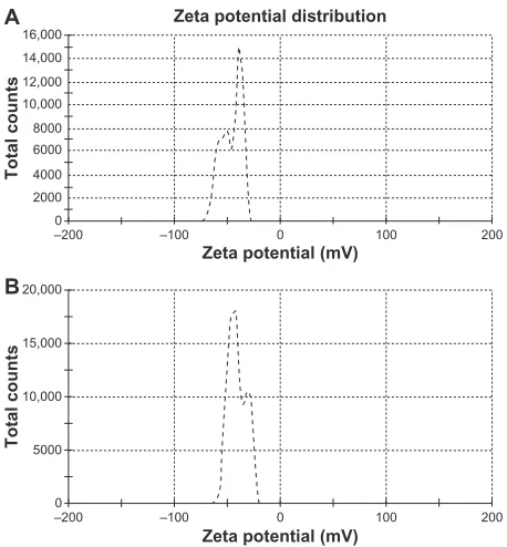
The mean particle size, polydispersity index and zeta potential of the SLNs with and without drug were measured by photon correlation spectroscopy, as summarized in Table 1. The mean particle sizes of 165.8 nm and 97.44 nm were obtained for drug-loaded and free SLNs, respectively (Table 1). The increase in size after drug loading must be due to incorporation of the drug in the nanoparticle matrix.23
The polydispersity index is a ratio providing information about the homogeneity of particle size distribution in a given system, and ideally, this should be ,0.3.29 Triamcinolone
acetonide-loaded and free SLN had polydispersity index values of 0.254 and 0.337, respectively, indicating that the nanoparticles had a narrow size distribution, and suggesting nanoparticle monodispersity.30,31 The zeta potential is a key
factor for evaluation of the stability of a colloidal dispersion.23
When the absolute value of the zeta potential is higher than 30 mV for a colloidal formulation, the particles are likely to be electrochemically stable under the investigated condition.25
Zeta potential values of −41.6 mV and −43.0 mV were obtained for the drug-loaded and free SLNs, respectively (Figure 1). As Table 1 demonstrates, as time passes, increases are observed in size, polydispersity index, and zeta potential as a result of light and other factors that cause gelation.32 However, it is obvious
from these values that the prepared nanosuspension has an acceptable electrochemical and physical stability potential for 3 and 6 months of storage.
Similarly, in order to evaluate the stability of SLNs lyophilized powder after 6 months, 10 mg of sample was
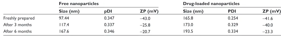
Table 1 Particle size polydispersity index and zeta potential for drug-loaded and free solid lipid nanoparticles in colloidal suspension
Free nanoparticles Drug-loaded nanoparticles
Size (nm) pDI ZP (mV) Size (nm) PDI ZP (mV)
Freshly prepared 97.44 0.347 −43.0 165.8 0.254 −41.6
After 3 months 117.4 0.337 −25.8 173.0 0.329 −40.0
After 6 months 167.6 0.346 −20.7 193.5 0.334 −23.3
Abbreviations: PDI, polydispersity index; ZP, zeta potential.
International Journal of Nanomedicine downloaded from https://www.dovepress.com/ by 118.70.13.36 on 23-Aug-2020
Dovepress N-glutaryl phosphatidylethanolamine lipid nanoparticles
dispersed in distilled water and characterized. The values for particle diameter, polydispersity index, and zeta potential were 223.3 nm, 0.321, and −30.1, respectively. These results indicate that, using sucrose as a cryoprotectant, the particle size remains in the nanometric range.
In order to obtain a scanning electron microscopic image, we used freshly prepared SLN loaded with triamcinolone acetonide, and micrographs of the samples containing drug-loaded SLNs are shown in Figure 2. The micrographs demonstrated a spherical shape and a smooth surface, with a particle size in the nanometric range.25
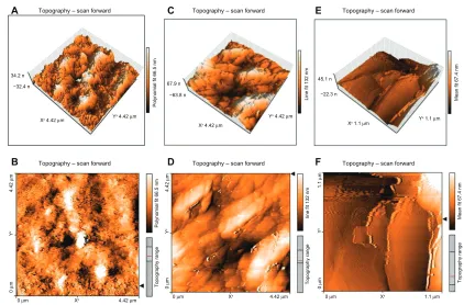
The atomic force microscopy technique has been widely used to obtain size, shape, and surface morphological infor-mation about nanoparticles. It is capable of resolving surface details down to 0.01 nm and producing a contrasted and three-dimensional image of the sample.23 A drop of diluted
aqueous suspension (without preliminary ultrafiltration) was placed on a mica slide and dried out at room temperature for 24 hours. Figure 3 gives the tapping mode atomic force microscopic images of freshly prepared SLN containing triamcinolone acetonide after 24 hours (A and B), 48 hours (C and D), and 1 week (E and F). The nanoparticle images demonstrated spherical particles of 50–600 nm in diameter, but only 66.5 nm in height. After 48 hours, the nanoparticles were uniformly grown on mica slides, as illustrated in Figure 3 (C and D). One week after spreading SLNs onto the mica plates, no particles could be distinguished in the atomic
force microscopic images, which showed homogeneous and flat shapes, as illustrated in Figure 3 (E and F).33 This
result is not surprising, considering the well known ability of particles to fuse during evaporation of water consequent to the molecular diffusion on the surface of the particles. Moreover, the presence of free surfactant in the suspension may increase the characteristic size.33
–2000 –100 0
2000 4000 6000 8000 10,000 12,000 14,000 16,000
100 200
–200 –100 0 100 200
Total counts
Total counts
Zeta potential (mV)
Zeta potential (mV) Zeta potential distribution
0 5000 10,000 15,000 20,000
B A
Figure 1 Zeta potential of (A) drug-loaded solid lipid nanoparticles and (B) free solid lipid nanoparticles.
Figure 2 scanning electron micrographs of freshly prepared solid lipid nano-particles loaded with triamcinolone acetonide; (A) 3.00 K× resolution, (B and C) 20.00 K× resolution.
International Journal of Nanomedicine downloaded from https://www.dovepress.com/ by 118.70.13.36 on 23-Aug-2020
Dovepress
Kashanian et al
A C E
34.2 n −32.4 n
Xx 4.42 µm
0 µm Xx 4.42 µm
0
µ
m
4.42
µ
m
Y
x
Topography range
Polynomial fit 66.5 nm
0 µm Xx 4.42 µm
0
µ
m
4.42
µ
m
Y
x
Topography range
line fit 132 nm
0 µm Xx 1.1 µm
0
µ
m
1.1
µ
m
Y
x
Topography range
Mean fit 67.4 nm
Topography – scan forward Topography – scan forward Topography – scan forward
Yx 4.42 µm
67.9 n −63.8 n
Polynomial fit 66.5 nm
Line fit 132 nm Mean fit 67.4 nm
Xx 4.42 µm
Yx 4.42 µm
45.1 n −22.3 n
Xx 1.1 µm Y x 1.1 µm
B Topography – scan forward D Topography – scan forward F Topography – scan forward
Figure 3 Atomic force microscopy of solid lipid nanoparticles loaded with triamcinolone acetonide. (A) and (B) are solid lipid nanoparticles loaded with triamcinolone acetonide after 24 hours. (C) and (D) are solid lipid nanoparticles loaded with triamcinolone acetonide after 48 hours remaining on a mica slide. (E) and (F) are solid lipid nanoparticles loaded with triamcinolone acetonide after 1 week remaining on a mica slide. A, C, and D are three-dimensional images of the multiparticles and B, D, and F
are the two-dimensional pictures of the multiparticles.
Differential scanning calorimetry thermograms for triam-cinolone acetonide, tripalmitin glyceride, a physical mixture of triamcinolone acetonide, tripalmitin glyceride, modified phosphatidylethanolamine, and lyophilized SLNs are shown in Figure 4. A sucrose thermogram is also shown in this figure to demonstrate peaks of lyophilized SLNs. The triamcinolone acetonide powder showed a sharp melting process with an onset temperature of 284.72°C and a peak of 286.04°C, with a melting enthalpy of 64.73 J/g. This was confirmed by the presence of a melting peak for triamcinolone acetonide in the physical mixture. However, no melting process was observed for the lyophilized SLN suspension. This suggested that there was no crystalline triamcinolone acetonide in the SLN suspension, and triamcinolone acetonide might be present in an amorphous state. Similar results have been reported by previous researchers, stating that rapid quenching of the nanoemulsion does not allow the drug to crystallize.26,27 An
endothermic peak of the sucrose used as a cryoprotectant was observed at 170.21°C in the triamcinolone acetonide-SLN curve. Meanwhile, the melting peak temperature of tripalmitin glyceride in lyophilized SLN (57.73°C) was diminished when compared with the temperature of the bulk lipid (64.38°C). The decrease in melting point can be attributed to the small size (nanometer range), high specific
surface area, and presence of a surfactant.15,30 This can also
be attributed to the Kelvin effect and is described by the Thomson equation.3 Moreover, the lower melting enthalpy
value of 35.72 J/g indicates a less ordered lattice arrange-ment of lipids within the nanoparticles compared with the bulk materials.9
In vitro drug release
In this study, the in vitro release of triamcinolone acetonide from SLNs was investigated for 60 hours at 37°C using a Franz diffusion cell (Figure 5). Due to the low water solubility of triamcinolone acetonide, 20% ethanol was added to the phosphate-buffered solution at four pH values of 4.0, 5.0, 6.0, and 7.4.24 Our results showed that up to 50% was released
over the test period. The prolonged and lower release of SLNs after 12 hours can be explained by slower diffusion of triamcinolone acetonide from the inner solid core matrix of the formulations. However, the initial fast release was detected at a lower pH. In this experiment, we studied the release profile of pure drug compared with nanoparticles in acidic pH, and the release half-life of drug release was significantly diminished compared with nanoparticles (about 25 minutes).
The release data were analyzed using the following Higuchi kinetic equation:25 Q
t = k·t0.5, where Qt is the
International Journal of Nanomedicine downloaded from https://www.dovepress.com/ by 118.70.13.36 on 23-Aug-2020
Dovepress N-glutaryl phosphatidylethanolamine lipid nanoparticles
percentage of drug released at time t, and k is the release rate constant. Regarding the release model of all formulations, it was found that the prolonged release characteristic of triam-cinolone acetonide was well fitted to Higuchi’s square root model, as has been reported for drug-loaded SLN systems.25,34,35
Linear fits were obtained, indicating that the release profile of triamcinolone acetonide from homogenous and granular matrix systems is diffusion-controlled. The R2 values from
in vitro release kinetics and the K values or release rate
constant obtained from the Higuchi model plot are presented in Table 2. Furthermore, as shown in Figure 6 and the in vitro release data, we found that the burst release was increased with a decline in pH that demonstrates a change in structure of the phospholipids and release of the terminal anhydride. Several previous studies have been conducted to identify a pH-sensitive carrier preparation, and often the amount of drug release from SLNs was small compared with our results, and the values for common SLNs without modification were
0.0 10 mV
A B C D E
20.0 40.0 60.0 80.0 100.0 120.0 140.0 160.0 180.0 200.0 220.0 240.0 260.0 280.0 300.0
Temp (C)
Figure 4 Differential scanning calorimetry thermograms of (A) tripalmitin glyceride, (B) a physical mixture of tripalmitin glyceride, triamcinolone acetonide, and modified phosphatidylethanolamine, (C) lyophilized solid lipid nanoparticle suspension, (D) sucrose, and (E) triamcinolone acetonide.
0
0 10 20 30 40
Time (h)
% cumulative release of TA
50 60 70
pH = 7.4 pH = 6 pH = 5 pH = 4
10 20 30 40 50 60
Figure 5 Drug release from nanoparticles after 60 hours in four different types of medium, each value represents the mean of three experiments ± standard deviation.
Abbreviation: TA, triamcinolone acetonide.
International Journal of Nanomedicine downloaded from https://www.dovepress.com/ by 118.70.13.36 on 23-Aug-2020
Dovepress
Kashanian et al
below 25%.8,25,27 Subedi et al investigated pH-sensitive SLNs
and used them for doxorubicin delivery; in their work, 30% of the drug was released after 24 hours.8
Venkateswarlo and Manjunath investigated SLNs of tripalmitin and used them as clozapine carriers, and in their work, up to 20% of drug was released after 48 hours.25 Liu
et al investigated SLNs containing triamcinolone acetonide in mixtures of monoglycerides, diglycerides, and triglycerides of palmitic acid and stearic acid, and found that the drug was released more slowly compared with our prepared carrier.24
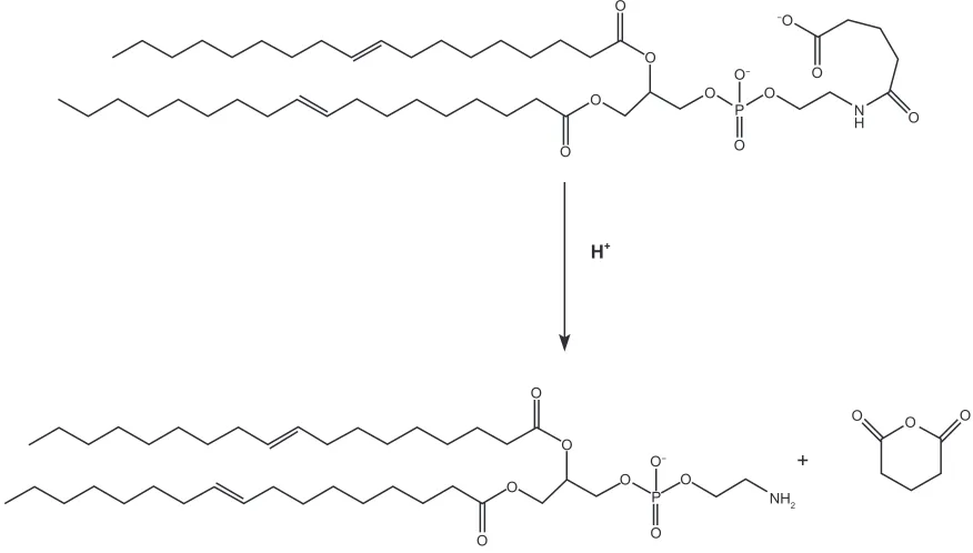
The simple mechanism for terminal release of anhydride from modified polyethylene is shown in Figure 6. This pathway was faster at an acidic pH of 4. Nevertheless, it is certain that this carrier requires further in vitro and in vivo studies.
Conclusion
As mentioned above, SLNs offer an attractive means of drug delivery, particularly for poorly water-soluble drugs. They combine the advantages of polymeric nanoparticles, fat emul-sions, and liposomes. In this study, modified N- glutaryl-phos-phatidylethanolamine was prepared, used for SLN preparation,
and characterized. The results indicate that the carrier has suitable stability in colloidal form and may increase drug release under acidic conditions. Homogenization followed by ultrasonication is suitable for producing small SLNs with an average diameter of 165.8 nm. Using a Zetasizer, atomic force microscopy, and SEM, the diameter and morphology of the particles was investigated. Zeta potential data indicate the stability of the nanosuspension. After 6 months, Zeta-sizer data did not indicate any obvious changes in diameter, polydispersity index, and zeta potential. An in vitro study indicated an increase in burst release and a pH decrement as well. In vitro release kinetics coincide with the Higuchi equation. These observations suggest that the present system offers an exciting mode of target delivery for potent lipophilic anticancer drugs and anti-inflammatory agents.
Acknowledgments
The authors acknowledge the instructors of the School of Pharmacy at Kermanshah University of Medical Sciences, with special thanks to Miss Farnaz Ahmadi and Miss Marjan Mohebbi from the analysis laboratory.
Table 2 r2 and K for drug-release experiment in four types of receptor medium
Receptor pH 4 5 6 7.4
r2 0.989 ± 0.0034 0.981 ± 0.0046 0.985 ± 0.007 0.980 ± 0.0025
K 0.119 ± 0.013 0.102 ± 0.018 0.092 ± 0.011 0.086 ± 0.018
Notes: r2 determination coefficient and k dissolution rate constant (µg/mL/h−1/2). each value represents the mean of three experiments ± standard deviation.
O
O
O
O
O O O
O
O
O
O O
O
NH2 +
O O
O−
O−
H+
−O
O O
O N H O
P
P
Figure 6 Terminal release of anhydride from modified phosphatidylethanolamine.
International Journal of Nanomedicine downloaded from https://www.dovepress.com/ by 118.70.13.36 on 23-Aug-2020
International Journal of Nanomedicine
Publish your work in this journal
Submit your manuscript here: http://www.dovepress.com/international-journal-of-nanomedicine-journal The International Journal of Nanomedicine is an international,
peer-reviewed journal focusing on the application of nanotechnology in diagnostics, therapeutics, and drug delivery systems throughout the biomedical field. This journal is indexed on PubMed Central, MedLine, CAS, SciSearch®, Current Contents®/Clinical Medicine,
Journal Citation Reports/Science Edition, EMBase, Scopus and the Elsevier Bibliographic databases. The manuscript management system is completely online and includes a very quick and fair peer-review system, which is all easy to use. Visit http://www.dovepress.com/ testimonials.php to read real quotes from published authors.
Dovepress
Dove
press
N-glutaryl phosphatidylethanolamine lipid nanoparticles
Disclosure
The authors report no conflicts of interest in this work.
References
1. Muller RH, Mader K, Gohla S. Solid lipid nanoparticles (SLN) for controlled drug delivery – a review of the state of the art. Eur J Pharm Biopharm. 2000;50:161–177.
2. Wissing SA, Kayser O, Muller RH. Solid lipid nanoparticles for parenteral drug delivery. Adv Drug Deliv Rev. 2004;56:1257–1272. 3. Kaura IP, Bhandari R, Bhandari S, Kakkar V. Potential of solid lipid
nano-particles in brain targeting. J Control Release. 2008;127:97–109. 4. Chen DB, Yang TZ, Lu WL, Zhang Q. In vitro and in vivo study of two
types of long-circulating solid lipid nanoparticles containing paclitaxel.
Chem Pharm Bull. 2001;49:1444–1447.
5. Heiati H. Development and characterization of solid lipid nanoparticles for delivery of bioactive materials [PhD thesis]. Montreal: University of Montreal; 1996.
6. Torchilin VP, editor. Nanoparticulates as Drug Carriers. London, UK: Imperial College Press; 2006.
7. Wong HL, Bendayan R, Rauth AM, Li Y, Wu XY. Chemotherapy with anticancer drugs encapsulated in solid lipid nanoparticles. Adv Drug Deliv Rev. 2007;59:491–504.
8. Subedi RK, Kang KW, Choi HK. Preparation and characterization of solid lipid nanoparticles loaded with doxorubicin. Eur J Pharm Sci. 2009;37:508–513.
9. Lai F, Sinico C, De Loqu A, Zaru M, Müller RH, Fadda AM. SLN as a topical delivery system for Artemisia arborescens essential oil: In vitro antiviral activity and skin permeation study. Int J Nanomed. 2007;2: 419–425.
10. Andresen TL, Jensen SS, Jorgensen K. Advanced strategies in liposomal cancer therapy: Problems and prospects of active and tumor specific drug release. Prog Lipid Res. 2005;44:68–97.
11. Reddy JA, Low PS. Enhanced folate receptor mediated gene therapy using a novel pH-sensitive lipid formulation. J Control Release. 2000;64:27–37.
12. Dong L, Hoffman AS. A novel approach for preparation of pH-sensitive hydrogels for enteric drug delivery. J Control Release. 1991; 15:141–152.
13. Oh KT, Lee ES. Cancer-associated pH-responsive tetracopolymeric micelles composed of poly (ethylene glycol)-b-poly (L-histidine)-b-poly (L-lactic acid)-b-poly(ethylene glycol). Polym Adv Technol. 2008;19: 1907–1913.
14. Matsumura Y, Maeda H. A new concept for macromolecular therapeutics in cancer chemotherapy: Mechanism of tumoritropic accumulation of proteins and the antitumor agent SMANCS. Cancer Res. 1986;6:193–210.
15. Derakhshandeh K, Soheili M, Dadashzadeh S, Saghiri R. Preparation and in vitro characterization of 9-nitrocamptothecin loaded long circulating nanoparticles for delivery in cancer patients. Int J Nanomedicine. 2010;5:1–59.
16. Dadashzadeh S, Derakhshandeh K, Hoseinishirazi F. 9-Nitrocamptothecin polymeric nanoparticles: Cytotoxicity and pharmacokinetic studies of lactone and total forms of drug in rats. Anticancer Drugs. 2008; 19:805–811.
17. Mehnert W, Mader K. Solid lipid nanoparticles: Production, character-ization and applications. Adv Drug Deliv Rev. 2001;47:165–196.
18. Garcia-Fuentes M, Alonso MJ, Torres D. Design and characterization of a new drug nanocarrier made from solid-liquid lipid mixtures.
J Colloid Interface Sci. 2005;285:590–598.
19. Hou DZ, Xie CS, Huang KJ, Zhu CH. The production and characteristics of solid lipid nanoparticles (SLNs). Biomaterials. 2003;24:1781–1785. 20. Zhang N, Ping QN, Huang GH, Xua WF. Investigation of lectin-modified insulin liposomes as carriers for oral administration. Int J Pharm. 2005;294:247–259.
21. Drummond DC, Daleke DL. Synthesis and characterization of N- acylated, pH-sensitive ‘caged’ aminophospholipids. Chem Phys Lipids. 1995;75:27–41.
22. Lin X, Li X, Zheng LQ, Yu L, Zhang Q, Liu W. Preparation and characterization of monocaprate nanostructured lipid carriers. Colloids Surf A Physicochem Eng Asp. 2007;311:106–111.
23. Hu FQ, Jiang SP, Du YZ, Yuan H, Ye YQ, Zeng S. Preparation and characterization of stearic acid nanostructured lipid carriers by solvent diffusion method in an aqueous system. Colloids Surf B Biointerfaces. 2005;45:167–173.
24. Liu W, Hu M, Liu W, Xue C, Xu H, Yang XL. Investigation of the carbopol gel of solid lipid nanoparticles for the transdermal iontophoretic delivery of triamcinolone acetate. Int J Pharm. 2008;364:135–141. 25. Ruktanonchai U, Bejrapha P, Sakulkhu U, et al. Physicochemical
characteristics, cytotoxicity, and antioxidant activity of three lipid nanoparticulate formulations of alpha-lipoic acid. AAPS Pharm Sci Tech. 2009;10:227–234.
26. Bhaskar K, Anbu J, Ravichandiran V, Venkateswarlu V, Rao YM. Lipid nanoparticles for transdermal delivery of flurbiprofen: Formulation, in vitro, ex vivo and in vivo studies. Lipids Health Dis. 2009;8:1–15. 27. Venkateswarlu V, Manjunath K. Preparation, characterization and in
vitro release kinetics of clozapine solid lipid nanoparticles. J Control Release. 2004;95:627–638.
28. Joshi M, Misra A. Dry powder inhalation of liposomal ketotifen fumarate: Formulation and characterization. Int J Pharm. 2001;223:15–27. 29. Pathak P, Nagarsenker M. Formulation and evaluation of lidocaine
lipid nanosystems for dermal delivery. AAPS Pharm Sci Tech. 2009;10: 985–992.
30. Ali H, El-Sayed K, Sylvester P, Nazzal S. Molecular interaction and localization of tocotrienol-rich fraction (TRF) within the matrices of lipid nanoparticles: Evidence studies by differential scanning calorimetry (DSC) and proton nuclear magnetic resonance spectroscopy (1H NMR). Colloids Surf B Biointerfaces. 2010;77:286–297. 31. Derakhshandeh K, Erfan M, Dadashzadeh S. Encapsulation of
9-nitro-camptothecin, a novel anticancer drug, in biodegradable nanoparticles: Factorial design, characterization and release kinetics. Eur J Pharm Biopharm. 2007;66:34–41.
32. Freitas C, Muller RH. Effect of light and temperature on zeta potential and physical stability in solid lipid nanoparticle (SLN) dispersions. Int J Pharm. 1998;168:221–229.
33. Igartua M, Saulnier P, Heurtault B, et al. Development and characterization of solid lipid nanoparticles loaded with magnetite. Int J Pharm. 2002;233:149–157.
34. Jenning V, Thunemann AF, Gohla SH. Characterization of a novel solid lipid nanoparticle carrier system based on binary mixtures of liquid and solid lipids. Int J Pharm. 2000;199:167–177.
35. Tiyaboonchai W, Tungpradit W, Plianbangchang P. Formulation and characterization of curcuminoids loaded solid lipid nanoparticles. Int J Pharm. 2007;337:299–306.
International Journal of Nanomedicine downloaded from https://www.dovepress.com/ by 118.70.13.36 on 23-Aug-2020