PPAR expression and function during vertebrate development
LILIANE MICHALIK, BÉATRICE DESVERGNE, CHRISTINE DREYER
1, MATHILDE GAVILLET,
RICARDO N. LAURINI
2and WALTER WAHLI
*Institut de Biologie Animale, Université de Lausanne, Switzerland, 1Max Planck Institut fuer Entwicklungsbiologie, Tuebingen, Germany
and 2University Hospital San Joao, Porto, Portugal
ABSTRACT The peroxisome proliferator activated receptors (PPARs) are ligand activated recep-tors which belong to the nuclear hormone receptor family. As with other members of this superfamily, it is thought that the ability of PPAR to bind to a ligand was acquired during metazoan evolution. Three different PPAR isotypes (PPARα, PPARβ, also called δ, and PPARγ) have been identified in various species. Upon binding to an activator, these receptors stimulate the expression of target genes implicated in important metabolic pathways. The present article is a review of PPAR expression and involvement in some aspects of Xenopus laevis and rodent embryonic develop-ment. PPARα and β are ubiquitously expressed in Xenopus early embryos but become more tissue restricted later in development. In rodents, PPARα, PPARβ and PPARγ show specific time- and tissue-dependent patterns of expression during fetal development and in the adult animals. PPARs are implicated in several aspects of tissue differentiation and rodent development, such as differentiation of the adipose tissue, brain, placenta and skin. Particular attention is given to studies undertaken by us and others on the implication of PPARα and β in rodent epidermal differentiation.
KEY WORDS:
PPAR, Xenopus leavis, rodent, tissue differentiation
0214-6282/2002/$25.00 © UBC Press
Printed in Spain
www.ijdb.ehu.es
*Address correspondence to: Dr. Walter Wahli. Institut de Biologie Animale, Université de Lausanne, Bâtiment de Biologie, CH-1015 Lausanne, Switzerland. Fax: + 41-2-1692-4115. e-mail: [email protected]
Abbreviations used in this paper: ACS, Acyl-CoA synthase; LBD, Ligand Binding
Domain; NHR, Nuclear Hormone Receptor; PPAR, Peroxisome Proliferator Activated Receptor; PPRE, PPAR response element; RXR, Retinoid X Receptor.
Introduction
PPARs are ligand-inducible transcription factors and belong to the nuclear hormone receptor (NHR) superfamily. Based on sequence homology with previously identified members of this superfamily, three different PPAR isotypes (PPARα, β/δ or FAAR or NUC1, and
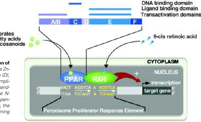
γ; NR1C1, NR1C2, NR1C3, respectively, Nuclear Receptor Nomen-clature Commitee, 1999) have been identified in the early 1990’s in Xenopus laevis and the mouse (Dreyer et al., 1992; Issemann and Green, 1990). Since then, PPARα, β/δ and γ have also been identified in human, rat, fish, hamster and chicken, each isotype having a unique spatio-temporal tissue distribution (reviewed in Desvergne and Wahli, 1999). Like the other members of the super-family, PPARs are organized into four domains (Fig. 1). The DNA-binding domain (C domain) is extremely well conserved and its Zn-finger structure is the signature of the members of the NHR super-family. The DNA binding domain is linked to the C-terminal ligand binding domain (E/F domain, LBD) by the hinge region (D domain). The E/F domain is implicated in the dimerization of PPARs with RXR and in the ligand-dependent transactivation function of the receptor, whereas the N-terminal domain of the protein (A/B domain) is involved in the ligand-independent regulation of receptor activity. Binding of PPARs to their ligands induces conformational changes which allow co-repressor release and co-activator recruitment.
describe the pattern of expression of PPARα, β and γ during development of Xenopus laevis, rodents and human. The last part will finally concentrate on the best characterized PPAR functions in tissue differentiation and vertebrate development.
I. Evolution of the PPAR Nuclear Hormone Receptors
The NHR superfamily includes ligand-activated transcription factors, as well as members called orphan receptors for which no ligand has yet been identified, or possibly do not exist for a very few of them. All these receptors are phylogenetically related proteins. Sequence alignment of the DNA and ligand-binding domains of the known orphan and ligand-binding receptors has allowed the elabo-ration of a phylogenetic tree showing the evolutionary relationship between the members of the NHR family (reviewed in Escriva et al., 2000). This analysis sugests that the NHR appeared early during evolution, since both orphan and liganded receptors are present in all metazoan phyla. The large number of members in the superfam-ily is likely to result from two waves of gene duplication. The first wave happened before the arthropod/vertebrate divergence and has generated the ancestors of the NHR subfamilies, for instance PPARs, RARs, RXRs. The second wave of duplication is verte-brate-specific and led to a diversification inside the subfamilies, with the emergence of the presently known isotypes such as PPARα, β and γ. Based on evolutionary relationship, six groups of nuclear receptors could be defined. PPARs belong to group I, together with the VDR (Vitamin D receptor), TR (Thyroid hormone receptor), RAR (Retinoic acid receptor) and several orphan recep-tors (Dreyer et al., 1993; reviewed in Escriva et al., 2000). Interest-ingly, members within a group are obviously sequence-related but often have very different functions and ligands (e.g. PPAR and TR). In contrast, some receptors, like RXR and RAR belonging to two different groups, group II and I respectively, share similar functions and ligands, although being evolutionary distant. Taken all to-gether, these data favor the hypothesis of a common ancestral nuclear receptor which did not have any ligand (orphan nuclear receptor). Following gene duplication, and during the last 600 Myrs, the newly emerging receptors would have acquired ligand-binding capacities in an independent fashion (reviewed in Escriva et al., 2000). Once this capacity was acquired, each receptor
probably further evolved and refined its specificity for a given ligand. PPARs are one example of receptors which have probably followed this evolutionary route. As mentioned above, they can be activated by diverse ligands. However, and very interestingly, each PPAR isotype has its own properties with regard to ligand binding. For instance, the eicosanoid PGJ2 and the synthetic thiazolidinediones are selective PPARγ ligands, whereas the fibrate hypolipidemic drugs preferentially bind to PPARα. Polyun-saturated fatty acids can bind and activate all three PPAR isotypes, however with different affinities and efficiencies. Moreover, differ-ences in affinities can vary strikingly across species for given ligands and isotypes (Krey et al., 1997). Therefore, it is likely that PPARα, β and γ emerged from a common PPAR with broad ligand-binding specificity, itself derived from the ancestral orphan receptor. Each PPAR isotype then evolved by mutations leading to a more specific range of ligands across species. This hypoth-esis is further supported by the comparison of Xenopus, rodents and human PPARα, β and γ sequences, which shows that the ligand-binding domain, in which the evolutionary rate was the fastest in these receptors (reviewed in Escriva et al., 2000), mediates the differences in ligand binding among species (Keller et al., 1997).
II. PPAR Expression Profiles During Vertebrate
Develop-ment
The first step in understanding the physiological function of a protein is often the study of its pattern of expression. In short, the three PPAR isotypes are expressed in a variety of cell types having ectodermal, mesodermal or endodermal embryonic origins, with different, yet overlapping, spatio-temporal expression patterns. This chapter will summarize the patterns of expression of each PPAR isotype in Xenopus laevis, rodents and human.
PPAR Expression During Xenopus laevis Development
Xenopus PPARα, β and γ cDNA were first isolated from ovary (PPARα, β) or liver (PPARγ) Xenopus laevis libraries, on the basis of sequence similarities with the DNA-binding domain of the estrogen receptor (ER) (Dreyer et al., 1992; Dreyer et al., 1993). While the PPARα and β mRNAs and proteins are present in
Xenopus oocytes throughout oogenesis as well as in the embryo (see the Xenopus PPARα protein in Fig. 2), PPARγ mRNA is detected in significant amounts only in post-embryonic stages (Dreyer et al., 1993; reviewed in Dreyer and Ellinger-Ziegelbauer, 1996). During embryogenesis, the maternal pool of PPARα and β mRNAs are replaced by the zygotic transcripts at the neurula (PPARβ) or the tailbud (PPARα) stages. In adult Xenopus organs, PPARα and PPARβ (the prevalent isotype) are expressed ubiqui-tously (testes, liver, kidney, fat body, muscle, brain, spleen) (Dreyer et al., 1993; reviewed in Dreyer and Ellinger-Ziegelbauer, 1996). PPARγ shows a much more restricted pattern of expression and is present mainly in the adipose tissue, and to lower levels in the kidney, and the liver. A larger PPARγ mRNA, a likely equivalent of the mouse PPARγ2, is detected specifically in the fat body (Dreyer et al., 1992; Dreyer et al., 1993).
PPAR Expression During Rodent Development
We and others have investigated the distribution of the PPARs in the rat and mouse fetal and adult tissues, mainly at the mRNA level. Overall, the results are similar in both species (Beck et al., 1992; Braissant et al., 1996; Braissant and Wahli, 1998; Escher et al., 2001; Kliewer et al., 1994). During fetal development, PPARα and γ transcripts appear late during development in both species (day 13.5 of gestation), with a pattern of expression similar to their adult distribution. PPARα is present in the liver, the kidney, the intestine, the heart, the skeletal muscle, the adrenal gland and the pancreas. PPARγ expression is restricted to the brown adipose tissue (day 18.5 of gestation), and to the central nervous system (day 13.5 to 15.5 of gestation). Compared to the two other isotypes, PPARβ is expressed ubiquitously and earlier during fetal develop-ment. The rat PPARβ transcript is already present in the ectoderm and the visceral and parietal endoderms at day 8.5 of gestation, and the murine PPARβ mRNA was observed as soon as embryonic
day 9.5. In summary, the PPARβ gene is expressed broadly, and was detected in all the organs tested. More recently, the PPAR distribution was examined in mouse embryos at the protein level, and overall the data confirm the transcript expression profiles (Keller et al., 2000). In the rodent adult organs, the distribution of PPARα is similar to its fetal pattern of expression. In summary, PPARα is expressed in cells with high catabolic rates of fatty acids and peroxisomal metabolism, such as in hepatocytes and cardiomyocytes. PPARγ remains restricted to the brown and white adipose tissues, and is expressed at lower levels in the intestinal mucosa, the retina, the skeletal muscle and lymphoid organs. Similarly to its fetal distribution, the PPARβ transcript is present in all the organs tested, and is often more abundant than the PPARα and γ transcripts (Desvergne and Wahli, 1999; Escher et al., 2001). In addition, and very interestingly, PPARβ expression is induced in the uterus at the time of mouse blastocyst implantation (Lim et al., 1999).
Finally, it is interesting to note that the expression of the three PPAR isotypes peaks in the rat central nervous system between day 13.5 and 18.5 of gestation (Braissant and Wahli, 1998). Whereas PPARβ remains highly expressed in this tissue, the expression of PPARα and γ decreases postnatally in this area. Similarly, we also found that the three isotypes are transiently coexpressed in the epidermis during mouse fetal development, and that their expression becomes restricted to the hair follicles in the adult epidermis (Michalik et al., 2001). The functions of the PPARs in the brain and the epidermis will be discussed in further detail below.
PPAR Expression During Human Development
Less data are available about the expression of the PPARs during human development (Auboeuf et al., 1997; Mukherjee et al., 1997; Palmer et al., 1998). These data indicate that human PPARα is expressed in the adult liver, heart, kidney, large intestine and
C
Fig. 2. PPARα expression profile in Xenopus laevis. (A,B) Western blot analysis of the Xenopus PPARα protein in Xenopus embryo (A) or in adult Xenopus tissues (B), using a polyclonal antibody specific for Xenopus PPARα. (A) Embryonic stages: stage 2, 2 cell embryo; stage 8, blastula; stages 10-12, gastrula; stages 15-20, neurula; stages 23-28, tailbud; stage 33, early tadpole. (B) Adult tissues: Ki, kidney, Li, liver; Pa, pancreas; He, heart; Br, brain; Sp, spleen; Te, testis; Mu, muscle; Lu, lung. (C) Xenopus PPARα was detected by wholemount immunostaining of Xenopus laevis kidney using the same polyclonal antiserum, followed by embedding in Technovit and sectioning as described (Dreyer and Ellinger-Ziegelbauer, 1996). (C1)
immunofluorescence; (C2) DAPI staining. Magnification bar, 20µm.
A
skeletal muscle. PPARβ mRNA is present ubiquitously, with a higher expression in the digestive tract and the placenta. PPARγ is abundantly expressed in the white adipose tissue, and is present at lower levels in the skeletal muscle, the heart and the liver. Surprisingly, and in contrast to rodents, human PPARγ seems to be absent from lymphoid tissues, eventhough PPARγ has been shown to be present in macrophages in human atheroma (reviewed in Rosen and Spiegelman, 2001). The expression of PPARα, β and
γ in the human fetal digestive tract between week 7 and 23 of gestation has been studied at the protein level (Huin et al., 2000). The three isotypes are expressed as early as week 7 of gestation in cell types of endodermal and mesodermal origin. PPARα, β and
γ are present at similar levels in the ileum from week 12 to 20, whereas they show a more spatio-temporal specific expression pattern in the other part of the digestive tract (Huin et al., 2000).
III. Functions of PPARs in Vertebrate Development
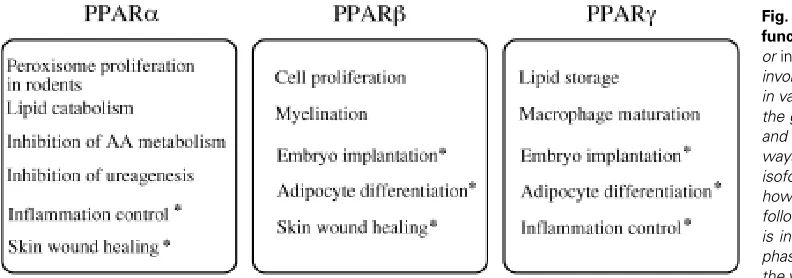
In agreement with the hints given by their respective tissue distribution, specific roles for PPARs have emerged from both in vitro and in vivo models. The three PPARs have distinct but often complementary functions. They are involved in multiple physiological pathways summarized in Fig. 3, including energy homeostasis and inflammation control (reviewed in Desvergne and Wahli, 1999). Particular attention will be given here to the roles of PPARs in mouse and rat tissue differentiation and development. In this context, the most documented functions of PPARs concern the involvement of PPARβ and γ in adipose tissue differentiation, PPARγ during the placenta formation, PPARβ in the development of the central ner-vous system, and finally, the involvement of PPARs in the maturation of the epidermis.
PPARβ and γ are Implicated in the Differentiation of White Adipose Tissue
As mentioned above, relatively high levels of PPARγ in rodents are almost restricted to the white and brown adipose tissues, and the importance of PPARγ in the differentiation of these tissues is well documented. Particularly, the role played by PPARγ in the white adipose tissue has been extensively studied, and it has
become clear that PPARγ is one of the essential transcription factors implicated in adipocyte differentiation. Numerous in vitro studies initially showed that activation of ectopically expressed PPARγ by its ligands is sufficient to induce the differentiation of preadipocytes or of fibroblasts into adipocytes (reviewed in Rosen and Spiegelman, 2001). In addition to these PPARγ gain-of-function experiments, PPARγ deficient ES cells or embryonic fibroblasts were shown to be unable to undergo adipocyte differen-tiation, suggesting that PPARγ is not only sufficient, but is also required to allow adipogenesis (Kubota et al., 1999; Rosen et al., 1999). Due to the lethality of the PPARγ -/- embryos, alternative mouse models had to be constructed to study the role of PPARγ in vivo. In one of these models, a PPARγ null mouse surviving to term was obtained after selective rescue of the placental defect (Barak et al., 1999). In this pup, the brown and the white adipose tissues were absent, whereas the heterozygous counterparts developed both types of adipose tissues. This observation, although based on a single PPARγ null pup, strongly suggests that PPARγ is neces-sary for adipogenesis in vivo. This is further supported by the observation that in mice, which are chimeric for PPARγ wild-type and null cells, the PPARγ deficient cells are unable to participate in the development of the adipose tissue. On the contrary, most of the other tissues examined were chimeric for both cell types, even those having the same embryonic origin as that of the adipose tissue (Rosen et al., 1999).
Mainly because the broad tissue distribution of PPARβ does not offer any clue about its putative functions, the roles of PPARβ remain elusive. However, increased expression of PPARβ in the early phase of adipogenesis raised the question of its participation in this differentiation process. Recently, Bastie and coll (reviewed in Grimaldi, 2001) have shown that the activation of ectopically expressed PPARβ by long chain fatty acids induces the expression of a set of genes including PPARγ in fibroblasts, finally leading to adipogenesis. Although PPARβ activation is not sufficient to in-duce terminal adipocyte differentiation, its expression and activity are necessary to initiate the adipogenesis program. Moreover, a dominant negative form of PPARβ in the same cell culture model severely inhibits final adipogenesis (reviewed in Grimaldi, 2001). Thus, these authors suggest that PPARβ is an important player in
mediator of adipogenesis upon fatty acid activation, and would be involved in the activation of PPARγ expression (reviewed in Grimaldi, 2001). Differentiation of the adipose tissue in rodent and human initially happens around birth, but this tissue can be remodeled in the adult organism upon nutritional or physiological changes. The identification of PPARβ and γ as key players of the adipogenesis program is of very high medical interest for human obesity. For instance, a PPARγ antagonist able to strongly reduce adipocyte differentiation was recently reported (Oberfield et al., 1999), open-ing the route for putative new treatments.
PPARγ is Necessary for the Normal Differentiation of Mouse Placenta
PPARβ and γ have been linked to embryo implantation during rodent gestation. However, since the function of PPARβ in this organ is not much documented yet, this paragraph will focus on the description of the involvement of PPARγ in placental formation.
Trials in generating PPARγ null mice have revealed unexpected functions for PPARγ in murine placental differentiation (Barak et al., 1999; Kubota et al., 1999). Indeed, the PPARγ null fetuses only survive until midgestation and die by day 10.0 of development. Analysis of the developping embryos revealed that the PPARγ -/-embryos are still present and alive at day 9.5 of gestation. They are similar (see below) to their wild type counterparts, and their placenta has undergone normal differentiation. However, exami-nation of embryos at day 10.0 shows clear placental alterations, suggesting that PPARγ becomes necessary within a short period of time. Despite of a normal expression of differentiation markers, PPARγ null placentas exhibit vascular anomalies. Fetal vessels do not properly invade the labyrinth, and the maternal vessels are dilated and ruptured (Barak et al., 1999). At the ultrastructural level, mutant PPARγ placentas also show poorly differentiated chorionic villi and loose contacts between the fetal endothelium and the trophoblast. Altogether, these defects probably result in severly impaired fetal-maternal exchanges, therefore leading to lethality (Barak et al., 1999). In addition to these vascular defects, the placenta of the PPARγ null embryos also fails to accumulate lipid droplets in the labyrinthine barrier, in contrast to that of wild type
fetuses. Probably as a consequence of the placental dysfunction, there is an alteration of cardiac development. It is noteworthy that this is the only obvious defect in the formation of the fetus organs. Indeed, analysis of day 9.5 embryos only revealed myocardial thinning, whereas no other growth or developmental retardation was noted in the PPARγ deficient fetuses (Barak et al., 1999). The hypothesis that PPARγ lethality is indeed due to placental dysfunc-tion is further supported by the successful tetraploid placental rescue (Barak et al., 1999). After rescue, null embryos were recovered after midgestation, and one fetus was able to survive to birth, suggesting that involvement in placenta differentiation is indeed the only essential function for PPARγ during mouse fetal development. However, the only null mouse which survived to term after placental rescue was sacrificed for analysis one week after birth because of clear health deterioration. It was devoid of adipose tissue, exhibited fatty liver and hemorrhages, suggesting that the presence of PPARγ is essential also during perinatal maturation of the pups.
Finally, in addition to the function of PPARγ during mouse development, very little information is available concerning human embryogenesis. PPARγ is expressed in human placenta at term, and its ligands are able to stimulate the activity of the receptor in a trophoblastic cell line. However, the effects of these ligands on the differentiation of these cells in culture is controversial. Troglitazone was shown to stimulate primary trophoblasts differentiation, whereas PGJ2 did inhibit the process in the same cells (Schaiff et al., 2000; Waite et al., 2000). Therefore, the putative function of PPARγ in human placenta remains to be elucidated.
Involvement of PPARβ in Brain Development
As mentioned above, the three PPAR isotypes are co-ex-pressed in the nervous system during late rat embryogenesis, and PPARβ is the prevalent isotype. During postnatal maturation and in the adult animal, only PPARβ remains expressed at significant levels in this tissue, with the exception of the retina, where the three receptors are still present (Braissant et al., 1996; Braissant and Wahli, 1998; Cullingford et al., 1998). Eventhough this pattern of expression, which is isotype specific and regulated during develop-adipocyte differentiation upon stimulation
by long chain fatty acids, and that its activa-tion is a very early step towards adipogen-esis. The phenotype of the PPARβ null mice further supports this hypothesis. With regard to adipocyte differentiation, these in vivo data indeed reinforce the idea that PPARβ is involved in this process, since the PPARβ null mice appeared to have smaller fat stores (Peters et al., 2000).
Eventhough the regulation of adipogen-esis is only partially understood, a model including PPARβ and γ plus three addi-tional major adipogenic transcription fac-tors can be drawn explaining the transcrip-tional events leading to adipocyte differen-tiation. This model suggests that C/EBPβ and δ initiate the cascade and induce PPARγ expression. PPARγ would in turn increase C/EBPα expression, and the combination of their activity induces terminal adipocyte differentiation. PPARβ is likely to be the
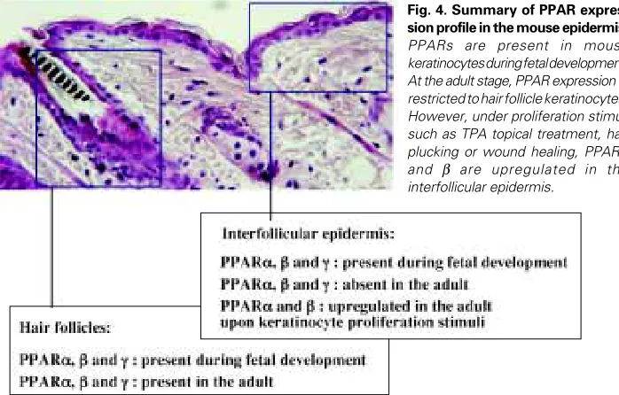
Fig. 4. Summary of PPAR expres-sion profile in the mouse epidermis.
ment, suggests that the PPARs may play a role during the forma-tion of the central nervous system, their funcforma-tions in this tissue are still poorly understood. In order to study this aspect of PPAR biology, we used a model of reaggregated fetal brain cell cultures prepared from rat telencephalon. Very interestingly, these neural cell aggregates offer a three-dimensional culture model, in which the network of neural cells progressively acquires characteristics resembling that of the brain, including cell migration, synaptogenesis and myelination (Basu-Modak et al., 1999). Using this model, we confirmed previous in vivo observations showing that PPARβ is the prevalent isoform in the brain, present in all cell types, whereas PPARα is expressed at very low levels predominantly in the astrocytes. Because of the involvement of PPAR in the regulation of lipid metabolism, which is very important in the brain, and given the key role of acyl-CoA synthetases (ACSs) in fatty acid utilization, we used the reaggregated cell model to study the putative regula-tion of ACS expression by PPARβ (Basu-Modak et al., 1999). We demonstrated that ACS2 and PPARβ have overlapping expression patterns, and that ACS2 expression is regulated by PPARβ at the transcriptional level, providing the first identification of a PPARβ target gene. This result strongly suggests that PPARβ participates in the regulation of lipid metabolism in the brain. This hypothesis is further supported by the observation that PPARβ null mice, mainly females, exhibit an altered myelination in the corpus callosum (Peters et al., 2000). Interestingly, such a defect was not observed in other regions of the central nervous system, and the expression of mRNA encoding proteins involved in the myelination process remained unchanged in the mutant mice. Despite the fact that its
expression was shown to be PPARβ-dependent, the expression of ACS2 was similar in the PPARβ null versus wild type mice, based on normalized data using the actin mRNA levels (Peters et al., 2000). However, results recently obtained in our group (Tan et al., 2001) showed that the expression of actin itself is different in the PPARβ null and wild type mice, probably explaining why ACS2 was not identified as a PPARβ target in vivo (Peters et al., 2000). Thus, and eventhough the underlying mechanisms remain to be investi-gated, these results indicate that the PPARβ isotype is implicated in the development of the murine central nervous system.
PPARs are Involved in Epidermal Maturation
The Epidermal Maturation Process. The epidermis is a multistratified
epithelium, in which the basal layer consists of progenitor undiffer-entiated keratinocytes. As they migrate from the basal to the uppermost layer, the keratinocytes undergo a vectorial differentia-tion program, which includes biochemical differentiadifferentia-tion, the se-quential expression of various structural proteins (e.g. keratins, involucrin and loricrin) and the processing and reorganization of lipids (e.g. sterols, free fatty acids, and sphingolipids) which will provide a hydrophobic barrier to the body. The maturation of the epidermis happens in the latest stages of vertebrate fetal develop-ment and is completed before term.
Many nuclear hormone receptors and their respective ligands have been implicated in the regulation of skin development. For instance, thyroid hormones, glucocorticoids and estrogen acceler-ate the skin barrier maturation, whereas testosterone delays the process (Aszterbaum et al., 1993; Hanley et al., 1996a; Hanley et
TABLE 1
SUMMARY OF THE MORPHOLOGICAL AND BIOCHEMICAL EVENTS OF THE DEVELOPMENT OF THE MOUSE EPIDERMIS
Developmental stages Morphology of the epidermis Cornified layer proteins Lipids Barrier function Epidermal PPAR expression (interfollicular)
E 13.5 Single undifferentiated layer (basal layer) + periderm K5; K14 Periderm PPARα +++ PPARβ +++ PPARγ + E 14.5 Single undifferentiated layer (basal layer)+ periderm. Hair follicles K5; K14 Involucrin Periderm PPARα +++
PPARβ +++
PPARγ +
E 15.5 Second layer (spinous layer). Hair follicles K5; K14;K1; K10 Involucrin Periderm PPARα +++
PPARβ +++
PPARγ +
E 16.5 Several layers + periderm. Hair follicles K5; K14; K1; K10 Involucrin Lipid lamellar Periderm PPARα ++
Loricrin granule formation PPARβ ++
PPARγ ++ E 17.5 Formation of the granular layer. Hair follicles K5; K14; K1; K10 Involucrin Lipid lamellar Epidermis PPARα ++ Loricrin Profilaggrin granule formation PPARβ ++
and extrusion PPARγ ++
E 18.5 Fully differentiated epidermis (6 to 8 layers). K5; K14; K1; K10 Involucrin Lipid lamellar Epidermis PPARα ++
Periderm detached Hair follicles Loricrin. Filaggrin granule formation PPARβ +++
and extrusion PPARγ ++
Newborn Fully differentiated (6 to 8 layers) epidermis. K5; K14; K1; K10 Involucrin Lipid lamellar Epidermis PPARα +
Hair follicles Loricrin. Filaggrin granule formation PPARβ +++
and extrusion PPARγ ++
+ 5 Fully differentiated (6 to 8 layers) epidermis. Hair growth K5; K14; K1; K10 Involucrin Lipid lamellar Epidermis PPARα + Loricrin. Filaggrin granule formation PPARβ ++
and extrusion PPARγ ++
Adult Fully differentiated (3 to 4 layers) epidermis. Hair growth K5; K14; K1; K10 Involucrin Lipid lamellar Epidermis Below detection level Loricrin. Filaggrin granule formation
and extrusion
al., 1996b). Retinoids are known to affect keratinocyte differentia-tion, although the consequences of retinoid treatments are differ-ent when applied in vitro or in vivo. In vivo however, the specific ablation of the RXRα isotype in the murine epidermis was recently demonstrated to have very severe consequences on the hair follicle cycle and the epidermal maturation (Li et al., 2000; Li et al., 2001).
PPAR Gene Expression During Epidermal Differentiation. The de-scription of the implication of PPAR in epidermal maturation is more recent. We and others have described the presence of PPARα, β and γ in rodent keratinocytes (Braissant et al., 1996; Braissant and Wahli, 1998; Hanley et al., 1999). Each isotype has a specific pattern of expression, during development and in the various layers of the epidermis, suggesting non redundant func-tions for the three receptors. We recently demonstrated that PPARα, β and γ transcripts are already present in the mouse epidermis at fetal day 13.5 (Fig. 4; Michalik et al., 2001). Their expression decreases after birth to be undetectable in the interfollicular epidermis in the adult animals, whereas the three isotypes remain expressed in the hair follicles. Interestingly, PPARα and PPARβ expression is upregulated in the adult epidermis upon proliferation stimuli (TPA topical application, hair plucking) (Figs. 4,5; Michalik et al., 2001). As shown in Table 1, the transient expression of PPARα, β and γ in the interfollicular epidermis during
mouse fetal development parallels all the major events of the maturation of the epidermal barrier. This includes the expression of the differentiation markers (such as involucrin, loricrin, filaggrin) and changes in lipid metabolism (apparition of the lipid granules). Whether some of these events are regulated through PPARs remains to be elucidated. Despite of some discrepancies in the reported data, the three PPAR isotypes have also been identified in human keratinocytes (Matsuura et al., 1999; Rivier et al., 1998; Westergaard et al., 2001). The PPARβ transcript seems to be the prevalent isotype, and its expression remains high during the differentiation of human keratinocytes. PPARα and γ are ex-pressed at lower levels, and their expression have been reported to increase upon differentiation (Matsuura et al., 1999; Rivier et al., 1998; Westergaard et al., 2001).
Effects of PPAR Ligands on Keratinocyte Differentiation. Consistent with the presence of PPARα in the epidermis during rodent fetal development, PPARα ligands were shown to accelerate rat epider-mal maturation in vitro (Hanley et al., 1998) and in utero (Hanley et al., 1999), whereas PPARβ and γ activators had no effects. In addition, PPARα ligands were recently reported to induce epider-mal differentiation and to restore epiderepider-mal homeostasis in hyperproliferative mouse epidermis (Komuves et al., 2000a; Komuves et al., 2000b). In human keratinocytes however, the results are rather different. Indeed, PPARα activators were
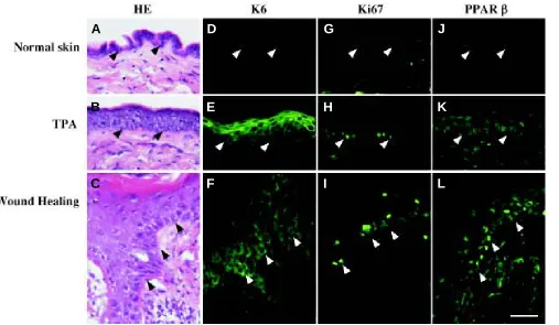
re-Fig. 5. Immunodetection of PPARβ and proliferation markers in adult mouse skin. (A-C) Histological staining (hematoxylin/eosin: HE) of adult mouse skin frozen sections. (D-F) Immunodection of Keratin 6 keratinocyte specific proliferation marker (K6) on adult mouse skin frozen sections. (G-I)
Immunodection of Ki67 nuclear proliferation marker on adult mouse skin frozen sections. (J-L) Immunodection of PPARβ on adult mouse skin frozen sections, using an anti-mouse PPARβ A/B domain polyclonal antibody. In unchallenged adult mouse interfollicular epidermis, Keratin 6 and PPARβ are absent, whereas the Ki67 protein is present in a few basal keratinocytes. The expression of the three proteins is strongly upregulated upon proliferation stimuli, e.g. after TPA topical application (TPA) or at the edges of a skin wound (Wound Healing, day three after an injury). Magnification bar, 20µm. Arrowheads point to the epidermis-dermis interface.
A
B
C
D
E
F
G
H
I
J
K
ported to have no effect on keratinocyte cell line differentiation (Westergaard et al., 2001), but to influence lipid metabolism in an in vitro human skin model (Rivier et al., 2000). The PPARβ selective ligand L165041 was able to induce the expression of differentiation markers in a human keratinocyte cell line, whereas the PPARγ ligand BRL had negligible effects (Westergaard et al., 2001). However, and very interestingly, the same authors de-scribed that PPARβ and γ ligands have a synergistic effect on human keratinocyte cell line differentiation when added simulta-neously. Overall, the effects of the PPAR ligand on keratinocyte differentiation appear to be quite different across species. As mentioned above, the PPARs exhibit important species specificity in ligand binding which, in addition to differences in the models used, could account for these apparent discrepancies.
Study of PPAR Functions in Epidermal Differentiation In Vivo. In addition to ligand assays in vitro or in vivo, important indications on the role of PPARs in epidermis homeostasis were obtained from PPAR mutant mouse models. To address the hypothesis of the involvement of PPAR in the differentiation of the epidermis during mouse fetal development, we looked at the skin maturation in PPAR mutant embryos during late fetal development (embry-onic day 14.5, 16.5 and 18.5), at the time of the formation of a competent epidermal barrier. Sections of PPARα null or PPARβ heterozygous embryonic skin were compared to their respective wild type controls at the histological level. Surprisingly, these mutant mice showed a normal skin architecture upon histological staining at all the embryonic stages examined. All the character-istic layers were present in the epidermis of PPARα and β mutant embryos, with no major defect in their thickness and organization (data not shown). No major difference was observed either after examination of the expression of epidermal differentiation mark-ers in the PPARα and β deficient epidermis (data not shown). These results suggest that the mouse fetal epidermis is able to undergo normal maturation in the absence of PPARα, and in conditions where PPARβ expression is decreased by half. Simi-larly, the PPARγ heterozygous animals, or the PPARγ null mice born after placental rescue, do not exhibit any obvious defect in epidermal maturation either (Barak et al., 1999). Moreover, PPARγ deficient cells are able to participate in the development of the epidermis in mouse chimeras comprising PPARγ null and wild type cells, suggesting no or little contribution of PPARγ in epider-mal differentiation (Rosen et al., 1999).
Consistent with earlier characterization of the PPARα null mice (Komuves et al., 2000a; Lee et al., 1995), we did not see any major defect after examination of skin sections of PPARα null adult animals. However, histological and immunostaining examination of the epidermis of the PPARβ adult mutant mice indicated a slight but significant increase in the keratinocyte proliferation rate in the PPARβ heterozygous mice compared to the wild type control animals (Michalik et al., 2001). This difference was even more striking upon TPA topical application on the epidermis of these animals. The hyperplastic response usually observed after TPA topical application on the epidermis was indeed much more pronounced in the PPARβ mutant animals, strongly suggesting a defect in the control of keratinocyte proliferation in the whole animal (Michalik et al., 2001). An impaired control of keratinocyte proliferation in PPARβ deficient keratinocytes was reported as well by Peters and colleagues in a PPARβ null mouse model (Peters et al., 2000). These in vivo data
demonstrate that the PPARβ isotype is implicated in the control of keratinocyte proliferation in the whole animal.
Because in vitro and in vivo observations strongly suggest a link between PPAR and keratinocyte differentiation and prolifera-tion, we studied the expression and the involvement of PPAR during adult skin wound repair. In this situation, the mature skin is disrupted, and the covering of the wound by a new epithelium starts within hours after the injury. A fully differentiated epithelium, and thus a competent protective epidermis, will eventually be reconstituted. The re-epithelialization of a wound involves initially the proliferation of the keratinocytes and their migration to form the neo-epithelium. Stratification and differentiation/maturation follows to restaure the normal adult epithelium structure. Using in situ hybridization, we demonstrated that PPARα and PPARβ, but not PPARγ expression is upregulated in the keratinocytes at the edges of the skin wound. In Fig. 5, we confirm that the upregulation of PPARβ also occur at the protein level. The PPARα transcript is present transiently in this area during the early inflammatory phase of the healing, whereas PPARβ remains expressed until completion of the process (Michalik et al., 2001). Consistent with this pattern of PPAR expression in the epidermis during wound healing, and using PPARα, β and γ mutant mouse models, we showed that PPARα and β, but not PPARγ, are necessary for the normal healing of an excisional skin wound (Michalik et al., 2001). The PPARα null mice indeed exhibit a transient delay in skin healing during the inflammatory phase of the process, whereas in the PPARβ mutant mice the healing was postponed for 2 to 3 days compared to the wild type animals. In both cases, the delay observed in the skin repair is consistent with the pattern of expression of the respective PPAR isotype as analysed during skin wound repair (Michalik et al., 2001). These results revealed important but non redundant roles of PPARα and β in the regen-eration of the skin in adult mouse, with the involvement of PPARα the early inflammatory phase, whereas PPARβ plays a role during the whole healing process.
Taken all together, our data suggest, very interestingly, that in the same animal, a PPAR mutation has no obvious effect during fetal development in the epidermis, but affects epidermal regen-eration at the adult stage.
Conclusions
differentiation in vivo might still be underestimated, because of functional redundancies or lethality in the null mouse lines. In these cases, the analysis of mouse lines in which specific tissues are deficient for a given PPAR isotype will likely reveal additional unexpected functions for these nuclear receptors.
Acknowledgements
We thank Nathalie Deriaz, Mai Perroud, Tatiana Favez and Brigitte Glaeser for excellent assistance. This work was supported by the Swiss National Science Foundation (grants to Walter Wahli and to Béatrice Desvergne), by the Etat de Vaud, by the Human Frontier Science Program Organization and by Parke-Davis Pharmaceutical Research.
References
NUCLEAR RECEPTOR NOMENCLATURE COMMITEE. 1999. A unified nomencla-ture system for the nuclear receptor superfamily. Cell. 97:161-3.
ASZTERBAUM, M., FEINGOLD, K. R., MENON, G. K. and WILLIAMS, M. L. (1993). Glucocorticoids accelerate fetal maturation of the epidermal permeability barrier in the rat. J Clin Invest 91:2703-2708.
AUBOEUF, D., RIEUSSET, J., FAJAS, L., VALLIER, P., FRERING, V., RIOU, J. P., STAELS, B., AUWERX, J., LAVILLE, M. and VIDAL, H. (1997). Tissue distribution and quantification of the expression of mRNAs of peroxisome proliferator-activated receptors and liver X receptor-alpha in humans: no alteration in adipose tissue of obese and NIDDM patients. Diabetes 46:1319-1327.
BARAK, Y., NELSON, M. C., ONG, E. S., JONES, Y. Z., RUIZ-LOZANO, P., CHIEN, K. R., KODER, A. and EVANS, R. M. (1999). PPAR gamma is required for placental, cardiac, and adipose tissue development. Mol Cell 4:585-595.
BASU-MODAK, S., BRAISSANT, O., ESCHER, P., DESVERGNE, B., HONEGGER, P. and WAHLI, W. (1999). Peroxisome proliferator-activated receptor beta regu-lates acyl-CoA synthetase 2 in reaggregated rat brain cell cultures. J Biol Chem 274:35881-35888.
BECK, F., PLUMMER, S., SENIOR, P. V., BYRNE, S., GREEN, S. and BRAMMAR, W. J. (1992). The ontogeny of peroxisome-proliferator-activated receptor gene expres-sion in the mouse and rat. Proc R Soc Lond B Biol Sci 247:83-87.
BRAISSANT, O., FOUFELLE, F., SCOTTO, C., DAUCA, M. and WAHLI, W. (1996). Differential expression of peroxisome proliferator-activated receptors (PPARs): tissue distribution of PPAR-alpha, -beta, and -gamma in the adult rat. Endocrinology 137:354-366.
BRAISSANT, O. and WAHLI, W. (1998). Differential expression of peroxisome proliferator-activated receptor-alpha, -beta, and -gamma during rat embryonic development. Endocrinology 139:2748-2754.
CULLINGFORD, T. E., BHAKOO, K., PEUCHEN, S., DOLPHIN, C. T., PATEL, R. and CLARK, J. B. (1998). Distribution of mRNAs encoding the peroxisome proliferator-activated receptor alpha, beta and gamma and the retinoid X receptor alpha, beta and gamma rat central nervous system. J Neurochem70:1366-1375.
DESVERGNE, B. and WAHLI, W. (1999). Peroxisome proliferator-activated receptors: Nuclear control of metabolism. Endocrine Reviews 20:649-688.
DREYER, C., KREY, G., KELLER, H., GIVEL, F., HELFTENBEIN, G. and WAHLI, W. (1992). Control of the peroxisomal beta-oxidation pathway by a novel family of nuclear hormone receptors. Cell 68:879-887.
DREYER, C., KELLER, H., MAHFOUDI, A., LAUDET, V., KREY, G. and WAHLI, W. (1993). Positive regulation of the peroxisomal beta-oxidation pathway by fatty acids through activation of peroxisome proliferator-activated receptors (PPAR). Biology of the Cell 77:67-76.
DREYER, C. and ELLINGER-ZIEGELBAUER, H. (1996). Retinoic acid receptors and nuclear orphan receptors in the development of Xenopus laevis. Int J Dev Biol 40:255-262.
ESCHER, P. and WAHLI, W. (2000). Peroxisome proliferator-activated receptors: insight into multiple cellular functions. Mutat Res 448:121-138.
ESCHER, P., BRAISSANT, O., BASU-MODAK, S., MICHALIK, L., WAHLI, W. and DESVERGNE, B. (2001). Rat PPARs: quantitative analysis in adult rat tissues and regulation in fasting and refeeding. Endocrinology 142:4195-4202.
ESCRIVA, H., DELAUNAY, F. and LAUDET, V. (2000). Ligand binding and nuclear receptor evolution. Bioessays 22:717-727.
GRIMALDI, P. A. (2001). The roles of PPARs in adipocyte differentiation. Prog Lipid Res 40:269-281.
HANLEY, K., RASSNER, U., ELIAS, P. M., WILLIAMS, M. L. and FEINGOLD, K. R. (1996a). Epidermal barrier ontogenesis: maturation in serum-free media and acceleration by glucocorticoids and thyroid hormone but not selected growth factors. J Invest Dermatol 106:404-411.
HANLEY, K., RASSNER, U., JIANG, Y., VANSOMPHONE, D., CRUMRINE, D., KOMUVES, L., ELIAS, P. M., FEINGOLD, K. R. and WILLIAMS, M. L. (1996b). Hormonal basis for the gender difference in epidermal barrier formation in the fetal rat. Acceleration by estrogen and delay by testosterone. J Clin Invest 97:2576-2584. HANLEY, K., JIANG, Y., HE, S. S., FRIEDMAN, M., ELIAS, P. M., BIKLE, D. D., WILLIAMS, M. L. and FEINGOLD, K. R. (1998). Keratinocyte differentiation is stimulated by activators of the nuclear hormone receptor PPARalpha. J Invest Dermatol 110:368-375.
HANLEY, K., KOMUVES, L. G., BASS, N. M., HE, S. S., JIANG, Y., CRUMRINE, D., APPEL, R., FRIEDMAN, M., BETTENCOURT, J., MIN, K., ELIAS, P. M., WILL-IAMS, M. L. and FEINGOLD, K. R. (1999). Fetal epidermal differentiation and barrier development In vivo is accelerated by nuclear hormone receptor activators. J Invest Dermatol 113:788-795.
HUIN, C., CORRIVEAU, L., BIANCHI, A., KELLER, J. M., COLLET, P., KREMARIK-BOUILLAUD, P., DOMENJOUD, L., BECUWE, P., SCHOHN, H., MENARD, D. and DAUCA, M. (2000). Differential expression of peroxisome proliferator-activated receptors (PPARs) in the developing human fetal digestive tract. J Histochem Cytochem 48:603-611.
ISSEMANN, I. and GREEN, S. (1990). Activation of a Member of the Steroid Hormone Receptor Superfamily by Peroxisome Proliferators. Nature 347:645-650. KELLER, H., DEVCHAND, P. R., PERROUD, M. and WAHLI, W. (1997). PPAR-alpha
structufunction relationships derived from species-specific differences in re-sponsiveness to hypolipidemic agents. Biological Chemistry 378:651-655. KELLER, J. M., COLLET, P., BIANCHI, A., HUIN, C., BOUILLAUD-KREMARIK, P.,
BECUWE, P., SCHOHN, H., DOMENJOUD, L. and DAUCA, M. (2000). Implica-tions of peroxisome proliferator-activated receptors (PPARS) in development, cell life status and disease. Int J Dev Biol 44:429-442.
KERSTEN, S., DESVERGNE, B. and WAHLI, W. (2000). Roles of PPARs in health and disease. Nature 405:421-424.
KERSTEN, S. and WAHLI, W. (2000). Peroxisome proliferator activated receptor agonists. In New approaches to drug development (Eds Jolles, P.) Birkhauser Verlag, Switzerland 89:141-151.
KLIEWER, S. A., FORMAN, B. M., BLUMBERG, B., ONG, E. S., BORGMEYER, U., MANGELSDORF, D. J., UMESONO, K. and EVANS, R. M. (1994). Differential expression and activation of a family of murine peroxisome proliferator-activated receptors. Proc Natl Acad Sci U S A 91:7355-7359.
KOMUVES, L. G., HANLEY, K., LEFEBVRE, A. M., MAN, M. Q., NG, D. C., BIKLE, D. D., WILLIAMS, M. L., ELIAS, P. M., AUWERX, J. and FEINGOLD, K. R. (2000a). Stimulation of PPARalpha promotes epidermal keratinocyte differentiation in vivo. J Invest Dermatol 115:353-360.
KOMUVES, L. G., HANLEY, K., MAN, M. Q., ELIAS, P. M., WILLIAMS, M. L. and FEINGOLD, K. R. (2000b). Keratinocyte differentiation in hyperproliferative epider-mis: topical application of PPARalpha activators restores tissue homeostasis. J Invest Dermatol 115:361-367.
KREY, G., BRAISSANT, O., L’HORSET, F., KALKHOVEN, E., PERROUD, M., PARKER, M. G. and WAHLI, W. (1997). Fatty acids, eicosanoids, and hypolipidemic agents identified as ligands of peroxisome proliferator-activated receptors by coactivator-dependent receptor ligand assay. Mol Endo 11:779-791.
KUBOTA, N., TERAUCHI, Y., MIKI, H., TAMEMOTO, H., YAMAUCHI, T., KOMEDA, K., SATOH, S., NAKANO, R., ISHII, C., SUGIYAMA, T., ETO, K., TSUBAMOTO, Y., OKUNO, A., MURAKAMI, K., SEKIHARA, H., HASEGAWA, G., NAITO, M., TOYOSHIMA, Y., TANAKA, S., SHIOTA, K., KITAMURA, T., FUJITA, T., EZAKI, O., AIZAWA, S., KADOWAKI, T. and ET AL. (1999). PPAR gamma mediates high-fat diet-induced adipocyte hypertrophy and insulin resistance. Mol Cell 4:597-609. LEE, S. S., PINEAU, T., DRAGO, J., LEE, E. J., OWENS, J. W., KROETZ, D. L., FERNANDEZ, S. P., WESTPHAL, H. and GONZALEZ, F. J. (1995). Targeted disruption of the alpha isoform of the peroxisome proliferator-activated receptor gene in mice results in abolishment of the pleiotropic effects of peroxisome proliferators. Mol Cell Biol 15:3012-3022.
temporally controlled RXRalpha mutations in mouse epidermis. Nature 407:633-636.
LI, M., CHIBA, H., WAROT, X., MESSADDEQ, N., GERARD, C., CHAMBON, P. and METZGER, D. (2001). RXRalpha ablation in skin keratinocytes results in alopecia and epidermal alterations. Development 128:675-688.
LIM, H., GUPTA, R. A., MA, W. G., PARIA, B. C., MOLLER, D. E., MORROW, J. D., DUBOIS, R. N., TRZASKOS, J. M. and DEY, S. K. (1999). Cyclo-oxygenase-2-derived prostacyclin mediates embryo implantation in the mouse via PPARdelta. Genes Dev 13:1561-1574.
MATSUURA, H., ADACHI, H., SMART, R. C., XU, X., ARATA, J. and JETTEN, A. M. (1999). Correlation between expression of peroxisome proliferator-activated receptor beta and squamous differentiation in epidermal and tracheobronchial epithelial cells. Mol Cell Endocrinol 147:85-92.
MICHALIK, L., DESVERGNE, B., TAN, N. S., BASU-MODAK, S., ESCHER, P., RIEUSSET, J., PETERS, J. M., KAYA, G., GONZALEZ, F. J., ZAKANY, J., METZGER, D., CHAMBON, P., DUBOULE, D. and WAHLI, W. (2001). Impaired skin wound healing in peroxisome proliferator-activated receptor (PPAR)alpha and PPARbeta mutant mice. J Cell Biol 154:799-814.
MUKHERJEE, R., JOW, L., CROSTON, G. E. and PATERNITI, J. R. J. (1997). Identification, characterization, and tissue distribution of human peroxisome proliferator-activated receptor (PPAR) isoforms PPARgamma2 versus PPARgamma1 and activation with retinoid X receptor agonists and antagonists. J Biol Chem 272:8071-8076.
OBERFIELD, J. L., COLLINS, J. L., HOLMES, C. P., GOREHAM, D. M., COOPER, J. P., COBB, J. E., LENHARD, J. M., HULL-RYDE, E. A., MOHR, C. P., BLANCHARD, S. G., PARKS, D. J., MOORE, L. B., LEHMANN, J. M., PLUNKET, K., MILLER, A. B., MILBURN, M. V., KLIEWER, S. A. and WILLSON, T. M. (1999). A peroxisome proliferator-activated receptor gamma ligand inhibits adipocyte differentiation. Proc Natl Acad Sci U S A 96:6102-6106.
PALMER, C. N. A., HSU, M. H., GRIFFIN, K. J., RAUCY, J. L. and JOHNSON, E. F. (1998). Peroxisome proliferator activated receptor-alpha expression in human liver. Mol Pharmacol 53:14-22.
PETERS, J. M., LEE, S. S., LI, W., WARD, J. M., GAVRILOVA, O., EVERETT, C., REITMAN, M. L., HUDSON, L. D. and GONZALEZ, F. J. (2000). Growth, adipose, brain, and skin alterations resulting from targeted disruption of the mouse peroxi-some proliferator-activated receptor beta(delta). Mol Cell Biol 20:5119-5128. RIVIER, M., SAFONOVA, I., LEBRUN, P., GRIFFITHS, C. E., AILHAUD, G. and
MICHEL, S. (1998). Differential expression of peroxisome proliferator-activated receptor subtypes during the differentiation of human keratinocytes. J Invest Dermatol 111:1116-1121.
RIVIER, M., CASTIEL, I., SAFONOVA, I., AILHAUD, G. and MICHEL, S. (2000). Peroxisome proliferator-activated receptor-alpha enhances lipid metabolism in a skin equivalent model. J Invest Dermatol 114:681-687.
ROSEN, E. D., SARRAF, P., TROY, A. E., BRADWIN, G., MOORE, K., MILSTONE, D. S., SPIEGELMAN, B. M. and MORTENSEN, R. M. (1999). PPAR gamma is required for the differentiation of adipose tissue in vivo and in vitro. Mol Cell 4:611-617.
ROSEN, E. D. and SPIEGELMAN, B. M. (2001). PPARgamma: a nuclear regulator of metabolism, differentiation, and cell growth. J Biol Chem 276:37731-37734. SCHAIFF, W. T., CARLSON, M. G., SMITH, S. D., LEVY, R., NELSON, D. M. and
SADOVSKY, Y. (2000). Peroxisome proliferator-activated receptor-gamma modu-lates differentiation of human trophoblast in a ligand-specific manner. J Clin Endocrinol Metab 85:3874-3881.
TAN, N. S., MICHALIK, L., NOY, N., YASMIN, R., PACOT, C., HEIM, M., FLUHMANN, B., DESVERGNE, B., WAHLI, W. (2001). Critical role of PPARβ/δ in keratinocyte response to inflammation. Genes Dev. 15: 3263-3277.
WAITE, L. L., PERSON, E. C., ZHOU, Y., LIM, K. H., SCANLAN, T. S. and TAYLOR, R. N. (2000). Placental peroxisome proliferator-activated receptor-gamma is up-regulated by pregnancy serum. J Clin Endocrinol Metab 85:3808-3814. WESTERGAARD, M., HENNINGSEN, J., SVENDSEN, M. L., JOHANSEN, C.,