R E S E A R C H A R T I C L E
Open Access
ISPD
mutations account for a small proportion
of Italian Limb Girdle Muscular Dystrophy cases
Francesca Magri
1†, Irene Colombo
2†, Roberto Del Bo
1, Stefano Previtali
3, Roberta Brusa
1, Patrizia Ciscato
2,
Marina Scarlato
3, Dario Ronchi
1, Maria Grazia D
’
Angelo
4, Stefania Corti
1, Maurizio Moggio
2, Nereo Bresolin
1and Giacomo Pietro Comi
1*Abstract
Background:Limb Girdle Muscular Dystrophy (LGMD), caused by defectiveα-dystroglycan (α-DG) glycosylation, was recently associated with mutations in Isoprenoid synthase domain-containing (ISPD) and GDP-mannose pyrophosphorylase B (GMPPB) genes. The frequency ofISPDandGMPPBgene mutations in the LGMD population is unknown.
Methods:We investigated the contributions ofISPDandGMPPBgenes in a cohort of 174 Italian patients with LGMD, including 140 independent probands. Forty-one patients (39 probands) from this cohort had not been genetically diagnosed. The contributions ofISPDandGMPPBwere estimated by sequentialα-DG immunohistochemistry (IHC) and mutation screening in patients with documentedα-DG defect, or by direct DNA sequencing of both genes when muscle tissue was unavailable.
Results:We performedα-DG IHC in 27/39 undiagnosed probands: 24 subjects had normalα-DG expression, two had a partial deficiency, and one exhibited a complete absence of signal. Direct sequencing ofISPDandGMPPBrevealed two heterozygousISPDmutations in the individual who lackedα-DG IHC signal: c.836-5 T > G (which led to the deletion of exon 6 and the production of an out-of-frame transcript) and c.676 T > C (p.Tyr226His). This patient presented with sural hypertrophy and tip-toed walking at 5 years, developed moderate proximal weakness, and was fully ambulant at 42 years. The remaining 12/39 probands did not exhibit pathogenic sequence variation in either gene.
Conclusion:ISPDmutations are a rare cause of LGMD in the Italian population, accounting for less than 1 % of the entire cohort studied (FKRPmutations represent 10 %), whileGMPPBmutations are notably absent in this patient sample. These data suggest that the genetic heterogeneity of LGMD with and withoutα-DG defects is greater than previously realized.
Keywords:Isoprenoid synthase domain-containing gene, GDP-mannose pyrophosphorylase B, Limb Girdle Muscular Dystrophy,α-dystroglycan glycosylation
Background
Limb girdle muscular dystrophies (LGMDs) are a hetero-geneous group of inherited progressive muscle disorders characterized by progressive shoulder and pelvic gir-dle muscle weakness variably associated with cardiac,
respiratory, and cognitive involvement [1]. The num-ber of genes involved in these disorders has exponen-tially increased in recent years, and now up to 30 different forms have been described, inherited both with autosomal dominant (7 forms) and autosomal recessive (23 forms) patterns [2].
In particular, a high number of genes involved in
α-dystroglycan (α-DG) glycosylation have been associ-ated with LGMD. α-DG is a highly glycosylated core component of the dystrophin glycoprotein complex, and forms a link between the sarcolemma and the extracellu-lar matrix [3]. To date, mutations in 14 genes [4–18], all
* Correspondence:[email protected] †Equal contributors
1Dino Ferrari Centre, Department of Neurological Sciences, University of Milan, I.R.C.C.S. Foundation Cà Granda, Ospedale Maggiore Policlinico, via F. Sforza 35, 20122 Milan, Italy
Full list of author information is available at the end of the article
coding for putative or demonstrated glycosyltransferase, have been associated with muscular dystrophies (referred to as secondary dystroglycanopathies) [19], while only a couple of cases have been associated with mutations in
DAG1, the gene that encodes both α-dystroglycan and
β-dystroglycan [20, 21]. Dystroglycanopathies are char-acterised by a broad variety of clinical phenotypes, ranging from congenital muscular dystrophy (CMD), with or without brain and eye involvement as Walker-Warburg syndrome (WWS) and muscle eye brain dis-ease (MEB), to LGMD, as summarised by Godfrey et al. [22]. Mutations in most of these genes are mainly associ-ated with severe or congenital conditions, with few not-able exceptions: FKRP mutations account for a variable proportion of LGMD depending on ethnic background (from 6 % in the Italian population [1, 23] to 38 % in the Danish population [24]). On the other hand, LGMD phenotypes caused by mutations inPOMT1 (LGMD2K) [25], FKTN (LGMD2L) [26], POMT2 (LGMD2N) [27],
POMGNT1 (LGMD2O) [22], DAG1 [21], and DPM3
[10] have been reported in a very limited number of patients.
ISPD, a gene located on chromosome 7p21, encodes the Isoprenoid synthase domain-containing protein and has been implicated in the initial step of the O-mannosylation of α-DG. Mutations in this gene were first identified within the most severe spectrum of dystroglycanopathies, WWS and MEB cases [13, 14], although more recently they have also been associated with milder phenotypes [28, 29]. In a paediatric cohort of dystroglycanopathies with British and Turkish back-ground, ISPD mutations have been found to cause LGMD in seven probands, including four LGMD cases with normal cognitive development (LGMD – no MR); two LGMD cases with cerebellar involvement (LGMD-CRB); and one case of LGMD with mental retardation, but without structural brain abnormalities (LGMD-MR) [28]. ISPD mutations were also detected in two Italian LGMD families that presented with disease onset during the first two decades of life, late motor impairment, and no functional or structural brain involvement [28]. Muscle biopsy revealed dystrophic features and α-DG reduction at immunohistochemistry [28, 29]. According to this description, forms of LGMD caused by mutations in ISPD are described as LGMD2S [2]. Intra-familial variability has also been described [30].
ISPDmutations account for 9–11 % of the most severe dystroglycanopathy variants (comprising CMD, WWS, and cobblestone lyssencephaly) in three large cohorts from different ethnic backgrounds [13, 14, 17]. The preva-lence of ISPD mutations has not yet been estimated in LGMD cohorts: at this time, only 12 patients withISPD mutation and this phenotype have been described. Similar considerations apply to disease phenotypes caused by
GMPPBmutations, initially shown to be causally linked to
MEB/FCMD-like syndrome [16, 31], and more recently to a wider phenotypic spectrum that includes infantile phe-notypes with mental retardation [16] and adult-onset LGMD with normal cognition [32].
The aim of this study is to establish the prevalence of
ISPDand GMPPB mutations within an Italian cohort of
LGMD patients.
Methods
Patient selection and characterization
From a cohort of 174 Italian LGMD patients (140 families), all followed at a single neuromuscular centre, we selected 41 patients (39 families) without a molecular diagnosis. Written informed consent was obtained (and preserved in original) from all patients or their caregivers at first evaluation, with explicit consent to future use for research purposes, in accordance with the Declaration of Helsinki. This protocol was approved by the Research Ethics Board of IRCCS Foundation Ca’ Granda Ospedale Maggiore Policlinico. The patients have been previously screened for the following genes: MYOT, LMNA, CAV3, DNAJB6,
and TNPO3, if autosomal dominant transmission was
supported by family history; CAPN3, DYSF, SGCA,
SGCB, SGCG, SGCD, FKRP, ANO5, FKTN,andLAMA2
in sporadic or autosomal recessive cases.
LARGE, POMT1, POMT2, POMGnT1 were also screened in selected cases.
Patients were defined as affected with LGMD if they fulfilled the following criteria: clinical phenotype charac-terised by progressive muscle weakness and wasting af-fecting primarily the shoulder girdle and pelvic muscles, in keeping with the diagnostic criteria for LGMD [33]; and dystrophic features at muscle biopsy. All patients had undergone systematic clinical characterisation, in-cluding comprehensive neurological [Medical Research Council (MRC) and functional scales], cardiac (electro-cardiogram and echo(electro-cardiogram), and respiratory (spir-ometry and nocturnal saturimetry) assessments. Data about clinical and familial history were also collected. All specimens were obtained from the Skeletal Muscle, Peripheral Nerve, DNA and Cell Line Bank of the Neuro-muscular Unit, Fondazione IRCCS Ca’ Granda Ospedale Maggiore Policlinico, University of Milan. Written in-formed consent was obtained and preserved in the original form from all individuals or their caregivers when primary diagnostic procedures were performed, with ex-plicit consent for future use for research purposes, accord-ing to the Declaration of Helsinki.
Muscle biopsy analysis
nitrogen, and stained histochemically according to stand-ard procedures [34]. We reviewed muscle biopsies from cases without a genetic characterization, which included data about α-DG immunohistochemical (IHC) analysis (performed with the clone VIA4-1; Merck Millipore, UK). In muscle samples in which α-DG IHC had not been conducted previously, this study was performed using the antibody cited above if a muscle tissue sample was available.
Molecular analysis
Genomic DNA was extracted from peripheral blood samples according to standard procedures (Flexi Gene DNA Handbook, Qiagen).
ISPDand GMPPBanalysis were performed in patients
who exhibited α-DG deficiency at muscle IHC and in patients who had LGMD inclusion criteria, but whose muscle sample was unavailable at the time of this inves-tigation. Mutations in FKRP, the most common LGMD gene involved in α-DG glycosylation [1, 32, 35], were also ruled out for the first group of patients.
All exons and flanking intronic regions were directly sequenced using an ABI PRISM 3100 XL Genetic Analyzer (Applied Biosystems) and previously published primer [13, 16].
The pathogenic nature of new mutations was con-firmed by screening 100 healthy control subjects. The intronic mutation that leads to abnormal mRNA splicing was investigated through transcript analysis. The paren-tal origin of each mutation was assessed through analysis of parental genomic DNA, when available. Amino acid conservation was confirmed by comparison with se-quences from different species.
We isolated mRNA from muscle tissue using Eurozol; the cDNA was produced through reverse transcription polymerase chain reaction (Ready-To-Go RT-PCR kit, Amersham Pharmacia) and analyzed by amplification, cloning, and sequencing. Mutations were named ac-cording to the Leiden Muscular Dystrophy database (www.dmd.nl).
Results
Relative frequency of ISPD and GMPBB mutations in an Italian cohort
Among a single-centre cohort of 174 Italian LGMD patients (140 probands), 41 patients (39 probands) were without a genetic diagnosis. For 12/39 probands, we dir-ectly analyzed ISPDandGMPPBbecause muscle biopsy samples were unavailable for α-DG IHC analysis: none contained mutations. For 27/39 probands with avail-able muscle biopsy, we performed α-DG IHC analysis, followed by ISPD and GMPPB sequencing in cases that exhibited any glycosylation defect: of 27 individuals, 24 had normal muscleα-DG expression, two had a partial
deficiency, and one exhibited a complete absence of signal. The latter three cases were analysed for FKRP and GMPPB showing wild-type sequences. Only one of them had ISPD-causative mutations. Noteworthy, this latter proband was the only case with a muscle biopsy that exhibited complete absenceα-DG labelling (Fig. 1).
Including this new case, the 102/140 probands with a confirmed genetic diagnosis of LGMD had mutations in the following genes: 2/102 (2 %) LMNA, 6/102 (6 %)
CAV3, 29/102 (28 %) CAPN3, 27/102 (25 %) DYSF, 5/ 102 (5 %) SGCG, 12/102 (12 %) SGCD, 5/102 (5 %)
SGCB, 1/102 (1 %)SGCE, 8/102 (8 %)FKRP5/102 (5 %)
ANO5, and 1/102 (1 %)LAMA2. The molecular data of these patients are listed in a separate table [see Additional file 1]. Overall,ISPDmutations account for 0.9 % of genet-ically characterized LGMD probands (Fig. 2a). Among our paediatric LGMD cohort, defined as patients with disease onset before 10 years of age (33 patients; 27 probands)
ISPD mutations account for 4 % of cases. Relative frequencies in the paediatric onset population are indi-cated in Fig. 2b; sarcoglycanopathies (51 %), calpaino-pathies (19 %), and LGMD2I (11 %) are the most frequent forms.
Molecular analysis
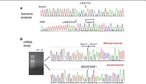
The ISPD-mutated patient carried two compound heterozygous mutations (Fig. 3a). The first mutation (c.676 T > C; p.Tyr226His) was a missense substitution in exon 3. Vuillaumier-Barrot et al. reported this muta-tion previously [17] in a heterozygotic foetal case af-fected with cobblestone lissencephaly; it was associated with a large deletion involving exons 4 through 6. The concerned amino acid is highly conserved among species. The second mutation (c.836-5 T > G) was a novel intronic substitution in IVS5. Its effect was in-vestigated through transcript analysis, which revealed the production of two different transcripts: the wild-type transcript, and a shorter transcript corresponding to an isoform with lower molecular weight. cDNA cloning demonstrated that the smaller isoform corre-sponds to a transcript in which the deletion of exon 6 results in the production of an out-of-frame transcript (Fig. 3b). No alternative splicing could be demon-strated because the full-length transcript is completely encoded by the other allele, which contains a missense mutation.
Clinical findings
Fig. 2Relative frequency ofISPDmutations.aThe relative frequency of different mutations in the entire sample.ISPDmutations account for approximately 1 % of the probands.bThe relative frequencies of different mutations in the sample of patients younger than 10 years old.ISPD
mutations account for 4 % of the probands
reported. The patient’s prenatal history, delivery, and psycho-motor development were unremarkable. At the time of his first evaluation, neurological examination re-vealed only mild sural hypertrophy. Muscle strength was normal overall, with the exception of mild weakness at the pelvic girdle with positive Gowers’ sign. Creatine kinase levels ranged from 630 to 1200 IU/L (normal values <200 IU/L). Neurophysiological investigation re-vealed signs of mild myopathy at needle examination. The patient underwent a quadriceps muscle biopsy, which indicated a dystrophic process (Fig. 4). Consider-ing these clinical and morphological findConsider-ings, a diagnosis of Becker muscular dystrophy was initially supposed; however, later IHC analyses and Western blotting with monoclonal antibodies directed against dystrophin (Novocastra, 28 Newcastle Upon Tyne, UK) did not re-veal any abnormalities. Furthermore, IHC analysis of caveolin-3 (Transduction Laboratories, Lexington, KY) and sarcoglycans (Transduction Laboratories, Lexington, KY) demonstrated normal staining. Dysferlin Western blot (Novocastra antibody) did not demonstrate any re-duction of protein.
The patient’s motor performance has been stable since the age of 14 years, when the patient first com-plained of running difficulty and muscle cramps during exercise. Over the following decades, muscle weakness demonstrated a progressive course. At his last clinical
examination, at the age of 42 years, he showed mild weakness at the neck flexors (MRC: 4) with moderate proximo-distal weakness at all four limbs (MRC: del-toids 3, brachial biceps 4, triceps 3, wrist extensors/ flexors 4, sartorius 2, hip extra- and intra-rotators 3, quadriceps 3, tibialis anterior 4). He had universally de-creased deep tendon reflexes, myopathic facies, bilateral scapular winging, and mild scoliosis. He walked with a waddling-type gait, and was able to climb stairs with double support. Ability to run had been lost (he walked 10 meters in 6.19 seconds). Regarding pulmonary in-volvement, spirometry indicated that his forced vital capacity was 3.35 L (71 %); nocturnal oxygen saturation was normal. His cardiac evaluation (echocardiogram) documented a right bundle branch block without any abnormalities of contractility. No cognitive impairment was observed (the patient acquired a degree in Engin-eering and worked as an engineer). We were unable to perform magnetic resonance imaging studies because of the patient’s severe claustrophobia.
Discussion
Mutations in genes involved in α-DG glycosylation have been associated with a broad spectrum of disorders ranging from severe CMD to milder LGMD phenotypes [36]. An increasing number of genes involved in these disorders have been discovered recently, enlarging the
spectrum of molecular heterogeneity of dystroglycanopa-thies. Among them,ISPDand GMPPBappear to follow
FKRP as relevant genes in the LGMD population, ac-cording to earlier reports [16, 28].
It should be kept in mind that variations in relative frequency for a gene may be population-dependent. This is the case for the FKRP gene, which accounts for ap-proximately 6 % of LGMD in Italian patients [1, 23], compared with 19 % among the British population [37] and 38 % among the Danish population [24]. Regarding other genes responsible for secondary dystroglycanopa-thies, only sporadic cases with LGMD phenotype have been reported thus far [10, 16, 21, 25–28]. In particular,
ISPDmutations appear to be responsible for a relatively high proportion of dystroglycanopathies within the most severe clinical spectrum [13, 14, 17], although they have also been described in a few LGMD cases [28–30].
We analysed a large cohort of Italian LGMD patients in order to estimate the frequency ofISPDand GMPPB mutations and their associated clinical picture. In our LGMD sample, only a small proportion of undiagnosed cases (3/27, 11 %) exhibited reduction ofα-DG staining. This finding suggests that if we exclude mutations in the
FKRP gene, the other forms associated with defects of
α-DG glycosylation are much more rare. Forms of LGMD caused by mutations in ISPD were also rare overall in our cohort, as they represent 0.9 % of genetic-ally defined cases. This proportion increases to 4 % if we consider the group of patients with onset before 10 years of age (which is mainly patients with sarcoglycanopathies, calpainopathies, and LGMD2I). Interestingly, GMPPB mutations were absent from our cohort.
Overall, our patient carrying ISPD mutations pre-sented a very mild LGMD phenotype compared with other cases described in the literature. He presented with early onset at 5 years of age with abnormal gait on tiptoe, and complained of his first motor limitation (im-pairment of his ability to run) at 14 years of age. Muscle weakness demonstrated a slowly progressive course with preserved independent ambulation at 42 years of age. Motor performances in the previously reported ISPD-mutated cases were variable, ranging from supported standing to independent running [29]; however, loss of independent ambulation (or ambulation for very short distances) has been reported universally in patients who had their last follow-up in adult age [28, 29]. In particu-lar, the four cases of LGMD without central involvement described by Cirak et al. [29] presented a more severe Duchenne-like phenotype: they have early onset (1.5 to 3 years), higher creatine kinase levels, and severe pro-gression (3 of 4 were non-ambulant at 12 years of age). Regarding cardiac impairment in patients with mutated
ISPD, one adult patient exhibited a cardiac conduction defect in a likely history of previous myocardic ischemia [28], and three of four LGMD children described by Cirak et al. exhibited decreased contractility without any conduction defects [29]. Respiratory impairment has been described in a minority of patients affected with
CMD with α-dystroglycan deficiency [36, 38]; however, decreased pulmonary volumes have been detected among both paediatric and adult patients withISPD mu-tations [28, 29]. Interestingly, our patient did not exhibit any cardiac or respiratory involvement and he was fully ambulant in his forties, featuring the mildest symptoms on the ISPD-mutated spectrum reported thus far. Fur-thermore, our case confirms that absence of cognitive impairment is common in patients withISPDmutations and LGMD phenotype [28, 29].
ISPD pathogenic mutations are generally located in the first exons of the gene. Furthermore, LGMD pheno-types are generally associated with milder mutations, such as missense and in-frame mutations, compared with CMD presentations.
In our patient, we detected two heterozygous muta-tions located in the first exons, namely one missense substitution and one intronic change that caused alter-ation of splicing and production of an out-of-frame tran-script. The missense mutation had also been described in a foetal presentation with cobblestone lissencephaly, in association with a large deletion of three exons (exons 4 through 6) [17]. We can argue that our patient’s relatively mild phenotype is correlated with the compen-satory action of other enzymes that have been implicated inα-DG glycosylation.
Immunohistochemical analysis revealed a complete absence ofα-DG staining in our case. Among dystroglyca-nopathies, good correlation between α-DG staining and disease course was demonstrated in only a few forms
(POMT1,POMT2, andPOMGnT1-mutated cases), and is
absent in patients with mutations in FKRP and FKTN [39]. As previously reported,α-DG labelling was severely reduced or absent in all patients with mutations in ISPD, irrespective of clinical severity [13, 14, 28, 29]. Our case, in whom clinical phenotype did not correlate with the absence ofα-DG, further supports this lack of correlation.
Conclusion
Overall, ISPD mutations are a rare cause of LGMD in the Italian population, and account for approximately 1 % of our entire cohort of genetically characterised LGMD (in comparison,FKRPmutations are responsible for up to 8 %). If we consider patients with paediatric onset, the frequency of LGMD2S increases to 4 % of molecularly diagnosed forms. At the time of this writing, no cases with adult onset have been reported. GMPPB mutations were absent in our cohort. Furthermore, re-duction ofα-DG staining is not frequent among LGMD cases; it accounts for only 11 % of biopsies from genetic-ally undiagnosed patients. However, considering the increasing number of genes involved in α-DG glycosyla-tion and the genetic overlap between congenital muscu-lar dystrophies and LGMD, α-DG IHC analysis should
be always performed in cases of undiagnosed LGMD in order to detect reduction of the protein level, which can then be investigated further.
Additional file
Additional file 1:Molecular characterization of the LGMD cohort. The table illustrates the detailed molecular data of the 102 molecularly diagnosed patients. (PDF 68 kb)
Abbreviations
LGMD:Limb Girdle Muscular Dystrophy;α-DG:α-dystroglycan; CMD: Congenital muscular dystrophy; WWS: Walker-Warburg syndrome; MEB: Muscle eye brain disease;FKRP:Fukutin related protein;ISPD: Isoprenoid synthase domain-containing gene; GMPPB: GDP-mannose pyrophosphorylase CK, Creatine kinase; MRI: Magnetic Resonance Imaging; MRC scale: Medical Research Council Scale; IHC: Immunohistochemical; WB: Western blot.
Competing interests
The authors declare that they have no competing interests.
Authors’contributions
FM and IC defined the study design and drafted the manuscript. RD conducted the molecular genetic studies. PC conducted the muscle biopsy analysis and immunoassays. RB, MS, and SP participated in patient selection. SC, MM, and NB revised the manuscript. GPC conceived of the study, participated in its design and coordination, and helped to draft the manuscript. All authors read and approved the final manuscript.
Acknowledgements
This research received funding support from Telethon Grant GUP 10006. Telethon Genetic Biobanks Network GTB07001E was the source of the DNA used in this study.
Author details
1Dino Ferrari Centre, Department of Neurological Sciences, University of Milan, I.R.C.C.S. Foundation Cà Granda, Ospedale Maggiore Policlinico, via F. Sforza 35, 20122 Milan, Italy.2Neuromuscular and Rare Disease Unit, Department of Neuroscience, Foundation IRCCS Ca’Granda Ospedale Maggiore Policlinico, University of Milan, via F. Sforza 35, 20132 Milan, Italy. 3
Inspe, Division of Neuroscience, San Raffaele, Via Olgettina 60, Milan, Italy. 4IRCCS E. Medea, Bosisio Parini, Italy.
Received: 7 April 2015 Accepted: 14 September 2015
References
1. Guglieri M, Magri F, D’Angelo MG, Prelle A, Morandi L, Rodolico C, et al. Clinical, molecular, and protein correlations in a large sample of genetically diagnosed Italian limb girdle muscular dystrophy patients. Hum Mutat. 1999;2:258–66.
2. Magri F, Brajkovic S, Govoni A, Brusa R, Comi GP. Revised genetic classification of Limb Girdle Muscular Dystrophies. Curr Mol Med. 2014; 14:834-943. 3. Muntoni F, Brockington M, Blake DJ, Torelli S, Brown SC, et al. Defective
glycosylation in muscular dystrophy. Lancet. 2002;360:1419–21.
4. Beltrán-Valero De Bernabé D, Currier S, Steinbrecher A, Celli J, van Beusekom E, van der Zwaag B, et al. Mutations in the O-mannosyltransferase gene POMT1 give rise to the severe neuronal migration disorder Walker-Warburg syndrome. Am J Hum Genet. 2002;71:1033–43.
5. van Reeuwijk J, Janssen M, van den Elzen C, Beltran-Valero de Bernabé D, Sabatelli P, Merlini L, et al. POMT2 mutations cause alpha-dystroglycan hypoglycosylation and Walker-Warburg syndrome. J Med Genet. 2005;42:907–12.
6. Yoshida A, Kobayashi K, Manya H, Taniguchi K, Kano H, Mizuno M, et al. Muscular dystrophy and neuronal migration disorder caused by mutations in a glycosyltransferase, POMGnT1. Dev Cell. 2001;1:717–24.
8. Brockington M, Yuva Y, Prandini P, Brown SC, Torelli S, Benson MA, et al. Mutations in the fukutin-related protein gene (FKRP) identify limb-girdle muscular dystrophy 2I as a milder allelic variant of congenital muscular dystrophy MDC1C. Hum Mol Genet. 2001;10:2851–9.
9. Longman C, Brockington M, Torelli S, Jimenez-Mallebrera C, Kennedy C, Khalil N, et al. Mutations in the human LARGE gene cause MDC1D, a novel form of congenital muscular dystrophy with severe mental retardation and abnormal glycosylation of alpha-dystroglycan. Hum Mol Genet.
2013;12:2853–61.
10. Lefeber DJ, Schonberger J, Morava E, Guillard M, Huyben KM, Verrijp K, et al. Deficiency of Dol-P-Man synthase subunit DPM3 bridges the congenital disorders of glycosylation with the dystroglycanopathies.
Am J Hum Genet. 2009;85:76–86.
11. Lefeber DJ, de Brouwer AP, Morava E, Riemersma M, Schuurs-Hoeijmakers JH, Absmanner B, et al. Autosomal recessive dilated cardiomyopathy due to DOLK mutations results from abnormal dystroglycan O-mannosylation. PLoS Genet. 2011;7(12):e1002427.
12. Barone R, Aiello C, Race V, Morava E, Foulquier F, Riemersma M, et al. DPM2-CDG: a muscular dystrophy-dystroglycanopathy syndrome with severe epilepsy. Ann Neurol. 2012;72:550–8.
13. Roscioli T, Kamsteeg EJ, Buysse K, Maystadt I, van Reeuwijk J, van den Elzen C, et al. Mutations in ISPD cause Walker-Warburg syndrome and defective glycosylation ofα-dystroglycan. Nat Genet. 2012;44:581–5.
14. Willer T, Lee H, Lommel M, Yoshida-Moriguchi T, de Bernabe DB, Venzke D, et al. ISPD loss-of-function mutations disrupt dystroglycan O-mannosylation and cause Walker-Warburg syndrome. Nat Genet. 2012;44:575–80. 15. Buysse K, Riemersma M, Powell G, van Reeuwijk J, Chitayat D, Roscioli T, et al.
Missense mutations inβ-1,3-N-acetylglucosaminyltransferase 1 (B3GNT1) cause Walker-Warburg syndrome. Hum Mol Genet. 2012;22:1746–54.
16. Carss KJ, Stevens E, Foley AR, Cirak S, Riemersma M, Torelli S, et al. Mutations in GDP-mannose pyrophosphorylase B cause congenital and limb-girdle muscular dystrophies associated with hypoglycosylation of α-dystroglycan. Am J Hum Genet. 2013;93:29–41.
17. Vuillaumier-Barrot S, Bouchet-Séraphin C, Chelbi M, Devisme L, Quentin S, Gazal S, et al. Identification of mutations in TMEM5 and ISPD as a cause of severe cobblestone lissencephaly. Am J Hum Genet. 2012;91:1135–43. 18. Manzini MC, Tambunan DE, Hill RS, Yu TW, Maynard TM, Heinzen EL, et al.
Exome sequencing and functional validation in zebrafish identify GTDC2 mutations as a cause of Walker-Warburg syndrome. Am J Hum Genet. 2012;91:541–7.
19. Wells L. The o-mannosylation pathway: glycosyltransferases and proteins implicated in congenital muscular dystrophy. J Biol Chem. 2012;288:6930–5. 20. Geis T, Marquard K, Rödl T, Reihle C, Schirmer S, von Kalle T, et al.
Homozygous dystroglycan mutation associated with a novel muscle-eye-brain disease-like phenotype with multicystic leucodystrophy.
Neurogenetics. 2013;205–13.
21. Hara Y, Balci-Hayta B, Yoshida-Moriguchi T, Kanagawa M, Beltrán-Valero De Bernabé D, Gündeşli H, et al. A dystroglycan mutation associated with limb-girdle muscular dystrophy. N Engl J Med. 2012;364:939–46. 22. Godfrey C, Clement E, Mein R, Brockington M, Smith J, Talim B, et al.
Refining genotype phenotype correlations in muscular dystrophies with defective glycosylation of dystroglycan. Brain. 2007;130:2725–35. 23. Boito CA, Melacini P, Vianello A, Prandini P, Gavassini BF, Bagattin A, et al.
Clinical and molecular characterization of patients with limb-girdle muscular dystrophy type 2I. Arch Neurol. 2005;62:1894–9.
24. Sveen ML, Schwartz M, Vissing J. High prevalence and phenotype-genotype correlations of limb girdle muscular dystrophy type 2I in Denmark. Ann Neurol. 2006;59:808–15.
25. Balci B, Uyanik G, Dincer P, Gross C, Willer T, Talim B, et al. An autosomal recessive limb girdle muscular dystrophy (LGMD2) with mild mental retardation is allelic to Walker-Warburg syndrome (WWS) caused by a mutation in the POMT1 gene. Neuromuscul Disord. 2005;15:271–5. 26. Godfrey C, Escolar D, Brockington M, Clement EM, Mein R,
Jimenez-Mallebrera C, et al. Fukutin gene mutations in steroid-responsive limb girdle muscular dystrophy. Ann Neurol. 2006;60:603–10.
27. Biancheri R, Falace A, Tessa A, Pedemonte M, Scapolan S, Cassandrini D, et al. POMT2 gene mutation in limb-girdle muscular dystrophy with inflammatory changes. Biochem Biophys Res Commun. 2007;363:1033–7. 28. Tasca G, Moro F, Aiello C, Cassandrini D, Fiorillo C, Bertini E, et al.
Limb-girdle muscular dystrophy withα-dystroglycan deficiency and mutations in the ISPD gene. Neurology. 2013;80:963–5.
29. Cirak S, Foley AR, Herrmann R, Willer T, Yau S, Stevens E, et al. ISPD gene mutations are a common cause of congenital and limb-girdle muscular dystrophies. Brain. 2013;136:269–81.
30. Baranello G, Saredi S, Sansanelli S, Savadori P, Canioni E, Chiapparini L, et al. A novel homozygous ISPD gene mutation causing phenotype variability in a consanguineous family. Neuromuscul Disord. 2015;25:55–9.
31. Raphael AR, Couthouis J, Sakamuri S, Siskind C, Vogel H, Day JW, et al. Congenital muscular dystrophy and generalized epilepsy caused by GMPPB mutations. Brain Res. 2014;1575:66–71.
32. Cabrera-Serrano M, Ghaoui R, Ravenscroft G, Johnsen RD, Davis MR, Corbett A, et al. Expanding the phenotype of GMPPB mutations. Brain. 2015;138:836–44.
33. Beckmann JS, Brown RH, Muntoni F, Urtizberea A, Bonnemann C, Bushby KM. The 66th/67th ENMC sponsored International Workshop: the limb-girdle muscular dystrophies, 26–28 March 1999, Naarden, The Netherlands. Neuromuscul Disord. 1999;9:436–45.
34. Dubowitz V, Sewry CA, Oldfors A. Muscle biopsy: a practical approach. 4th ed. Edinburgh: Saunders/Elsevier; 2013.
35. Brown SC, Winder SJ. Dystroglycan and dystroglycanopathies: report of the 187th ENMC Workshop 11–13 November 2011, Naarden, The Netherlands. Neuromuscul Disord. 2012;22:659–68.
36. Muntoni F. Walker-Warburg syndrome and limb girdle muscular dystrophy; two sides of the same coin. Neuromuscul Disord. 2005;5:269–70. 37. Norwood FL, Harling C, Chinnery PF, Eagle M, Bushby K, Straub V.
Prevalence of genetic muscle disease in Northern England: in-depth analysis of a muscle clinic population. Brain. 2009;132(Pt 11):3175–86.
38. Pane M, Messina S, Vasco G, Foley AR, Morandi L, Pegoraro E, et al. Respiratory and cardiac function in congenital muscular dystrophies with alpha dystroglycan deficiency. Neuromuscul Disord. 2012;22:685–9. 39. Jimenez-Mallebrera C, Torelli S, Feng L, Kim J, Godfrey C, Clement E, et al.
A comparative study of alpha-dystroglycan glycosylation in dystroglycanopathies suggests that the hypoglycosylation of alpha-dystroglycan does not consistently correlate with clinical severity. Brain Pathol. 2009;19:596–611.
Submit your next manuscript to BioMed Central and take full advantage of:
• Convenient online submission
• Thorough peer review
• No space constraints or color figure charges
• Immediate publication on acceptance
• Inclusion in PubMed, CAS, Scopus and Google Scholar
• Research which is freely available for redistribution