M E T H O D O L O G Y
Open Access
Screening for
in planta
protein-protein
interactions combining bimolecular fluorescence
complementation with flow cytometry
Kenneth Wayne Berendzen
1, Maik Böhmer
2, Niklas Wallmeroth
1, Sébastien Peter
3, Marko Vesi
ć
1, Ying Zhou
1,
Franziska Katharina Elisabeth Tiesler
1, Frank Schleifenbaum
3and Klaus Harter
1*Abstract
Understanding protein and gene function requires identifying interaction partners using biochemical, molecular or genetic tools. In plants, searching for novel protein-protein interactions is limited to protein purification assays, heterologousin vivosystems such as the yeast-two-hybrid or mutant screens. Ideally one would be able to search for novel protein partners in living plant cells. We demonstrate that it is possible to screen for novel protein-protein interactions from a random library in protoplastedArabidopsisplant cells and recover some of the interacting partners. Our screen is based on capturing the bi-molecular complementation of mYFP between an YN-bait fusion partner and a completely random prey YC-cDNA library with FACS. The candidate interactions were confirmed usingin plantaBiFC assays andin plantaFRET-FLIM assays. From this work, we show that the well characterized protein Calcium Dependent Protein Kinase 3 (CPK3) interacts with APX3, HMGB5, ORP2A and a ricin B-related lectin domain containing protein At2g39050. This is one of the firstrandomin plantascreens to be successfully
employed.
Keywords:FACS, BiFC,In planta,In vivo, Protein-protein interaction screen, CPK3
Background
Identifying interaction partners for proteins and expanding the list of known gene products that interact with a particu-lar protein are crucial to studying protein function. Several methods exist for searching for novel protein interaction partners in an unbiased way, for example yeast two-hybrid [1], split-ubiquitin [2], and complex yeast screening assays [3]. Yet, while these methods are very useful, few attempts have been made for establishing library-scale screens in planta. However, anin planta screening method is po-tentially more reliable in regards of minimizing unspecific behaviors observed in heterologous systems, should allow for proper protein modifications, and presumably lead to discovering more functionally relevant interaction partners. A non-random libraryin plantascreen has been developed using the split-luciferase system and a high-throughput 96-well protoplast transformation method [4]. It relies on
screening defined plasmid pools, making it possible to de-termine many interactions in a small amount of space.
Other protein complementation assays exist that would lend themselves also to the establishment of high-throughput assays besides split-luciferase, for example dihy-drofolate reductase (DHFR), split-ubiquitin and bimolecular fluorescence complementation (BiFC) [5,6]. BiFC generates fluorescence derived from the association of fragments of a fluorescent protein that are fused to interacting proteins once brought within proximity of one another. BiFC has been heralded as a very robust and reliable method for the detection of novel protein interactions in vivo, some of which can occurviaintermediate complex-associated pro-teins and not direct binding [6,7]. An attempt at using BiFC in a‘high-throughout’in plantascreen was used for testing 58 core cell cycle proteins [8,9]. This screen however was conducted in tobacco epidermal cells and is not suited for screening hundreds or thousands of interactions. Coupling of BiFC with flow cytometry has been shown to be a very sensitive method for screening applications, for detecting * Correspondence:[email protected]
1
Universität Tübingen, ZMBP, Plant Physiology, Auf der Morgenstelle 1, D-72076 Tübingen, Germany
Full list of author information is available at the end of the article
weak interactions between SH3 domains and partners in bacteria [10], and for plant cells [11-13].
Here, we present a method for the identification of un-known protein-protein interactions that occurin planta. This method is based on the detection of capture of YFP-BiFC emission by FACS between a bait fusion pro-tein and a random fusion library. We have used proto-plasts fromArabidopsisdark-grown cell culture, but the method should be applicable for protoplasts derived from any tissue. Establishing the method required test-ing of different YFP-fragment fusions for both bait and library, as well as determining flow cytometric detection limits. We illustrate our observations and present an ex-ample screen along with interaction confirmation using independent in vivo BiFC measurements, and in vivo FRET-FLIM measurements. We conclude with a thor-ough discussion of the results, including the advantages, disadvantages and possible screening improvements.
Results Screen design
Brief summary
The novelin plantaprotein-protein library screen using BiFC technology is depicted in Figure 1. The screen is based on recovering plasmid DNA from a random, plas-mid encoded cDNA library that has been transfected along with a bait plasmid into living plant protoplasts. Protein interactions are observed in whole cells by detecting complemented YFP using a flow cytometer and are collected by Fluorescence Assisted Cell Sorting (FACS). Transfected plasmid DNA that is present in the collected protoplasts is isolated and transformed into bacteria. Plasmids from these bacteria are re-isolated, pooled and transfected again with the bait-plasmid into plant protoplasts; positives are identified and collected as just described. From there, plasmids from individual bacterial colonies are tested against the bait for BIFC in plant protoplasts. The plasmid DNA from those trans-fection events with positive BIFC signals are then sequenced to identify the cloned cDNA whose encoded proteins represent the set of putative interactors with the bait protein.
The screen
The optimized screen is shown in Figure 1 and described in the following text in detail. As we worked with Arabidopsis thaliana, we recombined an Arabidopsis third-flower stage seedling cDNA library into the pE-SPYCE plasmid (see Methods for specific details) which carries an ampicillin bacteria resistance marker. The coding-sequence of a bait of interest is cloned into a plasmid to create a BAIT-YN fusion protein that has a bacterial marker other than ampicillin. In our case, we used a bait plasmid that carried a spectinomycin
100 101 102 103 104
FL 1 Log 100
101
102
103
104
FL2Log
100 101 FL 1 Log102 103 104
100
101
102
103
104
FL
2L
og
100 101 FL 1 Log102 103 104
100
101
102
103
104
FL
2Log
100 101 102 103 104
FL 1 Log 10 0 10 1 10 2 10 3 10 4
FL
2
Lo
g
TT320ACTTTGAAAC330TCCGGCACCACC340G
1.
2.
3.
4.
6.
5.
7.
8. 9.
100 101 102 103 104
FL 1 Log 100
101
102
103
104
FL
2Log
*
*
100 101 102 103 104
FL 1 Log 100
101
102
103
104
FL
2Log
100 101 FL 1 Log102 103 104
100
101
102
103
104
FL2L
og
Figure 1Design for performingin vivoBiFC screens by
DNA transfection in protoplasts.The screen design is
bacterial marker and an YN173 BiFC fragment. The lo-cation of the tag, C- or N-terminus is dependent on the protein of interest, but for this work, the bait fusion was C-terminal. Once the cloning is completed, the plasmids need to be highly purified and concentrated; for ex-ample, by cesium chloride purification or an equivalent method.
For this screen, 5 μg of the purified bait plasmid was co-transfected along with 15 μg purified pPE-SPYCE-cDNA library plasmid DNA per transfection into pro-toplasts (Figure 1.1). We generated propro-toplasts from Arabidopsis cell-culture as described in [12]. The trans-fected protoplasts were incubated for 24 to 36 h at 26°C in the dark. After the incubation period, the protoplasts with a positive BiFC signal were detected and sorted by FACS. The maximum number of BiFC positive cells was obtained by plotting the primary YFP fluorescence chan-nel against the secondary YFP fluorescence chanchan-nel and choosing those cells that had significant shifts in the YFP channel over the autofluorescence. Exact FACS parameters can slightly vary between machine set-ups, but any protocol that approximates our protocol should work (see Methods for FACS details).
The protoplasts were sorted directly into a 2 ml eppen-dorf tube containing 300μl Edwards DNA extraction buf-fer [14] at 20°C (Figure 1.2). The collected protoplasts were thoroughly mixed, iso-propanol was added at a 1:1 ratio and mixed, followed by incubation at −20°C for 30’ to precipitate DNA, centrifuged at 13000 rpm at 4°C for 45’ on a bench-top micro-centrifuge, washed 500μl with cold 75% ethanol, centrifuged again at 4°C for 15’and the resultant precipitate was air-dried for 15’on the bench at room-temperature to be finally resuspended in 20μl Milli-pore purified water.
The purified DNA extracted from the protoplasts was transformed into highly chemically competent bacterial cells and selected on ampicillin at 28°C to select for library plasmids (Figure 1.3). Specifically, chemically competent NEB 10-β (New England Biolabs) cells were used as they have been optimized for high transformation efficiency with large plasmids and the library plasmid without an insert is ~8 kb. Fifty micro-liters of NEB 10-β cells were thawed on ice and added to 10μl of the DNA precipitate, followed by an incubation at 4°C for 30’then a 30”heat-shock at 42°C, 2 min incubation on ice, and a longer incubation for 2 hrs at 37°C in 800 μl SOC medium with shaking. The transformation was plated out on two large (145 x 20 mm) Petri dishes containing LB-agar with 100μg /ml ampicillin and placed at 28°C to se-lect for colonies with the pE-SPYCE-cDNA library vector. To recover plasmid from colonies on a plate, the bac-teria were mixed and removed in 10 ml LB directly to the plate; the LB contained selection antibiotic. Plasmid DNA was isolated using a DNA Maxi-prep kit and
transfected into protoplasts along with the bait-plasmid (Figure 1.4) as described above. The positive BiFC cells were sorted by FACS (Figure 1.5) and processed just as described to isolate bacteria transformed with the library plasmid.
Plasmid DNA from single colonies was tested indi-vidually after this second round of protoplast transfec-tion / FACS / bacteria transformatransfec-tion. The plasmid DNA from each colony was purified using commercial midi-DNA preparation columns. This plasmid DNA from each colony was transfected individually along with the bait encoding plasmid (Figure 1.6) and screened for BIFC by flow cytometry (Figure 1.7). BiFC expressing cells identified from this analysis were those carrying plasmids encoding the putative bait interactors. The plasmid DNA from those positive colonies was sent for sequencing (Figure 1.8) and cDNAs were re-cloned into virgin plasmids (Figure 1.9) for confirming the inter-action and continued analysis.
The screen takes about 3 to 4 weeks to positively iden-tify individual colonies. The difficult and more time con-suming part is the obligatory re-cloning of the cDNAs. Repeated attempts at rescuing the cDNAs from the iso-lated pE-SPYCE vectors by BP recombination reactions failed. As there were typically multiple plasmids inside each colony, visible in the DNA sequence trace files as minor peaks (Additional File 1), one might presume that this interfered with the recombination reaction, but this was not confirmed. Nevertheless, a dominate sequence could be identified for most of the positive signals, and this sequence corresponded to a clear, singular cDNA sequence as determined by BLAST analysis against the ArabidopsiscDNA banks. Thus, this dominate sequence was presumed to encode for the interacting partner. According to our data this was the correct assumption (see case study screen below). The cDNAs encoding for the putatively interacting partners could be amplified by PCR either from the recovered plasmid DNA, the ori-ginal pPE-SPYCE-cDNA library or from freshly won cDNA from Arabidopsis leaf material. The screen ends in the confirmation of the BiFC interactions using alter-native methods.
BiFC sensitivity
Cytometric sensitivity limit
SLAC1(SLOW ANION CHANNEL-ASSOCIATED 1) [16] and FLS2 (FLAGELLIN-SENSITIVE 2) [17], as well as an additional nuclear localized protein, ARR2 (ARABIDOPSIS RESPONSE REGULATOR 2) [18].
Free mYFP and the different mYFP fusion proteins, including those restricted to the plasma membrane, were clearly detectable by FACS for all of the tested fusion proteins. BiFC derived signals of bZIP63 and T14-3c could be detected by FACS and these are localized to the nucleus and cytoplasm/nucleus,
respectively (Figure 2).These results indicated that all of the full-length mYFP-fusion proteins could be detected by flow cytometry and this observation should be applicable to other proteins with the same intercellular distributions. BiFC expression is esti-mated to be 10x weaker than the full-length mYFP but the homodimers of bZIP63 and T14-3c were well detected. As a note, our results are limited to our FACS set-up, but should be good starting points for other laboratories using similar technology. 100 101 102 103 104
FL 1 Log 100
101
102
103
104
F
L
2
Log
100 101 102 103 104
FL 1 Log 100
101
102
103
104
FL
2
L
o
g
100 101 102 103 104
FL 1 Log 100
101
102
103
104
FL
2
L
o
g
100 101 102 103 104
FL 1 Log 100
101
102
103
104
FL
2
L
o
g
100 101 102 103 104
FL 1 Log 100
101
102
103
104
F
L
2 Lo
g
100 101 102 103 104
FL 1 Log 100
101
102
103
104
F
L
2 Lo
g
100 101 102 103 104
FL 1 Log 100
101
102
103
104
F
L
2
Log
100 101 102 103 104
FL 1 Log 100
101
102
103
104
F
L
2 Lo
g
100 101 102 103 104
FL 1 Log 100
101
102
103
104
F
L
2 Lo
g
YFP Fusions
BiFC
A
E
D
C
B
F
H
G
I
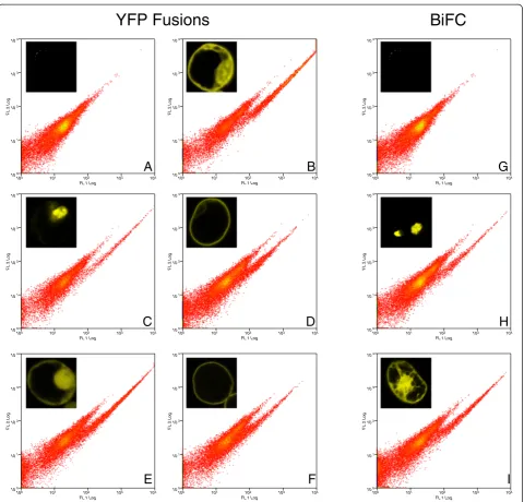
Figure 2Detection sensitivity of YFP fusion proteins.We tested our cytometer for its detection sensitivity of different protein fusions. One
can see from comparison with the mock negative control, that YFP expressing protoplasts can be clearly identified as a cloud of cells shifted
to the right in the FL1 channel.A.Mock transfected (water only),B.CPK3-mYFP,C.ARR2-mYFP,D.FLS2-mYFP,E.free mYFP,F.SLAC1-mYFP,
Screening specificity and background signals
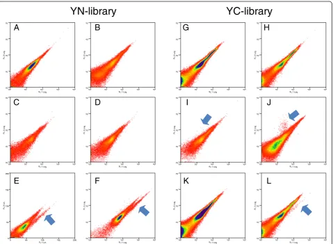
BiFC interactions are purported to be irreversible [7] and require many tests to control for spontaneous asso-ciation of the YFP fragments [15]. Techniques designed for reducing non-specific background in confocal set-ups [15] were not suitable for designing this screen to capture rare, weak signals. Therefore we used both the
YN154 and YC155 fragments to generate Arabidopsis
cDNA libraries. These libraries were then tested to iden-tify which library that would generate BiFC signals that could be attributed to specific protein-protein interac-tions (Figure 3). The first library that we generated and tested was with the YN fragment. The YN154 fragment in fusion with cDNAs or alone, or in combination with free YC fragments did not show any BiFC signals in transfected protoplasts (Figure 3B, C, D). While
seemingly encouraging, the free YN fragment would however spontaneously associate when the YC fragment was fused to any protein as exemplified by spontaneous association of free YN with bZIP63-YC or a non-functional control, mRFP-YC (Figure 3E, F). This is best explained by the fact that while bacterially purified YC fragments are mostly insoluble [19,20] and this is likely the case with free YC fragmentsin vivo as well, the YN fragment is stable enough to cause some non-specific complementation. Although we thought that perhaps the non-specific background would be drowned out by spe-cific bait interactions detectable in flow cytometry as strong fluorescence signals, a preemptive screen using the YN154-cDNA-librarytaking only strong BiFC signals detected by
FACS from a bZIP63-YC / YN154-cDNA-library
co-transfection, resulted in the identification of BiFC signals
100 101 102 103 104
FL 1 Log 100 101 102 103 104 FL 2 L o g
100 101 102 103 104
FL 1 Log 100 101 102 103 104 FL 2 L o g
YN-library
YC-library
100 101 102 103 104
FL 1 Log 100 101 102 103 104 FL 2 L o g
100 101 102 103 104
FL 1 Log 100 101 102 103 104 FL 2 L o g
100 101 102 103 104
FL 1 Log 100 101 102 103 104 FL 2 L o g
100 101 102 103 104
FL 1 Log 100 101 102 103 104 FL 2 L o g
100 101 102 103 104
FL 1 Log 100 101 102 103 104 FL 2 L o g
100 101 102 103 104
FL 1 Log 100 101 102 103 104 FL 2 L o g
100 101 102 103 104
FL 1 Log 100 101 102 103 104 FL 2 L o g
100 101 102 103 104
FL 1 Log 100 101 102 103 104 FL 2 L o g
100 101 102 103 104
FL 1 Log 100 101 102 103 104 FL 2 L o g
0 64 128 192 256 FL 1 Lin
0 64 128 192 256 FL 2 L in
G
H
I
L
J
K
A B
C
E
F
D
Figure 3Screening controls for the YN-cDNA and YC-cDNA libraries.X-axis is YFP fluorescence and Y-axis is autofluorescense; arrows show
and indicate positive mYFP-BiFC signals.A.Mock transfected cells.B.Cells transfected withYN-Libraryalone.C.Cells transfected withfree YN
fragment alone.D.Cells co-transfected withYN-Libraryandfree YCfragment.E.Cells co-transfected withbZIP63-YCandYN-Library; a clear
YFP-BiFC signal was detected.F.Cells co-transfected withmYFP-YCandYN-Library; a clear YFP-BiFC signal was detected.G.Mock transfected cells.
H.Cells transfected with theYC-Libraryalone.I.Cells co-transfected withmRFP-YNandYC-Library;only mRFP signal was detectable.J.Cells
co-transfected withmCherry-YN173andYC-Library; only mCherry signal was detectable.K.Cells co-transfected withbZIP63-YNandYC-Library.
that were derived from empty or out-of-frameYN154-Library plasmids (not shown). This indicated that the screen could not be conducted in this orientation.
In light of the knowledge gained from the YN-library, the YC-library was expected to perform better, since any empty or out-of-frame plasmids should not give any BiFC signal. Indeed this was the case, as only YC fusions, and not just YC fragments, resulted in positive BiFC signals using the large-scale transformation method (Figure 3G-L). What was surprising, however, was that we were not able to obtain a BiFC signal with bZIP63-YN / YC-Library co-transfections . Therefore, a longer YN-fragment including the 8th-beta strand (YN173) known to have better complementation efficiency [21] and is therefore brighter, was generated in order to overcome this deficiency. We did not take brighter YFPs derivates such as VENUS or mCitrine due to their higher rates of spontaneous BiFC association [15,20] as they would have led to unspecificity during the screen. An additional control fusion construct YN173-mCherrywas made and also tested against theYC-library. Neither the YN154-mRFP nor the mCherry-YN173 co-transfection with free YC or the YC-Library lead to any non-specific BiFC (Figure 3I, J). Thereafter, we decided in addition to using the brighter YN173 fragment that it would be helpful to take a protein whose expression do-main is more widespread than bZIP63 which is only found in the nucleus. Therefore, we choose a protein, Calcium Dependent Protein Kinase 3 (CPK3; AT4G23650) that is fairly well characterized and known to be present in both the cytoplasm and the nucleus and interact with proteins in both compartments [22,23]. While the CPK3-YN173 fu-sion protein showed no YFP fluorescence by itself in transfected protoplasts, clear BiFC derived signals were observed when CPK3-YN173 was co-transfected along with theYC-Library. Additionally, the lack of any detect-able interactions of the YC-library with YN154-mRFP and YN173-mCherry suggested that the interactions observed by CPK3-YN173 were due to specific interaction with YC protein fusions from the library. These results indicate that the screening conditions had been met: the detection of specific interactions in a rare-event analysis. We there-fore followed CKP3-YN173 through a complete screen as it fulfilled all prerequisites required for a successful BiFC screen (Figure 3).
Screening the YC-library: screening with CPK3, a case study
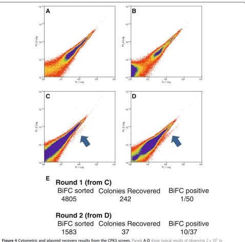
Screening results
The screen with bait CPK3-YN173 was conducted accord-ing to Figure 1. The CPK3-YN173 fusion was negative for self-complementation (Figures 3K, 4A). Furthermore, BiFC signals were detected in the protoplast population,
when CPK3-YN173 was co-transformed with the YC
-cDNA library. 2.5 x 10⁶ transfected protoplasts were
screened for BiFC signals resulting in 4805 sorted events in the first round (Figures 3L, 4B). Two-hundred and forty-two (242) bacterial colonies were obtained from the DNA preparation of these 4805 sorted protoplasts, a
‘recovery-rate’of 5%. Of those, only 1 out of 50 tested by flow cytometry in pair-wise challenges with the bait showed a positive BiFC signal (i.e. only 2%). Therefore, the plasmid DNA was purified from all 242 colonies and chal-lenged with the bait plasmid in protoplasts to enrich for plasmids that carry protein fusions that specially interact with the CPK3-YN173 bait. The second round of 2.5 x 10⁶ transfected protoplasts resulted in 1588 positive BiFC events from which 37 colonies were obtained, a‘ recovery-rate’of 2%. However, this time significantly more YC fu-sion constructs from singular colonies lead to positive BiFC events with the bait construct were recovered: 10 out of the 37 had positive BiFC signals, meaning that 27% had encoded fusion proteins putatively interacting with the bait (Figures 4B, 5A). Thus, a total of eleven colonies had been found containing plasmids encoding for inter-action partners with the CPK3-YN173 bait fusion protein whose in-frame fusions were confirmed by DNA sequen-cing. From these 11 plasmids, only 8 delivered readable DNA sequence trace files, as the other three had multiple plasmids inside as judged by strongly overlapping peak trace signals. From those 8, a clear sequence correspond-ing to a specific Arabidopsis ORF was identified by BLAST although there were minor sub-traces in some of the sequence traces (Additional File 1). This indicated that although a major plasmid species had been discov-ered present in each bacterial colony, the bacteria still picked up other plasmids as well. The majority of the ORF-matching sequences that were obtained included or started near the start-codon. The prey inserts were recal-citrant to re-cloning into expression vectors via Gateway™ recombination which would have allowed us to analyze a single plasmid species; thus, we were not able to deter-mine the complete ORF coverage for all of the clones. Nevertheless, the sequences were sufficient for assigning gene identity. Minor plasmid species present in the same bacterial colony were assumed to be off-targets. We therefore decided to clone the full-length ORF of each gene coding for each putative interaction partner using the corresponding cDNAs obtained fromArabidopsis tis-sue. The putative interacting proteins of CPK3-Y173 are listed in Table 1.
YC clone 1 (AT4G35000) contained the ascorbate
peroxidase 3 (APX3) cDNA. APX3 has been shown to
mitochondrial ATP synthase (EC 3.6.3.14) beta-subunit that has been previously purified along with mitochon-dria [26]. The insert of clone 4 (AT3G14420) encoded a putative glycolate oxidase (GLO1), that has also been co-purified with peroxisomes [27] and is involved in H2O2 production. Clone 5 (AT4G22540) encoded oxysterol binding protein-related protein 2A (ORP2A). Oxysterol binding proteins (OBPs) are thought to
control sterol traffic between membranes [28]. Clone 6 (AT2G39050) carried a cDNA encoding a ricin B-related lectin domain that is commonly associated with mem-branes [29]. Clone 7 (AT4G35570) encoded the high mobility group B protein 5(HMGB5). HMGB5 is a chro-matin associated protein involved in controlling DNA architecture influencing transcription [30]. Finally, clone 8 (AT1G67980) encoded an enzyme with putative
100 101 102 103 104
FL 1 Log
100
101
102
103
104
100 101 102 103 104
FL 1 Log
100
101
102
103
104
100 101 102 103 104
FL 1 Log
100
101
102
103
104
100 101 102 103 104
FL 1 Log
100
101
102
103
104
Round 1 (from C)
BiFC sorted Colonies Recovered
BiFC positive
4805
242
1/50
Round 2 (from D)
BiFC sorted Colonies Recovered
BiFC positive
1583
37
10/37
A
B
C
D
E
F
L
2 Log
F
L
2 Log
F
L
2 Log
F
L
2 Log
Figure 4Cytometric and plasmid recovery results from the CPK3 screen.PanelsA-Dshow typical results of observing 2 x 10⁵to
4 x 10⁵events; more than 2.5 x 10⁶events were analyzed per FACS session.A.Mock transfected (water only),B.CPK3-YN173transfected alone
(no YFP signal is produced),C.First Round, initial co-transfection ofCPK3-YN173andYC-cDNAlibrary,D.Second Round, co-transfection of the
fished library plasmids of the first round, pooled challenged with baitCPK3-YN173,E.Number of positive BIFC cells sorted, the number of
caffeoyl-CoA O-methyltransferase activity (CCoAMT, EC 2.1.1.104). It is most likely involved in the phenylpro-panoid pathway. Quite remarkably, many of these fished proteins have domains that are membrane associated.
BiFC validation of putative CPK3 interaction partners
To validate and quantify the BiFC interactions, the iden-tified prey cDNAs were cloned to virgin YC-fusion plas-mids and individually tested against YN-CPK3 fusions
under the conditions as those used for the screening. After re-cloning of all of the cDNAs into the different vectors, their potential for generating positive YFP derived BiFC was tested pair-wise against the bait ver-sions in small-scale transfections (Figure 5B).
The screen was made with the YN-fragment fused to the C-terminus of CPK3. It is known CPK3 can be myr-istoylated at the N-terminus and associates with mem-branes [23,31,32]. However, CPK3 is also known to be
CPK3-YN173
YN154-CPK3
100 101 102 103 104
FL 1 Log 100
101
102
103
104
FL 2 Log
100 101 102 103 104
FL 1 Log 10 0 101 102 103 104 F
L 2 Log
100 101 102 103 104
FL 1 Log 100 101 102 103 104 F
L 2 Log
100 101 102 103 104
FL 1 Log 100
101
102
103
104
FL 2 Log
100 101 102 103 104
FL 1 Log 100 10 1 102 103 104 F
L 2 Lo
g
100 101 102 103 104
FL 1 Log 10 0 101 102 103 104 F
L 2 Log
100 101 102 103 104
FL 1 Log 100 101 102 103 104 F
L 2 Log
100 101 102 103 104
FL 1 Log 100 101 102 103 104 FL 2 L og
100 101 102 103 104
FL 1 Log 100 101 102 103 104 FL 2 L og
100 101 102 103 104
FL 1 Log 100
101
102
103
104
FL 2 Log
100 101 102 103 104
FL 1 Log 100 101 102 103 104 F
L 2 Lo
g
100 101 102 103 104
FL 1 Log 100 10 1 102 103 104 F
L 2 Lo
g
100 101 102 103 104
FL 1 Log 100 101 102 103 104 F
L 2 Log
100 101 102 103 104
FL 1 Log 100
101
102
103
104
FL 2 Log
100 101 102 103 104
FL 1 Log 100 101 102 103 104 F
L 2 Log
100 101 102 103 104
FL 1 Log 100 101 102 103 104 F
L 2 Log
100 101 102 103 104
FL 1 Log 100
101
102
103
104
FL 2 Log
100 101 102 103 104
FL 1 Log 100 10 1 102 103 104 F
L 2 Lo
g
100 101 102 103 104
FL 1 Log 100 101 102 103 104 FL 2 L og
100 101 102 103 104
FL 1 Log 10 0 101 102 103 104 F
L 2 Log
100 101 102 103 104
FL 1 Log 100 101 102 103 104 F
L 2 Log
100 101 102 103 104
FL 1 Log 100
101
102
103
104
FL 2 Log
100 101 102 103 104
FL 1 Log 100 10 1 102 103 104 F
L 2 Lo
g
100 101 102 103 104
FL 1 Log 10 0 101 102 103 104 F
L 2 Log
100 101 102 103 104
FL 1 Log 100 101 102 103 104 F
L 2 Log
Initial Screening
Follow-up Screening
No Bait No Bait No Bait
#1
#5
#2
#6
#3
#7
#4
#8
#1
#5
#2
#6
#3
#7
#4
#8
#1
#5
#2
#6
#3
#7
#4
#8
100 101 102 103 104 FL 1 Log 100 101 102 103 104 FL 2 Log
A
B
C
n.d. Fluorescence Index 100 0 200 300 400 500 6001 2 3 4 5 6 7 8
-CPK3YN173
A C C C B B B C C
+ + + + + + + + + BAIT-YC Fluorescence Index 100 0 200 300 400 500 600 CPK3YN173
BAIT-YC 1 3 4 5 6 7 8
-+ + + + + + + +
A B B B B B B B
Figure 5Cytometric plots of testing for BIFC between bait CPK3-YN173 and fished YC fusion proteins. A.Cytometric plots are shown
from the second round screening that lead to 8 positive colonies (3 were eliminated due to technical difficulties, see text).B.Subsequent testing
of the 8 candidate proteins after re-cloning of the candidate cDNAs and tested against CPK3-YN173 or YN154-CPK3.C.The YFP BiFC index was
present in or around the nucleus [22,23]. CPK3-mYFP localized in the cytoplasm and in and around the nu-cleus in transiently transformed Arabidopsis protoplasts and Nicotiana benthamiana epidermal leaf cells under our conditions (Figure 2; Figure 6). The recorded fluor-escence pattern does not exclude the specific association of CPK3 with membranes or other cell compartments nor is it different from previous publications. Neverthe-less, it cannot be excluded that the cloning linker inter-fered with CPK3´s myristoylation. We, therefore, also tested an YN154-CPK3 fusion that definitely masks the CPK3 myristoylation site to appraise its effect on the interactions with the fished genes and compared it to the CPK3-YN173 version.
Three biological replicates were done with three tech-nical transfection replicates per experiment for quantify-ing BiFC fluorescence by analytical flow cytometry. The same trend was observed in all three experiments. Detailed statistical results for Student’s t-test and Dun-net’s test are given in Additional file 2. The cDNAs of clone 2 (AT2G29670), 3 (AT5G08680), 4 (GLO1) and 8 (CCoAMT) produced no BiFC with CPK3. In contrast, clone 6 (AT2G39050), 5 (ORP2A), and 7 (HMGB5) and showed some weak (and occasionally significant) BiFC with CPK3. On the other hand, clone 1 (APX3)
inter-acted very strongly and irrefutably with CPK3
(Figure 5C). These interactions, as measured by quanti-fied BiFC, were observed for the CPK3-YN173 as well as for the CPK3 construct, albeit that the YN154-CPK3 signals were radically weaker overall compared to CPK3-YN173. For those proteins that showed no BiFC, no detectable protein was observed by western blotting, and it is presumed these proteins were not expressed in the protoplasts derived from theArabidopsisdark-grown cell culture for unknowable reasons although the inter-action conditions were maintained the same as the screening conditions.
Interaction validation with FRET-FLIM
To substantiate the BiFC protein-protein interactions in planta,fluorescence resonance energy transfer– fluores-cence lifetime (FRET-FLIM) measurements were carried out. This was also necessary as BiFC complexes are sup-posedly irreversible [7] and could form from non-specific interactions [15]. FRET only occurs over a limited range of 100 Å, and is theoretically only possible when there are actual protein-protein associations [33]. Thus FRET-FLIM allows one to determine if associations are real, in vivo protein-protein interactions and reveal how dynamic or transient the associations are. To study FRET-FLIM, we switched from the Arabidopsis proto-plast system to tobacco leaves using a transient Agrobac-teriumtransformation method.
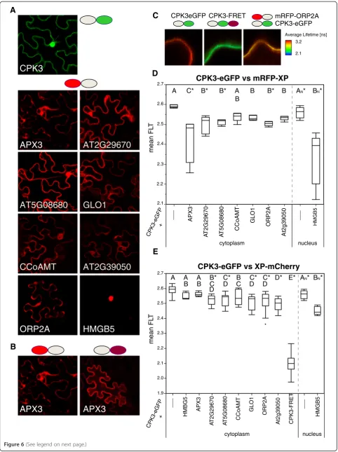
We used the pABind; [34] vector set that involves clon-ing cDNAs into a terminal donor (eGFP) or a C-terminal mCherry. In addition, we fused the fished pro-teins to an N-terminal mRFP [35] to mirror the screening orientation. Simply put, GFP is excited and if it is near mRFP or mCherry, FRET from the GFP to the mCherry molecule results in a reduction of the GFP’s fluorescence lifetime [33]. The lifetime of eGFP is then measured within a small window from which one can calculate the average fluorescence lifetime (Figure 6C). The experiment was repeated for at least five independent cells per sample. The expression of the fusion protein was induced by estra-diol application (Figure 6A, B).
We had previously shown that free proteins did not ex-hibit any non-specific FRET-FLIM of donor to acceptor [35]. We first confirmed that the CPK3-eGFP localized to the cytoplasm and the nucleus (Figure 6A) as all of the proteins interacting in BiFC were localized to these com-partments (Figure 6A). FRET-FLIM measurements were conducted in a cytoplasmic space, except for HMGB5 that was exclusively localized in the nucleus (see Additional file 3 for comparison of mCherry and mRFP fusions).
Table 1 Table of putatively CPK3 interacting proteins
Colony Number AGI Identifier Common Name Description
1 AT4G35000 APX3 Encodes a microsomal ascorbate peroxidase; scavenges hydrogen peroxide
2 AT2G29670 AT2G29670 tetra-trico peptide repeat (TPR) containing protein
3 AT5G08680 AT5G08680 Encodes one of three mitochondrial ATP synthase beta-subunits
4 AT3G14420 GLO1 miscrosomal glycolate oxidase, involved in the production of hydrogen peroxide
5 AT4G22540 ORP2A oxysterol binding protein (OSBP); sterol trafficking, affecting membrane fluidity and permeability and influencing secretory events
6 AT2G39050 AT2G39050 ricin B-related lectin domain containing protein; traffics from the ER, via the Golgi complex, to the vacuole
7 AT4G35570 HMGB5 chromatin-associated protein that binds to the minor groove of short stretches of A/T-rich B-form DNA
8 AT1G67980 CCoAMT Encodes S-adenosyl-L-methionine: transcaffeoyl Coenzyme A 3-O-methyltransferase
The colony number as in Figure5Ais given for consistency along with the gene AGI identifier and common name of the plasmid predominately present in the
plasmid prep. The dominate cDNA sequence was re-cloned from fresh Arabidopsis cDNA for confirmatory BiFC (data in Figure5B, C) and FRET-FLIM experiments
CPK3-eGFP vs mRFP-XP
2.1 2.2 2.3 2.4 2.5 2.6 2.7
mean FL
T
APX3
A
T2G29670
___
A
T5G08680 CCoAMT
GLO1
ORP2A
At2g39050
HMGB5
___
nucleus cytoplasm
1.9 2.0 2.1 2.2 2.3 2.4 2.5 2.6 2.7
mean FL
T
CPK3-eGFP vs XP-mCherry
APX3
A
T
2G29670
CPK3-eGFP +
A
T
5G08680 CCoAMT
GLO1
ORP2A
At2g39050
HMGB5
___
nucleus cytoplasm
CPK3-FRET
HMBG5
A A
B
C* B* B* B B* B AN* BN*
A A A B E*
B B
B* C D
C*
D C
D* C*
D C* D
BN*
AN*
CPK3eGFP CPK3-FRET mRFP-ORP2A
Average Lifetime [ns] 3.2
2.1
C
D
CPK3
HMGB5
APX3
AT2G29670
AT5G08680
GLO1
ORP2A
AT2G39050
CCoAMT
APX3
APX3
A
B
E
CPK3-eGFP
___
CPK3-eGFP +
We first tested for FRET-FLIM using the same fusion protein orientations as those used in the screen. Detailed statistical results for Student’s t-test and Dunnet’s test are given in Additional file 2. According to the FRET-FLIM data and using Student’s t-test (α= 0.05), all sam-ples except for CCoAMT caused a significant reduction in the fluorescence lifetime of GFP. A more stringent significance test, Dunnett's Method showed that a frac-tion of those were significantly different. If we restrict ourselves to Dunnett’s Method, then APX3, AT2G29670, AT5G08680, ORP2A, and HMGB5 all showed FRET-FLIM reductions with CPK3. APX3 and HMGB5 inter-acted strongest with CPK3 as they showed the greatest reduction in the fluorescence lifetime of the GFP donor.
We also tested the CPK3-eGFP for FRET-FLIM with the putative interactors C-terminally fused to mCherry (Figure 6E). AT2G29670, AT5G08680, GLO1, ORP2A, AT2G39050, and HMGB5 showed significant reductions in the GFP fluorescence lifetime, indicating that they also interacted with CPK3-GFP. In contrast to the N-terminal mRFP fusion with does interact with CPK3-eGFP, the C-terminal mCherry fusion of APX3 (APX3-mCherry) did not. This could be due to a block of APX3´s C-terminal peroxisome targeting motif [24] by mCherry. Interestingly, compared to mRFP-APX3, APX3-mCherry was more uniformly distributed in the cytoplasm (also see Additional file 3). Thus, APX3-mCherry is presumably not able to associate with micro-somal compartments any longer (Figure 6B). Based on these observations, APX3 requires its C-terminal target-ing signal for interaction with CPK3.
We performed additional negative controls by measuring the fluorescence lifetime of CPK3-eGFP in the cytoplasm when HMGB5 was present in the nucleus. As shown in Figure 6E no significant reduc-tion was observed (Figure 6E). Summed together, APX3, ORP2A and HMGB5 all interacted via BiFC and FRET-FLIM with CPK3 under the same screen-ing orientation with RTB-like AT2G39050 also bescreen-ing a suitable candidate.
Interaction validation with yeast-2-hybrid
To further support the idea that interacting proteins were found, we also tested the putative CPK3 interac-tions with the yeast-two-hybrid assay. We were not able to validate any of the interactions in the yeast-two-hybrid assay (Additional file 4). We repeated the trans-formation four times and at different temperatures (28°C and 16°C) but we could not obtain any interaction. Fur-thermore, the strongest candidate, APX3 was lethal to the yeast cells as very few colonies were obtained at each transformation and for those that were transformed, they did not express the protein (Additional file 4). Other than APX3 and AT5G08680, all of the putative interacting proteins were expressed in the yeast system. Although perhaps alternating the tags in the yeast sys-tem or manipulating the screening environment might be helpful to rule out any other over-expression effects of the fished proteins, we conclude that the proteins found to interact with CPK3 in this screen cannot be routinely detected in a standard yeast-two-hybrid assay.
Discussion
Challenges of thein vivoBiFC screen
We have showed that it is possible to find proteins from a random library that interact with a bait proteinin vivo and in planta. This was done by observing protein-protein interactions measured by BiFC of mYFP. Al-though this screen is one of the few purely random, in planta screens ever established, its efficacy is not per-fect. We had to come up with a method that would allow the identification of a single population of interact-ing proteins from a sinteract-ingle protoplast. We used several techniques that were impossible to avoid but are difficult to control: transfection of protoplasts, recovery of plas-mids from protoplasts and transformation of bacteria with plasmid. The first condition is obvious: in order to make a screen in living plant tissue, it is necessary to have a method that has the capacity of performing many interactions simultaneously and we choose protoplast transfection. However, there is no way to exclusively
(See figure on previous page.)
Figure 6Results of FRET-FLIM measurements of CPK3 with candidate interaction proteins. A.Example expression domains of CPK3-eGFP
and mRFP-XP fusion proteins as seen in tobacco epidermal cells.B.Comparison of the localization pattern of APX3 with either an N-terminal
mRFP fusion (left) or a C-terminal mCherry fusion (right). The unhampered fusion is the N-terminal mRFP fusion which shows an uneven
expression domain unlike the C-terminal mCherry fusion.C.Example of false-colored FRET-FLIM imaging sectors. Stronger FRET-FLIM results in a
reduction in the average lifetime. The negative control in CPK3-eGFP alone; the positive control is a CPK3-eGFP-mCherry (called CPK3-FRET)
fusion; and real sample of CPK3-eGFP co-expressed with mRFP-ORP2A.D.The averages of the average lifetime (from the measurement sector)
are shown as box plots for the CPK3-eGFP in the presence of mRFP-XP fusions. Letters are significance classes based on Students t-test; any
sample not connected by a letter is significantly different from the CPK3-eGFP control. Significance difference to the control using Dunnett’s
Method is indicated by a *.E.The averages of the average lifetime (from the measurement sector) are shown as box plots for the CPK3-eGFP in
the presence of XP-mCherry fusions. Letters are significance classes based on Students t-test; any sample not connected by a letter is significantly
different from the CPK3-eGFP control. Significance difference to the control using Dunnett’s Method is indicated by a *. Cytoplasm and Nucleus
introduce a single plasmid into a protoplast, the nature of the method means that each protoplast takes up mul-tiple plasmids. Thus methods that would rely on PCR (single cell PCR) for single cells or pooling of the cells (454 sequencing) would most likely not eliminate false-positives. An attempt to dilute the mixed library DNA to some statically desirable transfection rate of 1:1 of bait to prey would be dependent on the transfection rate, which varies from batch to batch, and most likely pro-duce undetectable protein amounts. An alternative to our random library approach would be to use defined grids of prey plasmids that could be transfected into the protoplasts as binary pools; such an approach was already performed using the split-luciferase system [4]. This method of course, requires a cloned or clonable li-brary ORF set. While this worked for the split-luciferase as the detection of luciferase activity has good signal-to-noise ratios, the BiFC system relies on weaker mYFP emission, and this emission must exceed that of the plant autofluorescence. Thus, we found that the screen-ing was only possible usscreen-ing a flow cytometer where we can compare YFP versus autofluorescence to detect those cells expressing BiFC of YFP.
Due to the chanciness of the protoplast transfection and the DNA recovery, we choose to stay with the re-covery of the plasmid DNA first. At the beginning of the screen design, we had anticipated that we would be able to isolate a single bacterial colony carrying a single plas-mid type that could be screened pair-wise against the bait and that plasmid’s cDNA would be available for im-mediate downstream cloning via GatewayTMtechnology. Unfortunately, although only few bacterial colonies were obtained, those that were obtained also carried multiple, independent plasmids as well. Furthermore, the majority of colony forming units derived by plasmid recovery from the protoplasts did not have interacting prey (Figure 4B). This suggested to us that although the plas-mid recovery from positive BiFC cells sorted by FACS did lead to a slight enrichment of the BiFC generating plasmids (Figure 4E), other plasmids were still present in those protoplasts and also consequently in the bacteria. Our solution to the multiple-plasmid problem was an enrichment of the actual plasmids encoding the interact-ing protein by isolatinteract-ing all of the plasmids from the first round and transfect them against the bait once again and sort again by FACS on BiFC of YFP. This strategy worked, as the positive interaction rate went up from 2% to 27%.
In the end we utilized the protoplast transfection and the recovery of the bacterial plasmid DNA without any additional interventions to identify novel bait interacting partners and recovered the cDNA by PCR and cloning. Together these observations mean that the screen is not saturated. For example, the one positive found in the
first round of the CPK3 screen was not found in the sec-ond round (Figure 4). This also indicated that there were probably many more plasmids still present encoding for other CPK3-interacting partners that had been not detected. Nevertheless, we conclude that it is possible to recover interacting proteins encoded on bacterial plas-mids from anin plantascreen.
The majority of observed BiFC signals was surprisingly weak and only detectable in the flow cytometer. One would have expected that some strong BiFC interactions should have been detected as it was the case for APX3. The best explanation for the low BiFC-YFP signals dur-ing the screendur-ing is that, in order for enough YFP signal to be detected, it must exceed a certain concentration in the cell before the total fluorescence intensity is detect-able above the autofluorescence. Reducing the YFP by using BiFC fragments already reduces the total detect-able protein [15]. For those fusion proteins whom are less abundant in a cell or have localizations that restrict their abundance (for example those in the nucleus, plasma membrane or Golgi apparatus), it means that it is not possible to screen for any type of protein. It is not only remarkable that could we recover transfected plas-mids from plant protoplasts after 36 hrs, but that it was also possible to identity some interacting partners with a bait construct. According to the data presented here, the in vivo in planta BiFC screen provides a lucrative alter-native to search for novel protein-protein interactions that can, according to our data, only be foundin planta.
Putative interaction partners of CPK3
BiFC CPK3-prey interactions were too weak to be observed in the microscope; thus we were not able to show where the BiFC interactions were taking place in-side the cell. We used two fluorescence based methods to substantiate the protein-protein interactions found in the screen: biased BiFC and FRET-FLIM. I wanted to say that theoretically BIFC interactions could be through an un-seen partner or due to trapping, but this sentence ended up a bit self-contradictory the way is it currently written. We used GFP/mCherry or GFP/mRFP donor/acceptor FRET pairs that have been shown to perform very well in vivo [35,41]. Proteins (and their fluorophore fusion) must be within 1 to 10 nm distance for FRET to occur [42], which is the typical distance found for interacting proteins. Similarly, BiFC has been discussed to occur over a distance around 7 nm [43] [44]. Both BiFC and FRET-FLIM support four previously uncharacterized protein interactions of CPK3.
APX3 (#1, AT4G35000) showed the strongest BiFC with the CPK3 baits in both orientations, N-terminal and C-terminal SPYNE (Figure 5C). APX3 is targeted to peroxisomes [24,45], but has been shown to be retarded in the cytoplasm by AKR2A [46]. In the FRET-FLIM studies, the N-terminal mRFP-APX3 fusion showed the strongest FRET efficiency with CPK3-eGFP (Figure 6D). mRFP-APX3 was clearly non-homogenous in its sub-cellular distribution as its C-terminal transmembrane domain [24] was not masked. In contrast, the C-terminal APX3-mCherry was mis-localized to the cyto-plasm (Figure 6B; Additional file 3) and showed no interaction with CPK3 in FRET-FLIM (Figure 6E). This evidence combined with the very strong BiFC makes a good argument that the screen was able to find a major interactor of CPK3. Interestingly, APXs are important for scavenging ROS (H2O2) and APX3 could provide the link proposed for CPK3 and CPK6 in regulating ROS and NADPH activation in guard cell function [36].
ORP2A (#5, AT4G22540) is a predicted oxysterol binding protein (OSBP). ORP2A significantly interacted with CPK3 in BiFC experiments (Figure 5C). It also interacted preferentially with CPK3 in tobacco epidermal cells as shown by FRET-FLIM. OSBPs are involved in sterol trafficking [47] affecting membrane fluidity and permeability and influencing secretory events. OSBPs are known to bind to oxysterols, which compose minor amounts of sterols in plants [48], but OSBPs are known in other species to bind to different lipids including phosphoinositides, ergosterol, and cholesterol (refer-ences in [28]). Interestingly, there is some evidence that OSBPs are involved in the regulation of processes like Ca2+ uptake and transcriptional control, both processes which relate directly to CPK3. Mechanisms how newly synthesized sterols reach the plasma membranefrom the ER are unclear in plants and OSBPs are possible
candidates. ORP2A is well expressed in many tissues and is somewhat regulated by stresses [28]. CPK3 was shown to target ER associated proteins Calnexin and Calreticulin [23], the latter of which regulates Ca2+ stores and signaling from the ER.
AT2G39050 (#6) is a ricin B-related lectin domain con-taining protein. Ricin is a heterodimeric plant protein that is toxic to mammalian and many other eukaryotic cells by binding to membrane localized galactose-containing recep-tors [29]. Ricin is composed to two subunits, ricin toxin A (RTA) and B (RTB). RTA is catalytically-active and removes a specific residue from the 28 S ribosomal RNA [49]. Dur-ing its synthesis in plant cells ricin traffics from the ER, via the Golgi complex, to the vacuole [29]. AT2G39050 showed significant interaction with CPK3 in BiFC. The FRET-FLIM studies also support the interaction of this protein with CPK3. That CPK3 is also membrane associated and that ricin moves through the ER to the vacuole strongly sup-ports the interaction with CPK3.
HMGB5 (AT4G35570) belongs to the class of high mo-bility group (HMG) proteins and are, after histones, the second most abundant type of chromosomal proteins [50]. HMGs have an‘AT-hook’that binds to the minor groove of short stretches of A/T-rich B-form DNA independent of the nucleotide sequence [51]. Unlike histones, HMG pro-teins are very dynamic and some even shuttle in and out of the nucleus in animal and plant cells [50,52]. HMGB5 is predominantly found inside the nucleus [30] and is ex-tremely mobile within the nucleus [52]. HMGB5 showed a significant BiFC interaction with CPK3 inArabidopsis pro-toplasts. The FRET-FLIM experiments in tobacco epider-mal cells also substantiate the interaction between CPK3 with HMGB5 in the nucleus.
Among CPK3’s roles in the regulation of plasma membrane-localized ion-channels, it is known to have roles in phosphorylating nuclear transcription factors [32,53,54], other DNA-binding proteins [23,55]) and many RNA asso-ciated proteins [23]. This suggests that CPK3 has a role in regulating gene expression before, during and after tran-scription and that it may also have a role in chromatin regulation in conjugation with, for instance, HMGB5.
We could exclude AT2G29670 (#2), AT5G08680 (#3), GLO1 (#4, AT3G14420) and CCoAMT (#8, AT1G67980) as true interaction partners for CPK3 as they did not show interaction any via BiFC in Arabidopsisprotoplasts where the screen was conducted and therefore we conclude they do not meet inArabidopsis cells and thus do not interact with each other (see Additional file 5 and 6 for details).
Conclusions
HMGB5, ORP2A, and AT2G39050 (Ricin-B). None of these interactions have been observed before. Our ap-proach, therefore, is one of the firstin plantarandom li-brary screens shown to work. Although this method is not suited for high-throughput screens, it still is an alter-native to search for novel interactions that may or may not be caught with other screening methods, as in our case with the yeast-two-hybrid system. And, as the screen is in planta, one still has the opportunity to treat the cells with elicitors, hormones or pharmaceuticals, as well as use protoplasts from mutant plant lines to screen for interactions that maybe dependent on such conditions.
Methods
Protoplast transfections
Protoplasts were transformed either in a large-scale (7.5 x 10⁵ to 1 x 10⁶protoplasts per transfection) or in small-scale (6.0 x 10⁴protoplasts per transfection). Proto-plasts were generated from 3-day-old Arabidopsis Col-0 cell suspension culture. The suspension culture was main-tained in a 250–300 ml Erlenmeyer flask, in the dark, at 24°C-26°C and 120 rpm (constant shaking) in 50 ml MSCol Medium (0.43% w/v MS salts, 0.1% w/v Nicotin acid, 0.1% w/v Pyridoxin-HCl, 1% w/v Thiamin-HCl, 10% w/v myo-Inositol, 3% w/v sucrose, pH = 5.8 with KOH and 0.1% v/v 2,4-D added after autoclaving). The proto-plasts were prepared and transformed by the PEG method according to the protocols of [56,57], which were recently summarized and described by us in full detail in [58].
Large-scale transformations were performed as described in [12,58] in 14 ml round-bottom Falcon tubes. In short, the cells were collected by centrifugation (max. 100 g) and the cell walls are removed by incubation in cell wall digestion solution (1% cellulase, 0.25% macerozym, 8 mM CaCl2, 0.4 M mannitol, pH 5.5, filter sterilized) for 6 hrs. The cells are washed and resuspended in W5 (154 mM NaCl, 125 mM CaCl2, 5 mM KCl, 5 mM glu-cose, pH 5.8–6.0, autoclaved) and kept at 4°C for 20–30 min. Thereafter the cells are transferred to MMM (15 mM MgCl2, 0.1% MES, 0.5 M mannitol, pH 5.8, auto-claved) and ready for PEG transfection. The cells were transfected with 20 ug DNA in water and PEG solution (40% PEG 4000, 0.4 M mannitol, 0.1 M Ca(NO3)2, pH 8–9 (the pH needs 1–2 h to stabilize), autoclaved). After the transfection process, the transfected cells were stored in the dark at 24°C-26°C in standard K3 protoplast medium (see [58] for details of K3 medium preparation) before analysis (16 to 36 hrs later).
Small-scale transformations were performed in 96 well round-bottom PP plates (Roth) using the protocol described here. Protoplasts were generated as in [58], but once in W5 medium, were incubated for 30 min at 4°C and either processed immediately or stored over-night at 4°C. The cells were sieved through a 70 μM
filter (Becton-Dickinson), re-counted and resuspended to 2 x 10⁶/cells/ml in MMG media (0.4 M mannitol, 15 mM MgCl2). CsCl purified plasmid DNA or the equivalent was added to each well (up to 5 μg) in a vol-ume of 9μl, followed by 30μl of filtered protoplast cells (6 x 10⁴), and mixed well by gently knocking the palm against the plate about 10 times on each side. Once mixed, 30μl of PEG solution (6.75 mM Ca(NO3)24H2O, 270 mM mannitol, 17.5 ml H2O, 38.5 % w/v PEG1500, [pH9.5 with 0,1 mM KOH], filter sterilized and stored in small aliquots) was added to each well and mixed thor-oughly as just described and incubated for 10 min on the bench. Thereafter, 30 μl of MMG is added to each well and mixed completely as described, followed by quick addition of 250 μl of standard K3 protoplast medium [58] and mixed thoroughly for approx. 30 s or until any precipitated DNA had been dissolved. The plate was then covered with Nescofilm (Fisher Scientific) and incubated overnight at 26°C before analyses. For large-scale or high-throughput analyses, plasmid DNA was prepared with commercial DNA purification col-umns; for screening of putative interactors, standard mini-DNA preparations [59] or mini-DNA preparation kits were used. Protoplasts were incubated for 16 to 36 hrs before flow cytometric analysis or FACS; longer incubation times resulted in more positive signals and were necessary for detecting BiFC library interactions. BiFC fluorescence index was calculated as in [12], and statistical tests were performed in JMP9 (SAS).
Transient expression in tobacco leaves
A single colony of transformedAgrobacterium tumefaciens was inoculated in 5 ml of YEB-Medium (0.5% beef extract, 0.5% sucrose, 0.1% yeast extract, 0.05% MgSO47H2O) con-taining Rif/Gent/ and vector-specific antibiotic at 28°C overnight. In the morning, 1 ml of the pre-culture was taken and re-inoculated into 5 ml of the same Medium. The same was done for Agrobacteriumstrain carrying the p19 RNAi-suppressor protein from tomato bushy stunt virus [60]. Each culture was collected in a 15-ml Falcon Tube and centrifuged at 4000 rpm for 20 min. Bacteria pellets were then resuspended in AS-Medium (10 mM MgCl2, 10 mM MES [5.6], 150μM acetosyringone) to an optical density at 600 nm of about 0.7-0.8. The resuspended bacteria (two potential interaction partners and p19 strain) were mixed 600 ml each, a 1:1:1 ratio, in a 2 ml Eppendorf tube and incubated for 0.5-1 hour at 4°C.
after the bacterial inoculation, the pABind vectors [34] and the N-terminal mFRP vector pB7WGR2,0 (Plant Systems Biology, Gent) were induced by application of estradiol by brushing a 20μM estradiol (in 0.1% Tween-20) solution onto the abaxial leaf surface. FRET mea-surements were performed 24 to 48 hours after estradiol application.
FACS
FACS and flow cytometric analyses were performed with a MoFlo (2007; Beckman-Coulter). mGFP, mYFP, mRFP or mCherry were excited with a 50 mW 488 nm argon laser. GFP and YFP were detected in FL1 (510–550 nm), Auto-fluorescence in FL2 (565–605 nm) and RFP/mCherry in FL3 (605 – 650 nm). RFP and mCherry expression was cross checked with a 50 mW 532 nm solid-state laser and detected behind a 585/30 bandpass for RFP or 613/20 for mCherry. Transfected protoplasts were sieved through 40μM (BD) before FACS or 70μM filters before analysis. Sorts and analyses were run approximately under 31.0/ 30.0 psi (sample/sheath) using a 100μM nozzle; sheath was 1x PBS at pH 7.0.
Protoplasts identified with a detectable BiFC signal were sorted directly into Edwards’s Buffer [14] in a 2 ml eppendorf tube at 20°C. The collected protoplasts were thoroughly mixed, then iso-propanol was added 1:1, incubated at −20°C for 30’, centrifuged at 13000 rpm at 4°C for 45’, washed with cold 75% ethanol, centrifuged for at 4°C for 15’. The resultant precipitate was air-dried for 15’ on the bench at room-temperature and resus-pended in 20 μl Millipore purified water. 50 μl chem-ically competent NEB 10-beta (New England Biolabs) cells were thawed on ice and added to 10 μl DNA ex-traction, further incubated for 30’ followed by a 30” heat-shock at 42°C, then 2 min on ice, and incubated for 2 hrs at 37°C in 800 μl SOC medium. The transform-ation was then plated out on two large (145 x 20 mm) petri dishes containing LB-agar with 100μg /ml ampicil-lin to select for colonies with the pE-SPYCE-cDNA li-brary vector. To recover plasmids from colonies on a plate, the bacteria were mixed and removed in 10 ml LB with selection antibiotic added to the plate. The plasmid DNA was then purified using commercial midi-DNA preparation columns.
Cloning
35S::cDNA::YN173AcV5 (SpecR) constructs were con-structed with multi-site GW vectors (pEntryL4R1-P35S, pH7m34GW or pB7m34GW) from Plant Systems Biol-ogy (Gent). The pENTR-R2L3-YN173AcV5 was gener-ated by amplifying from N-terminus until the 173 amino acids (fwd: B2 + gaATGGTGAGCAAGGGCGAG and rev top strand (YFP, AcV5, stop): CGCCACAACATCGAG GAC-TCTTGGAAAGATGCGAGCGGCTGGTCTTGA
t + B3) of mYFP by PCR and cloned via BP-reaction into pDONRP2R-P3 (Invitrogen). pUC-SPYC/NE-mRFP vecr-tors are described in [12]; pUC SPYC/NE vecvecr-tors are those described in [6]; pPE-SPYC/NE::cDNA library was made by cloning the pSPORT-P (KanR) Arabidopsis cDNA library (Supescript Arabidopsisthird-flower stage seedlings #11474012) into pDONR222 (resultant titer 5.1 x 10⁶cfu/ml) and recombining it with pE-SPYCE/SPYNE ([61]; AmpR) vectors to make YFP-fragment fusions (re-sultant titers: pE-SPYNE-cDNA at 3.36 x 10⁶ cfu/ml; pE-SPYCE-cDNA at 3.95 x 10⁶ cfu/ml). The libraries were grown on 30 cm diameter LB plates plus ampicillin at 28°C for 18 hrs. The bacteria were harvested from the plates using LB liquid plus antibiotic followed by plasmid purification using a Qiagen Giga-prep kit following the manufacturer’s instructions.
After sequencing interacting partner clones in pE-SPYCE, the matching cDNAs were cloned from cDNA produced from Arabidopsis (Col-0) leaf material. The full-length cDNAs were cloned into either pENTR/D-TOPO (Invitrogen) or pDONR207 (Invitrogen) and subsequently recombined into target Destination vectors as needed. FRET-FLIM vectors were pABindGFP / mCherry / GFP:: mCherry [34] as C-terminal tag fusions. N-terminal mRFP fusions were made with pB7WGR2.0 (Plant Systems Biol-ogy, Gent). Additional clones not mentioned explicitly were provided by collaborators and are listed in the Acknowledgements.
FRET-FLIM and microscopy
FRET-FLIM measurements were carried out as previ-ously described in [35] with the addition that the pres-ence of the acceptor was confirmed. Statistical tests were performed in JMP9. Confocal Laser Scanning Micros-copy was performed as described in [35].
Yeast-two-hybrid
Additional files
Additional file 1:Phred DNA trace files of original prey plasmids.
DNA trace files from the original plasmid mini-preps that encoded for proteins that theoretically interacted with the bait protein.
Additional file 2:Significance Tests for BiFC and FRET-FLIM
quantifications.P-values for all significance tests that were mentioned in
the text or in figures 5 and 6.
Additional file 3:Confocal Localization Images of prey fusion
proteins with mCherry or mRFP.Confocal images of prey fusion
proteins expressed in tobacco epidermal cells with enlarged insets of APX3 fusions.
Additional file 4:Yeast-two-hybrid assays of CPK3 with prey
proteins.Representative yeast-two-hybrid assays performed with CPK3
verses prey proteins on selective and non-selective media. Six independent colonies were analyzed per combination. Western blots are also shown for all proteins and the band corresponding to the full-length protein is indicated with an asterisk.
Additional file 5:Additional discussion text for non-interacting
prey proteins.Additional discussion is provided for the four prey
proteins that we consider not to be interaction partners of CPK3.
Additional file 6:Confocal Localization Images of CPK3-eGFP with
tetra-trico peptide repeat (TPR) AT2G29670.Confocal images of of
CPK3-eGFP with tetra-trico peptide repeat (TPR) illustrating conglomerates of AT2G29670 that led to the exclusion of CPK3-eGFP.
Competing interests
The authors declare that they have no competing interests.
Authors' contributions
KWB and KH designed, conducted and were funded to perform the experiment. KWB, MB, NW, YZ, FKET, and MV performed the molecular laboratory work. FS and SP built the custom-built confocal stage scanning
microscope and conducted thein vivoFRET-FLIM measurements. All authors
read and approved the final manuscript.
Acknowledgments
We thank Silke Robatzek for providing us with the FLS2 clone. We thank Klaus Palme and Alexander Dovzhenko for initial help with the 96 well transformation method. We are also grateful to Caterina Barancato (ZMBP Central Facility‘Transformation Unit’) for performing the large scale transformations. We appreciate Christina Chaban and Katja Schütze for providing us with the pENTR-bZIP63 construct. We also thank Katrin Weckermann and Claudia Oecking for providing us with pSPYC/NE-mRFP and pSPYC/NE-T14-3c vectors. Detlef Weigel supplied us with pMD143-ENTR-mCherryNLS. We also thank Claudia Haß for the p35S::ARR2-mYFP construct.
This work was supported by the DFG (SPP1212, HA 2146 / 9–1) to KH and
KB. The CPK3 cDNA was prepared in Julian Schroeder's laboratory (supported by NIH R01GM060396).
Author details
1Universität Tübingen, ZMBP, Plant Physiology, Auf der Morgenstelle 1, D-72076 Tübingen, Germany.2University of California, San Diego, Division of Biological Sciences, Cell and Developmental Biology Section & Ctr for Mol. Genetics 0116, 9500 Gilman Drive #0116, La Jolla, CA 92093-0116, USA. 3Universität Tübingen, ZMBP, Biophysical Chemistry, Auf der Morgenstelle 18, D-72076 Tübingen, Germany.
Received: 17 April 2012 Accepted: 26 May 2012 Published: 12 July 2012
References
1. Braun P, Tasan M, Dreze M, Barrios-Rodiles M, Lemmens I, Yu H, Sahalie JM, Murray RR, Roncari L, de Smet AS,et al:An experimentally derived confidence score for binary protein-protein interactions.Nat Methods 2009,6:91–97.
2. Grefen C, Obrdlik P, Harter K:The determination of protein-protein interactions by the mating-based split-ubiquitin system (mbSUS).
Methods Mol Biol2009,479:217–233.
3. Young KH:Yeast two-hybrid: so many interactions, (in) so little time.
Biol Reprod1998,58:302–311.
4. Fujikawa Y, Kato N:Split luciferase complementation assay to study protein-protein interactions in Arabidopsis protoplasts.Plant J2007,52:185–195.
5. Morell M, Ventura S, Avilés FX:Protein complementation assays:
Approaches for the in vivo analysis of protein interactions.FEBS Lett 2009,583:1684–1691.
6. Walter M, Chaban C, Schütze K, Batistic O, Weckermann K, Näke C,
Blazevic D, Grefen C, Schumacher K, Oecking C,et al:Visualization of protein interactions in living plant cells using bimolecular fluorescence complementation.Plant J2004,40:428–438.
7. Kerppola TK:Bimolecular Fluorescence Complementation (BiFC) Analysis
as a Probe of Protein Interactions in Living Cells.Annu Rev Biophys2008,
37:465–487.
8. Boruc J, Inze D, Russinova E:A high-throughput bimolecular fluorescence
complementation protein-protein interaction screen identifies functional Arabidopsis CDKA/B-CYCD4/5 complexes.Plant Signal Behav2010,
5:1276–1281.
9. Boruc J, Van den Daele H, Hollunder J, Rombauts S, Mylle E, Hilson P, Inze D, De Veylder L, Russinova E:Functional modules in the Arabidopsis core cell cycle binary protein-protein interaction network.Plant Cell2010,
22:1264–1280.
10. Morell M, Espargaro A, Aviles FX, Ventura S:Detection of transient protein-protein interactions by bimolecular fluorescence complementation: the Abl-SH3 case.Proteomics2007,7:1023–1036. 11. Li M, Berendzen KW, Schoffl F:Promoter specificity and interactions between early and late Arabidopsis heat shock factors.Plant Mol Biol 2010,73:559–567.
12. Li M, Doll J, Weckermann K, Oecking C, Berendzen KW, Schoffl F:Detection of in vivo interactions between Arabidopsis class A-HSFs, using a novel BiFC fragment, and identification of novel class B-HSF interacting proteins.Eur J Cell Biol2010,89:126–132.
13. von Behrens I, Komatsu M, Zhang Y, Berendzen KW, Niu X, Sakai H,
Taramino G, Hochholdinger F:Rootless with undetectable meristem 1
encodes a monocot-specific AUX/IAA protein that controls embryonic seminal and post-embryonic lateral root initiation in maize.Plant J2011,
66:341–353.
14. Edwards K, Johnstone C, Thompson C:A simple and rapid method for the
preparation of plant genomic DNA for PCR analysis.Nucleic Acids Res 1991,19:1349.
15. Kerppola TK:Visualization of molecular interactions using bimolecular fluorescence complementation analysis: Characteristics of protein fragment complementation.Chem Soc Rev2009,38:2876. 16. Geiger D, Scherzer S, Mumm P, Marten I, Ache P, Matschi S, Liese A,
Wellmann C, Al-Rasheid KA, Grill E,et al:Guard cell anion channel SLAC1 is regulated by CDPK protein kinases with distinct Ca2+ affinities.Proc Natl Acad Sci USA2010,107:8023–8028.
17. Haweker H, Rips S, Koiwa H, Salomon S, Saijo Y, Chinchilla D, Robatzek S,
von Schaewen A:Pattern recognition receptors require N-glycosylation
to mediate plant immunity.J Biol Chem2010,285:4629–4636.
18. Hass C, Lohrmann J, Albrecht V, Sweere U, Hummel F, Yoo SD, Hwang I,
Zhu T, Schafer E, Kudla J, Harter K:The response regulator 2 mediates ethylene signalling and hormone signal integration in Arabidopsis.EMBO J2004,23:3290–3302.
19. Ottmann C, Weyand M, Wolf A, Kuhlmann J:Applicability of superfolder
YFP bimolecular fluorescence complementation in vitro.Biol Chem2009,
390:81–90.
20. Robida AM, Kerppola TK:Bimolecular fluorescence complementation
analysis of inducible protein interactions: effects of factors affecting protein folding on fluorescent protein fragment association.J Mol Biol 2009,394:391–409.
21. Shyu YJ, Liu H, Deng X, Hu CD:Identification of new fluorescent protein fragments for bimolecular fluorescence complementation analysis under physiological conditions.Biotechniques2006,40:61–66.
22. Dammann C:Subcellular Targeting of Nine Calcium-Dependent Protein
Kinase Isoforms from Arabidopsis.Plant Physiol2003,132:1840–1848. 23. Mehlmer N, Wurzinger B, Stael S, Hofmann-Rodrigues D, Csaszar E, Pfister B,