The Thirty-Third AAAI Conference on Artificial Intelligence (AAAI-19)
Difficulty-Aware Attention Network with
Confidence Learning for Medical Image Segmentation
Dong Nie,
1,2Li Wang,
2Lei Xiang,
2,3Sihang Zhou,
2,4Ehsan Adeli,
5Dinggang Shen
2 1Department of Computer Science, University of North Carolina at Chapel Hill, NC 27514, USA 2Department of Radiology and BRIC, University of North Carolina at Chapel Hill, NC 27514, USA3Institute for Medical Imaging Technology, School of Biomedical Engineering, Shanghai Jiao Tong University, Shanghai, China 4College of Computer, National University of Defense Technology, Changsha, China
5Stanford University, Stanford, CA 94305, USA
Abstract
Medical image segmentation is a key step for various appli-cations, such as image-guided radiation therapy and diagno-sis. Recently, deep neural networks provided promising solu-tions for automatic image segmentation; however, they often perform good on regular samples (i.e., easy-to-segment sam-ples), since the datasets are dominated by easy and regular samples. For medical images, due to huge inter-subject vari-ations or disease-specific effects on subjects, there exist sev-eral difficult-to-segment cases that are often overlooked by the previous works. To address this challenge, we propose a difficulty-aware deep segmentation network with confidence learning for end-to-end segmentation. The proposed frame-work has two main contributions: 1) Besides the segmenta-tion network, we also propose a fully convolusegmenta-tional adver-sarial network for confidence learning to provide voxel-wise and region-wise confidence information for the segmenta-tion network. We relax the adversarial learning to confidence learning by decreasing the priority of adversarial learning, so that we can avoid the training imbalance between gener-ator and discrimingener-ator. 2) We propose a difficulty-aware at-tention mechanism to properly handle hard samples or hard regions considering structural information, which may go be-yond the shortcomings of focal loss. We further propose a fusion module to selectively fuse the concatenated feature maps in encoder-decoder architectures. Experimental results on clinical and challenge datasets show that our proposed net-work can achieve state-of-the-art segmentation accuracy. Fur-ther analysis also indicates that each individual component of our proposed network contributes to the overall performance improvement.
Introduction
The recent development of deep learning has largely boosted the state-of-the-art segmentation methods (Long et al. 2015; Ronneberger et al. 2015). Among them, fully convolutional networks (FCN) (Long et al. 2015), a variant of convolu-tional neural networks (CNN), is a recent popular choice for semantic image segmentation in both computer vision and medical image fields (Long et al. 2015; Ronneberger et al. 2015; Yu et al. 2017; Pan et al. 2017; Yang et al. 2017; Xiao et al. 2017). FCN trains neural networks in an end-to-end fashion by directly optimizing intermediate feature lay-ers, which makes it outperform the traditional methods that
Copyright c⃝2019, Association for the Advancement of Artificial Intelligence (www.aaai.org). All rights reserved.
often regard the feature learning and segmentation as two separate tasks. UNet (Ronneberger et al. 2015), an evolu-tionary variant of FCN, has achieved excellent performance by effectively combining high-level and low-level features in the network architecture. Compared to FCN, UNet can improve the localization accuracy, especially near organ boundaries.
Though being effective in most cases, the above-mentioned deep segmentation networks cannot properly handle the hard-to-segment samples (or regions) since the training of the network is inclined to be dominated by the easy-to-segment samples. This easy-to-segment sample dominance phenomenon often occurs in medical image seg-mentation tasks due to the irregular distribution of some medical images which may be caused by the different ab-normal degree of the lesion or the imaging factors, such as different vendor devices or imaging protocols.
Several works have been proposed in the literature to ad-dress the aforementioned challenges (Shrivastava, Gupta, and Girshick 2016; Lin et al. 2017; Zhou et al. 2017). To achieve better performance on hard-to-segment (or detect) samples, (Shrivastava, Gupta, and Girshick 2016) proposed a simple strategy to automatically select hard samples for further tuning the networks. To prevent the vast number of easy samples from overwhelming the networks during train-ing, (Lin et al. 2017) proposed focal loss for detection and achieved promising results. In another work, (Zhou et al. 2017) introduced focal loss for the biomedical image seg-mentation. However, the focal loss has some shortcomings when applied to medical image segmentation due to its us-age of predicted probability on the samples as the hard-or-easy evaluator which could neglect the structural informa-tion and also suffer from multi-category competiinforma-tion issues. We argue that the widely used adversarial learning strategies may contribute to building a better evaluator.
discriminator estimates the probability of a sample coming from the training data or the generator. It is shown that ad-versarial learning can help improve the segmentation accu-racy (Moeskops et al. 2017; Kohl et al. 2017); however, it is challenging to train such a GAN framework due to the diffi-culty of balancing the generator and discriminator (i.e., since discriminator has an easier job compared to the generator, we may face vanishing gradient for the generator) (Good-fellow et al. 2014; Arjovsky, Chintala, and Bottou 2017; Gulrajani et al. 2017; Mao et al. 2017). Though various methods have been proposed to solve this problem (Ar-jovsky, Chintala, and Bottou 2017; Gulrajani et al. 2017; Mao et al. 2017), this issue has been alleviated but still not solved (Mescheder, Geiger, and Nowozin 2018).
To overcome such issues, we propose a difficulty-aware attention mechanism based on confidence learning for med-ical image segmentation. Our framework is composed of two subnetworks: 1) segmentation network and 2) confi-dence network. Specifically, apart from the segmentation network, we propose a fully convolutional confidence learn-ing scheme (i.e., uslearn-ing confidence network), which is in-spired by the concept of adversarial learning, to learn how well the local regions are segmented (i.e., the confidence map generated by the confidence network can provide us the trustworthy and untrustworthy regions in the segmented la-bel map from the segmentation network). Based on the con-fidence map, we propose a difficulty-aware attention mech-anism to adaptively assign region-level and voxel-level im-portance for training the network. Since we can adopt a difficulty-aware mechanism to further train the segmenta-tion network, the easy-sample dominance issue can be al-leviated accordingly. Our proposed algorithm has been ap-plied to several medical image segmentation tasks, such as prostate segmentation, which is critical for guiding both biopsy and cancer radiation therapy, and brain tissue seg-mentation, which can help diagnose the brain lesions. Ex-perimental results indicate that our proposed algorithm can significantly improve the segmentation accuracy, compared to other state-of-the-art methods. In addition, our proposed
fully convolutional confidence learninganddifficulty-aware attention mechanismstrategies are proved to be effective.
To summarize, we propose a novel difficulty-aware at-tention mechanism to overcome the limitations of training for FCN (or UNet) in medical image segmentation tasks. Specifically, our proposed method has two main contribu-tions over FCN (or UNet):
1) We apply a fully convolutional adversarial network to pro-vide voxel-wise and region-wise confidence information for the segmentation network. More importantly, we re-lax the adversarial learning to confidence learning, which can alleviate the training imbalance problem for the su-pervised generative adversarial network.
2) With confidence learning, we propose a difficulty-aware mechanism to largely alleviate the overwhelming effect of easy samples during training networks, which goes be-yond the shortcomings of focal loss for medical image segmentation. Experiments on several clinical datasets and ablation studies demonstrate the effectiveness of our
proposed method.
Method
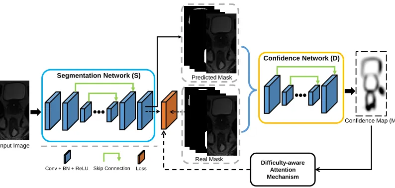
As mentioned in the introduction, the proposed method con-sists of two sub-networks, i.e., 1) segmentation network (de-noted as S) and 2) confidence network (denoted as D). The architecture of our proposed framework is presented in Fig. 1, in which we conduct the fully convolutional confi-dence learning to avoid the training imbalance of GAN and design the difficulty-aware mechanism to alleviate the easy-sample dominance issue for training the segmentation net-work.
To ease the description of the proposed algorithm, we first give the formal notation used throughout the paper. Given a labeled input imageX ∈ RH×W×T with corresponding
ground-truth label map Y ∈ ZH×W×T, we encode it to
one-hot format P ∈ RH×W×T×C (by converting the
la-bel mapY into C binary label maps with one-hot encod-ing), whereC is the number of semantic categories in the dataset. The segmentation network outputs the class proba-bility maps Pˆ ∈ RH×W×T×C. The segmented label map
can be obtained byYˆ = arg maxP.ˆ
In the following subsections, we first introduce the seg-mentation network. Then, we describe the confidence net-work with fully convolutional adversarial learning, followed by the difficulty-aware attention mechanism. Finally, we de-scribe the implementation details.
Segmentation Network
As shown in Fig. 1, the segmentation network can be any end-to-end segmentation network, such as FCN (Long et al. 2015), UNet (Ronneberger et al. 2015), VNet (Milletari et al. 2016), or DSResUNet (Yu et al. 2017) (a UNet-like structure with residual learning, element-wise addition of skip con-nection, and deep supervision). In this paper, we adopt an enhanced UNet as the segmentation network. Specifically, we replace all the convolutional layers but the last one with the residual modules (He et al. 2016), apply dilated residual module in the intermedia layers between encoder and de-coder (the feature maps with the smallest size) (Yu, Koltun, and Funkhouser ), utilize the transformation modules in the long skip connections (Nie et al. 2018), inject deep super-vision at three scales in the decoder path (Merkow et al. 2016), and propose channel attention module to better fuse the concatenated information from lower layers and higher layers (Hu, Shen, and Sun ).
Training segmentation network with hybrid loss: The class imbalance problem is usually serious in medical im-age segmentation tasks. To overcome it, we propose using a generalized multi-class Dice loss (Sudre et al. 2017) as the training loss for our segmentation network, as defined below in Eq. (1):
LDice(X,P;θS) = 1−2
C ∑
c=1 πc
H ∑
h=1 W ∑
w=1 T ∑
t=1
Ph,w,t,cPh,w,t,cˆ
C ∑
c=1 πc
H ∑
h=1 W ∑
w=1 T ∑
t=1
Ph,w,t,c+Pˆh,w,t,c ,
Real Mask Segmentation Network (S)
Conv + BN + ReLU Loss
Input Image
Skip Connection
Predicted Mask
Confidence Map (M)
Difficulty-aware Attention Mechanism
Confidence Network (D)
Figure 1: Illustration of the architecture of the proposed framework. This framework consists of a segmentation network (S), a confidence network (D), and the difficulty-aware attention mechanism. Note, we pursuea perfect Din this framework.
whereπc is the class balancing weight of categoryc, and
θScontains the parameters of segmentation network. We set
πc= 1/
(H
∑
h=1 W
∑
w=1 T
∑
t=1 Ph,w,t,c
)2
.Pˆ is the predicted
proba-bility maps from the segmentation network:Pˆ =S(X, θs).
Besides, we also use the multi-category cross entropy loss to form the voxel-wise measurement, as shown in Eq. (2):
LCE(X, Y;θS) =− H ∑
h=1 W ∑
w=1 T ∑
t=1 C ∑
c=1
I{Yh,w,t,c}logPˆh,w,t,c
(2)
To this end, the hybrid loss which leverages both losses for training the segmentation network can be concluded as in Eq. (3):
LHyb =LDice+LCE (3)
Fully Convolutional Confidence Learning
Adversarial learning has been shown to be effective in improving the segmentation network (Luc et al. 2016; Moeskops et al. 2017; Hung et al. 2018). More impor-tantly, it can provide a better hard-easy sample evaluator with proper adjustment. Thus, we decide to incorporate ad-versarial learning in our architecture to further improve the segmentation network.
In the classical adversarial networks, the discriminator is mostly a CNN-based network with the output probabil-ity of an input image belonging to be the real (Sabokrou et al. 2018). Obviously, the conventional discriminator only provides a global confidence over the entire image domain, without providing any confidence at the local region, e.g., voxel-wise confidence. To address this issue, we propose using an FCN-based network to model the discriminator and name it as confidence network. The output of confi-dence network is called as conficonfi-dence map (M) with size
H×W×T×1, which indicates locally whether automatic segmentation is similar to the ground-truth segmentation. We argue that the confidence network can learn the struc-tural information that can be used to regularize the output of segmentation network (Hung et al. 2018). In this paper, a simplified version of typical UNet (Ronneberger et al. 2015) is used as the architecture of confidence network. Specifi-cally, to save memory, we only keep one convolutional layer at each stage and also half the number of feature maps in the convolution layers across the network except the last one.
However, non-convergence and model collapse issues usually occur in GAN’s training, which are often explained as an imbalance between the discriminator and the genera-tor. Though widely researched, this problem still exists in training GAN (Mescheder 2018; Mescheder, Geiger, and Nowozin 2018; Kodali et al. 2017). To avoid the imbalance, we relax the adversarial learning to confidence learning after analyzing the role of the discriminator in the GANs.
Relaxing the adversarial learning to confidence learning:
framework and the classical GANs, i.e., besides the adver-sarial learning from the discriminator, the segmentation net-work can provide strong supervision signals to train itself.
As mentioned before, the GAN framework has a big chal-lenge of imbalance training. Since the discriminator is much easier to be perfectly trained than the generator and thus will result in few training signals for the generator from discriminator (Arjovsky, Chintala, and Bottou 2017), which will eventually lead to non-convergence and model collapse. However, it is a different situation in our case since the seg-mentation network has training supervision signals from it-self which can provide continuous support to improve the segmentation network. To this end, we propose to relax ad-versarial learning to confidence learning for avoiding the training imbalance by adjusting the role of the discrimina-tor:we place the role of confidence learning prior to that of adversarial learning. In other words,we reformulate the original min-max game to a maximization of discriminator with a soft constraint over the generator.
With this strategy, we can discover the difficulty degree of each local region of being segmented and can thus provide difficulty-aware information to guide the training of the seg-mentation network. To this end, the segseg-mentation network can be further improved, which will in return boost the dis-criminator. As a result, the adversarial learning can be for-mulated as asoft constraintto work as a high-order potential regularization for the segmentation network.
Training the confidence network: The training objective of the confidence network is the summation of binary cross-entropy loss over the image domain, as shown in Eq. (4). Here, we useSandDto denote the segmentation and con-fidence networks, respectively.
LD(X,P;θD) =LBCE(D(P, θD),1) +LBCE(D(S(X), θD),0),
(4)
whereLBCE
(
ˆ
Q,Q)=−
H ∑ h=1 W ∑ w=1 T ∑ t=1
Qh,w,tlog
(
ˆ
Qh,w,t
)
+ (1−Qh,w,t) log
(
1−Qˆh,w,t
)
(5)
whereXandPrepresent the input data and its correspond-ing manual label map (one-hot encodcorrespond-ing format), respec-tively.θDis network parameters for the confidence network.
Adversarial loss of the segmentation network: For seg-mentation network, besides the hybrid loss as defined in Eq. (3), there is another loss fromD used as “variational” regularization term working as a soft constraint, which aims at enforcing higher-order consistency between ground-truth segmentation and automatic segmentation. In particular, the adversarial loss (“ADV”) to improveS and foolD can be defined by Eq. (6):
LADV (X, θS) =LBCE(D(S(X;θS)),1) (6)
Difficulty-Aware Attention Mechanism
Focal loss has been shown effective to alleviate the over-whelming effect of easy samples in many computer vision tasks, such as image detection and segmentation (Lin et al.
2017; Zhou et al. 2017). The success of focal loss can be at-tributed to its strategy that it tries to pay more attention on the recognized hard samples (regions) and less attention to the easy ones. The key point is how to recognize difficult samples (regions). Focal loss utilizes the predicted proba-bility of a sample as the indicator of the difficulty degree, which may lead to some potential problems in medical im-age segmentation tasks. Firstly, training may be unstable due to the dominance of a certain class. Secondly, easy and hard samples may also have similar focal weights due to the po-tential multi-class competition. Thirdly, focal loss only pro-vides voxel-level attention and ignores region-level atten-tion. Lastly, only predicted mask may not really indicate the hard regions without considering the original input image of the segmentation network. These potential problems are mostly caused by the fact that the focal loss uses predicted probability from the segmentation network as the standard to determine whether it is a hard or easy sample. To overcome the above-mentioned problems, we would prefera more pro-fessional easy-or-hard representer.
The previously described confidence learning provides us with a solution to better recognize the easy-or-hard sam-ples. The confidence map produced by the confidence net-work contains the easy-or-hard information. Also, since con-fidence network is actually a binary classification model, it will avoid the multi-category competition issue. More im-portantly, the confidence map contains information from both the original input image and predicted probability mask, and thus it can provide structural information about the easy-or-hard samples (regions).
To this end, we propose a difficulty-aware attention mechanism to better represent the easy-or-hard information. Specifically, we design a difficulty-aware hybrid loss using region-level and voxel-level attentions from both predicted probability mask and confidence map.
At first, we propose an organ-level attention based gener-alized Dice loss to depict the region-level difficulty, which is shown in Eq. (7).
LF Dice= 1−2
C ∑
c=1
πc(1−dscc)r
H ∑ h=1 W ∑ w=1 T ∑ t=1
Ph,w,t,cPh,w,t,cˆ
C ∑
c=1
πc(1−dscc)r
H ∑ h=1 W ∑ w=1 T ∑ t=1
Ph,w,t,c+Ph,w,t,cˆ
(7)
where dscc is the average Dice similarity coefficient of aspecific category c, e.g., a certain organ or tissue.γ is the organ-level attention parameter with a range of [0,5]. Fol-lowing (Lin et al. 2017), we setγto2in this paper.
The voxel-level difficulty-aware attention from the confi-dence map (M) is formulated (based on Eq. 2) in Eq. (8):
LF CE(X, Y;θS) =
− H ∑ h=1 W ∑ w=1 T ∑ t=1 C ∑ c=1
I{Yh,w,t, c}Fh,w,tlogPˆh,w,t,c
(8)
where
F= (1−M)β (9)
Now we can define the difficulty-aware attention mecha-nism with the hybrid loss as Eq. (10).
LDamHyb=LF Dice+LF CE (10)
With the difficulty-aware hybrid loss in Eq. (10), we can pay more attention in the lower confidently (hard) seg-mented regions. Note, it is different from focal loss which is defined based on the probability map (P) from the segmen-tation network.
Total loss for segmentation network
By summing the above losses, the total loss to train the seg-mentation network can be defined by Eq. (11).
LSeg=LDamHyb+λ1LADV (11)
whereλ1is the scaling factor for the regularization term of adversarial learning. It is selected as a very small value (i.e.,
0.005in our case) since it works as soft constraint.
Implementation Details
Pytorch1 is adopted to implement our proposed framework shown in Fig. 1. Since we desire a perfect discriminator (D), we do not adopt the traditionally used strategies to limit the D(Radford, Metz, and Chintala 2015). We adopt Adam al-gorithm to optimize the networks. The input size of the seg-mentation network is64×64×16. The network weights are initialized by the Xavier algorithm (Glorot and Bengio 2010) and weight decay is set to be 1e-4. For the network biases, we initialize them to 0. The learning rates for the segmentation network and the confidence network are both initialized to 5e-3, followed by decreasing the learning rate 2 times for theS, and 5 times for theDevery 3 epochs dur-ing the traindur-ing. A Titan X GPU server is utilized to train the networks.
Experiments
To evaluate the proposed method, we apply our algorithm on three different datasets. The first dataset is our own pelvic dataset and the other two are both publicly available chal-lenge datasets which will be introduced in later subsections. The pelvic dataset consists of 50 prostate cancer patients from a Cancer Hospital, each with one T2-weighted MR image and corresponding manually-labeled map by a med-ical expert. The images were acquired with 3T magnetic field strength, while different patients were scanned with different MR image scanners (i.e., Siemens Medical Sys-tems and Philips Medical SysSys-tems). Under such a situation, the challenge for the segmentation task increases since both shape and appearance differences are large. The prostate, bladder, and rectum in all MRI scans have been manu-ally segmented, which serve as the ground truth for evalu-ating our segmentation method. The image size is mostly
256 ×256 ×(120∼192), and the voxel size is mainly
1×1×1mm3.
Five-fold cross-validation is used to evaluate our method. Specifically, in each fold of cross-validation, we randomly
1
https://github.com/pytorch/pytorch
MALF SSAE UNet VNet DSResNet Proposed
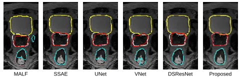
Figure 2: Pelvic organ segmentation results of a typical sub-ject by different methods. Orange, silver and pink contours indicate the manual ground-truth segmentations, and yellow, red and cyan contours indicate automatic segmentations.
chose 35 subjects as the training set, 5 subjects as the valida-tion set, and the remaining 10 subjects as the testing set. We use sliding windows to go through the whole MRI for pre-diction for a testing subject. Unless explicitly mentioned, all the reported performance by default is evaluated on the test-ing set. As for evaluation metrics, we utilize Dice Similarity Coefficient (DSC) and Average Surface Distance (ASD) to measure the agreement between the manually and automati-cally segmented label maps.
Comparison with state-of-the-art methods
To demonstrate the advantage of our proposed method, we compare our method with other five widely-used methods on the same dataset as shown in Table 1: 1) multi-atlas label fusion (MALF), 2) SSAE (Guo et al. 2016), 3) UNet (Ron-neberger et al. 2015), 4) VNet (Milletari et al. 2016), and 5) DSResUNet (Yu et al. 2017). Also, we present the perfor-mance of our proposed method.
Table 1 quantitatively compares our method with the five state-of-the-art segmentation methods. We can see that our method achieves better accuracy than the five state-of-the-art methods in terms of both DSC and ASD, especially for the prostate and rectum which are believed more difficult to segment. The VNet works well in segmenting bladder and prostate, but it cannot work very well for rectum (which is often more challenging to segment due to the long and nar-row shape). Compared to UNet, DSResUNet improves the accuracy by a large margin, indicating that residual learn-ing and deep supervision brlearn-ing performance gain. We also visualize some typical segmentation results in Fig. 2, which further show the superiority of our proposed method.
Impact of the Difficulty-aware Attention
Mechanism
Table 1: DSC and ASD on the pelvic dataset by different methods.
Method DSC (%) ASD (in mm)
Bladder Prostate Rectum Bladder Prostate Rectum
MALF 86.69(6.81) 79.28(8.72) 76.43(11.88) 1.641(.360) 2.791(.930) 3.210(2.112) SSAE 91.75(3.10) 87.07(4.24) 86.38(4.41) 1.089(.231) 1.660(.490) 1.701(.412) UNet 89.57(2.83) 82.22(5.88) 81.04(5.31) 1.214(.216) 1.917(.645) 2.186(0.850) VNet 92.61(1.84) 86.40(3.61) 83.16(4.12) 1.023(.186) 1.725(.457) 1.969(.449) DSResUNet 94.43(.90) 88.24(2.01) 86.91(3.24) .914(.168) 1.586(.358) 1.586(.405) Proposed 97.48(.65) 92.11(1.70) 91.05(2.47) .850(.146) 1.297(.276) 1.387(.346)
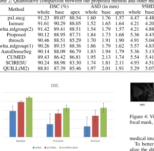
Table 2: Quantitative comparison between our proposed method and other methods on the prostate challenge testing dataset.
Method DSC (%) ASD (in mm) 95HD aRVD Score(std)
whole base apex whole base apex whole base apex whole base apex
pxl mcg 91.23 89.07 88.54 1.60 1.76 1.57 4.47 4.48 3.64 2.08 -0.07 2.23 88.98(3.41) Isensee 91.61 90.29 88.05 1.52 1.65 1.64 4.21 4.20 3.85 3.42 1.86 3.48 88.84(2.94) whu mlgroup(2) 91.42 89.41 88.51 1.54 1.79 1.57 4.21 4.88 3.82 5.27 4.00 6.43 88.72(4.36) Proposed 90.12 88.95 87.71 1.84 1.73 1.68 5.36 4.43 3.99 4.99 2.19 6.65 88.28(3.02) tbrosch 90.46 88.51 85.29 1.70 1.91 1.90 4.91 5.04 4.57 2.14 7.22 -4.93 87.24(4.46) whu mlgroup(1) 90.26 89.15 88.36 1.86 1.79 1.62 5.57 4.83 3.90 9.74 10.73 9.64 87.04(5.79) AutoDenseSeg 90.14 88.09 86.79 1.83 1.94 1.79 5.36 5.13 4.32 4.53 5.19 2.05 87.19(4.25) CUMED 89.43 86.42 86.81 1.95 2.13 1.74 5.54 5.41 4.29 6.95 11.04 15.18 86.65(4.42) SCIRESU 90.24 88.98 83.30 1.74 1.81 2.11 4.93 4.51 5.34 6.01 8.18 -7.33 86.41 (3.49) QUILL(M2) 88.81 87.39 85.46 1.97 2.01 1.91 5.29 5.07 4.35 6.97 4.76 5.85 85.93(4.97)
0.75 0.8 0.85 0.9 0.95 1
Bladder Prostate Rectum
DSC
UNet enUNet enUNet+dam
Figure 3: Average Dice ratios of different methods.
train UNet and enUNet). Actually, in our case, the widely used techniques injected to the basic UNet contribute most to the performance gain. The effectiveness of difficulty-aware attention mechanism is also confirmed by the im-proved performance as shown in Fig. 3. It is worth noting that our proposed difficulty-aware attention mechanism con-tributes more performance gain for prostate and rectum com-pared with the bladder. It is consistent with our assumption that difficulty-aware attention mechanism could pay more attention to difficult samples (regions) and thus can handle difficult samples (regions) much better.
Comparing with the Focal Loss
Since our proposed difficulty-aware attention mechanism is designed based on the focal loss, it is necessary to investigate the difference of the proposed module against focal loss for
MRI Ground Truth Predicted Difficulty-aware mask Focal mask
Figure 4: Visualization of the difficulty-aware mask and the focal mask, obtained after training the network for 5 epochs.
medical image segmentation.
To better understand the two strategies, we firstly visu-alize the difficulty-aware mask (i.e.,(1−M)) and the fo-cal mask (i.e.,(1−Pˆ
)
) in Fig. 4. The focal mask mainly focuses on the regions with low predicted probability from segmentation network which needs more attention. Since it is directly related with predicted probability map, it can re-flect the difficult regions more precisely invoxel-level. On the contrary, difficulty-aware mask reflects the difficulty re-gions in a morestructuredmanner, in which it focuses more on the regions with lower confidence ratios from confidence network. The reason behind it is that we have a professional hard-or-easy recognizer: TheDcan represent the input con-taining both the predicted probability mask from segmen-tation network and the original input image by confidence learning so that we can have a more expert hard-or-easy rep-resentation, as expressed in Eq. (12):
M =D(Pˆ∪X) (12)
where∪denotes the concatenation operation.
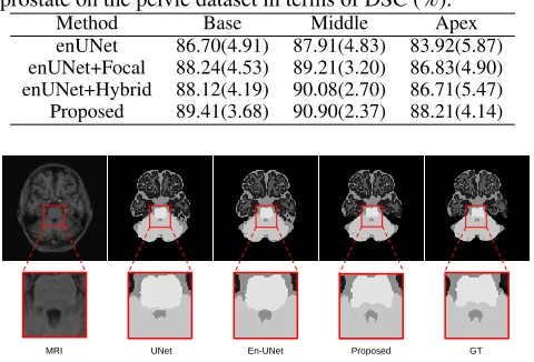
Table 3: Comparison of different strategies to segment prostate on the pelvic dataset in terms of DSC (%).
Method Base Middle Apex
enUNet 86.70(4.91) 87.91(4.83) 83.92(5.87) enUNet+Focal 88.24(4.53) 89.21(3.20) 86.83(4.90) enUNet+Hybrid 88.12(4.19) 90.08(2.70) 86.71(5.47) Proposed 89.41(3.68) 90.90(2.37) 88.21(4.14)
MRI UNet En-UNet Proposed GT
Figure 5: Comparison of segmentation results with different methods and the manual ground-truth on a sample subject.
is traditionally thought to be hard to segment. To make a fair comparison, we use the same architecture (enUNet) as the basis to conduct the experiments. Due to computational times, we only do a two-fold cross-validation for these com-parison experiments. To better depict the difficult parts of the prostate, we partition the prostate into three parts: apex (first 1/3 of the prostate volume), base (last 1/3 of the prostate vol-ume) and middle (the rest). The performance of the enUNet with different strategies is listed in Table 3.
As described in Table 3, the focal loss can help improve the performance, especially for the base and apex parts of the prostate, since it pays more attention to the hard voxels. The hybrid loss described in Eq. (3) can achieve similar per-formances with the focal loss since the hybrid loss can cap-ture the organ struccap-ture as well as the voxel-level informa-tion. The proposed method (difficulty-aware attention mech-anism) achieves the largest performance gain, since it can not only capture the difficult regions in a structured way but also absorb the advantage of the hybrid loss. This demon-strates that the proposed difficulty-aware attention mecha-nism can work better than the focal loss in medical image segmentation tasks.
Validation on MR Brain Challenge Dataset
We further validate our proposed method on MR Brain dataset2. This dataset contains 7 subjects, each with T1 MRI, Flair and manually labeled ground truth map. The task is to segment each voxel into one of the following (tissue) types: background, cortical gray matter (CGM), basal gan-glia (BG), white matter (WM), WM lesion (WML), cere-brospinal fluid in the extracerebral space (CSF), ventricle (V), cerebellum (C), brain stem (BS), infarction, and other.
We conduct the experiment in a leave-one-out manner. We visualize one typical slice of a sample in Fig. 5 to make a qualitative comparison. The proposed method can capture
2
http://mrbrains18.isi.uu.nl/
better contour which is usually considered as hard regions compared with the UNet and enUNet; this again proves the effectiveness of our proposed method. The quantitative com-parison (the proposed mechanism can improve the average performances by about 3.5% in terms of DSC) also indicates the success of the proposed modules.
Validation on Prostate Challenge Dataset
We also evaluate our proposed method on the prostate seg-mentation challenge dataset whose ground-truth label maps are hidden from the participants. The official evaluation met-rics used in this challenge include the DSC, the average over the shortest distance between the boundary (surface) points of the volumes (ABD or ASD), the percentage of the abso-lute difference between the volumes (aRVD), and the 95% Hausdorff distance (95HD). It is worth noting that the orga-nizers not only calculate the evaluation metrics on the whole prostate, but also on the apex and base parts of the prostate that are believed to be the most difficult regions for segmen-tation. In addition, an overall score (shown in the last col-umn) combining the above-mentioned evaluation metrics is also provided to rank the submitted methods (please refer to (Litjens et al. 2014) for the details about the evaluation metrics).
The quantitative results of our method and our competi-tors are shown in Table 2. (Note, the results were directly ob-tained from the organizers). Currently, there are more than 150 teams successfully submitting their results and listed in the leaderboard. Note we only list top 10 teams in the Ta-ble for convenience, and please refer the whole leaderboard through this link3. Our proposed method ranks 4th in terms of the overall score among all the participants. It is worth noting that the top 3 methods all ensemble their results from different deep networks. In contrast, our submission is a sin-gle model as presented in this paper. More importantly, our proposed method presents a much lower standard deviation value compared to the other top 8 methods. (Note, the low-est standard deviation comes from the 2nd ranked team who ensembles results from 20 deep networks), which further indicates the effectiveness and robustness of our proposed method.
More importantly, our proposed method achieves a very competitive performance on the base and apex parts which are thought to be the most difficult segmented regions, and it further proves that our designed difficulty-aware attention mechanism indeed contributes to the gain of performance.
Conclusions
In this paper, we presented a novel difficulty-aware atten-tion deep networks to segment medical images. Specifically, we proposed fully convolutional confidence learning to re-lax the adversarial learning so that we can largely alleviate the training imbalance between discriminator and genera-tor, and the discriminator can thus provide wonderful confi-dence information. Based on that, difficulty-aware attention mechanism was proposed to effectively address the easy-to-segment sample dominance issue in a more structured way,
3
which goes beyond the shortcomings of focal loss for train-ing medical image segmentation networks. By integrattrain-ing these components into the framework, our proposed frame-work achieved significant improvement in terms of both ac-curacy and robustness on three datasets.
Acknowledgments. This work was supported by the Na-tional Institutes of Health under Grant R01 CA206100.
References
Arjovsky, M.; Chintala, S.; and Bottou, L. 2017. Wasserstein gan.
arXiv preprint arXiv:1701.07875.
Glorot, X., and Bengio, Y. 2010. Understanding the difficulty of training deep feedforward neural networks. InAISTATS, 249–256. Goodfellow et al., I. 2014. Generative adversarial nets. InNIPS. Gulrajani, I.; Ahmed, F.; Arjovsky, M.; Dumoulin, V.; and Courville, A. C. 2017. Improved training of wasserstein gans. InNIPS, 5767–5777.
Guo et al., Y. 2016. Deformable mr prostate segmentation via deep feature learning and sparse patch matching. IEEE TMI35:1077– 1089.
He, K.; Zhang, X.; Ren, S.; and Sun, J. 2016. Deep residual learn-ing for image recognition. InCVPR, 770–778.
Hu, J.; Shen, L.; and Sun, G. Squeeze-and-excitation networks. Hung, W.-C.; Tsai, Y.-H.; Liou, Y.-T.; Lin, Y.-Y.; and Yang, M.-H. 2018. Adversarial learning for semi-supervised semantic segmen-tation.arXiv preprint arXiv:1802.07934.
Kodali, N.; Abernethy, J.; Hays, J.; and Kira, Z. 2017. On conver-gence and stability of gans.arXiv preprint arXiv:1705.07215. Kohl et al., S. 2017. Adversarial networks for the detection of aggressive prostate cancer.arXiv preprint arXiv:1702.08014. Lin et al., T.-Y. 2017. Focal loss for dense object detection.arXiv preprint arXiv:1708.02002.
Litjens et al., G. 2014. Evaluation of prostate segmentation algo-rithms for mri: the promise12 challenge.MedIA18(2):359–373. Long et al., J. 2015. Fully convolutional networks for semantic segmentation. InCVPR, 3431–3440.
Luc, P.; Couprie, C.; Chintala, S.; and Verbeek, J. 2016. Se-mantic segmentation using adversarial networks. arXiv preprint arXiv:1611.08408.
Mao, X.; Li, Q.; Xie, H.; Lau, R. Y.; Wang, Z.; and Smolley, S. P. 2017. Least squares generative adversarial networks. InICCV, 2813–2821. IEEE.
Merkow, J.; Marsden, A.; Kriegman, D.; and Tu, Z. 2016. Dense volume-to-volume vascular boundary detection. InMICCAI, 371– 379. Springer.
Mescheder, L.; Geiger, A.; and Nowozin, S. 2018. Which training methods for gans do actually converge? InICML, 3478–3487. Mescheder, L. 2018. On the convergence properties of gan training.
arXiv preprint arXiv:1801.04406.
Milletari et al., F. 2016. V-net: Fully convolutional neural net-works for volumetric medical image segmentation. In3DV, 565– 571. IEEE.
Moeskops et al., P. 2017. Adversarial training and dilated convolu-tions for brain mri segmentation.arXiv preprint arXiv:1707.03195. Nie, D.; Trullo, R.; Lian, J.; Petitjean, C.; Ruan, S.; Wang, Q.; and Shen, D. 2017. Medical image synthesis with context-aware gen-erative adversarial networks. InMICCAI, 417–425. Springer.
Nie, D.; Wang, L.; Adeli, E.; Lao, C.; Lin, W.; and Shen, D. 2018. 3-d fully convolutional networks for multimodal isointense infant brain image segmentation.IEEE Transactions on Cybernetics. Pan, T.; Wang, B.; Ding, G.; and Yong, J.-H. 2017. Fully convo-lutional neural networks with full-scale-features for semantic seg-mentation.
Radford, A.; Metz, L.; and Chintala, S. 2015. Unsupervised rep-resentation learning with deep convolutional generative adversarial networks.arXiv preprint arXiv:1511.06434.
Ronneberger et al., O. 2015. U-net: Convolutional networks for biomedical image segmentation. InMICCAI, 234–241. Springer. Sabokrou, M.; Pourreza, M.; Fayyaz, M.; Entezari, R.; Fathy, M.; Gall, J.; and Adeli, E. 2018. Avid: Adversarial visual irregularity detection.ACCV.
Shrivastava, A.; Gupta, A.; and Girshick, R. 2016. Training region-based object detectors with online hard example mining. InCVPR, 761–769.
Sudre et al., C. H. 2017. Generalised dice overlap as a deep learn-ing loss function for highly unbalanced segmentations. InDLMIA. Springer.
Xiao, H.; Wei, Y.; Liu, Y.; Zhang, M.; and Feng, J. 2017. Trans-ferable semi-supervised semantic segmentation. arXiv preprint arXiv:1711.06828.
Xue, Y.; Xu, T.; Zhang, H.; Long, L. R.; and Huang, X. 2018. Segan: Adversarial network with multi-scale l 1 loss for medical image segmentation.Neuroinformatics1–10.
Yang, X.; Yu, L.; Wu, L.; Wang, Y.; Ni, D.; Qin, J.; and Heng, P.-A. 2017. Fine-grained recurrent neural networks for automatic prostate segmentation in ultrasound images. InAAAI, 1633–1639. Yu et al., L. 2017. Volumetric convnets with mixed residual con-nections for automated prostate segmentation from 3d mr images. InAAAI.
Yu, F.; Koltun, V.; and Funkhouser, T. A. Dilated residual networks. Zhang, Y.; Yang, L.; Chen, J.; Fredericksen, M.; Hughes, D. P.; and Chen, D. Z. 2017. Deep adversarial networks for biomedical image segmentation utilizing unannotated images. InMICCAI, 408–416. Springer.
Zhou, X.-Y.; Shen, M.; Riga, C.; Yang, G.-Z.; and Lee, S.-L. 2017. Focal fcn: Towards small object segmentation with limited training data.arXiv preprint arXiv:1711.01506.