http://dx.doi.org/10.4236/aim.2016.63014
How to cite this paper: Watanabe, K., Horinishi, N., Matsumoto, K., Tanaka, A. and Yakushido, K. (2016) A New Evaluation Method for Antibiotic-Resistant Bacterial Groups in Environment. Advances in Microbiology, 6, 133-151.
http://dx.doi.org/10.4236/aim.2016.63014
A New Evaluation Method for
Antibiotic-Resistant Bacterial
Groups in Environment
Katsuji Watanabe
1*, Naoto Horinishi
1, Kunimasa Matsumoto
1, Akihiro Tanaka
2,
Kenichi Yakushido
31Department of Life, Environment and Materials Science, Fukuoka Institute of technology, Fukuoka, Japan 2National Agricultural Research Center for Kyushu-Okinawa Region, National Agriculture and Food Research Organization, Kumamoto, Japan
3National Agricultural Research Center, National Agriculture and Food Research Organization, Tsukuba, Japan
Received 31 January 2016; accepted 8 March 2016; published 11 March 2016
Copyright © 2016 by authors and Scientific Research Publishing Inc.
This work is licensed under the Creative Commons Attribution International License (CC BY). http://creativecommons.org/licenses/by/4.0/
Abstract
In the present manuscript it was presented whether spreading of antibiotic resistant bacterial groups in environment could be monitored by our newly developed method by enumerating anti-biotic resistant bacterial groups in various biological wastes and composts. Although the numbers were not so high, diverse kinds of colistin resistant bacteria (25 mg∙L−1) were included in row cat-tle feces (1.78 × 10⁴ MPN g−1) and cattle feces manure (>3.84 × 10⁴ MPN g−1). Compost originated from leftover food (>44.8 × 10⁴ MPN g−1) and shochu lee (>320 × 104 MPN g−1) included higher numbers of chlortetracycline resistant Pseudomonas sp., (25 mg∙L−1), and row cattle feces included higher numbers of chlortetracycline resistant Enterobacteriacea (15.7 × 10⁴ MPN g−1), which mostly consisted from Pantoea sp. or Xenorhobdus doucetiae. Numbers of multi drug resistant bacteria, resistant to 25 mg·L−1 ofciprofloxacin, streptomycin, chloramphenicol, and ampicillin,
were the highest in row cattle feces (>143.6 × 10⁴ MPN g−1), followed by cattle feces manure (4.19 × 10⁴ MPN g−1), and shochu lee (0.36 × 10⁴ MPN g−1), which included diverse kinds of bacterial group. The present results indicated that higher numbers of multi drug resistant bacteria were typically found in row cattle feces, and the method was found suitable to enumerate and identify them. These results suggested that the method might become their environmental risk evaluation method.
Keywords
Colistin Resistant Bacteria, Chlortetracycline Resistant Bacteria, Multi Drug Resistant Bacteria, Multiple Enzyme Restriction Fragment Length Polymorphism Analysis, The Most Probable
*
Number Method, Microchip Electrophoresis System
1. Introduction
At present, spreading of antibiotic resistant bacteria is becoming a major public health problem in all over the world [1]. As the susceptibility tests using bacterial isolates, with respect to specific nosocomial bacteria, were indispensable not only to search effective antibiotic for patient but also to prevent their nosocomial infection, the method has been used as a standard surveillance method for their risk assessment [2]-[4]. However their spread-ing area was recently expandspread-ing over various environments, such as drinkspread-ing water [5]-[7], or vegetables [8]-[10], or daily products [11]-[13], due to their overuse not only as therapeutic agent in human and veterinary medicine but also as growth promotor in animal husbandry [14] [15], and the susceptibility tests and taxonomy determinations must be broadly expanded over a large numbers of environmentally important bacterial groups in order to know what kinds of antibiotic bacteria will be numerically dominant and then has a higher environmen-tal risk [5]-[11] [13] [15]-[18].
With respect to the antibiotic bacteria, their taxonomic positions had no relation to the antibiotic resistance which was irregularly evolved by acquiring diverse kinds of resistant genes. Therefore molecular-based analysis method could not be used for their risk assessment because bacterial phylogenetic positions estimated by the unculture-based community analysis methods, such as DGGE or t-RFLP or clone library sequencing or pyro- sequencing [19], had no-relation to those of the resistant bacterial groups. Although sequence-based metage-nomics [20] [21] and real time PCR [22]-[25] targeting resistant gene afforded the information of the diversity or copy numbers of the resistant gene, they had no relation to the phylogenetic positions nor phenotypic proper-ties of the resistant bacterial groups.
As prime feature of resistant bacteria distinguished from susceptible one was an ability to survive and proli-ferate under antibiotic, which was also concerned with one of their risk, we thought that environmental risk of antibiotic resistant bacteria in might be evaluated by identifying and quantifying bacteria grown under applica-tion of antibiotic. Until now, we had presented a new method to provide numbers of each taxonomically differ-ent bacterial groups in the former papers [26] [27]. By the method, sample having simple microbial diversity such as food and aquatic sample could be analyzed without cultivation, that having the huge microbial diversity such as soil and manure required cultivation before analysis for exact phylogenetic estimation [26] [27]. In this manuscript, bacterial groups resistant to colistin, chlortetracycline, and multi drugs, in row cattle feces, cattle feces manure, shochu lee, and compost originated from leftover food were identified and enumerated by the method adding these antibiotics.
2. Materials and Methods
2.1. Samples
Row cattle feces (R), which would be converted into cattle feces manure, was collected from a cow barn for daily cattle in National Agricultural Research Center for Kyushu-Okinawa Region, where daily cattle were breeding in Japanese standard method [28]. Cattle feces manure (M) was provided from farmer around National Agricultural Research Center for Kyushu-Okinawa Region. Shochu lee (S), residual aqueous solution of sweet potato after fermentation and distillation of shochu, was obtained from Akashi Shuzo Co. Ltd. (Miyazaki, Japan), which would be converted into compost. Compost originated from leftover food (L) was collected in compost-ing facility in Kumamoto prefecture (Kumamoto, Japan).
2.2. MPN and Used Antibiotics
incubation at 30˚C, bacterial DNA in each vial was extracted as described previously and purified by conven-tional methods [26] [27].
2.3. Maintaining the Integrity of the Specifications
Using the V2 forward primer (41f; 5 ‘GCTCAGATTGAACGCTGGCG3’), and the V6 reverse primer (1066r; 3 ‘GTCGAGCACAACACTTTACA5’) [29], 16S rDNA of about 1070 bp length was amplified as described pre-viously [30] [31]. Their restriction fragment lengths were measured by microchip electrophoresis systems (Cosmo-i SV1200; Hitachi Electronics Engineering Co., Ltd. Tokyo Japan, or MCE-202 MultiNA; Shimadzu Co., Ltd. Kyoto Japan) after digestion of the PCR product (10 μl) using each restriction enzyme, HaeIII or HhaI or Rsa I (10 units, Takara Bio Co. Ltd. Shiga Japan) in buffer solution (10× Low salt buffer, Takara Bio Co. Ltd.) and 5 folds dilution by de-ionized water as described previously [30] [31].
2.4. Reference Database Used for the Phylogenetic Estimation
The reference database used for this research included 30,844 post-amplification sequence files for the 41f/ 1066r primers, which were mainly re-edited from small subunit rRNA files in RDP II release 9_61 [32]under 5 - bases mismatches in the both in primer annealing sites and were consisted from 1379 bacterial genera, including uncultured and unidentified bacteria.
2.5. Data Processing for Multi-Template DNA and Phylogenetic Estimation
As each MPN vials included multi-template DNAs originated from heterogeneous bacteria, the measured MERFL digested from the homogeneous 16S rDNA was selected among the mixed MERFLs digested from the heterogeneous 16S rDNA as described previously [26]. Because all the reference MERFLs were originated from the homogeneous 16S rDNA sequence. The major RFs (represented as H inTables 1-3) were those with the highest relative mole concentration (ratio of fluorescent intensity to fragment size). After subtraction of the ma-jor RFs from the mixed heterogeneous RFs, the 2nd mama-jor RFs were similarly selected (represented as M in
Tables 1-3). After subtraction of the 2nd major RFs from the remained heterogeneous RFs, the 3rd major RFs were similarly selected (represented as L inTables 1-3). The similarity between the measured RFLP (A) and the reference RFLP (B) was calculated as described previously [30] [31] based on the pairwise distance (DAB)
ac-cording to Nei and Li [33]. The pairwise distance of the MERFLPs (DABME) was an average of all the DABs for
used restriction enzymes. Similarity (%) was (1-DABME) × 100 (Tables 1-3). In the phylogenetic estimation,
combinations of the 2 restriction enzymes was used when the identical reference MERFL (100% similarity) was not found using all of the measured MERFL for the 3 restriction enzymes. When the identical reference MERFL to the measured MERFL for 2 restriction enzymes was not found, the reference MERFL having the highest si-milarity (over 80% ) to the measured MERFL was indicated in most cases (Tables 1-3) [30] [31].
2.6. Enumeration of Antibiotic Resistant Bacterial Groups by MPN
By five-tube, three-decimal-dilution experiment, MPNs of each antibiotic resistant bacterialgroups (A~K) were estimated (Tables 4-6). Using FDA’s Bacterial Analytical Manual [34], confidence limits were obtained and shown in the Tables.
3. Results
3.1. Phylogenetic Estimation of Antibiotic Resistant Bacteria
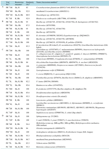
Affiliations of 67 MERFLs of colistin resistant bacteria (P) in each MPN vials were summarized inTable 1. All of the 67 MERFLs were divided into 67 OTUs, then ratio of total number of the OTUs to the total number of MERFLs was 100% (diversity of MERFLs), which was the highest among all the samples analyzed until now
Table 1. Affiliation of colistin resistant bacteria grown in serially diluted LB medium by MERFLa.
Vial No.b
Restriction enzymesc
Similarity (%)d
Name (Accession number)e
A PR−41M Ha, R 83 Corynebacterium glutamicum (BX9217148, BX927150, BX927152, BX927156)
PM−44H Ha, Hh 92.9 uncultured Corynebacterium (AM420211)
PM−51H Ha, R, Hh 88.9 Nocardiopsis sp. (AF361322)
PL−21H R, Hh 92.9 Rhodococcus erythropolis (AB177886, AY168580)
B PR−33H Ha, R, Hh 95 Bacillus sp. (AY461745, AY461746, AY461756), B. thuringiensis (AY461762)
PR−33M Ha, Hh 87.5 B.oleronius (AF393508)
PR−35H Ha, R, Hh 95 Bacillus sp. (AY461742, AY461750)
PR−51H R, Hh 100 Bacillus sp. (AF326359)
PM−54H Ha, R, Hh 90.5 B. oleronius (AY988598, X82492) Exguobacterium sp. (DQ246625)
PM−53H R, Hh 100 Bacillus sp. (AY461742, AY461750, AJ878858)
PC−31M Ha, R, Hh 100 Bacillus sp. (AY566219, AY583458), B. pallidus (Z26930)
PL−32M Ha, R 83.3 B. laevolacticus (B.lvolact3), B. racemilacticus (D16278), Gracilibacillus halotolerans (Grb. haltol)
C
PR−22M Ha, Hh 100 Clostridium(D14140), Lactobacillus aviaries sp. (AY957603), C. malenominatum (M58808) (M59099), Anaerococcus hydrogenalis
PS−45M Ha, R 100 C. collagenovorans (C. colgenvo), C. grantii (C. grantii), C. kluyveri (M59092, CP000673),
Anaeroplasma bactoclasticum (M25049) (fJ)
PL−31L Ha, Hh 100 C.butyricum (M59085), Ureaplasma diversum (D78650), U. canigenitalium (D78648)
PR−34H Ha, R, Hh 86 Alicyclobacillus hesperidum (AB059678, AB059679), A. sacchari (AB262020)
PR−23H Ha, Hh 100 A. pomorum (AB089840), Streptococcus mutans (AF139601), Enterococcus faecalis (AY94256)
PR−22H Ha, R 100 Syntrophomonas wolfei (CP000448)
PL−24H Ha, R 100 S. erecta (DQ86234), S. sporosyntrop (DQ112186)
PR−24L Ha, Hh 100 Paenibacillus peoriae (D78476), Bacillus brevis (X60612), B. edaphicus (AB045093)
PL−33L Ha, R, Hh 82.2 P. lautus (D85394, D85609)
PM−43L Ha, Hh 85.6 Ruminococcus albus (AY445592),
PL−44M Ha, R 100 R. productus (AY937379), Bacillus edaphicus (B. edaphicu) (fB)
PR−21H Ha, R, Hh 85.6 Desulfotomaculum nigrificans (AB026550)
PR−24M Ha, R, Hh 93.3 Desulfosporosinus orientis (Ds.orient2)
PR−45H R, Hh 100 Leuconostoc gelidum (AB004661)
PM−44L Ha, R 100 Lactobacillus vaccinostercus (M58813) (AB218801), L. bifermentans (M58809), L. coryniformis
PR−32H Ha, R, Hh 100 Slenomonas ruminantium (AB198430, AB198432, AB198433, AB198438), Megamonas
hypermegale (AJ420107)
PL−31Hg Ha, R 100 Staphylococcus aureus (CP000730), S. cohnii (Stp.cohni3)
D PR−41H Ha, R 100 Sphingomonas sp. (Y12803)
PL−31Hg R, Hh 100 S. mali (Y09638), S. pruni (Y09637), S. asaccharolyticas (Y09639)
PR−54H R, Hh 94.4 Methylobacrerium sp. (Mlb.sp.PK1, Mlb.sp.PR6), Methylosporovibrio methanica (Mls.methan)
PM−52H Ha, R, Hh 84 Orientia tsutsugamuchi (AM494475)
PM−43H Ha, R 100 Acidosphaera rubrifaciens (D86512), Erythrobacter longus (Erb. longus)
PL−44H Ha, R, Hh 93.3 Rhodopseudomonas acidophilas (M34128)
PR−21M Ha, R, Hh 86.7 uncultured beta proteobacterium (AB294945)
PS−45L Ha, Hh 83.3 Telluria mixta (X65589)
Continued
E PR−34M Ha, Hh 83.3 Pseudomonas sp. (DQ279343), P. stutzeri (U26262),
PR−55H Ha, R, Hh 86.9 Pseudomonas sp. (AM410901), Hahella chejuensis (CP000155)
PS−44H Ha, R, Hh 91.7 Pseudomonas sp. (AM111028)
PS−45H Ha, R 100 P. putida (DQ232745)
PL−33M Ha, R, Hh 94.8 P. caricapapayae (D84010)
F PR−44H Ha, R, Hh 85 Vibrio sp. (DQ173039), Methylobacillus flagellates (CP000284)
PM−43M R, Hh 100 Vibrio sp. (DQ146975), V. harveyi (AY911396,AY911387), V. carchariae (X74693),
G PR−32M Ha, Hh 100 Xenorhabdus indica (AM040494)
PR−52H Ha, R 93 Photobacterium profundum (CR378665, CR378680), Thermoanaebacterium therm (Tbm. thslf) (fC), Eubacterium yurii (Eub. yurii) (fC).
PR−53H R, Hh 92.9 uncultured gamma proteobacteria (AF445671)
PM−44M Ha, R 100 Marinobacter sp. (AB089803), Pseudomonas sp. (AM110949), Sporolactobacillus
laevis (D16287) (fC)
PL−35M Ha, R, Hh 88.9 Nitrococcus mobilis (L35510,)
PL−24M Ha, R 100 Nitrococcus mobilis (Nc. mobilis), Legionella hackeliae (Leg. hackel)
H PM−51M Ha, Hh 100 Desulfovibrio marrakechensis (AM947130)
PL−33H Ha, R, Hh 91.7 uncultured delta proteobacteria (AY771945)
PL−34H Ha, R, Hh 90.5 Desulfobacterium sp. (DQ146482)
PL−35H Ha, R, Hh 85.7 Stigmatella erecta (AJ233933), S. aurantiaca (AJ233936, AJ233937)
PL−35L Ha, R 92.9 Desulfomonile tiedjei (M26635)
PL−21M Ha, R 100 Corallococcus coralloides (AY072739)
J PR−31H R, Hh 100 uncultured Gemmatimonadetes (AY9211783, AY921939, AY921994, AY922110)
PM−41H Ha, R, Hh 90.5 uncultured Acidobacteria bacterium (AY922163)
PL−34M Ha, R, Hh 84.1 Deinococcus murrayi (Y13042)
K PR−24H Ha, R, Hh 93.3 uncultured rumen bacterium (AB034009, AB185580)
PR−35M Ha, Hh 100 uncultured bacterium (AB240503)
PM−53M R, Hh 83.3 uncultured bacterium (AY854278)
PM−54M Ha, Hh 92.9 uncultured bacterium (AY768822, DQ251791)
PS−44M R, Hh 100 halophilic bacterium(AB042504)
PR−23M R, Hh 83.3 Adiantum pedatum (AF244549)
PM−42M Ha, R 94.4 Olavius loisae endosymbiont (AF104475)
PM−42L Ha, R 90 marine psychrophile IC079 (U85854)
a
Grouping was based on affiliation by MERFL; Actinobacteria (A), Bacillus spp. (B), the other Firmicutes (C), α, β-Proteobacteria (D), Pseudomonas
sp. (E), Enterobacteriacea (F), the other γ-Proteobacteria (GG), δ, ε-Proteobacteria (H), Cytophaga (I), the other bacteria (J), and unidentified or un-cultured bacterial group (K). bThe 1st letter in vial indicates used antibiotics; “P” stands for colistin (polymyxin E). The 2nd letter in vial indicates samples; “R” stands for row cattle feces, “M” stands for cattle feces manure, “S” stands for shochu lee, and “L” stands for compost originated from leftover food. Exponential of vial number represents the decimal dilution of the vial. The 2nd number of vial number (1 - 5) represents number in 5 replicates for the each decimal dilution. “H” of last letter represents MERFL originating from the major 16S rDNA, “M” represents from the 2nd ma-jor 16S rDNA, and “L” represents from the 3rd mama-jor 16S rDNA. c Restriction enzymes used for similarity search; “Ha”, “R”, and “Hh” stand for Hae
III, Rsa I, and Hha I. For the measured MERFLP which had no completely identical theoretical MERFLP, the theoretical MERFLP having the highest similarity using all the RFLPs was presented with the similarity as described in the materials and method. dThe similarity between the measured RFLP
(A) and the reference RFLP (B) was calculated based on the pairwise distance (DAB) according to Nei and Li [33]. eSpecies name (accession number)
of the theoretical MERFL having the highest similarity with the measured MERFL. fThe theoretical MERFL (accession number) having the same
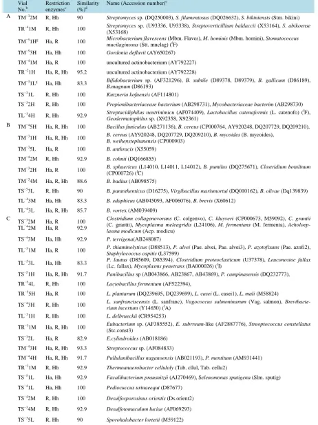
Table 2. Affiliation of chlortetracycline resistant bacteria grown in serially diluted LB medium by MERFLa.
Vial No.b
Restriction enzymesc
Similarity (%)d
Name (Accession number)e
A TM−32M R, Hh 90 Streptomyces sp. (DQ250003), S. filamentosus (DQ026632), S. bikiniensis (Stm. bikini)
TR−41M R, Hh 100 Streptomyces sp. (U93336, U93338), Streptoverticillium baldaccii (X53164), S. abikoense (X53168)
TM−51Hg Ha, R 100 Microbacterium flavescens (Mbm. Flaves), M. hominis (Mbm. homini), Stomatococcus
mucilaginosus (Stt. muclag) (fF) TM−53H Ha, Hh 100 Gordonia defluvii (AY650267)
TM−41M Ha, R 100 uncultured actinobacterium (AY792227)
TR−21H Ha, R, Hh 95.2 uncultured actinobacterium (AY792228)
TM−31Lg Ha, Hh 83.3 Bifidobacterium sp. (AF321296), B. subtile (D89378, D89379), B. gallicum (D86189),
B.magnum (D86193)
TS−31L R, Hh 100 Kutzneria kofuensis (AF114801)
TS−32H R, Hh 100 Propionibacteriacease bacterium (AB298731), Mycobacteriaceae bacterim (AB298730)
TL−34H R, Hh 92.9 Streptacidiphilus neutrinimicu (AF074409), Lactobacillus catenaformis (L. catenofo) ( fF),
Geodermatophilus sp. (X92358, X92361)
B TM−45H Ha, R, Hh 100 Bacillus funiculus (AB271136), B. cereus (CP000764, AY920248, DQ207729, DQ209210),
TM−31H Ha, R, Hh 100 B. cereus (AY920248, DQ207729, DQ209210), B. mycoides (B. mycoides),
B. weihenstephanensis (CP000903) TM−35L Ha, R 100 B. anthracis (X55059)
TM−42M R, Hh 92.9 B. cohnii (DQ166855)
TM−32H Ha, R 100 B. sphaericus (L14010, L14011, L14012), B. pumilus (DQ275671), Clostridium botulinum (CP000726) (fC)
TM−34M Ha, R, Hh 88.6 B. badius (AB098575)
TS−53L R, Hh 90 B. pantothenticus (D16275), Virgibacillus marismortui (DQ010162), B. olivae (Dq139839)
TL−43M Ha, Hh 83.3 B. edaphicus (AB045093, AF006076), B. brevis (X60612)
TL−43L Ha, R, Hh 85.7 B. vortex (AM039409) C
TS−32M TL−42M
Ha, R Ha, R
100 92.9
Clostridium collagenovorans (C. colgenvo), C. kluyveri (CP000673, M59092), C. grantii
(C. grantii), Mycoplasma meleagridis (L24106), M. fermentans (M. fermenta), Acholoep-lasma modicum (Acp. modicu)
TS−43M Ha, Hh 92.9 P. terrigena(AB248087)
TL−51M Ha, R 100 P. thiaminolyticus (D88513), P. alvei (Pae. alvei, Pae. alvei3), P. azotofixans (Pae. azofi2),
Staphylococcus capitis (L37599)
TL−53L Ha, Hh 83.3 P. lautus (D85609, D85394), Clostridium proteoclasticum (U37378), Leuconostoc fallax (Lc. fallax), Mycoplasms penetrans (BA000026) (fI)
TS−51H Ha, R, Hh 91.7 Panibacillus sp (AB043866, AB23867, AB43869), P. campinasensis (DQ232773),
TR−54L R, Hh 100 Lactobacillus fermentum (AF522394),
TR−55H Ha, R 100 L. plantarum (DQ239695, DQ239699), L. casei (L. casei1), L. mali (M58824)
TS−43H R, Hh 100 L. sanfranciscensis rium incertum (Y14650) ((L. sanfranc), fA) Vagococcus salmoninarum (Vag. salmon),
Brevibacte-TL−31H R, Hh 100 L. delbrueckii (CR954253)
TR−31M Ha, R, Hh 100 Eubacterium (Stc.const3) sp. (AF385552), E. subrreum-like (AF2887776), Streoptococcus constellatus
TS−32L Ha, R 82.9 E.cylindroides (AB018186)
TM−43H Ha, R, Hh 93.3 Streptococcus sp. (AF084833)
TM−44H Ha, R, Hh 91.7 Pullulanibacillus naganoensis (AB021193), P. mentitum (AM931441)
TR−21M R, Hh 92.9 Thermoanaerobacter celluloly (Tab. cllul, Tab. cellu2)
TS−51L Ha, Hh 92.9 Facalibacterium prausnitzii (AJ270469), Selenomonas sputigena (Slm. sputig)
TS−41L Ha, Hh 100 Pediocuccus urinaeequi (D87677)
TS−42M R, Hh 100 Desulfosporosinus orientis (Ds.orient2)
TS−34M R, Hh 92.9 Desulfotomaculum luciae (AF069293)
Continued
TL−53M Ha, R, Hh 88.9 Leuconostoc fallax (AB362604, AF218797), Nitrosospira multiformis (Nss.multi2) (fD)
TL−32H Ha, R, Hh 95.2 Staphylococcus aureus (BA000033, CP000255, CP000703, CP000730), Macrococcus
carouselicus (Y15713)
TL−33H Ha, R, Hh 95.2 Halobacillus(BA000028) sp. (AY966034, DQ089675), Oceanobacillus sp. (DQ190427), O. iheyensis
D TM−42H Ha, R, Hh 95.2 Rhizobium sp. (AJ294417), R. gallicum (AF417559), R. mangolense (U89818)
TM−34H Ha, R, Hh 95.2 R. leguminosarum (AF417563), Ochrobactrum sp. (DQ133574) TS−55H
TM−41H Ha, R Ha, R, Hh
92.9
93.3 Rhodopseudomonas acidophila (M34128) TM−51Hg Ha, Ha 100 Methylobacterium fujisawaensis (AJ250801)
TM−31M Ha, Hh 100 Acidocella aluminiidurans (AB362219)
TR−33M Ha, Hh 100 Neisseria animalis (Nis.animal), N. flavescens (L06168), N. denitrificans (Nis. dentri), N.
elongata (Nis. elong2), Kingella oralis (L06164) TS−53M Ha, R 94.4 Pandoraea sputorum (AM921627)
TS−31M Ha, Hh 100 Chromobacterium violaceum (AE016825, M25510), C. subtsugae (AY344056)
TC−54L Ha, Hh 90 Matsuebacter sp. (AB024305)
TC−41M R, Hh 100 Bordetella sp. (AB039335) E TS−54H R, Hh 100
Pseudomonas sp. (AF456214, AJ391194, AM111052, AY014824, DQ200851), P. amygdali
(AB021378), P. cichorii (Z76658), P. corrugata (D84012), P. stutzeri (AJ288148), P. en-tomophila (CT573326), Alkalilimnicola halodurans (AJ404972) (f
G) TS−44H Ha, R, Hh 81
TS−41H Ha, R, Hh 90.5
TS−42H Ha, R, Hh 90.5
TS−45H Ha, R, Hh 88.6
TS−34H Ha, R, Hh 93.3
TS−35H Ha, R, Hh 93.3
TL−51H Ha, R, Hh 93.3
TL−54H Ha, R, Hh 93.3
TL−55H Ha, R, Hh 93.3
TL−41H R, Hh 100
TL−42H Ha, R, Hh 93.3
TL−43H Ha, R, Hh 95.2
TL−44H Ha, R, Hh 93.3
TL−45H Ha, R, Hh 88.9
TM−33L Ha, Hh 90 Pseudomonas sp. (D87346), P. otitidis (AY953247)
TS−52H Ha, R 100 Pseudomonas sp. (AY998984), P. syringae (CP000058, CP000075), P. fluorescens (CP000094), P. stutzeri (U65012)
TS−33H R, Hh 100 P. putida (AY958233), Pseudomonas sp. (DQ205299, DQ227388)
TS−35M R, Hh 100 Pseudomonas sp. (AM934700), Advenella sp. (AY569461) (fD)
TL−52H R, Hh 100 Pseudomonas sp. (AM184269, AY573031), P. aureofaciens (D84008), P. chlororaphis (D84011)
F TR−52H Ha, R, Hh 100
Pantoea sp. (DQ094146), Xenorhobdus doucetiae (DQ211702) TR−53H Ha, R, Hh 100
TR−41H Ha, R, Hh 100
TR−42H Ha, R, Hh 100
TR−31H Ha, R, Hh 100
TR−32H Ha, R, Hh 100
TR−34H Ha, R, Hh 100
Continued
TR−42M Ha, R, Hh 95.2 Vibrio wodanis (AJ132227), V. logei (AY292928), V. fischeri (X74702), Enterobacter sp. (AY941832)
TR−33H Ha, R, Hh 100 V. fischeri (CP00020), Klebsiella sp. (DQ229100)
TM−35H Ha, R, Hh 95.2 Yersinia aldovae (Yer. aldova), Y. intermedia (Yer.intme2), Serratia sp. (AM050059) G TR−52M Ha, R 83.3 uncultured gamma proteobacteria (AB294936)
TR−54H Ha, R, Hh 100 Moritella sp. (AB183497)
TR−34M Ha, R 90 Frateuria aurantia (Frt. aurant)
TS−53H R, Hh 100 Halomonas campisalis (DQ077910)
TS−31H R, Hh 100 Thicapsa roseopersicina (AF113000), Rheinheimera baltica (AJ002006) H TL−44M
TL−45M R, Hh Ha, Hh
100
94.4 uncultured Enrotheonella sp. (AY897125) TR−55M Ha, R 100 uncultured delta proteobacteria (AY921877)
TL−33M R, Hh 100 uncultured delta proteobacteria (AB425060)
TL−52M Ha, Hh 90 Arcobacter sp. (AJ271654)
TL−53H Ha, R,Hh 82.2 Helicobacter pullorum (AF047850) I TS−55M
TL−55M Ha, R Ha, R
92.9
100 Bacteroidetes bacterium (AM932279) TR−32L Ha, Hh 100 uncultured Bacteroidetes (AY921921)
TM−51M Ha, Hh 100 uncultured Cytophaga sp. (DQ070792)
TR−33L R, Hh 100 Prevotella sp. (AF385558), Sulfurivirga caldicularium (AB245479, AB245480) (fG)
TL−31M Ha, R 92.9 Hymenobacter sp. (AB251884) J TS−54M R, Hh 100 Mycoplasma imitans (L24103)
TS−54L Ha, R 90 M. gallinaceum (L24104), M. pullorum (M. pollorum)
TS−45M Ha, R, Hh 95.2 M. penetrans (BA000026)
TM−35M Ha, R 100 Ureaplasma canigenitalium (D78648)
TS−41M Ha, R, Hh 100 Acholeplasma modicum (Acp. modicu)
TL−54M Ha, R 100 A. polakii (AF031479)
TM−51Hg R, Hh 100 Fusobacterium mecrophorum (AF044948)
TM−31Lg Ha, R 83.3 Spirochaeta bajacaliformiensis (M71239), Leptospira fainei (U60594), L. inadai (Z21634),
Holospora obtusa (X58198) (fG)
TR−53M Ha, Hh 92.9 Verrucomicrobic bacterium (AB331888)
TR−32M Ha, R 90 Leptospira fainei (U65094), L. inadai (Z21634)
K TM−44M Ha, R 100 uncultured bacterium (AF382142, AY344400)
TR−35M Ha, R, Hh 84.1 uncultured bacterium (AB294747, AJ48807)
TS−33M R, Hh 100 uncultured bacterium (AM777948)
TS−43L Ha, Hh 90 unidentified bacterium (AY796034)
TL−33L R, Hh 100 Oenothera berteriana (Oeno ber_M)
TL−35H R, Hh 100 Uncultured Green Bay ferroma (AF293008)
a
Grouping was based on affiliation by MERFL; Actinobacteria (A), Bacillus spp. (B), the other Firmicutes (C), α, β-Proteobacteria (D), Pseudomonas
sp. (E), Enterobacteriacea (F), the other γ-Proteobacteria (GG), δ, ε-Proteobacteria (H), Cytophaga (I), the other bacteria (J), and unidentified or un-cultured bacterial group (K). bThe 1st letter in vial indicates used antibiotics; “T” stands for chlortetracycline. The 2nd letter in vial indicates samples; “R” stands for row cattle feces, “M” stands for cattle feces manure, “S” stands for shochu lee, and “L” stands for compost originated from leftover food. Exponential of vial number represents the decimal dilution of the vial. The 2nd number of vial number (1 - 5) represents number in 5 replicates for the each decimal dilution. “H” of last letter represents MERFL originating from the major 16S rDNA, “M” represents from the 2nd major 16S rDNA, and “L” represents from the 3rd major 16S rDNA. cRestriction enzymes used for similarity search; “Ha”, “R”, and “Hh” stand for Hae III, Rsa
I, and Hha I. For the measured MERFLP which had no completely identical theoretical MERFLP, the theoretical MERFLP having the highest simi-larity using all the RFLPs was presented with the simisimi-larity as described in the materials and method. dThe similarity between the measured RFLP (A) and the reference RFLP (B) was calculated based on the pairwise distance (DAB) according to Nei and Li [33]. eSpecies name (accession number) of
the theoretical MERFL having the highest similarity with the measured MERFL. fThe theoretical MERFL (accession number) having the same
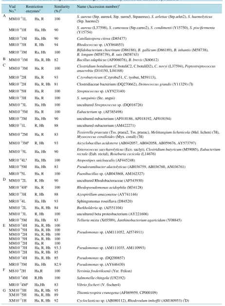
Table 3. Affiliation of multi drug resistant bacteria grown in serially diluted LB medium by MERFLa. Vial No.b Restriction enzymesc Similarity (%)d
Name (Accession number)e
A
MM10−51L Ha, R 100 S. aureus (Stp. haemo2) (Stp. aureu4, Stp. sureu5, Stpaureus), S. arlettae (Stp.arlet2), S. haemolyticus
MR10−41H Ha, Hh 90 S. aureus (Y15754)(L37598), S. camosusu (Stp.carno2), S. condimenti (Y15750), S. piscifermenta
MR10−51M Ha, Hh 90 Catellatospora citrea (D85477)
MR10−43H R, Hh 94 Rhodococcus sp. (AY864653)
MR10−33M Ra, Hh 100 Bifidobacterium choerinum (D86186), B. gallicum (D86189), B. inhantis (M58738),
B. longum (M58739), B. suis (M58743)
B MM10−21M Ha, R, Hh 82 Bacillus edaphicus (AF006076), B. brevis (X60612) C
MM10−33M Ha, R 100 Clostridium botulinum (C.botuliC2, C.botuliD2), C. novyi (L37594), Peptostreptococcus
anaerobiu (D14150, L04168)
MR10−52H Ha, R 93 C.tyrobutyricum (C.tyrobu51, C. tyobut, M59113),
MR10−32H Ha, R, Hh 81 Clostridiaceae bacterium (DQ270662), Deinococcus grandis (Y11329) (fI)
MR10−45H Ha, R 100 Streptococcus sp. (AY923140)
MR10−23H Ha, R 100 S. sanguinis (Stc. angui)
MM10−43L Ha, Hh 100 uncultured Streptococcus sp. (DQ016726)
MM10−55M Ha, R 100 Eubacterium sp. (AF385498)
MR10−43M Ha, Hh 90 uncultured eubacterium (AF018186, AF018192, AF018194)
MR10−31L R, Hh 88 uncultured eubacterium (AM422271)
MM10−42M Ha, R 83 Tissierella praecuta (Tss. praea2, Tss. praeac), Melittangium lichenicola (Mel. lichen) ( fH),
Myxococcus coralloides (Myx. corall) (fH)
MM10−23Mg R, Hh 93 Aicyclobacillus acidoterre (AB042057, AB042058, AB059676, AY573797)
MM10−25L Ha, Hh 90 Enterococcus saccharolyticus (Eco. saclyt), Clostridium butyricum (M59085), Eubacterium
rectale (Eub. rectal), Roseburia cecicola (L14676)
MR10−44Lg Ha, Hh 100 Atopostipes suicloacalis (AF445248)
MR10−45M Ha, Hh 83 Pseudoramibacter alactolyticus (AB036759, AB036760, AN036761)
MR10−45L Ha, R 100 Paenibacillus sp. (AB043868, AM162327) D MM10−42L R, Hh 90 uncultured Rhodobacteraceae (AF543930)
MR10−44Hg Ha, R 100 Rhodopseudomonas acidophila (M34128)
MR10−33H R, Hh 88 Azospirillum amazonense (AY741146)
MR10−24L Ha, Hh 93 Sphingomonas roseiflava (D84520)
MM10−32L Ha, R, Hh 84 Burkholderia sp. (AJ551104)
MM10−33L R, Hh 100 uncultured beta proteobacterium (AY221606)
MR10−55M Ha, Hh 83 Telluria mixta (X65589), Janthinobacterium agaricidam (Y08845) E MM10−54H
MM10−55H MM10−42H MM10−35H
Ha, R, Hh Ha, R, Hh Ha, R, Hh Ha, R, Hh
100 100 100 100
Pseudomonas sp. (AM111052, AJ574911)
MM10−52H MM10−33H MM10−32H
Ha, R Ha, R, Hh Ha, R, Hh
100 93.3 85
Pseudomonas sp. (AM111035, AM110993)
MM10−34H Ha, R, Hh 85 Pseudomonas sp. (DQ200857)
MR10−25M Ha, Hh 82.9 Pseudomonas sp. (AY646430) F MS10−52H Ha,R 100 Yersinia frederiksenii (Yer. Friksn)
MM10−54M R,Hh 100 Salmonella chingola (U92192)
MR10−24Mg Ha,Hh 83 Vibrio fischeri (V. fischer4) G XM10−23H
XM10−25H
Ha, R, Hh Ha, R, Hh
95
89 Thiomicrospira crunogena (AF069959, CP000109)
Continued
XM10−22H R, Hh 88 Haemophilus influenza (AF224305, AF224306, AY613568, AY613580)
XR10−51H Ha, R, Hh 89 Stenotrophomonas maltophilia (AB180661, AB294554, AB294555, DQ141193)
XR10−54Mg Ha, Hh 93 Oceanospirillum maris (AB006771), Pelagicoccus mobilis (AB286015) (fJ)
XR10−42H Ha, Hh 86 uncultured gamma proteobacteria (AJ567535, AJ567542)
XR10−44Hg R, Hh 100 Halorhodospira halophile (CP000544)
XR10−31H Ha, R 100 Buchnera aphidicola (AJ296759) H XM10−25M
XR10−44M XR10−25H
Ha, R, Hh Ha, Hh Ha, Hh
82 83 93
Angiococcus disciformis (Ang. discif)
XM10−51M R, Hh 83 Desulfovibrio africanus (M37315)
XR10−52M R, Hh 83 D. salexigens (Dsv. salexi)
XM10−43H Ha, Hh 90 uncultured delta proteobacteria (AF154094)
XR10−24H Ha, R, Hh 81 Pelobacter propionicus (CP000482, X70954)
XS10−52M Ha, R, Hh 86 Arcobacter sp. (AJ271654)
XR10−41M Ha, Hh 100 uncultured epsilon proteobacteria (AB235370, AM712353) I XM10−23L
XR10−51L
Ha, R, Hh Ha, R
93 83
Bacteroides sp. (AF139525), B. thetaiotaomicron (AE016936, AE016937, M58763),
Colwellia sp. (DQ027051) (fG) XM10−53H Ha, R, Hh 84 Flavobacterium sp. (AM934661)
XR10−52L Ha, R 88 uncultured Cytophagales bacteria (AF361196)
XR10−24Mg Ha, R 83 Prevotella sp. (AB166777) J XM10−43M
XM10−35M XR10−22H
Ha, R Ha, R Ha, R 100 100 100
Entomoplasma freundtii (AF036954), Encarsia pergandiella (AF319783), Bacteroidetes endocymbiont (AY753170) (fI)), Cardinium endosymbiont (AY327472)(fI), Bacillus tipchiralis (AF039408) (fB)
XM10−22M Ha, R 90 Spiroplasma leptinotarsae (AY189305) XR10−44Lg
XR10−25L
R, Hh R, Hh
100
90 S. mirum (M24662), S. citri (Spp. cit2HP), S. poulsonii (Spp. poulsn) XR10−32L Ha, R 90 S.linguale (M62789),
XM10−52L Ha, Hh 90 Ureaplasma cati (D78649), U. felinum (D78651) XS10−52L
XR10−55H
Ha, Hh Ha, R
83
93 uncultured planctomycetes (AM040106) XM10−23Mg Ha, R 93 Aquificales str. (AF255598, AF255597)
XM10−21L Ha, R 83 Heroetosiphan aurantiacus (M34117), Chlamydomonas reinhardtii (BK000554) ( fK),
Leptospira fainei (U60594)
XR10−54Mg Ha, R 93 uncultured cyanobacterium (AY874085, AJ431339), uncultured eubacterium (AF018194)
XR10−42L Ha, Hh 93 Arthrospira platensis (DQ279767, DQ279768, DQ279769, DQ279770) K XR10−54H Ha, Hh 100
uncultured bacterium (AY869688) XR10−35H Ha, Hh 100
XM10−51H Ha, Hh 100
uncultured bacterium (AF072927, AJ867657, AY661977, AY571416) XM10−52M Ha, R, Hh 100
XM10−34M Ha, R, Hh 100 XR10−32M Ha, Hh 100
XM10−34L Ha, R 83 Melosira varians (AJ536464), Phaeodactylum tricomutum (DQ174248)
XR10−35M Ha, R 100 Plantago sericea (AJ389621)
a
Grouping was based on affiliation by MERFL; Actinobacteria (A), Bacillus spp. (B), the other Firmicutes (C), α, β-Proteobacteria (D), Pseudomonas
sp. (E), Enterobacteriacea (F), the other γ-Proteobacteria (GG), δ, ε-Proteobacteria (H), Cytophaga (I), the other bacteria (J), and unidentified or un-cultured bacterial group (K). bThe 1st letter in vial indicates used antibiotics; “X” stands for multi drugs, ciprofloxacin, streptomycin, chloramphenicol, and ampicillin. The 2nd letter in vial indicates samples; “R” stands for row cattle feces, “M” stands for cattle feces manure, “S” stands for shochu lee, and “L” stands for compost originated from leftover food. Exponential of vial number represents the decimal dilution of the vial. The 2nd number of vial number (1 - 5) represents number in 5 replicates for the each decimal dilution. “H” of last letter represents MERFL originating from the major 16S rDNA, “M” represents from the 2nd major 16S rDNA, and “L” represents from the 3rd major 16S rDNA. cRestriction enzymes used for similari-ty search; “Ha”, “R”, and “Hh” stand for Hae III, Rsa I, and Hha I. For the measured MERFLP which had no completely identical theoretical MERFLP, the theoretical MERFLP having the highest similarity using all the RFLPs was presented with the similarity as described in the materials and method. dThe similarity between the measured RFLP (A) and the reference RFLP (B) was calculated based on the pairwise distance (D
AB)
ac-cording to Nei and Li [33]. eSpecies name (accession number) of the theoretical MERFL having the highest similarity with the measured MERFL. f
OUT (Table 2), which lowered the diversity of the MERFLs. Affiliations of 80 MERFLs of multi drug resistant bacteria (X) in each MPN vials were summarized inTable 3. All of the 80 MERFLs were divided into 62 OTUs, then ratio of total number of the OTUs to the total number of MERFLs was 77.5%, which was also lower than that of P (Table 3). Some MERFLs of the group E, group G, group H, group I, group J, and group K were placed in the same OUTs (Table 3).
The precisions of the affiliations of each MERFLs were lower than those of the former studies [26] [27]. Al-though, ratio of the MERFLs having 100% similarity to the reference MERFLs with respect to the major MERFL (37%;Tables 1-3) was lower than that of the 2nd major MERFLs (48.0%;Tables 1-3), that of the 3rd major MERLs (30%) was not so lower than that of the 2nd MERFLs and higher than those of the former studies
[26]. The lower precision of the major MERFL and higher precision of the 3rd major MERFLs was caused from lower ratio of Bacillus spp. as the followings; 8 MERFLs in colistin resistant bacteria (Table 1), 9 MERELs in chlortetracycline resistant bacteria (Table 2), and 1 MERFL in multi-drug resistant bacteria (Table 3). Be-cause16S rDNA of Bacillus spp. was preferentially amplified and increased the relative mole concentration of the major MERFL, and decreased those of the 2nd and 3rd major MERFLs [26] [27], which increased the preci-sion of the major MERFLPs, and decreased those of the 2nd and 3rd major MERFLPs.
3.2. Enumeration of Each Antibiotic Resistant Bacterial Groups by Mpn
As colistin was bactericidal to gram-negative bacteria and little to no effect on gram-positive bacteria, gram pos-itive bacterial groups (A to C) were eliminated from antibiotic resistant bacteria (Table 4,Figure 1). Numbers of the resistant bacteria was the highest in cattle feces manure (M; >3.84 × 10⁴ MPN g−1 dry matter), followed by row cattle feces (R; 1.78 × 10⁴ MPN g−1), shochu lee (S; 1.46 × 10⁴ MPN g−1), and compost originated from leftover food (L; 0.24 × 10⁴ MPN g−1) (Figure 1). α, β-proteobacteia (D; 1.04 × 10⁴ MPN g−1) was the numeri-cally dominant resistant bacteria in M, γ-proteobacteria (Pseudomonas spp. (E);0.44 ×10⁴ MPN g−1, Enterobac-teriaceae (F); 0.19 × 10⁴ MPN g−1, the other γ-Proteobacteria (G); 0.66 × 10⁴ MPN g−1) was the numerically do-minant in R, and α, β-proteobacteia (D; 0.36 × 10⁴ MPN g−1), and Pseudomonas spp. (E; 0.74 × 10⁴ MPN g−1) were the numerically dominant in S (Table 4,Figure 1).
Numbers of chlortetracycline resistant bacteria (T) was the highest in shochu lee (S; >320 × 104 MPN g−1),
Figure 1. Numbers of colistin resistant (25 mg∙L−1) bacterial groups estimated by MPN and MERFLP in cattle feces manure (M), row cattle feces (R), shochu lee (S), and compost originated from leftover food (L). Number of α,
β-Proteobacteria (D; ), Pseudomonas spp. (E; ), Enterobacteriacea (F; ), the other γ-Proteobacteria (G; ), δ,
followed by compost originated from leftover food (L; >44.8 × 10⁴ MPN g−1), row cattle feces (R; 16.6 × 10⁴ MPN g−1), and cattle feces manure (M; 13.7 × 10⁴ MPN g−1) (Table 5). In S, the other Firmucutes (C; 9.4 × 10⁴ MPN), Pseudomonas spp. (E; 9.4 × 10⁴ MPN g−1), and the other bacterial group (J; 18.6 × 10⁴ MPN,including
Mycoplasma spp), were the numerically dominant (Table 5, Figure 2). As 7 MERELs of Pseudomonas spp. (E) in S having the same MERFLs, they might proliferate preferentially than the other bacterial groups in S (Table 2). As Pseudomonas spp. (E) were the numerically dominant (>36.4 × 10⁴ MPN g−1) in L (Table 5) and 8 MERFLs of them also having the same MERFLs (Table 2), they might also proliferate preferentially in L. As Enterobacteriacea (F; 15.7 × 10⁴ MPN g−1) was the numerically dominant in R (Table 5) and 8 MERFLs of them also having the same MERFLs (Table 2), they might also proliferate preferentially in R (Table 2).
Numbers of multi drug resistant bacteria (X) was the highest in row cattle feces (R; >143.6 × 10⁴ MPN g−1), followed by cattle feces manure (M; 4.19 × 10⁴ MPN g−1), and shochu lee (S; 0.36 × 10⁴ MPN g−1) (Table 6,
Figure 3). In R, the other Firmicutes (C; 36.4 × 10⁴ MPN g−1), including Clostridium sp, and Streptcoccus sp, the other γ-proteobacteria (G; 10.3 × 10⁴ MPN g−1), and the other bacteria (J; 10.3 × 10⁴ MPN g−1), including
Spiroplasma sp, were the numerically dominant bacterial groups (Table 3, Table 6), while there was no bacteria which proliferated preferentially in R (Table 3). As Pseudomonas sp. (E; 0.88 × 10⁴ MPN g−1) was the numeri-cally dominant in M (Table 3, Table 6) and 4 MERFLs and 3 MERFLs of them had the same MERFLs, they were supposed to proliferate preferentially in M (Table 3).
4. Discussion
In the former studies, we had detected and enumerated polymyxin B resistant bacteria not only in field soils where liquid livestock feces had annually been applied [35] [36] but also in a paddy field soil where organic manure had annually been applied [26], although polymyxin B have mainly been used in hospitals and have never been used as AGP and there was no-report of the resistant bacteria except for nosocomial resistant bacteria in hospital [37]. Colistin (polymyxin E), which had a similar structure and the same site of action [37], had been used as AGP in Japan. The sample included diverse kinds of the resistant bacteria and no-specific numerically dominant bacterial group (Table 1), which might be caused from gradual proliferation of diverse kinds of bacte-ria having the lower resistance by a continual sub-therapeutic administration of the antibiotic as AGP. As higher
Figure 2.Numbers of chlortetracycline resistant (25 mg∙L−1) bacterial groups estimated by MPN and MERFLP in cat-tle feces manure (M), row catcat-tle feces (R), shochu lee (S), and compost originated from leftover food (L). Number of Actinobacteria (A; ), Bacillus spp.(B; ), the other Firmicutes (C; ), α, β-Proteobacteria (D; ), Pseudomonas
spp. (E; ), Enterobacteriacea (F; ), the other γ-Proteobacteria (G; ), δ, ε-Proteobacteria (H; ), Cytophaga (I; ), the other bacteria (J; ), and unidentified or uncultured bacterial group (H; ) were presented.
dosage of the antibiotics was used in this study (25l mg∙L−1) than those in the former studies (5 mg∙L−1), esti-mated numbers of the resistant bacteria in cattle feces manure (M; >3.84 × 10⁴ MPN), and row cattle feces (R; 1.78 × 10⁴ MPN) were lower than those estimated for the manure applied field soil (3.11 × 10⁶ MPN g−1) [26]
and those for field soils applied with liquid livestock feces (from 31.7 × 10⁶ CFU g−1 to 258 × 106 CFU g−1) [35] [36]. Although numerically dominant bacterial groups in M (unidentified bacterial group, α, β-proteobacteria;
Table 1, Table 4) were different from those of upland field soil (Prevotella spp. and Cytophagales) [26], colis-tin resistant gram negative bacteria detected in R and M in this study were concluded to be polymyxin B resis-tant bacteria in the former studies [26] [35] [36]. The antibiotic bacteria detected and enumerated in this study were supposed to have higher resistance to colistin than those of the former studies [26] [35] [36], and those of the reported resistant bacteria [37] because they could proliferate higher concentration (25 mg∙L−1) than the re-ported resistance breakpoints for Acinetobacter spp. (>2 or 4 mg∙L−1), that for Pseudomonas spp.(>4 mg∙L−1), and that for Enterobacteriacea (>2 mg∙L−1) [14] [37].
As chlortetracycline has widely been used for the past forty years as therapeutic agent for human and veteri-nary medicine but also as AGP, their numbers were higher than those of colistin resistant bacteria (Table 4, Ta-ble 5,Figure 1, Figure 2), which was coincident with the other report [14]. Although the numbers were unde-restimated due to an absence of MPN dilution vial higher than 10−6, the numbers were higher in shochu lee (S; >320 × 104 MPN g−1) and compost originated from leftover food (L; >44.8 × 10⁴ MPN g−1) (Table 5,Figure 2), where Pseudomonas spp. was not only the numerically dominant microorganisms but also proliferated pre-ferentially (Table 2,Table 5). In row cattle feces (R), specific resistant bacteria, Pantoea sp. or Xenorhobdus doucetiae, occupied the entire resistant bacterial group (Table 2,Table 5), which might suggest that therapeutic application of higher dosage of the antibiotic resulted in a rapid proliferation of this bacterial group in the cattle intestine. Although total number of the resistant bacteria in cattle feces manure (M; >13.7 × 10⁴ MPN g−1) were lower than those of the others (Table 5,Figure 2), the composition of the resistant bacterial groups (Table 2) were similar to those in the reported field soils, where continuous application of organic manure was supposed to cause accumulation of the resistant bacteria due to sub therapeutic use of the antibiotic as AGP [18]. As the concentration of applied chlortetracycline (25 mg∙L−1) was as the same level as those of the reported resistance breakpoints for Salmonella spp., E. coli, Camphylobacter spp., and Enterococcus spp. (>16 mg∙L−1) [14], the
Figure 3. Numbers of multi drug, ciprofloxacin (25 mg∙L−1), streptomycin (25 mg∙L−1), chloramphenicol (25 mg∙L−1), and ampicillin (25 mg∙L−1), resistant bacterial groups estimated by MPN and MERFLP in cattle feces
manure (M), row cattle feces (R), shochu lee (S), and compost originated from leftover food (L). Number of Actinobacteria (A; ), Bacillus spp.(B; ), the other Firmicutes (C; ), α, β-Proteobacteria (D; ), Pseu-domonas spp. (E; ), Enterobacteriacea (F; ), the other γ-Proteobacteria (G; ), δ, ε-Proteobacteria (H; ), Cytophaga (I; ), the other bacteria (J; ), and unidentified or uncultured bacterial group (H; ) were pre-sented.
enumerated resistant bacteria was estimated to be as same as the reported chlortetracycline resistant bacteria [14] [18] [25].
Although the numbers were underestimated due to lack of MPN dilution vial higher than 10−6, multi drug re-sistant bacteria was typically observed in row cattle feces (R) (Table 6,Figure 3). As their numbers in cattle feces manure (M) was 3% of that in R (Table 6), the most of the multi drug resistant bacterial groups in row feces might decrease during manuring process. Concentration of the used ampicillin (25 mg∙L−1) was higher than that of the reported resistance breakpoints for Haemophilus influenzae, Listeria monocytogenes, Neisseria meningitidis, and Pasteurella multocida(>1 mg∙L−1),those for Streptococcus pneumoniae, and Enterobacteria-cea (> 2 mg∙L−1), and those for Entrococcus spp.(>8 mg∙L−1) [14] [37]. Concentration of the used
chloramphe-nicol (25 mg∙L−1) was also higher than those for Haemophilus influenzae, and Moraxella catarrhalis (>2 mg∙L−1), those for Neisseria meningitidis (>4 mg∙L−1), those for Enterobacteriacea, Staphylococus spp, and
Streptococcus pneumoniae (>8 mg∙L−1) [37]. Concentration of the used ciprofloxacin (25 mg∙L−1) was much higher than those for Neisseria meningitidis (>0.03 mg∙L−1), those for N. gonorrhoeae, and Pasteurella multo-cida (>0.06 mg∙L−1),those for Haemophilus influenzae, Moraxella catarrhalis,and Camphylobacter jejuni/coli
(>0.5 mg∙L−1), those for Enterobacteriacea, Pseudomonas spp, Acintobacter spp, Staphylococus spp, and
Cory-nebacterium spp. (> 1 mg∙L−1), those for Streptococcus pneumoniae (>2 mg∙L−1), and Entrococcus spp. (>4 mg∙L−1) [37]. While concentration of the used streptomycin (25 mg∙L−1) was lower than those for Salmonella
spp. and E. coli (>64 mg∙L−1), or Enterococcusspp. (>1000 mg∙L−1) [14], the detected bacteria by the method was estimated to be one of the multi drug resistant bacteria. As the bacteria detected by this method had sur-vived and proliferated under co-application of higher concentrations of these antibiotics, they might have higher resistance than those detected by the ordinal susceptibility tests where each antibiotic was separately applied for evaluation [3] [4] [7] [15] [17] [37] [38].
5. Conclusions
Until now the risk of antibiotic resistant bacteria has mainly been evaluated by the susceptibility tests using iso-lates [7]-[18] or by using selective primer of resistant gene [5] [22]-[25]. Although the susceptibility test was in-dispensable to search what kinds of antibiotics were effective for specific bacterial group, it was difficult to use the method for the environmental risk assessment. Because the susceptibility tests and taxonomy determinations had to be expanded broadly over a large numbers of environmentally important bacterial groups, and it was dif-ficult to estimate numbers of the resistant bacterial group from these isolates due to the isolation bias [7]-[18]. Although the spreading of antibiotic resistant gene into various environments could be monitored by tracing re-sistant gene [22]-[25], the molecular-based analysis method could not be used for their environmental risk as-sessment because detected resistant gene had no relation to their phylogenetic positions nor phenotypic properties. Method presented here had the following superior properties as monitoring method for the antibiotic resistant hbacteria spreading into various environment, although some bacterial groups might be underestimated as de-scribed previously [26] [27]; 1) We could easily know what kinds of antibiotics had higher risk for emergence of resistant bacteria by changing the kinds and combinations of applied antibiotics without preliminary information
[26]. 2) We could easily know the environment where the number of the antibiotic resistant bacteria was high. Because false-negative results could be removed by using microbial DNA extracted after proliferation in the growth medium and decimal dilution vials of MPN where the effect PCR inhibiting substances included in var-ious environmental samples decreased as described prevvar-iously [26] [27]. 3) Because the susceptibility tests us-ing bacterial isolates were not required for the monitorus-ing, the risk of their community acquired infection might be evaluated safely by using it as stand-alone method.
The present results indicated that multi drug resistant bacteria might widely be spreading through animal husbandry. Their reduction method and spreading into environment will be presented in the following manu-scripts by using this method.
Acknowledgements
References
[1] World Health Organization (2012) The Evolving Threat of Antimicrobial Resistance: Options for Action. WHO, Ge-neva. http://www.who.int/patientsafety/implementation/amr/publication/en/
[2] Canadian Integrated Program for Antimicrobial Resistance Surveillance (2005) Canadian Integrated Program for An-timicrobial Resistance Surveillance Annual Report (2003). Health Canada, Ottawa.
http://www.phac-aspc.gc.ca/cipars-picra/2012/index-eng.php
[3] Jorgensen, J.H. and Ferraro, M.J. (1998) Antimicrobial Susceptibility Testing: General Principles and Contemporary Practices. Clinical Infectious Diseases, 26, 973-980.http://dx.doi.org/10.1086/513938
[4] Jorgensen, J.H. and Ferraro, M.J. (2009) Antimicrobial Susceptibility Testing: A Review of General Principles and Contemporary Practices. Clinical Infectious Diseases, 49, 1749-1755.http://dx.doi.org/10.1086/647952
[5] Samra, Z.Q., Naseem, M., Khan, S.J., Dar, N. and Athar, M.A. (2009) PCR targeting of Antibiotic Resistant Bacteria in Public Drinking Water of Lahore Metropolitan, Pakistan. Biomedical and Environmental Sciences, 22, 458-463.
http://dx.doi.org/10.1016/S0895-3988(10)60002-5
[6] Armstrong, J.L., Shigeno, D.S., Calomiris, J.J. and Seidler, R.J. (1981) Antibiotic-Resistant Bacteria in Drinking Water.
Applied and Environmental Microbiology, 42, 277-283.
[7] Blaak, H., Lynch, G., Italiaander, R., Hamidjaja, R.A., Schets, F.M. and de Roda Husman, A.M. (2015) Multidrug- Resistant and Extended Spectrum Beta-Lactamase-Producing Escherichia coli in Dutch Surface Water and Wastewater.
PLoS ONE, 10, e0127752. http://dx.doi.org/10.1371/journal.pone.0127752
[8] Oluyege, J.O., Oluwaniyi, T.T. and Ijasan, O.C. (2015) Composition of Antibiotic Resistant Bacteria from Irrigated Vegetable Farmland. Journal of Microbiology Research, 5, 161-168.
[9] Chellapandi, K., Ralte, L., Malsawmtluangi, L., Masih, L., Singh, K.K. and Boro, D. (2015) Assessing Prevalence of Antibiotic Resistant Microbes on Fresh Marketed Vegetables of Aizawl City. Malaysian Journal of Microbiology,11, 40-46.
[10] Nipa, M.N., Mazumdar, R.M., Hasan, M.M., Fakruddin, M., Islam, S., Bhuiyan, H.R. and Iqbal, A. (2011) Prevalence of Multi Drug Resistant Bacteria on Raw Salad Vegetables Sold in Major Markets of Chittagong City, Bangladesh.
Middle-East Journal of Scientific Research, 10, 70-77.
[11] Mahami, T., Odonkor, S., Yaro, M. and Adu-Gyamfi, A. (2011) Prevalence of Antibiotic Resistant Bacteria in Milk Sold in Accra. International Research Journal of Microbiology, 2, 126-132.
[12] Munsch-Alatossava, P. and Alatossava, T. (2007) Antibiotic Resistance of Raw-Milk-Associated Psychrotrophic Bac-teria. Microbiological Research, 162, 115-123. http://dx.doi.org/10.1016/j.micres.2006.01.015
[13] Kilonzo-Nthenge, A., Rotich, E. and Nahashon, S.N. (2013) Evaluation of Drug-Resistant Enterobacteriaceae in Retail Poultry and Beef. Poultry Science, 92, 1098-1107. http://dx.doi.org/10.3382/ps.2012-02581
[14] USDA (2011) NARMS USDA 2011 Report. http://www.ars.usda.gov/Main/docs.htm?docid=6750
[15] Sawant, A.A., Hegde, N.V., Straley, B.A., Donaldson, S.C., Love, B.C., Knabel, S.J. and Jayarao, B.M. (2007) Anti-microbial-Resistant Enteric Bacteria from Dairy Cattle. Applied and Environmental Microbiology, 73, 156-163.
http://dx.doi.org/10.1128/AEM.01551-06
[16] Burgos, J.M., Ellington, B.A. and Varela, M.F. (2005) Presence of Multidrug Resistant Enteric Bacteria in Dairy Farm Topsoil. Journal ofDairy Science, 88, 1391-1398. http://dx.doi.org/10.3168/jds.S0022-0302(05)72806-X
[17] DebMandal, M., Mandal, S. and Pal, N.K. (2011) Antibiotic Resistance Prevalence and Pattern in Environmental Bac-terial Isolates. The Open Antimicrobial Agents Journal, 3, 45-52. http://dx.doi.org/10.2174/1876518101103010045
[18] Ghosh, S. and LaPara, T.M. (2007) The Effects of Subtherapeutic Antibiotic Use in Farm Animals on the Proliferation and Persistence of Antibiotic Resistance among Soil Bacteria. Multidisciplinary Journal of Microbial Ecology, 1, 191-203. http://dx.doi.org/10.1038/ismej.2007.31
[19] Hu, H., Johani. K., Gosbell, I.B., Jacombs, A.S.W., Almatroudi, A., Whiteley, G.S., Deva, A.K., Jensen, S. and Vick-ery, K. (2015) Intensive Care Unit Environmental Surfaces Are Contaminated by Multidrug-Resistant Bacteria in Bio-films: Combined Results of Conventional Culture, Pyrosequencing, Scanning Electron Microscopy, and Confocal La-ser Microscopy. Journal of Hospital Infection, 91, 35-44. http://dx.doi.org/10.1016/j.jhin.2015.05.016
[20] Durso, L.M., Miller, D.N. and Wienhold, B.J. (2012) Distribution and Quantification of Antibiotic Resistant Genes and Bacteria across Agricultural and Non-Agricultural Metagenomes. PLoS ONE, 7, e48325.
http://dx.doi.org/10.1371/journal.pone.0048325
[21] Wichmann, F., Udikovic-Kolic, N., Andrew, S. and Handelsman, J. (2014) Diverse Antibiotic Resistance Genes in Dairy Cow Manure. mBio, 5, e01017-13. http://dx.doi.org/10.1128/mbio.01017-13
Swine or Dairy Manure? Applied and Environmental Microbiology, 80, 3258-3265.
http://dx.doi.org/10.1128/AEM.00231-14
[23] Faldynova, M., Videnska, P., Havlickova, H., Sisak, F., Juricova, H., Babak, V., Steinhauser, L. and Rychlik, I. (2013) Prevalence of Antibiotic Resistance Genes in Faecal Samples from Cattle, Pigs and Poultry. Veterinarni Medicina, 58, 298-304.
[24] Zhua, Y.G., Johnson, T.A., Sua, J.Q., Qiaob, M., Guob, G.X., Stedtfeld, R.D., Hashsham, S.A. and Tiedje, J.M. (2013) Diverse and Abundant Antibiotic Resistance Genes in Chinese Swine Farms. Proceedings of the National Academy of Sciences of the United States of America, 110, 3435-3440. www.pnas.org/cgi/doi/10.1073/pnas.1222743110
http://dx.doi.org/10.1073/pnas.1222743110
[25] Holman, D.B. and Chenier, M.R. (2013) Impact of Subtherapeutic Administration of Tylosin and Chlortetracycline on Antimicrobial Resistance in Farrow-to-Finish Swine. FEMS Microbiology Ecology, 85, 1-13.
http://dx.doi.org/10.1111/1574-6941.12093
[26] Watanabe, K., Horinishi, N. and Matumoto, K. (2015) Antibiotic-Resistant Bacterial Group in Field Soil Evaluated by a Newly Developed Method Based on Restriction Fragment Length Polymorphism Analysis. Advances in Microbiolo-gy, 5, 807-816. http://dx.doi.org/10.4236/aim.2015.512085
[27] Watanabe, K., Horinishi, N., Matumoto, K., Tanaka, A. and Yakushido, K. (2015) Bacterial Groups Concerned with Maturing Process in Manure Production Analyzed by a Method Based on Restriction Fragment Length Polymorphism Analysis. Advances in Microbiology, 5, 832-841. http://dx.doi.org/10.4236/aim.2015.513088
[28] Japan Livestock Industry Association (2011) Japanese Feeding Standard for Dairy Cattle. National Agriculture and Food Research Organization, Tsukuba. (In Japanese)
[29] Weidner, S., Arnold, W. and Puhler, A. (1996) Diversity of Uncultured Microorganisms Associated with the Seagrass
Halophila stipulacea Estimated by Restriction Fragment Length Polymorphism Analysis of PCR-Amplified 16S rRNA Genes. Applied and Environmental Microbiology, 62, 766-771.
[30] Watanabe, K., Okuda, M. and Koga, N. (2008) A Newly Developed System Based on Multiple Enzyme Restriction Fragment Length Polymorphism—An Application to Proteolytic Bacterial Flora Analysis. Soil Science and Plant Nu-trition, 54, 204-215. http://dx.doi.org/10.1111/j.1747-0765.2007.00230.x
[31] Watanabe, K. and Koga, N. (2009) Use of a Microchip Electrophoresis System for Estimation of Bacterial Phylogeny and Analysis of NO3
Reducing Bacterial Flora in Field Soils. Bioscience Biotechnology and Biochemistry, 73, 479-488.
http://dx.doi.org/10.1271/bbb.70712
[32] Cole, J.R., Chai, B., Farris, R.J., Wang, Q., Kulam-Syed-Mohideen, A.S., McGarrell, D.M., Bandela, A.M., Cardenas, E., Garrity, G.M. and Tiedje, J.M. (2007) The Ribosomal Database Project (RDP-II): Introducing myRDP Space and Quality Controlled Public Data. Nucleic Acids Research, 35, D169-D172. http://dx.doi.org/10.1093/nar/gkl889
[33] Nei, M. and Li, W.H. (1979) Mathematical Model for Studying Genetic Variation in Terms of Restriction Endonuc-leases. Proceedings of the National Academy of Sciences of the United States of America, 76, 5269-5273.
http://dx.doi.org/10.1073/pnas.76.10.5269
[34] Blodgett, R. (2010) FDA, Bacterial Analytical Manual, Appendix 2: Most Probable Number from Serial Dilutions.
http://www.fda.gov/Food/FoodScienceResearch/LaboratoryMethods/ucm109656.htm
[35] Watanabe, K. and Niimi, H. (2005) Changes in Soil Protease Activity and Numbers of Culturable Bacteria in Upland Fields by the Application of Liquid Livestock Feces. Soil Science and Plant Nutrition, 51, 491-496.
http://dx.doi.org/10.1073/pnas.76.10.5269
[36] Watanabe, K. (2008) Application of Multiple Enzyme Restriction Fragment Length Polymorphism Analysis and Mi-crochip Electrophoresis for Estimation of Antibiotic-Tolerant Bacterial Group. Journal of Pesticide Science, 33, 249-260. http://dx.doi.org/10.1584/jpestics.G08-04
[37] Gales, A.C., Reis, A.O. and Jones, R.N. (2001) Contemporary Assessment of Antimicrobial Susceptibility Testing Methods for Polymyxin B and Colistin: Review of Available Interpretative Criteria and Quality Control Guidelines.
Journal of Clinical Microbiology, 39, 183-190. http://dx.doi.org/10.1128/JCM.39.1.183-190.2001
[38] The European Committee on Antimicrobial Susceptibility Testing (2016) Breakpoint Table for Bacteria. Clinical Breakpoints, Bacteria (v 6.0).