© 2020 by the Serbian Biological Society How to cite this article: Paunović I, Šelemetjev S, Išić Denčić T, Đorić I, Janković 37 Miljuš J, Rončević J, Cvejić D. Coexistence of BRAFV600E mutation and EGFR
overexpression is highly associated with adverse clinicopathological features of papillary thyroid carcinoma. Arch Biol Sci. 2020;72(1):37-44.
Coexistence of BRAFV600E mutation and EGFR overexpression is highly associated with
adverse clinicopathological features of papillary thyroid carcinoma
Ivan Paunović1,2, Sonja Šelemetjev3, Tijana Išić Denčić3, Ilona Đorić3, Jelena Janković Miljuš3, Jelena Rončević3
and Dubravka Cvejić3,*
1Center for Endocrine Surgery, Clinical Center of Serbia, Dr Koste Todorovića 8, Belgrade, Serbia 2Faculty of Medicine, University of Belgrade, Dr Subotića Starijeg 6, Belgrade, Serbia
3Department for Endocrinology and Radioimmunology, Institute for the Application of Nuclear Energy-INEP, University of Belgrade, Banatska 31b, Belgrade, Serbia
*Corresponding author: [email protected]
Received: September 20, 2019; Revised: October 16, 2019; Accepted: October 17, 2019; Published online: October 30, 2019
Abstract: Papillary thyroid carcinoma (PTC) generally has a good prognosis, but in a subset of patients it progresses to aggressive forms. Analysis of molecular alterations in relation to clinical phenotype may help in risk stratification of patients by predicting tumor aggressiveness. We analyzed the expression profiles of epidermal growth factor receptor (EGFR) using immunohistochemistry and the presence of BRAF(V600E) mutation by mutant allele-specific PCR in PTC tissue samples (n=92) in relation to clinicopathological parameters. BRAFV600E was detected in 46.7% of patients and correlated with the presence of lymph node metastasis (LNM, p=0.035) and extrathyroid invasion (EI, p<0.0001). EGFR overexpression was detected in 52.2% of the patients and also correlated with LNM (p<0.0001) and EI (p=0.027). Among patients with a single alteration, the presence of BRAFV600E impacted EI, while EGFR overexpression alone had a greater impact on LNM. The strongest association with adverse features was found in PTC patients with coexisting BRAFV600E and EGFR overexpression (28.3%), among whom LNM and EI were evident in 73% and 69%, respectively (p<0.0001, for both). Thus, the coexistence of BRAFV600E mutation and EGFR overexpression identifies high-risk PTC patients, who should be con-sidered for combined molecular therapy offering a better long-term therapeutic outcome.
Keywords: epidermal growth factor receptor; BRAF V600E mutation; papillary thyroid carcinoma; predictive biomarkers; tumor progression
INTRODUCTION
Thyroid cancer is the most common malignancy of the endocrine system with increasing incidence during the last few decades [1,2]. Thyroid carcino-mas originating from the follicular epithelium are generally classified as differentiated (papillary and follicular) and undifferentiated (anaplastic) carci-nomas. The biological behavior of these subtypes of thyroid cancer is highly divergent, as reflected by differences in their patterns of metastases, clinical aggressiveness and outcome [3,4]. Papillary thyroid carcinoma (PTC) is the most frequent histological type of thyroid cancer, accounting for approximately 80% of all thyroid carcinomas. PTC generally has a very slow growth rate and despite its potential for
Recent understanding of the molecular patho-genesis of PTC has resulted from the identification of genetic alterations in various cell signaling pathways. Genetic alterations in the mitogen-activated protein
kinase (MAPK) pathway, such as BRAF and RAS point
mutations, and RET rearrangements, play important
roles in the initiation and progression of PTC [8,9]. BRAFV600E mutation is the most frequent genetic alteration in thyroid cancer, occurring exclusively in PTC and PTC-derived anaplastic thyroid carcinoma, about 45% and 25%, respectively [10]. The BRAF gene codes a cytoplasmic serine-threonine kinase called B-Raf, a key molecule in the MAPK signaling pathway, which regulates fundamental cell functions such as proliferation, migration, differentiation and survival [11]. The point V600E mutation of the BRAF gene (thymine-to-adenine transversion at position 179 lead-ing to valine-to-glutamate substitution at position 600) causes constitutive activation of the MAPK pathway and promotes tumor genesis and progression [12]. Sev-eral studies have associated the BRAFV600E mutation with adverse clinicopathological features, recurrence and poor outcome of PTC [13]. There are, however, significant discrepancies regarding the overall frequen-cy, its prevalence in PTC variants, and its relationship with clinicopathological parameters of poor outcome.
Epidermal growth factor receptor (EGFR) is a 170-KDa cell-surface glycoprotein comprised of an extracellular ligand-binding domain, a transmem-brane domain and an intracellular domain with in-trinsic tyrosine kinase activity [14,15]. Upon ligand binding, EGFR activates the intracellular signal transduction pathways, including Ras/Raf/mitogen-activated protein kinase (MAPK) and the phospha-tidylinositol 3-kinase (PI3K)/Akt) pathway, both of which are involved in promoting proliferation, sur-vival, angiogenesis and migration [16]. Deregulation of EGFR signaling due to receptor overexpression, autocrine ligand stimulation or activating mutations has been frequently implicated in several types of hu-man cancers and is associated with an advanced stage of malignancy characterized with metastatic compe-tence and poor prognosis [17].
To contribute to a better understanding of the molecular background of aggressive PTC behavior, we analyzed the BRAF mutation status and EGFR
expression profiles in a series of PTC tissue samples in relation to clinicopathological parameters of PTC and examined whether concomitant presence of the BRAFV600E mutation and EGFR overexpression contribute to more aggressive disease behavior.
MATERIALS AND METHODS
Patients and tissue samples
Thyroid tissue from patients who had undergone thyroid surgery was obtained from the Center for Endocrine Surgery, Clinical Center of Serbia, Bel-grade. Tissue sections from archival thyroid tissue blocks were stained with hematoxylin-eosin and re-viewed by the pathologist to confirm the diagnosis of PTC according to well established criteria [18]. The sample cohort consisted of 92 cases involving the lowing subtypes: 52 of classical histotype, 34 of fol-licular variant and 6 classified as others (3 of the tall cell variant, 2 of the Warthin-like variant and 1 of diffuse sclerosing variant). Information concerning gender and age, tumor size, presence of lymph node metastasis (LNM) and extrathyroid invasion (EI) was retrieved by reviewing the pathology reports. The to-tal cohort of PTC cases (n=92) included 70 females (76.1%) and 22 males (23.9%) aged 15-78 years at di-agnosis. Tumor size ranged from 6 to 60 mm. Lymph node metastasis was present in 40/92 (43.5%) and extrathyroid invasion in 36/92 (39.1%) of the PTC cases. Patients were staged according to the patholog-ical tumor-node-metastasis (pTNM) staging system in accordance with the American Joint Committee on Cancer [19]. All procedures were carried out in conformance with the Declaration of Helsinki ethical guidelines and approved by the Ethics Committee of the Clinical Center of Serbia, Belgrade.
Immunohistochemistry
binding. The primary antibody to EGFR was rabbit polyclonal IgG (EGFR 1005: SC-03, Santa Cruz Bio-technology, Dallas, TX, USA) used at dilution 1:100 with overnight incubation at 4°C. Secondary incuba-tion was carried out using biotinylated goat anti-rabbit IgG, followed by streptavidin-biotin-peroxidase com-plex (ABC, supplied by Vector Laboratories, Burlin-game, CA, USA). The reaction was visualized using 3,3’-diaminobenzidine tetrahydrochloride (DAB) so-lution as the chromogen. Slides were counterstained in hematoxylin, dehydrated in ascending ethanol, cleared in xylene and mounted with coverslips using permanent mounting medium. Tissue sections were analyzed using an Axio Imager 1.0 microscope (Carl Zeiss, Jena, Germany) supplied with a Canon A640 Digital Camera System. For negative controls, the pri-mary antibody was replaced with PBS and no positive staining was observed.
EGFR immunoreactivity on stained tissue sec-tions was independently assessed by two observers and scored as previously described [21] by evaluating both the distribution and intensity of staining, as fol-lows: (0), absence of staining; (1), weak widespread or focal (up to 40%) staining of tumor cells; (2), moder-ate staining in more than 40% of tumor cells and (3), strong staining in more than 40% of the cells. Cases scored as (0) and (1) presented a low expressing group, while cases scored as (2) and (3) were taken as a high expressing group, considered as EGFR overexpression.
Nucleic acid isolation
Total nucleic acids were extracted from FFPE sam-ples, as previously described [21]. The equivalent of 80 µm of paraffin embedded tissue shavings were de-paraffinized in xylene treatment for 10 min at 50°C. The specimens were then centrifuged briefly at room temperature and xylene was removed. The deparaf-finization step was repeated and pellets were washed in 1 ml of 100% ethanol three times. Tissue shavings were incubated overnight with 500 µL of digestion buffer (50 mmol Tris pH 8.0, 100 mmol EDTA, 100 mmol NaCl, 1% SDS) and 50 µg of Proteinase K at 55°C. Specimens were then incubated on ice for 10 min, treated with 300 µL 6 M NaCl to precipitate the protein and again incubated on ice for an additional
5 min. After 20-min centrifugation at 12000 x g at
4°C, the supernatant was treated with 1 volume of ice-cold isopropanol, incubated on ice for 20 min and centrifuged again. The pellet was then washed with
ice-cold 70% ethanol and centrifuged at 12000 x g for
15 min at 4°C. Ethanol was removed and the pellet was dissolved in 30 µL of nuclease-free water.
Mutational analysis
BRAF mutation was detected by mutant allele-specif-ic PCR amplifallele-specif-ication [22] using two forward primers: BRAF_f_a GTGATTTTGGTCTAGCTACAGT and BRAF_f_b GTGATTTTGGTCTAGCTACAGA (for wild type and BRAF T1799A transversion mutation, respectively), and a reverse primer, BRAF_r GGC-CAAAAATTTAATCAGTGGA. The initial denatur-ation was carried out at 94°C for 2 min, 94°C for 30 s, 58°C for 30 s and at 72°C for 30 s, for 35 cycles.
Statistical analysis
Statistical analysis was performed using SPSS soft-ware package for Windows (ver. 12.0.1, SPSS Inc., Chicago, IL, USA). Associations between immuno-histochemical results and clinicopathological data were determined using Fisher’s exact test, with p<0.05 considered statistically significant.
RESULTS
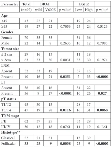
The BRAF mutation status of PTC patients was de-termined by performing mutant allele-specific PCR amplification on archival tissue samples. As shown in Table 1, the BRAFV600E mutation was detected in 43/92 (46.7%) cases. Its presence correlated sig-nificantly with LNM, (p=0.0351), EI, (p <0.0001) and pT status (p=0.0116). Among the 43 BRAFV600E-positive cases, 24 patients (55.8%) had LNM and 27 (62.8%) had EI at presentation.
was found in 48 out of 92 cases (52.2%) (Table 1). EGFR overexpression showed significant association with LNM (p<0.0001), EI (p=0.027) and pT status (p=0.0068). In the group of PTC patients with EGFR
overexpression, LNMs were present in 33/48 (68.8%) cases and EI in 26/48 (54.2%) cases.
Although both BRAFV600E mutation and EGFR overexpression significantly correlated with LNM, EI and pT status, BRAFV600E was more closely asso-ciated with EI, while EGFR overexpression showed stronger correlation with LNM.
High levels of expression of EGFR or the presence of BRAF V600E mutation were not significantly asso-ciated with advanced pTNM stages. It should be noted that in our cohort of PTC patients, almost half of the BRAF mutation positive cases (21/43, 48.8%) and highly positive EGFR cases (24/48, 50%), although having LNM or EI, were young patients, classified as stage I according to the pTNM classification proposed by the American Joint Committee on Cancer [19].
There were no statistically significant associations of BRAFV600E mutation or high EGFR expression with the age or gender of the patients, nor with the tumor size.
Interestingly, not only the presence of BRAFV600E mutation, but also high expression of EGFR showed statistically significant differences regarding the his-tomorphological growth pattern of PTC, both being more frequent in the classical than in the follicular variant of PTC (p=0.0038 and p<0.001, respectively).
Coexistence of BRAFV600E mutation and EGFR overexpression was found in 26 out of 92 patients (28.3%) and was associated with an aggressive phe-notype; among them 73% had LNM presence and Fig. 1. Immunohistochemical examination of the expression of EGFR in PTC tissue. A – weak cytoplasmic staining (short arrow), scored as 1, low expressing group; B – moderate cytoplasmic/membranous (long arrow) staining, scored as 2, high expressing group; C – strong cytoplasmic/membranous staining (thick arrow), scored as 3, high expressing group. Scores 2 and 3 were con-sidered as EGFR overexpression. ABC immune/hematoxylin-diaminobenzidine. Original magnifications: A x10; B and C x20. Scale bars correspond to 50 µm.
Table 1. BRAF mutation status and EGFR expression in correla-tion with clinicopathological parameters of PTC patients.
Parameter Total BRAF EGFR
(n=92) wild V600E p value* Low High p value* Age
<45 43 22 21 19 24
≥45 49 27 22 0.7056 25 24 0.5126
Gender
Female 70 35 35 34 36
Male 22 14 8 0.2635 10 12 0.7985
Tumor size
≤ 2 cm 29 16 13 11 18
> 2cm 63 33 30 0.8031 33 30 0.1974
LNM
Absent 52 33 19 37 15
Present 40 16 24 0.0351 7 33 <0.0001 EI
Absent 56 40 16 34 22
Present 36 9 27 <0.0001 10 26 0.027 pT status
T1/T2 45 30 15 28 17
T3/T4 47 19 28 0.0116 16 31 0.0068
TNM stage
I/II 62 37 25 33 29
III/IV 30 12 18 0.0761 11 19 0.1361
Histotype**
Classical 52 21 31 13 39
Follicular 33 25 9 0.0038 25 9 <0.0001 LNM – lymph node metastasis; EI – extrathyroid invasion;
69% had EI (Table 2). In contrast, in the group of pa-tients with wild-type BRAF and low EGFR expression (n=27), LNM was present in only two patients (7%) and EI was observed in only one patient (4%). Table 2. Coexistence of BRAFV600E mutation and EGFR over-expression (BRAFV600E / EGFR-high) in correlation with clini-copathological parameters of PTC patients.
Parameter BRAF wild/EGFR-low BRAFV600E/EGR-high
(n=27) (n=26) p value*
LNM
Absent 25 7
Present 2 19 <0.0001
EI
Absent 26 8
Present 1 18 <0.0001
pT status
T1/T2 20 7
T3/T4 7 19 0.0009
TNM stage
I/II 21 13
III/IV 6 13 0.0473
Histotype
Classical 3 20 <0.0001
Follicular 24 6
LNM – lymph node metastasis; EI – extrathyroid invasion; pT status and TNM stage according to AJCC [19]; *Fisher’s exact test, p <0.05 statistically significant (bolded)
Overall, among 92 PTC patients, 65 (70.7%) har-bored at least one of the two analyzed molecular alter-ations (either BRAFV600E mutation or EGFR overex-pression). BRAFV600E mutation alone was present in 17 cases, while EGFR overexpression occurred alone in 22 cases. The impact of a single molecular alteration on adverse clinicopathological features (lymph node metastasis and extrathyroid invasion) in comparison with the group of patients with concomitant BRAF V600E mutation and EGFR overexpression is given in Fig. 2. As shown, the presence of BRAFV600E muta-tion alone strongly affected EI, while EGFR overex-pression alone had a greater impact on LNM.
The obtained results were further evaluated by ROC analysis, as shown in Fig. 3. ROC curve analysis confirmed the above results. Thus, patients harboring BRAFV600E mutation and with EGFR overexpres-sion belong to the high-risk PTC group, as they sta-tistically significantly more often have LNM (Fig 3 A: AUC=0.670, p=0.005) and EI (Fig 3 B: AUC=0.679, p=0.004), than patients with none or a single alteration.
In the total cohort, among 40 patients with LNM, 38 (95%) had at least one of the two analyzed altera-tions and among 36 patients with EI, all but one (97%) had either the BRAV600E mutation (27 pa-tients) or EGFR overexpression (8 papa-tients).
DISCUSSION
The main molecular mechanisms underlying thyroid cancer have been clarified after identification of ge-netic alterations in the cellular signaling pathways, including the MAPK pathway and phosphatidylino-sitol-3 kinase (PI3K/AKT) pathway, both coupled to the receptor tyrosine kinases (RTKs) at the cell mem-brane. Molecular alterations in components of these pathways (gene mutations, rearrangements, gene amplifications and copy number gains) constitutively activate signaling cascades, causing uncontrolled cell Fig. 2. Frequency of lymph node metastasis (LNM) and extrathy-roid invasion (EI) in PTC patients divided into four groups with regard to the molecular background. BRAF wt=BRAF wild type, BRAFV600E=BRAF mutation, EGFR L=EGFR low expression, EGFR H=EGFR high expression (overexpression).
division, survival, migration and angiogenesis, lead-ing to malignancy [8,9].
Genetic alterations in the MAPK pathway caused
by BRAFandRAS point mutations and RET/PTC
rearrangements play important roles in the initia-tion and progression of PTC. These mutainitia-tions are found in more than 70% of PTCs and are almost al-ways mutually exclusive. The most common muta-tion, BRAFV600E, results in a constitutively active MAPK pathway and thyroid cancer progression. In many studies the presence of this mutation has been correlated with aggressive tumor characteristics, such as extrathyroid extension, advanced tumor stage at presentation, tumor recurrence, lymph node or dis-tant metastases, resistance to radioiodine and poor prognosis [10,23-25].
Since a subset of PTCs are highly aggressive can-cers that are often poorly responsive to radioiodine therapy, BRAF/MEK/ERK signaling, i.e. the MAPK pathway, has emerged as a promising target in these malignancies. Selective inhibitors of BRAF V600E have been proposed as a novel treatment for patients with thyroid cancer exhibiting this mutation, but have demonstrated a limited therapeutic effect [26]. The reasons for BRAF inhibitor resistance have been in-vestigated and found to be due to reactivation of the MAPK signaling pathway and a secondary activation of the PI3K/Akt pathway [27]. However, it is not clear whether the PI3K/Akt signaling pathway is activated after treatment with BRAF inhibitor or whether this kinase signaling pathway is already upregulated in some thyroid carcinomas.
Both PI3K/Akt and MAPK signaling path-ways are initiated at different RTKs, such as EGFR [15,16,28]. Aberrant EGFR signaling is caused by re-ceptor overexpression, autocrine ligand stimulation or activating mutations. In thyroid cancer, EGFR mu-tations are rare, but EGFR overexpression has been found in anaplastic thyroid carcinoma in association with dedifferentiation, an aggressive phenotype and poor outcome [29]. In PTC, high expression of EGFR has been associated with adverse clinicopathological features, i.e. local aggressiveness [30] and metastatic spreading [31]. Indeed, EGF has been shown to stim-ulate the proliferation, migration and invasiveness of
thyroid malignant cells in vitro through activation of
both MAPK and PI3K/Akt pathways [32].
In the present study, we analyzed BRAF muta-tion status and EGFR expression profiles in a series of clinical PTC tissue samples with the aim of ob-taining insight into the frequency of concomitant BRAFV600E mutation and EGFR overexpression and its relation to the aggressive clinical phenotype of PTC. Among our PTC patients, 65/92 (70.7%) harbored at least one of the two analyzed molecular alterations (either BRAFV600E mutation or EGFR overexpression). The impact of only one of them on adverse clinicopathological features (LNM and EI) was not the same: the presence of the BRAFV600E mutation alone strongly impacted EI, while EGFR overexpression alone had a greater impact on LNM. The coexistence of BRAFV600E mutation and EGFR overexpression that was found in 26 out of 92 patients (28.3%) was associated with a highly aggressive phe-notype: among them 73% had LNM and 69% had EI. Thus, although the BRAF mutation and EGFR overexpression are each implicated in an unfavorable disease course, their coexistence was associated with a particularly aggressive clinical phenotype in PTC patients.
The coexistence of BRAFV600E mutation and EGFR overexpression indicates that multiple sig-naling pathways are simultaneously active in some BRAFV600E-mutated thyroid carcinomas. Further-more, our results obtained in a clinical setting
con-firm the findings of a recent in vitro study in which
both active MAPK and PI3K/Akt pathways were found in BRAFV600E-mutated thyroid carcinoma cells in culture [33].
The cooccurrence of BRAFV600E mutation with EGFR overexpression, which allows growth promot-ing signals through both the MAPK and PI3K/Akt pathways, provides a synergistic effect for disease pro-gression. Therefore, inhibiting only a single prolifer-ation-survival signaling pathway for the treatment of BRAFV600E mutated thyroid carcinoma would not ensure a satisfactory outcome for these patients. Thus, the alternative proliferation pathway should be inhib-ited along with BRAF kinase inhibition for sustain-able growth arrest in these carcinomas.
have shown contradictory clinical responses to BRAF inhibitors. While selective BRAF inhibitors, such as vemurafenib and dabrafenib, have been shown to be effective in BRAF mutant melanoma, where they are Food and Drug Administration (FDA) approved as monotherapy, they have failed to demonstrate single-agent clinical activity in BRAF mutant colorectal car-cinoma patients. This difference in clinical results was shown to be due to the minimal expression of EGFR in melanoma cells in contrast to overexpression of EGFR in colorectal carcinoma cells, as reviewed [27].
In vitro studies have demonstrated that inhibition of EGFR signaling by monoclonal antibody to EGFR (cetuximab) or tyrosine kinase inhibitors (gefitinib or erlotinib) is synergistic with BRAF inhibition in colon carcinoma cells [34]. In addition, a clinical trial using vemurafenib in combination with cetuximab and irinotecan (EGFR inhibitors) displayed a valu-able clinical benefit and a reasonvalu-able toxicity profile in metastatic colorectal carcinoma patients [35].
Advanced BRAF mutated thyroid carcinomas are characterized by the loss of thyroid-specific charac-ters and poor responsiveness to radioiodine therapy
and thus require new therapeutic options [10]. In
vitro studies have demonstrated that thyroid malig-nant cells bearing BRAF mutations are, unlike mela-noma but similarly to colorectal cancer, less sensitive to BRAF inhibitors due to the activation of alterna-tive signaling pathways [33,36]. Furthermore, it has been reported that combined inhibition of BRAF and EGFR signaling in malignant thyroid cells (by vemurafenib and gefitinib) was more effective than vemurafenib or gefitinib single agents, and resulted in the induction of synthetic lethality [37,38]. These data, which are extremely relevant from a clinical perspective, suggest that combined BRAF and EGFR inhibition represents a potential new therapeutic op-tion for advanced BRAF-mutated thyroid carcinoma patients; however, this requires further validation in prospective, randomized clinical trials.
In conclusion, the coexistence of the BRAFV600E mutation and EGFR overexpression identifies a subset of high-risk BRAF-mutated PTC patients who should be considered for combined (anti-BRAF and anti-EG-FR) molecular therapy as it can provide an improved long-term therapeutic outcome.
Funding: This work was supported by the Ministry of Education, Science and Technological Development of the Republic of Serbia, Project No. 173050: “Molecular characterization of thyroid gland tumors: biological and clinical aspects.”
Acknowledgments: The authors are grateful to Dr. J. Anna
Nikolić for the language editing of the manuscript.
Author contributions: IP: conception, selection of patients, in-terpretation of clinical data; SŠ: drafting the article; TID: analysis and interpretation of the results; IĐ: interpretation of data, sta-tistics; JJM: experimental work, interpretation of data; ER: ex-perimental work, interpretation of data; DC: manuscript revision and final approval.
Conflicts of interest disclosure: The authors stated that they have no conflicts of interest regarding the publication of this article.
REFERENCES
1. Ito Y, Nikiforov YE, Schlumberger M, Vigneri R. Increasing incidence of thyroid cancer: controversies explored. Nat Rev Endocrinol. 2013;9:178-84.
2. Davies L, Welch HG. Current thyroid cancer trends in the United States. JAMA Otolaryngol Head Neck Surgery. 2014;140:317-22.
3. Li Volsi VA. Surgical Pathology of the Thyroid. Philadelphia: Saunders; 1990. 422 p.
4. Sipos JA, Mazzaferri EL. Thyroid cancer epidemiology and prognostic variables. Clin Oncol (R Coll Radiol). 2010;22:395-404.
5. Siironen P, Louhimo J, Nordling S, Ristimäki A, Mäenpää H, Haapiainen R, Haglund C. Prognostic factors in papillary thyroid cancer: an evaluation of 601 consecutive patients. Tumour Biol. 2005;26:57-64.
6. Ito Y, Miyauchi A. Prognostic factors and therapeutic strate-gies for differentiated carcinomas of the thyroid. Endocr J. 2009;56:177-92.
7. Choi H, Kasaian K, Melck A, Ong K, Jones SJ, White A, Wiseman SM. Papillary thyroid carcinoma: prognostic sig-nificance of cancer presentation. Am J Surg. 2015;210:298-301.
8. Nikiforov YE. Thyroid carcinoma: molecular pathways and therapeutic targets. Mod Pathol. 2008;21(Suppl 2):S37-43. 9. Xing M. Molecular pathogenesis and mechanisms of thyroid
cancer. Nat Rev Cancer. 2013;13:184-99.
10. Xing M. BRAF mutation in thyroid cancer. Endocr Relat Cancer. 2005;12:245-62.
11. Robinson MJ, Cobb MH. Mitogen-activated protein kinase pathway. Curr Opin Cell Biol. 1997;9:180-6.
12. Kimura ET, Nikiforova MN, Zhu Z, Knauf JA, Nikiforov YE, Fagin JA. High prevalence of BRAF mutations in thyroid cancer: genetic evidence for constitutive activation of the RET/PTC-RAS-BRAF signaling pathway in papillary thyroid carcinoma. Cancer Res. 2003;63:1454-7.
AP, Califano JA, Ringel MD, Zeiger MA, Sidransky D, Lad-enson PW. BRAF mutation predicts a poorer clinical prog-nosis for papillary thyroid cancer. J Clin Endocrinol Metab. 2005;90:6373-9.
14. Mendelsohn J, Baselga J. The EGF receptor family as targets for cancer therapy. Oncogene. 2000;19:6550-65.
15. Yarden Y. The EGFR family and its ligands in human cancer. Signaling mechanisms and therapeutic opportunities. Eur J Cancer. 2001;37(Suppl 4):S3-8.
16. Ullrich A, Schlessinger J. Signal transduction by receptors with tyrosine kinase activity. Cell. 1990;61:203-12. 17. Nedergaard MK, Hedegaard CJ,Poulsen HS. Targeting the
epidermal growth factor receptor in solid tumor malignan-cies. BioDrugs. 2012;26:83-99.
18. DeLellis RA, Lloyd RV, Heitz PU, Eng C. World Health Organization Classification of Tumours. Pathology and Genetics of Tumours of Endocrine Organs. Lyon: IARC Press; 2004. 320 p.
19. Shah JP, Ang K, Baatenburg RJ. Thyroid. In: Edge SB, Byrd DR, Compton CC, Fritz AG, Greene FL, Trotti A, editors. AJCC Cancer Staging Manual. 7th ed. New York: Springer-Verlag; 2010. p. 87-96.
20. Hsu SM, Raine L, Fanger H. Use of avidin-biotin-peroxidase complex (ABC) in immunoperoxidase techniques: a com-parison between ABC and unlabeled antibody (PAP) pro-cedures. J Histochem Cytochem. 1981;29:577-80.
21. Bartolome A, Boskovic S, Paunovic I, Bozic V, Cvejic D. Stomatin-like protein 2 overexpression in papillary thyroid carcinoma is significantly associated with high-risk clinico-pathological parameters and BRAFV600E mutation. APMIS. 2016;124(4):271-7.
22. Sapio MR, Posca D, Troncone G, Pettinato G, Palombini L, Rossi G, Fenzi G, Vitale M. Detection of BRAF mutation in thyroid papillary carcinomas by mutant allele-specific PCR amplification (MASA). Eur J Endocrinol. 2006;154:341-8. 23. Xing M, Alzahrani AS, Carson KA, Shong YK, Kim TY,
Viola D, Elisei R, Bendlová B, Yip L, Mian C, Vianello F, Tuttle RM, Robenshtok E, Fagin JA, Puxeddu E, Fugazzola L, Czarniecka A, Jarzab B, O’Neill CJ, Sywak MS, Lam AK, Riesco-Eizaguirre G, Santisteban P, Nakayama H, Clifton-Bligh R, Tallini G, Holt EH, Sýkorová V. Association between BRAF V600E mutation and recurrence of papillary thyroid cancer. J Clin Oncol. 2015;33:42-50.
24. Riesco-Eizaquirre G, Rodríguez I, De la Vieja A, Costa-magna E, Carrasco N, Nistal M, Santisteban P. The BRAFV600E oncogene induces transforming growth factor beta secretion leading to sodium iodide symporter repres-sion and increased malignancy in thyroid cancer. Cancer Res. 2009;69:8317-25.
25. Yang K, Wang H, Liang Z, Liang J, Li F, Lin Y. BRAFV600E mutation associated with non-radioiodine-avid status in distant metastatic papillary thyroid carcinoma. Clin Nucl Med. 2014;39:675-79.
26. Rahman MA, Salajegheh A, Smith RA, Lam AK. BRAF inhibitor therapy for melanoma, thyroid and colorectal can-cers: development of resistance and future prospects. Curr Cancer Drug Targets. 2014;14:128-43.
27. Lo RS. Receptor tyrosine kinases in cancer escape from BRAF inhibitors. Cell Res. 2012;22:945-7.
28. Katz M, Amit I, Yarden Y. Regulation of MAPKs by growth factors and receptor tyrosine kinases. Biochim Biophys Acta. 2007;1773:1161-76.
29. Landriscina M, Pannone G, Piscazzi A, Toti P, Fabiano A, Tortorella S, Occhini R, Ambrosi A, Bufo P, Cignarelli M. Epidermal growth factor receptor 1 expression is upregu-lated in undifferentiated thyroid carcinomas in humans. Thyroid. 2011;21:1227-34.
30. Fisher KE, Jani JC, Fisher SB, Foulks C, Hill CE, Weber CJ, Cohen C, Sharma J. Epidermal growth factor receptor over-expression is a marker for adverse pathologic features in pap illary thyroid carcinomas. J Surg Res. 2013;185:217-24. 31. Tang C, Yang L, Wang N, Li L, Xu M, Chen GG, Liu Z-M.
High expression of GPER, EGFR and CXCR1 is associated with lymph node metastasis in papillary thyroid carcinoma. Int J Clin Exp Pathol. 2014;7:3213-23.
32. Hoelting T, Siperstein AE, Clark OH, Duh QY. Epidermal growth factor enhances proliferation, migration, and inva-sion of follicular and papillary thyroid cancer in vitro and in vivo. J Clin Endocrinol Metab. 1994;79:401-8.
33. Rahman MA, Salajegheh A, Smith RA, Lam AK. Multiple proliferation-survival signalling pathways are simultane-ously active in BRAF V600E mutated thyroid carcinomas. Exp Mol Pathol. 2015;99:492-7.
34. Sundar R, Hong DS, Kopetz S, Yap TA. Targeting BRAF -Mutant Colorectal Cancer: Progress in Combination Strate-gies. Cancer Discov. 2017;7:558-60.
35. Hong DS, Morris VK, El Osta B, Sorokin AV, Janku F, Fu S, Overman MJ, Piha-Paul S, Subbiah V, Kee B, Tsimberidou AM, Fogelman D, Bellido J, Shureiqi I, Huang H, Atkins J, Tarcic G, Sommer N, Lanman R, Meric-Bernstam F, Kopetz S. Phase IB Study of Vemurafenib in Combination with Irinotecan and Cetuximab in Patients with Metastatic Colorectal Cancer with BRAFV600E Mutation. Cancer Dis-cov. 2016;6:1352-65.
36. Rahman MA, Salajegheh A, Smith RA, Lam AK. Inhibition of BRAF kinase suppresses cellular proliferation, but not enough for complete growth arrest in BRAF V600E mutated papillary and undifferentiated thyroid carcinomas. Endo-crine. 2016;54:129-38.
37. Notarangelo T, Sisinni L, Condelli V, Landriscina M. Dual EGFR and BRAF blockade overcomes resistance to vemu-rafenib in BRAF mutated thyroid carcinoma cells. Cancer Cell Int. 2017;17:86-94.

![Dichlorido{N′ [phenyl(pyridin 2 yl κN)methylidene]isonicotinohydrazide κ2N′,O}zinc](data:image/gif;base64,R0lGODlhAQABAIAAAP///wAAACH5BAEAAAAALAAAAAABAAEAAAICRAEAOw==)