Validation of Multiplex Serology detecting
human herpesviruses 1-5
Nicole BrennerID1,2
*, Alexander J. MentzerID3,4, Julia ButtID1, Angelika Michel1,
Kristina Prager1, Johannes Brozy1, Benedikt Weißbrich5, Allison E. Aiello6, Helen C. S. Meier7, Judy Breuer8, Rachael Almond9, Naomi Allen9,10, Michael Pawlita11,
Tim Waterboer1*
1 Infections and Cancer Epidemiology, German Cancer Research Center, Heidelberg, Germany, 2 Faculty
of Biosciences, Heidelberg University, Heidelberg, Germany, 3 The Wellcome Centre for Human Genetics, University of Oxford, Oxford, United Kingdom, 4 Big Data Institute, Li Ka Shing Centre for Health Information and Discovery, University of Oxford, Oxford, United Kingdom, 5 Institute of Virology and Immunobiology, Julius-Maximilians-Universita¨t Wu¨rzburg, Wu¨rzburg, Germany, 6 Department of Epidemiology, Gillings School of Global Public Health and Carolina Population Center, Chapel Hill, North Carolina, United States of America, 7 Joseph J. Zilber School of Public Health, University of Wisconsin-Milwaukee, Milwaukee, WI, United States of America, 8 Division of Infection and Immunity, University College London, London, United Kingdom, 9 UK Biobank, Stockport, United Kingdom, 10 Nuffield Department of Population Health, University of Oxford, Oxford, United Kingdom, 11 Molecular Diagnostics of Oncogenic Infections, German Cancer Research Center, Heidelberg, Germany
*[email protected](NB);[email protected](TW)
Abstract
Human herpesviruses (HHV) cause a variety of clinically relevant conditions upon primary infection of typically young and immunocompetent hosts. Both primary infection and reacti-vation after latency can lead to more severe disease, such as encephalitis, congenital defects and cancer. Infections with HHV are also associated with cardiovascular and neuro-degenerative disease. However, most of the associations are based on retrospective case-control analyses and well-powered prospective cohort studies are needed for assessing temporality and causality. To enable comprehensive investigations of HHV-related disease etiology in large prospective population-based cohort studies, we developed HHV Multiplex Serology. This methodology represents a low-cost, high-throughput technology that allows simultaneous measurement of specific antibodies against five HHV species: Herpes sim-plex viruses 1 and 2, Varicella zoster virus, Epstein-Barr virus, and Cytomegalovirus. The newly developed HHV species-specific (‘Monoplex’) assays were validated against estab-lished gold-standard reference assays. The specificity and sensitivity of the HHV species-specific Monoplex Serology assays ranged from 92.3% to 100.0% (median 97.4%) and 91.8% to 98.7% (median 96.6%), respectively. Concordance with reference assays was very high with kappa values ranging from 0.86 to 0.96 (median kappa 0.93). Multiplexing the Monoplex Serology assays resulted in no loss of performance and allows simultaneous detection of antibodies against the 5 HHV species in a high-throughput manner. a1111111111 a1111111111 a1111111111 a1111111111 a1111111111 OPEN ACCESS
Citation: Brenner N, Mentzer AJ, Butt J, Michel A,
Prager K, Brozy J, et al. (2018) Validation of Multiplex Serology detecting human herpesviruses 1-5. PLoS ONE 13(12): e0209379.https://doi.org/ 10.1371/journal.pone.0209379
Editor: Edward Gershburg, Rational Vaccines Inc,
UNITED STATES
Received: August 16, 2018
Accepted: December 4, 2018
Published: December 27, 2018
Copyright:©2018 Brenner et al. This is an open access article distributed under the terms of the
Creative Commons Attribution License, which permits unrestricted use, distribution, and reproduction in any medium, provided the original author and source are credited.
Data Availability Statement: All relevant data are
within the manuscript and its Supporting Information files.
Funding: DNHS was funded by the following
Introduction
Nine human herpesvirus (HHV) species have been identified, i.e. Herpes simplex viruses 1 (HSV-1, HHV-1) and 2 (HSV-2, HHV-2), Varicella zoster virus (VZV, HHV-3), Epstein-Barr virus (EBV, HHV-4), Cytomegalovirus (CMV, HHV-5), human herpesviruses 6 A and B (HHV-6 A and B), human herpesvirus 7 (HHV-7) and Kaposi’s sarcoma-associated herpesvi-rus (KSHV, HHV-8). According to genetic and biological properties, such as host cell tropism, theHerpesviridaefamily can be divided into three subfamilies,alphaherpesvirinae(HSV-1, HSV-2 and VZV),betaherpesvirinae(CMV, HHV-6 A/B, and HHV-7), and gammaherpesviri-nae(EBV and KSHV) [1]. Upon primary infection, human herpesviruses cause a variety of dis-eases, such as orolabial herpes and genital herpes (HSV-1, HSV-2), varicella (VZV), infectious mononucleosis (EBV) and exanthema subitum (HHV-6 A/B, HHV-7) [2,3,4,5]. Primary infection may be symptomatic or asymptomatic, depending on the infecting virus and the individual’s condition with respect to age and immunocompetence [2,5,6,7,8,9]. All herpes-viruses establish lifelong persistence in the infected host and undergo a life cycle with both lytic and latent phases [10]. Reactivation of latent infection may be symptomatic, e.g. in case of VZV reactivation as herpes zoster (i.e. shingles) in middle and older aged people [3]. In rare cases, both primary and latent HHV infection can cause severe disease such as HSV-1 enceph-alitis [11,12,13,14], congenital CMV infection [15], chronic active Epstein-Barr virus infec-tion [16], and EBV- or KSHV-related cancer [17]. EBV has been classified as Group I human carcinogen by the International Agency for Research on Cancer (IARC) and is causally associ-ated with Hodgkin’s, Burkitt’s and extranodal NK/T-cell lymphomas as well as nasopharyngeal cancer, while KSHV is classified as carcinogenic for Kaposi’s sarcoma and primary effusion lymphoma [17,18]. In addition, EBV and KSHV have been associated with mucosa-associated lymphoid tissue (MALT) lymphoma and multicentric Castleman’s disease, respectively [17].
As infections by herpesviruses are not reversible and illicit a humoral immune response, spe-cies-specific antibodies in serum can be used to detect whether individuals have been infected with HHV over their lifetime. Multiplex Serology is a fluorescent bead-based high-throughput method for simultaneous detection of antibodies against multiple pathogen-specific antigens in one reaction vessel using a very low sample volume [19]. Infectious disease assays have been estab-lished on this platform for a wide range of pathogens including human papillomaviruses [19], human polyomaviruses [20],Helicobacter pylori[21], hepatitis C virus [22], andStreptococcus gal-lolyticus subspecies galgal-lolyticus[23]. More than 40 antigens enabling simultaneous quantitation of antibodies against a variety of pathogens have been successfully included in Multiplex Serology panels in previous studies [24,25,26]. For efficient inclusion into such Multiplex Serology panels, newly developed pathogen-specific assays ideally consist of as few antigens as possible. Here, we report the development and validation of Multiplex Serology for HSV-1, HSV-2, VZV, EBV and CMV comprising 1 to 4 antigens each. Validation was conducted step-wise. First, each individual HHV specific assay was validated in monoplex format only comprising the HHV specific antigens, further called Monoplex Serology. In a second step, the validated HHV species-specific Monoplex Serology assays were combined and incorporated into a Multiplex Serology panel with various other pathogen-specific assays. Statistical performance of HHV species-specific assays in Multiplex Serology was re-evaluated and found to be maintained in multiplex format.
Material and methods
Antigen selection and cloning
Sixteen sequences from HHV proteins were selected as antigens for HHV species-specific anti-body measurement (Table 1). Most immunogenic and species-specific regions were chosen Centre (BRC). The views expressed are those of the
authors and not necessarily those of the NHS, the NIHR or the Department of Health. The funders had no role in study design, data collection and analysis, decision to publish, or preparation of the manuscript.
Competing interests: The authors have declared
according to the literature. Whenever possible, signal peptides and transmembrane regions were excluded from the recombinantly expressed proteins. For VZV antigens and EBNA-1 peptide, selected inserts (Table 1) were assembled into pGEX4T3tag vector (modified from pGEX4T3) via gene synthesis (eurofins Genomics, Ebersberg, Germany) [27]. These con-structs were also codon-optimized for expression inE.coli(Table 1).
Sequences for all other antigens were derived from genomic DNA (HSV-1, HSV-2, CMV) and cosmid clones (EBV), and used as templates for amplification via PCR. Corresponding primers were designed and subsequently ordered (eurofins Genomics). Sequences for viral antigens were cloned into pGEX4T3tag vector. HHV constructs were amplified in E. coli DH5α, commercially sequenced (eurofins Genomics / GATC Biotech, Konstanz, Germany) and validated against reference sequences from the NCBI nucleotide data base (Table 1).
Antigen expression
Antigens were expressed inE.colistrain BL21 as described previously [27]. Briefly, antigens were expressed as Glutathione S-Transferase (GST) fusion proteins (GST-X-tag) in
Table 1. Characteristics of selected HHV antigens.
HHV antigen(gene) function aa codon optimized1 accession no. Uniprot / DNA template NCBI reference
HSV-1
gD(US6) membrane glycoprotein 26–3402 - HSV-1 type 13 NC_001806
gG(US4) membrane glycoprotein 26–1892
-HSV-2
gD(US6) membrane glycoprotein 26–3392 - untyped genomic DNA3 EU445527.1
mgGunique(US4)4 membrane glycoprotein 344–546 - Z86099.2
VZV
gE(ORF68) envelope glycoprotein, cell-to-cell spread 31–1342 X P09259
-gI(ORF67) envelope glycoprotein, cell-to-cell spread 18–2952 X P09258
-IE63(ORF63) transcriptional regulator 1–278 X P09255
-EBV
EBNA-1 trunc(BKRF1) replication, latent viral infection 325–641 - EBV type 1 cosmid DNA3 NC_007605.1
EBNA-1 pep(BKRF1) 385–420 X P03211
-VCA p18(BFRF3)4 capsid protein 1–175 - M-ABA cosmid DNA3 NC_007605.1
ZEBRA(BZLF1)4 replication activator 1–244 - EBV type 1 cDNA clone3
EA-D(BMRF1) replication (polymerase accessory subunit) 1–403 - M-ABA cosmid DNA3
CMV
pp28(UL99) capsid protein 1–189 - genomic DNA, strain Towne3 FJ616285.1
pp52(UL44) DNA binding protein 1–432
-pp65(UL83) tegument protein 1–560 -pp150N5
(UL32) tegument protein 1–550
-1VZV and EBNA-1 peptide antigens were obtained via gene synthesis and sequence identity was confirmed by manufacturer
2transmembrane domain / signal peptide excluded
3templates kindly provided by Prof. Dr. Henri-Jacques Delecluse (HSV-1, EBV EBNA-1 trunc), Prof. Dr. Bertfried Matz (HSV-2), Prof. Dr. Wolfgang Hammerschmidt
(EBV ZEBRA), Dr. Georg Bornkamm (EBV EA-D, VCA p18), Dr. Stephan Bo¨hm (CMV)
4Upon alignment with NCBI nucleotide squences the following deviations were found. VCA p18: C500A (silent), A512G (silent); EA-D: C54T (silent); mgGunique:
A1048G (T!A), A1116G (silent), deletion nt 1276–1278 (deletion aa 431), deletion nt 1365–1406 (deletion aa 458–471); for all other antigens, sequenced nucleotide sequences match the corresponding NCBI reference sequences.
5N-terminus; C-terminus not additionally informative (unpublished data)
aa: amino acids
pGEX4T3tag vectors encoding N-terminally for GST and C-terminally for the last eleven amino acids of SV40 large T-antigen (tag). pGEX4T3tag carries the ampRgene for positive selection of transformed bacterial colonies in ampicillin-containing medium. In case of VZV, GST-gI-tag, expression was additionally performed in pDB.GST vector (DNASU, Arizona State University Biodesign Institute, DNASU Plasmid Repository, Tempe, Arizona, USA) encoding for a kanamycin resistance instead of ampicillin for selection of co-transformed colo-nies (co-expression of gE and gI). In both vectors, transcription of GST fusion proteins is inducible by IPTG (tac promotor).
After bacterial cell lysis, lysates were cleared and stored at -20˚C in 50% (v/v) glycerol. Quality control of expressed antigens was performed as described previously [22,27] and included protein gel electrophoresis followed by Coomassie staining, western blot staining for both C- and N-terminal tags to ensure expression of full length antigens and GST capture ELISA for relative quantitation of the fusion proteins [27].
Reference panels and reference assays
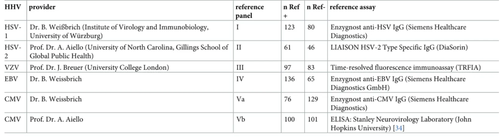
Human reference sera and details on the gold-standard reference assays are shown inTable 2. The numeration of the reference panels (RP) corresponds to the HHV numbering in Roman numerals (I-V). For CMV, two reference panels tested with different reference assays were available (denoted as reference panels Va and Vb). The reference sera were obtained from the Institute of Virology and Immunobiology of the University of Wu¨rzburg (HSV, EBV, CMV), from the Detroit Neighborhood Health Study (DNHS: HSV-2, CMV) and the Zoster Associ-ated Pain (ZAP) and Shingles UK (SUK) studies (VZV). Sera were sent to the DKFZ on dry ice and were stored at -20˚C until testing. The serum collections are described in detail below.
The reference serum panel obtained from the University of Wu¨rzburg was composed of two subgroups. Group 1 consisted of serum samples (n = 197; median age 16.9 years (range 0.3–84.6 years); 53% male) received by the viral diagnostic laboratory between 2007 and 2014 for analysis of herpesvirus IgG antibodies. This was part of routine work-up in patients before solid organ transplantation and in patients with malignant diseases before chemotherapy and potentially stem cell transplantation. Group 2 consisted of serum samples (n = 22; median age 1.2 years (range 0.4–25.6 years); 64% male) that were found to be negative for HHV6 IgG bodies in routine diagnostic testing. With few exceptions, all samples were tested for IgG anti-bodies against HSV, EBV, and CMV at the viral diagnostic laboratory. The sera were stored at
Table 2. Characteristics of reference serum panels.
HHV provider reference
panel
n Ref +
n Ref- reference assay
HSV-1
Dr. B. Weißbrich (Institute of Virology and Immunobiology, University of Wu¨rzburg)
I 123 80 Enzygnost anti-HSV IgG (Siemens Healthcare
Diagnostics)
HSV-2
Prof. Dr. A. Aiello (University of North Carolina, Gillings School of Global Public Health)
II 61 46 LIAISON HSV-2 Type Specific IgG (DiaSorin)
VZV Prof. Dr. J. Breuer (University College London) III 97 83 Time-resolved fluorescence immunoassay (TRFIA)
EBV Dr. B. Weissbrich IV 136 65 Enzygnost anti-EBV IgG (Siemens Healthcare
Diagnostics GmbH)
CMV Dr. B. Weissbrich Va 76 129 Enzygnost anti-CMV IgG (Siemens Healthcare
Diagnostics)
CMV Prof. Dr. A. Aiello Vb 100 101 ELISA: Stanley Neurovirology Laboratory (John
Hopkins University) [34]
n Ref+: number of reference assay positives n Ref-: number of reference assay negatives
-20˚C before shipping to the DKFZ. The use of human serum samples in this study was approved by the ethics committee of the medical faculty at the University of Wu¨rzburg. The need for consent was waived by the ethics committee.The DNHS is a longitudinal study of eco-logic factors that may influence mental and physical health in an urban setting. DNHS partici-pants are representative of Detroit, Michigan in terms of age, gender, race, income and educational attainment. Participants provided written informed consent for participation and study was approved by the University of Michigan and the University of North Carolina Insti-tutional Review Board (IRB #13–3999) [28]. The reference samples from DNHS were obtained from Wave 1 participants. Frozen serum samples stored at -70˚C were shipped on dry ice to the Stanley Neurovirology Laboratory of the John Hopkins University School of Medicine in Baltimore, Maryland to be tested for serum IgG antibodies to CMV and HSV-2 [29]. For each sample, the antibody levels were expressed as ratio of the optical density of a test sample to that of a standard sample assayed in each test run. Individuals were categorized as seronegative if their ratio value was<1.0 and seropositive if�1.0.
The VZV reference sera were collected from multiple sources. In the ZAP and SUK study, subjects presenting with acute zoster were followed up for 12 (ZAP) and 6 months (SUK) with serum samples obtained at four time points [30,31,32]. Additional sera from VZV positive asymptomatic blood donors were also included in the reference panel [33]. All samples were obtained under the UCLP DNA biobank ethical framework (REC reference: 17/LO/1530). Participants provided written informed consent.
Monoplex and Multiplex Serology
The reference sera were analyzed for antibodies against selected HHV antigens (Table 1) by species-specific Monoplex and Multiplex Serology, as described previously [19]. Briefly, HHV GST-tag fusion proteins were loaded onto glutathione casein-coated fluorescence-labelled polystyrene beads (COOH-beads xMAP Technology Microspheres, Luminex Corp. Austin, Texas, USA) byin situaffinity purification from lysate. Up to 100 bead sets are distinguishable by the Luminex flow cytometer via different ratios of two fluorescent dyes within the polysty-rene microspheres. Loading each antigen onto a specific bead set enables simultaneous mea-surement of antibodies against different antigens within one reaction vessel.
Detection of bound primary antibodies from serum took place with a biotinlyated goat-α -human IgM/IgG/IgA secondary antibody (1:1000, #109-065-064, Jackson Immunoresearch, West Grove, PA, USA) and subsequent incubation with streptavidin-R-phycoerythrin (1:750, PE-Streptavidin Conjugate, MOSS Inc., Pasadena, CA, USA) as reporter dye. Median Fluores-cence Intensities (MFI) from at least 100 detected beads per bead set (e.g. antigen) were calcu-lated for each serum. Monoplex Serology was conducted for each HHV species-specific assay in an individual experiment only comprising the species-specific antigens and GST-tag antigen for background subtraction in dilutions 1:100 and 1:1000. Optimal serum dilution was 1:1000 with the exception of VZV (1:100). In addition, performance of HHV Monoplex Serology assays were assessed in multiplex format by combining them with various pathogen-specific Monoplex Serology assays (e.g. human herpes viruses 6–8, human polyomaviruses, human papillomaviruses, human hepatitis B and C viruses) into a Multiplex Serology panel.
Statistical analysis
cut-off was determined to result in specificity and sensitivity of at least 85% analogous to Receiver Operating Characteristics analysis. This was achieved by gradually raising a working cut-off from a minimum of 30 MFI (dilution 1:1000) or 50 MFI (dilution 1:100) to optimize specificity and sensitivity. The optimum cut-off was determined to favor specificity, unless a further increase of the cut-off resulted in a disproportional loss in sensitivity. Thus, agreement with the reference assay was maximized. When multiple pathogen-specific antigens were included in the assay, sero-positivity against the respective pathogen (denoted as overall pathogen serosero-positivity) was addi-tionally determined by systematic investigations of antigen combinations. Antigen-specific cut-offs were adapted to optimize agreement with the reference assay if necessary as described above. In addition to sensitivity and specificty, Cohen’skappa (k)statistics to define agreement with the reference assay were calculated and evaluated as follows: 0.01<k<0.20: slight agreement, 0.21<k<0.40: fair agreement, 0.41<k<0.60: moderate agreement, 0.61<k<0.80 substantial agreement and 0.81<k<0.99: almost perfect agreement [35]. Sensitivity, specificity andkappa sta-tistics including 95% Confidence intervals (CI) were calculated using SAS 9.4.
Comparison of the Monoplex and Multiplex Serology performance on the corresponding reference serum panels was conducted by calculating Intraclass Correlation Coefficients (ICCs) using R 3.5.0 package ‘psych’ [36]. ICC(3,1) plus corresponding 95% CI are reported. ICCs were evaluated as follows: 0.01<ICC<0.49: poor reliability, 0.50<ICC<0.74: moderate reliability, 0.75<ICC<0.89: good reliability, 0.90<ICC<1.00: excellent reliability [37].
Results
Antigen development
Based on reported immunogenicity, antigen coverage by the reference assays and sequence homology, 2-4 antigens were selected for development and validation of HHV species-specific Monoplex Serology assays (Table 1). Antigens were expressed as recombinant GST-fusion pro-teins as described previously [27]. To ensure correct protein sequence, parental plasmids were sequenced. For most antigens, perfect agreement with the reference sequence (NCBI nucleo-tide database) was confirmed (Table 1). Only HSV-2 antigen mgGunique showed non-silent nucleotide sequence variations compared to strain HG52 resulting in one amino acid change, one single amino acid deletion and one 14 amino acid deletion.
Comparison of HHV species-specific Monoplex Serology assays with
reference serostatus
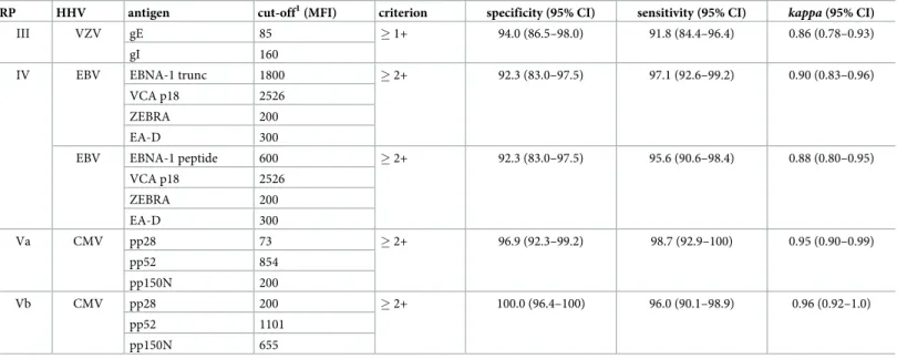
Six reference serum panels (RP I-IV, Va, Vb;Table 2) were analyzed by the corresponding HHV species-specific Monoplex Serology assay. For CMV, two reference panels using different gold-standard assays were available and tested (Va, Vb). Quantitative antibody reactivities (MFI) for each HHV antigen were compared against the corresponding reference serostatus (Fig 1). Where multiple species-specific antigens were included, overall seropositivity in Monoplex Serology was calculated by a combination of the included antigens. Cut-offs were determined by optimizing sensitivity and specificity. The performance characteristics (i.e. specificity, sensitivity andkappa
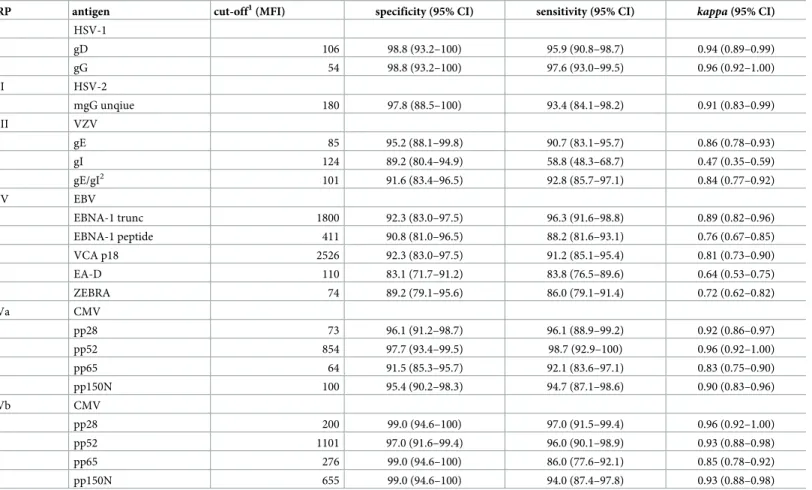
statistics) for the Monoplex Serology assays for HHV 1–5 compared with the gold-standard refer-ence assays are shown for each antigen (Table 3) and overall seropositivity (Table 4).
HSV species-specific Monoplex Serology validation
[38]. The sequence identity of the regions common to both HSV species is approximately 50% [38]. For HSV-2, gG is cleaved into a secreted (sgG) and a membrane anchored part (mgG) [38] of which the region unique to 2 (mgGu) was expressed and used as antigen in HSV-2 Monoplex Serology. For HSV-1 and -HSV-2 gD, sequence identity is approximately 80%. Thus, antibody responses against gD antigens are of high interest for assessing cross-reactivity between both assays.
HSV-1 and -2 Monoplex Serology assays were validated against a HSV species-unspecific (Enzygnost anti-HSV IgG), and a HSV-2 specific (LIAISON HSV-2 IgG) reference assay in RP I and II, respectively. No HSV-1 species-specific reference panel was available. Thus, an indi-rect approach using HSV-2 Monoplex Serology validation was pursued for HSV-1 Monoplex Serology validation.
The measured antibody reactivities against HSV-1 and -2 antigens based on RP I are shown inFig 1-I. Both HSV-1 antigens gD and gG discriminate well between reference assay positives and negatives, resulting in only one false-positive and 5 and 3 false-negatives, respectively. The resulting specificity is 98.8% for both HSV-1 antigens, while the sensitivity is 95.9% for gD and 97.6% for gG (Table 3). Based on RP I, HSV-2 gD also showed good capacity to distinguish between reference assay negatives and positives. However, in comparison to HSV-1 gD, a slightly larger overlap in measured antibody reactivities between reference assay negatives and positives was observed between approximately 10 and 100 MFI. HSV-2 mgGu showed very lit-tle seroreactivity in RP I, with only six sera showing antibody responses>100 MFI. Thus, no attempt was made to determine meaningful cut-offs for HSV-2 antigens in RP I.
HSV-2 Monoplex Serology was validated against a HSV-2-specific reference assay based on RP II (Fig 1-II). With a cut-off of 180 MFI, HSV-2 mgGu discriminated very well between ref-erence assay positives and negatives resulting in only one false-positive and 4 false-negatives, yielding a specificity and sensitivity of 97.8% and 93.4%, respectively.Kappastatistics showed almost perfect agreement with the reference assay (k= 0.91) (Table 3). For reference assay pos-itives, similar antibody reactivities were observed for HSV-2 gD and mgGu (approx. 200 to 10,000 MFI). However, high HSV-2 gD antibody reactivities were also observed for a substan-tial number of reference assay negative samples that were also negative for HSV-2 mgGu. Thus, no cut-off for HSV-2 gD was determined in RP II.
HSV-2 Monoplex Serology was successfully validated based on antigen mgGu. Based on the very low prevalence of HSV-2 mgGu, RP I contained very few (<5%) HSV-2 seropositives. Thus, the antibody reactivities measured with 2 gD most likely represent cross-reactivity with HSV-1 gD antibodies. This was confirmed by a high correlation of antibody reactivities between the homologous HSV-1 and -2 gD proteins observed in both RP I and RP II (S1 Fig). Thus, the gD antigens could not be validated for measurement of species-specific HSV antibodies but can be applied for detection of general (species-unspecific) HSV infection. Additionally, based on the small number of HSV-2 seropositives in RP I, this setting allowed us to indirectly validate HSV-1 Monoplex Serology based on antigen gG (RP I) although no HSV-1 species-specific reference panel was available. This is further supported by the low correlation of gG and mgGu (r = 0.13,S2 Fig).
VZV Monoplex Serology validation
The VZV Monoplex Serology antigen panel included glycoproteins E (gE) and I (gI) and immediate early protein 63 (IE63) and was validated against the time resolved fluorescence
Fig 1. Comparison of quantitative antibody measurements (MFI) with reference serostatus in HHV species-specific Monoplex Serology. I-Vb indicate
corresponding reference panels. gE/gI: co-loading of antigens gE and gI onto one bead set red lines: optimized cut-offs for single antigen performance; cut-offs were determined by optimizing specificity and sensitivity. MFI: Median Fluorescence Intensity.
immunoassay (TRFIA) [31]. Antigen gE differentiated well between reference assay negatives and positives, with only 4 false-positives and 9 false-negatives (Fig 1-III); specificity was 95.2% and sensitivity was 90.7% (Table 3).Kappastatistics showed almost perfect agreement with the reference assay (k= 0.86). gI Monoplex Serology resulted in a substantial number of false-posi-tives and even more false-negafalse-posi-tives compared to the reference assay (Fig 1-III). Using a cut-off to yield the minimum desired specificity of 85.0%, calculated specificity was 89.2% and sensi-tivity was 58.8% (Table 3). Antigen IE63 showed no capacity to discriminate between reference assay negative and positive sera (Fig 1-III). Thus, IE63 is not informative for VZV Monoplex Serology and no cut-off was determined.
VZV proteins gE and gI form hetero-dimers in infected cells [39]. Thus, different approaches were undertaken to assess whether individual antigen performance of gE can be improved by a combination with gI. Determination of overall VZV serostatus by seropositivity to gE and / or gI resulted in very similar statistics compared to gE alone (Tables3and4). To simulate hetero-dimerisation of gE and gI, two additional strategies were pursued to enable detection of VZV antibodies directed against epitopes jointly formed by gE and gI. First,
Table 3. Single antigen performance compared to corresponding reference panels in Monoplex Serology.
RP antigen cut-off1(MFI) specificity (95% CI) sensitivity (95% CI) kappa(95% CI)
I HSV-1
gD 106 98.8 (93.2–100) 95.9 (90.8–98.7) 0.94 (0.89–0.99)
gG 54 98.8 (93.2–100) 97.6 (93.0–99.5) 0.96 (0.92–1.00)
II HSV-2
mgG unqiue 180 97.8 (88.5–100) 93.4 (84.1–98.2) 0.91 (0.83–0.99)
III VZV
gE 85 95.2 (88.1–99.8) 90.7 (83.1–95.7) 0.86 (0.78–0.93)
gI 124 89.2 (80.4–94.9) 58.8 (48.3–68.7) 0.47 (0.35–0.59)
gE/gI2 101 91.6 (83.4–96.5) 92.8 (85.7–97.1) 0.84 (0.77–0.92)
IV EBV
EBNA-1 trunc 1800 92.3 (83.0–97.5) 96.3 (91.6–98.8) 0.89 (0.82–0.96)
EBNA-1 peptide 411 90.8 (81.0–96.5) 88.2 (81.6–93.1) 0.76 (0.67–0.85)
VCA p18 2526 92.3 (83.0–97.5) 91.2 (85.1–95.4) 0.81 (0.73–0.90)
EA-D 110 83.1 (71.7–91.2) 83.8 (76.5–89.6) 0.64 (0.53–0.75)
ZEBRA 74 89.2 (79.1–95.6) 86.0 (79.1–91.4) 0.72 (0.62–0.82)
Va CMV
pp28 73 96.1 (91.2–98.7) 96.1 (88.9–99.2) 0.92 (0.86–0.97)
pp52 854 97.7 (93.4–99.5) 98.7 (92.9–100) 0.96 (0.92–1.00)
pp65 64 91.5 (85.3–95.7) 92.1 (83.6–97.1) 0.83 (0.75–0.90)
pp150N 100 95.4 (90.2–98.3) 94.7 (87.1–98.6) 0.90 (0.83–0.96)
Vb CMV
pp28 200 99.0 (94.6–100) 97.0 (91.5–99.4) 0.96 (0.92–1.00)
pp52 1101 97.0 (91.6–99.4) 96.0 (90.1–98.9) 0.93 (0.88–0.98)
pp65 276 99.0 (94.6–100) 86.0 (77.6–92.1) 0.85 (0.78–0.92)
pp150N 655 99.0 (94.6–100) 94.0 (87.4–97.8) 0.93 (0.88–0.98)
1cut-offs determined by optimization of specificity and sensitivity
2gE/gI: co-loading of both antigens onto one bead set to simulate heterodimerisation
CI: confidence interval RP: reference panel
MFI: median fluorescence intensity
bacterial lysates containing VZV antigens gE and gI were mixed and simultaneously loaded onto the same bead set. Co-loading of gE and gI showed very similar detection characteristics as gE individually (Fig 1-IIIandTable 3). However, antibody reactivities for both reference assay negatives and positives were slightly increased. In a second approach, antigens gE and gI were co-expressed inE.coliyielding almost identical data compared to co-loading, or gE alone (S3 Fig).
Thus, VZV Monoplex Serology performance is largely driven by antigen gE. In RP III, no added benefit could be achieved by the various approaches to include gI.
EBV Monoplex Serology validation
EBV Monoplex Serology comprises a panel of four EBV proteins. In case of EBV nuclear anti-gen 1 (EBNA-1), two fragments of differing sizes were expressed, EBNA-1 truncated (EBNA-1 trunc) and EBNA-1 peptide (EBNA-1 pep) (Table 1). In addition, viral capsid antigen p18 (VCA p18), Z-Epstein-Barr virus replication activator (ZEBRA) and early antigen-diffuse (EA-D) were included in the EBV antigen panel. EBV Monoplex Serology was validated against the Enzygnost anti-EBV IgG assay. Among the reference assay seropositive sera (n = 136), 131 (96.3%) were seropositive for EBNA-1 trunc, 124 (91.2%) for VCA p18, 117 (86.0%) for EBNA-1 peptide, 110 (80.9%) for ZEBRA and 103 (75.7%) for EA-D (according to cut-offs shown inTable 4). Antigen-specific concordance with the reference assay is generally good (Fig 1-IV). However, for all antigens between 5 and 22 false-positives and/or false-nega-tives were observed. Specificity for individual antigens ranged between 83.1% for EA-D and 92.3% for both EBNA-1 trunc and VCA p18 (Table 3). Sensitivity is very similar, and between
Table 4. Overall HHV species-specific performance compared to corresponding reference panels in Monoplex Serology.
RP HHV antigen cut-off1(MFI) criterion specificity (95% CI) sensitivity (95% CI) kappa(95% CI)
III VZV gE 85 �1+ 94.0 (86.5–98.0) 91.8 (84.4–96.4) 0.86 (0.78–0.93)
gI 160
IV EBV EBNA-1 trunc 1800 �2+ 92.3 (83.0–97.5) 97.1 (92.6–99.2) 0.90 (0.83–0.96)
VCA p18 2526
ZEBRA 200
EA-D 300
EBV EBNA-1 peptide 600 �2+ 92.3 (83.0–97.5) 95.6 (90.6–98.4) 0.88 (0.80–0.95)
VCA p18 2526
ZEBRA 200
EA-D 300
Va CMV pp28 73 �2+ 96.9 (92.3–99.2) 98.7 (92.9–100) 0.95 (0.90–0.99)
pp52 854
pp150N 200
Vb CMV pp28 200 �2+ 100.0 (96.4–100) 96.0 (90.1–98.9) 0.96 (0.92–1.0)
pp52 1101
pp150N 655
�1+: seropositive against at least one antigen
�2+: seropositive against at least two antigens CI: confidence interval
RP: reference panel
MFI: median fluorescence intensity
1
cut-offs determined by optimizing specificity and sensitivity of overall HHV species seropositivity
83.8% for EA-D and 96.3% for EBNA-1 trunc.Kappastatistics showed substantial agreements for EBNA-1 pep, ZEBRA and EA-D and almost perfect agreement for VCA p18 and EBNA-1 trunc (k= 0.64 tok= 0.89).
Only 2 of the reference assay seropositive sera did not react with any or only one of the EBV antigens; 86 (63.2%) were seropositive against all four EBV antigens (EBNA-1 trunc, VCA p18, ZEBRA, EA-D), 29 (21.3%) against 3 and 17 (12.5%) against 2. Nine reference assay negative sera showed antibody responses against at least one antigen; in 5 of these, antibodies against multiple EBV antigens were detected. Determining overall EBV seropositivity by a combination of the antigens showed optimum specificity (92.3%) and sensitivity (97.1%) by seropositivity against at least 2 out of 4 EBV proteins (Table 4). Using this algorithm, almost perfect agreement between both EBV assays (k= 0.90) was reached. Inclusion of either EBNA-1 trunc or peptide in the algorithm showed to be equally specific, but slightly more sensitive when including EBNA-1 trunc (Table 4).
CMV Monoplex Serology validation
CMV Monoplex Serology is based on four proteins: pp52, pp28, pp65 and pp150 N-terminus (pp150N) and was validated against two different reference assays, an anti-CMV IgG ELISA based on commercially available virion proteins (RP Va) and the Enzygnost anti-CMV IgG (RP Vb). Antibody detection against individual CMV antigens pp28, pp52 and pp150N showed high concordance with both reference assays detecting a maximum of 6 false-positives or false-negatives (Fig 1-Va/Vb). Specificity ranged between 95.4% and 99.0% and sensitivity ranged between 94.0% and 98.7% (Table 3).Kappastatistics indicated almost perfect agree-ment with both reference assays (kappa0.90 to 0.96). Assay performance for CMV
antigen pp65 was slightly poorer in comparison with the other antigens showing a higher over-lap of measured antibody reactivities for reference assay positive and negative samples in both reference panels (Fig 1-Va/Vb). However,kappastatistics still indicated almost perfect agree-ment with both reference assays (Va,k= 0.83; Vb,k= 0.85) (Table 3).
Overall CMV serostatus was determined by seropositivity against at least two out of three CMV antigens (pp28, pp52, pp150N) as inclusion of pp65 did not additionally improve assay performance and is therefore dispensable. Specificity was 96.9% and 100.0% for the two refer-ence panels, while sensitivity was 98.7% and 96.0%, respectively (Table 4).
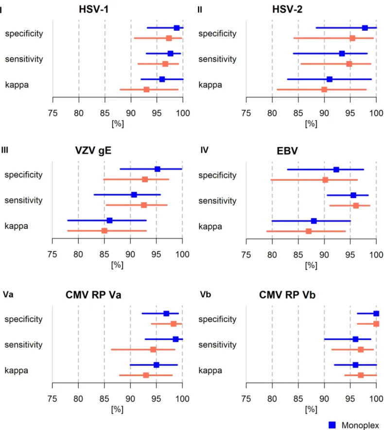
Comparison of performance of HHV Monoplex and Multiplex Serology
The reference sera were analyzed both in monoplex (i.e. one pathogen) and in multiplex (i.e. multiple pathogens) format in order to compare assay performance. The performance of HHV Multiplex Serology based on sensitivity, specificity andkappastatistics was evaluated in com-parison to the corresponding species-specific Monoplex Serology results (Fig 2). While some of the individual species-specific HHV assays showed slight differences in sensitivity and speci-ficity between the monoplex and multiplex format, the overall statistical performance of the species-specific assays in Multiplex Serology did not change. Sensitivity and specificity for overall seropositivity for all HHV species exceeded 90% in both monoplex and multiplex for-mat. A high concordance with the corresponding reference assays was maintained (k�0.85). A direct comparison of Monoplex versus Multiplex Serology performance was conducted using ICCs and showed good to excellent reliability (ICC: 0.82–0.99).
Discussion
Fig 2. Comparison of statistical performance of HHV species-specific assays in monoplex (blue) and multiplex (orange) format. Performance is shown for
overall seropositivity (EBV, CMV) and single antigens for HSV-1 (gG), HSV-2 (mgGu) and VZV (gE). I-Vb indicate corresponding reference panels. Cohen’skappa
The species-specific Monoplex Serology assays were validated using reference sera analyzed with gold-standard reference assays. For each species-specific assay, only a small number of false-positives and false-negatives was observed during validation. This resulted in a median specificity and sensitivity of 97.4% and 96.6%, respectively. The newly developed assays were found to be highly concordant with the established gold-standard reference assays (median
kappa0.93). Robust statistical assay characteristics of HHV species-specific serological assays were confirmed in a multiplex setting comprising a larger antigen panel including multiple additional pathogen-specific Monoplex Serology assays (Fig 2).
A minimum number of antigens per species-specific assay was pursued to facilitate the incorporation into larger Multiplex Serology panels comprising additional infectious disease antigens. By selecting a total of 10 antigens for the first 5 HHVs, this has been [e.g. [40,41,42,
43] and will be feasible in future seroepidemiological studies.
HSV glycoproteins gG are the ideal candidate antigens in serological assays aiming at HSV species-specificity due to reported low sequence identity [38], and low observed correlation of antibody responses in Multiplex Serology (S2 Fig). Thus, the evaluated HSV gG antigens most likely allow to measure species-specific antibody reactivities, while HSV-1 and 2 antigens gD detect non-species-specific HSV infection confirming previous reports [38,44]. HSV-1 Mono-plex Serology was indirectly validated against the HSV non-species-specific Enzygnost anti-HSV IgG assay. The assay was reported to be based on crude lysate of anti-HSV-1 infected cells [45]. Although HSV-1 Monoplex Serology is based on one antigen (gG) only, assay concor-dance is almost perfect (k= 0.96). This implies that missing glycosylation due to recombinant antigen expression inE.colidoes not seem to impair the immunogenicity of the epitopes.
HSV-2 Monoplex Serology was successfully validated against the LIAISON HSV-2 IgG chemi-luminescent immunoassay based on recombinant HSV-2 gG antigen [46]. As both the HSV-2 Monoplex Serology and the reference assay are based on the same HSV-2 protein, high concor-dance (k= 0.91) is not surprising. Only five discordantly classified samples were observed, poten-tially due to different expression systems or diverging antigen sequences. Sequencing of HSV-2 antigen mgGu revealed 3 deviations from HSV-2 strain HG52 on the protein sequence level: an amino acid exchange from Threonine to Alanine, a single amino acid deletion and a 14-amino acid deletion. This might indicate mutations inserted during PCR, errors during sequencing, or deviations in the parental DNA. In fact, our recombinant antigen is based on an untyped genomic DNA potentially representing a clinical isolate, or a different strain. Despite the detected potential sequence deviations, high concordance with the reference assay was reached. Thus, these devia-tions most likely do not result in conformational changes within immunogenic epitopes of HSV-2.
In reference panel I, 4 sera with HSV-2 mgGu antibody responses>180 MFI were detected. These were also seropositive against HSV-1 gG. Based on the low correlation between HSV-2 mgGu and HSV-1 gG (S2 Fig), we assume that these 4 seropositive individuals were co-infected by HSV-1 and HSV-2. This is further supported by the high HSV-1 preva-lence in the general population and their shared route of exposure [6,7]. However, these 4 individuals could also represent HSV-2 infected individuals with antibodies against the non-unique epitopes of HSV-2 gG cross-reacting with HSV-1 gG.
EBV Monoplex Serology was successfully validated against the Enzygnost anti-EBV IgG assay showing high concordance for overall EBV seropositivity (k= 0.88 andk= 0.90, depend-ing on the EBNA-1 antigen). The reference assay was reported to be based on a mixture of
panel, ICCs were calculated showing good to excellent reliability (ICCHSV1: 0.91 (95% CI 0.88–0.93), ICCHSV2: 0.93 (95% CI 0.89–0.95), ICCVZV: 0.93 (95% CI 0.91–
0.95), ICCEBV: 0.87 (95% CI 0.84–0.90), ICCCMV RPVa: 0.82 (95% CI 0.77–0.86), ICCCMV RPVb: 0.99 (95%CI 0.99–1.00)). kappa: Cohen’s kappa. ICC: Intraclass
correlation coefficient. CI: confidence interval.
EBV VCA, EBNA and EA antigens. The exact composition is unknown to the authors of this paper. However, the usage of probably overlapping antigen panels in EBV Monoplex Serology and the reference assay likely explains high concordance.
EBV Monoplex Serology uses antigens expressed during different stages of the EBV life cycle. While EBNA-1 is expressed during latent infection, ZEBRA, EA-D and VCA p18 are expressed during the lytic stage [17,47]. Detection of antibodies to EBV VCA and EBNA-1 were reported to distinguish EBV infection history; IgG and IgM antibodies against EBV VCA proteins mark acute infection, while presence of only IgG antibodies against VCA and EBNA-1 serologically defines past infection or late primary infection. This pattern in combination with IgM antibodies against VCA proteins also marks reactivation [47]. As a viral capsid pro-tein, VCA p18 has expectedly been presented to the immune system of all EBV infected indi-viduals upon primary infection. Thus, detection of 124 (91.2%) reference seropositive sera with VCAp18 antibody reactivities above the cut-off is consistent with its potential role as marker for acute and past infection. Among the reference assay seropositive sera, 109 (80.1%) and 121 (88.8%) sera were seropositive against VCA p18 and EBNA-1 pep or EBNA-1 trunc, respectively. Thus, these represent most likely past primary and latent infection. However, of the 124 VCA p18 seropositive sera, 110 were seropositive for either EA-D or ZEBRA, or both. High antibody reactivities against these antigens might represent markers of previous reactiva-tion as they are expressed early in the lytic stage and EA-D IgG antibodies were reported to be detectable only temporarily after the lytic phase of EBV infection [47]. Four out of 5 detected false-positive sera were seropositive against VCA p18 plus at least two other EBV antigens. This raises the question whether these sera are likely to be true EBV positives, and whether EBV Monoplex Serology might be slightly more sensitive than the reference assay.
Recently, Coghillet al. reported a risk stratification signature for nasopharyngeal carcinoma in Taiwan based on EBV IgG and IgA antibodies [48]. The possible detection of antibody pat-terns specific for past versus reactivated EBV infection, in combination with the findings by Coghillet al. imply the potential for future disease-specific EBV antibody patterns in Multiplex Serology based on separate measurements of IgG, IgM and IgA.
EBV elicits high antibody responses in infected individuals. For at least semi-quantitative measurement of antibodies within the dynamic range of the assay, serum dilutions must align with the expected antibody titers elicited by the pathogen. Thus, antibody measurements at high serum dilutions are recommended for EBV. Validation was performed at dilution 1:1000, i.e. the standard dilution of Multiplex Serology for the simultaneous measurement of antibodies against many pathogens. Inclusion of two EBNA-1 antigens of differential length in the algo-rithm defining overall EBV seropositivity did result in slightly higher sensitivity for EBNA-1 trunc. However, antibody measurements against the peptide show a wider dynamic range (Fig 1-IV). Thus, antigen EBNA-1 peptide instead of EBNA-1 trunc enables more quantitative anti-body measurements at dilution 1:1000 with only marginally reduced assay sensitivity.
VZV Monoplex Serology based on antigens gE and gI was validated against the TRFIA developed by McDonaldet al. [49]. The TRFIA is based on a sucrose density gradient centrifu-gation-purified extract of human embryo lung-cultured VZV strain Ellen detecting anti-VZV IgG [31]. Observed false-negative sera might not react with the antigens gE or gI, but another antigen of the VZV proteome present in the TRFIA. Sequences for expressed gE and gI anti-gens (strain Dumas) were compared with strain Ellen (reference assay) and were found to be highly concordant (min. 99%).
CMV Monoplex Serology performed very well in comparison with two independent refer-ence assays, the Enzygnost anti-CMV IgG and the CMV ELISA developed by the Stanley Neu-rovirology Laboratory [34]. The Enzygnost assay was reported to be based on inactivated antigens from CMV infected human fibroblasts, while the second CMV reference assay was reported to use purified CMV antigen [34,50,51,52]. The exact antigen compositions of both CMV reference assays are thus unknown to the authors of this paper. Despite potentially dif-ferent antigen composition used in the reference assays, very good agreement of CMV Mono-plex Serology with the two independent reference assays was observed. Additionally, the reported validation of the Enzygnost assay against a CMV IgG assay on the Abbott Architect platform [53] confirms robust and efficient detection of CMV infection with CMV Monoplex Serology.
CMV Monoplex Serology was applied to two different reference serum panels. Depending on the reference panel and corresponding reference assay, different cut-offs were found to optimize statistical characteristics per antigen. Thus, we conclude that cut-offs might not be directly transferable between studies. This might have multiple potential reasons such as differ-ences in the underlying study population, differential blood collection conditions and storage of serum specimens before testing, as well as potential assay drift and reagent performance over time. This can be accounted for by standard quality control and normalization proce-dures between studies. In addition, differential underlying reference assay characteristics can-not be excluded by only pair-wise comparison of CMV Monoplex Serology with each reference assay, and might have influenced the selected optimum cut-offs.
Similarly to the above described assays, antigens for species-specific Monoplex Serology assays were developed for human herpesviruses HHV-6A & B, HHV-7 and KSHV. To our knowledge, there are no universally applicable serological gold standard assays with suffi-ciently acceptable performance characteristics for clinical use available for these HHV species. Thus, the developed Monoplex Serology assays could not be validated so far.
Multiplex Serology uses a secondary antibody directed against human IgG, IgM and IgA antibodies. However, this set-up does not allow the discrimination between acute infection during which IgM antibodies are detectable and past infection marked by IgG antibodies. To allow for specific detection of IgM antibodies, the HHV Monoplex Serology assays could be adapted and re-validated using a secondary antibody detecting human IgM, and correspond-ing reference panels.
Herpesviruses utilize various mechanisms to interact with their hosts that may lead to can-cer initiation and progression. All HHV species have been associated with different types of cancer [10]. However, except for EBV and KSHV, scientific evidence for a role of herpesviruses in cancer development is controversially discussed [10,54]. In addition, human herpesviruses have been associated with coronary heart disease (HSV-1, VZV, EBV, CMV) and neurodegen-erative diseases such as Alzheimer’s disease (HSV-1) and multiple sclerosis (HHV-6, EBV) [55,56,57,58,59,60,61,62,63,64]. Large prospective cohort studies provide not only the sta-tistical power but also a suitable study design (i.e., pre-diagnostic exposure assessment) to study the role of human herpesviruses in disease etiology. Such large studies require cost-effi-cient methods to detect past or present viral infections, with minimal sample volume require-ments. The developed and validated HHV Multiplex Serology enables simultaneous and efficient detection of infections by human herpesviruses 1 to 5.
Supporting information
0.68.
Refstat: reference assay serostatus. MFI: Median Fluorescence Intensity. (TIF)
S2 Fig. Scatter plot of HSV-1 gG and HSV-2 mgGu antibody reactivities in reference panel II. For most sera, no correlation of antibody reactivities against HSV-1 gG and HSV-2 mgGu was observed. Some sera were reactive against both HSV-1 gG and HSV-2 mgGu most proba-bly representing co-infection of HSV-1 and HSV-2 instead of cross-reactivity.
MFI: Median Fluorescence Intensity. (TIFF)
S3 Fig. Comparison of quantitative antibody measurements (MFI) against antigen gE, co-loading of gE and gI (gE/gI) and co-expression of gE and gI (Coexpr) stratified by VZV ref-erence serostatus. Sera from RP III were tested at serum dilution 1:1000.
gE/gI: co-loading of antigens gE and gI. Coexpr: co-expression of antigens gE and gI. (TIFF)
Acknowledgments
The authors thank Claudia Brandel, Ute Koch, Monika Oppenla¨nder, Saskia Tomaschko, Daniela Hofmann and Julia Braun for excellent technical support, Prof. Dr. Henri-Jacques Delecluse, Prof. Dr. Wolfang Hammerschmidt, Dr. Georg Bornkamm, Dr. Stephan Bo¨hm for kindly providing DNA templates, and the Infectious disease expert working group: Prof. Dr. Adrian Hill, Dr. Michael Hill, Prof. Dr. Charles Bangham, Prof. Dr. Ray Borrow, Dr. Tim Brooks, Prof. Dr. Sir Rory Collins, Prof. Dr. Sir Brian Breenwood, Dr. Edward Guy, Dr. Katie Jeffrey, Dr. Dominic Kelly, Dr. Vivek Naranbhai, Dr. Tim Peakman, Dr. Richard Pebody, Prof. Dr. Tim Peto, Prof. Dr. Andrew Pollard, Prof. Dr. Graham Taylor, Prof. Dr. Robin Weiss, Dr. Denise Whitby, Dr. David Wyllie and Prof. Dr. Gary Clifford.
Author Contributions
Conceptualization: Rachael Almond, Naomi Allen.
Formal analysis: Nicole Brenner, Alexander J. Mentzer.
Investigation: Nicole Brenner, Alexander J. Mentzer.
Methodology: Julia Butt, Angelika Michel, Kristina Prager, Johannes Brozy, Michael Pawlita, Tim Waterboer.
Project administration: Alexander J. Mentzer.
Resources: Benedikt Weißbrich, Allison E. Aiello, Helen C. S. Meier, Judy Breuer.
Supervision: Michael Pawlita, Tim Waterboer.
Visualization: Nicole Brenner.
Writing – original draft: Nicole Brenner.
References
1. Grinde B. Herpesviruses: latency and reactivation—viral strategies and host response. Journal of oral microbiology. 2013 Oct; 5.
2. Gupta R, Warren T, Wald A. Genital herpes. Lancet (London, England). 2007 Dec; 370:2127–2137.
3. Arvin AM. Varicella-zoster virus. Clinical microbiology reviews. 1996 Jul; 9:361–381. PMID:8809466
4. Henle G, Henle W, Diehl V. Relation of Burkitt’s tumor-associated herpes-ytpe virus to infectious mono-nucleosis. Proceedings of the National Academy of Sciences of the United States of America. 1968 Jan; 59:94–101. PMID:5242134
5. Schwarzmann F, Ja¨ger M, Hornef M, Prang N, Wolf H. Epstein-Barr viral gene expression in B-lympho-cytes. Leukemia & lymphoma. 1998 Jun; 30:123–129.
6. Looker KJ, Magaret AS, May MT, Turner KME, Vickerman P, Gottlieb SL, et al. Global and Regional Estimates of Prevalent and Incident Herpes Simplex Virus Type 1 Infections in 2012. PloS one. 2015; 10:e0140765.https://doi.org/10.1371/journal.pone.0140765PMID:26510007
7. Looker KJ, Magaret AS, Turner KME, Vickerman P, Gottlieb SL, Newman LM. Global estimates of prev-alent and incident herpes simplex virus type 2 infections in 2012. PloS one. 2015; 10:e114989.https:// doi.org/10.1371/journal.pone.0114989PMID:25608026
8. Steininger C. Clinical relevance of cytomegalovirus infection in patients with disorders of the immune system. Clinical microbiology and infection: the official publication of the European Society of Clinical Microbiology and Infectious Diseases. 2007 Oct; 13:953–963.
9. Kimberlin DW. Human herpesviruses 6 and 7: identification of newly recognized viral pathogens and their association with human disease. The Pediatric infectious disease journal. 1998 Jan; 17:59–67; quiz 68. PMID:9469397
10. Alibek K, Baiken Y, Kakpenova A, Mussabekova A, Zhussupbekova S, Akan M, et al. Implication of human herpesviruses in oncogenesis through immune evasion and supression. Infectious agents and cancer. 2014 Jan; 9:3.https://doi.org/10.1186/1750-9378-9-3PMID:24438207
11. Whitley RJ. Viral encephalitis. The New England journal of medicine. 1990 Jul; 323:242–250.https:// doi.org/10.1056/NEJM199007263230406PMID:2195341
12. Smith MG, Lennette EH, Reames HR. Isolation of the virus of herpes simplex and the demonstration of intranuclear inclusions in a case of acute encephalitis. The American journal of pathology. 1941 Jan; 17:55–68.1. PMID:19970544
13. Zarafonetis CJ, Smadel JE. Fatal Herpes Simplex Encephalitis in Man. The American journal of pathol-ogy. 1944 May; 20:429–445. PMID:19970763
14. Bradshaw MJ, Venkatesan A. Herpes Simplex Virus-1 Encephalitis in Adults: Pathophysiology, Diagno-sis, and Management. Neurotherapeutics: the journal of the American Society for Experimental Neu-roTherapeutics. 2016 Jul; 13:493–508.
15. Swanson EC, Schleiss MR. Congenital cytomegalovirus infection: new prospects for prevention and therapy. Pediatric clinics of North America. 2013 Apr; 60:335–349.https://doi.org/10.1016/j.pcl.2012. 12.008PMID:23481104
16. Cohen JI, Jaffe ES, Dale JK, Pittaluga S, Heslop HE, Rooney CM, et al. Characterization and treatment of chronic active Epstein-Barr virus disease: a 28-year experience in the United States. Blood. 2011 Jun; 117:5835–5849.https://doi.org/10.1182/blood-2010-11-316745PMID:21454450
17. IARC. IARC Monographs on the Evaluation of Carcinogenic Risks to Humans; 2012. Available from: http://monographs.iarc.fr/ENG/Monographs/vol100B/index.php.
18. zur Hausen H, de Villiers EM. Cancer "causation" by infections–individual contributions and synergistic networks. Seminars in oncology. 2014 Dec; 41:860–875.https://doi.org/10.1053/j.seminoncol.2014.10. 003PMID:25499643
19. Waterboer T, Sehr P, Michael KM, Franceschi S, Nieland JD, Joos TO, et al. Multiplex human papillo-mavirus serology based on in situ-purified glutathione s-transferase fusion proteins. Clinical chemistry. 2005 Oct; 51:1845–1853.https://doi.org/10.1373/clinchem.2005.052381PMID:16099939
20. Gossai A, Waterboer T, Nelson HH, Michel A, Willhauck-Fleckenstein M, Farzan SF, et al. Seroepide-miology of Human Polyomaviruses in a US Population. American journal of epideSeroepide-miology. 2016 Jan; 183:61–69.https://doi.org/10.1093/aje/kwv155PMID:26667254
21. Michel A, Waterboer T, Kist M, Pawlita M. Helicobacter pylori multiplex serology. Helicobacter. 2009 Dec; 14:525–535.https://doi.org/10.1111/j.1523-5378.2009.00723.xPMID:19889070
23. Butt J, Werner S, Willhauck-Fleckenstein M, Michel A, Waterboer T, Zo¨rnig I, et al. Serology of Strepto-coccus gallolyticus subspecies gallolyticus and its association with colorectal cancer and precursors. International journal of cancer. 2017 Sep; 141:897–904.https://doi.org/10.1002/ijc.30765PMID: 28477334
24. Sankaranarayanan R, Prabhu PR, Pawlita M, Gheit T, Bhatla N, Muwonge R, et al. Immunogenicity and HPV infection after one, two, and three doses of quadrivalent HPV vaccine in girls in India: a multicentre prospective cohort study. The Lancet Oncology. 2016 Jan; 17:67–77. https://doi.org/10.1016/S1470-2045(15)00414-3PMID:26652797
25. Kreimer AR, Johansson M, Waterboer T, Kaaks R, Chang-Claude J, Drogen D, et al. Evaluation of human papillomavirus antibodies and risk of subsequent head and neck cancer. Journal of clinical oncology: official journal of the American Society of Clinical Oncology. 2013 Jul; 31:2708–2715.
26. Kreimer AR, Johansson M, Yanik EL, Katki HA, Check DP, Lang Kuhs KA, et al. Kinetics of the Human Papillomavirus Type 16 E6 Antibody Response Prior to Oropharyngeal Cancer. Journal of the National Cancer Institute. 2017 Aug; 109.
27. Sehr P, Zumbach K, Pawlita M. A generic capture ELISA for recombinant proteins fused to glutathione S-transferase: validation for HPV serology. Journal of immunological methods. 2001 Jul; 253:153–162. PMID:11384677
28. Simanek AM, Cheng C, Yolken R, Uddin M, Galea S, Aiello AE. Herpesviruses, inflammatory markers and incident depression in a longitudinal study of Detroit residents. Psychoneuroendocrinology. 2014 Dec; 50:139–148.https://doi.org/10.1016/j.psyneuen.2014.08.002PMID:25218654
29. Dickerson FB, Boronow JJ, Stallings C, Origoni AE, Ruslanova I, Yolken RH. Association of serum anti-bodies to herpes simplex virus 1 with cognitive deficits in individuals with schizophrenia. Archives of general psychiatry. 2003 May; 60:466–472.https://doi.org/10.1001/archpsyc.60.5.466PMID: 12742867
30. Scott FT, Leedham-Green ME, Barrett-Muir WY, Hawrami K, Gallagher WJ, Johnson R, et al. A study of shingles and the development of postherpetic neuralgia in East London. Journal of medical virology. 2003; 70 Suppl 1:S24–S30.
31. Maple PAC, Gray J, Breuer J, Kafatos G, Parker S, Brown D. Performance of a time-resolved fluores-cence immunoassay for measuring varicella-zoster virus immunoglobulin G levels in adults and com-parison with commercial enzyme immunoassays and Merck glycoprotein enzyme immunoassay. Clinical and vaccine immunology: CVI. 2006 Feb; 13:214–218. https://doi.org/10.1128/CVI.13.2.214-218.2006PMID:16467328
32. Scott FT, Johnson RW, Leedham-Green M, Davies E, Edmunds WJ, Breuer J. The burden of Herpes Zoster: a prospective population based study. Vaccine. 2006 Feb; 24:1308–1314.https://doi.org/10. 1016/j.vaccine.2005.09.026PMID:16352376
33. Warren-Gash C, Forbes H, Maple P, Quinlivan M, Breuer J. Viral load and antibody boosting following herpes zoster diagnosis. Journal of clinical virology: the official publication of the Pan American Society for Clinical Virology. 2018 Jun; 103:12–15.
34. Immuno-Biological Laboratories, Inc., Cytomegalovirus (CMV) IgG ELISA; 2014. Available from: https://www.ibl-america.com/pdf/elisa/IB19205.pdf.
35. Viera AJ, Garrett JM. Understanding interobserver agreement: the kappa statistic. Family medicine. 2005 May; 37:360–363. PMID:15883903
36. Revelle W. psych: Procedures for Psychological, Psychometric, and Personality Research. Evanston, Illinois; 2018. R package version 1.8.4. Available from:https://CRAN.R-project.org/package=psych.
37. Koo TK, Li MY. A Guideline of Selecting and Reporting Intraclass Correlation Coefficients for Reliability Research. Journal of chiropractic medicine. 2016 Jun; 15:155–163.https://doi.org/10.1016/j.jcm.2016. 02.012PMID:27330520
38. Tunba¨ck P, Bergstro¨ m T, Lo¨whagen GB, Hoebeke J, Liljeqvist JA. Type-specific reactivity of anti-glyco-protein G antibodies from herpes simplex virus-infected individuals is maintained by single or dual type-specific residues. The Journal of general virology. 2005 Feb; 86:247–251.https://doi.org/10.1099/vir.0. 80656-0PMID:15659743
39. Oliver SL, Sommer MH, Reichelt M, Rajamani J, Vlaycheva-Beisheim L, Stamatis S, et al. Mutagenesis of varicella-zoster virus glycoprotein I (gI) identifies a cysteine residue critical for gE/gI heterodimer for-mation, gI structure, and virulence in skin cells. Journal of virology. 2011 May; 85:4095–4110.https:// doi.org/10.1128/JVI.02596-10PMID:21345964
41. Bassig BA, Willhauck-Fleckenstein M, Shu XO, Koh WP, Gao YT, Purdue MP, et al. Serologic markers of viral infection and risk of non-Hodgkin lymphoma: A pooled study of three prospective cohorts in China and Singapore. International journal of cancer. 2018 Mar;.
42. Roura E, Castellsague´ X, Pawlita M, Travier N, Waterboer T, Margall N, et al. Smoking as a major risk factor for cervical cancer and pre-cancer: results from the EPIC cohort. International journal of cancer. 2014 Jul; 135:453–466.https://doi.org/10.1002/ijc.28666PMID:24338632
43. Aguilar R, Casabonne D, O’Callaghan-Gordo C, Vidal M, Campo JJ, Mutalima N, et al. Assessment of the Combined Effect of Epstein-Barr Virus and,javax.xml.bind.JAXBElement@2c06cccf, Infections on Endemic Burkitt Lymphoma Using a Multiplex Serological Approach. Frontiers in immunology. 2017; 8:1284.https://doi.org/10.3389/fimmu.2017.01284PMID:29123514
44. Turner KR, Wong EH, Kent CK, Klausner JD. Serologic herpes testing in the real world: validation of new type-specific serologic herpes simplex virus tests in a public health laboratory. Sexually transmitted diseases. 2002 Jul; 29:422–425. PMID:12170133
45. Zahariadis G, Severini A. Evaluation of a novel serology algorithm to detect herpes simplex virus 1 or 2 antibodies. Sexually transmitted diseases. 2010 Nov; 37:696–699.https://doi.org/10.1097/OLQ. 0b013e3181e2cdabPMID:20693937
46. p A DS. Herpes Simplex Virus HSV-1/2 IgG, HSV-2 IgG, HSV-1 IgG, HSV-1/2 IgM; 2018. Available from:http://www.diasorin.com/sites/default/files/allegati_prodotti/m0870004277_herpes_low_2.pdf.
47. De Paschale M, Clerici P. Serological diagnosis of Epstein-Barr virus infection: Problems and solutions. World journal of virology. 2012 Feb; 1:31–43.https://doi.org/10.5501/wjv.v1.i1.31PMID:24175209
48. Coghill AE, Pfeiffer RM, Proietti C, Hsu WL, Chien YC, Lekieffre L, et al. Identification of a Novel, EBV-Based Antibody Risk Stratification Signature for Early Detection of Nasopharyngeal Carcinoma in Tai-wan. Clinical cancer research: an official journal of the American Association for Cancer Research. 2018 Jan;.
49. McDonald SLR, Maple PAC, Andrews N, Brown KE, Ayres KL, Scott FT, et al. Evaluation of the time resolved fluorescence immunoassay (TRFIA) for the detection of varicella zoster virus (VZV) antibodies following vaccination of healthcare workers. Journal of virological methods. 2011 Mar; 172:60–65. https://doi.org/10.1016/j.jviromet.2010.12.021PMID:21192976
50. Ashley RL, Wu L, Pickering JW, Tu MC, Schnorenberg L. Premarket evaluation of a commercial glyco-protein G-based enzyme immunoassay for herpes simplex virus type-specific antibodies. Journal of clinical microbiology. 1998 Jan; 36:294–295. PMID:9431971
51. Buka SL, Tsuang MT, Torrey EF, Klebanoff MA, Bernstein D, Yolken RH. Maternal infections and sub-sequent psychosis among offspring. Archives of general psychiatry. 2001 Nov; 58:1032–1037. PMID: 11695949
52. Dickerson FB, Boronow JJ, Stallings C, Origoni AE, Ruslanova I, Yolken RH. Association of serum anti-bodies to herpes simplex virus 1 with cognitive deficits in individuals with schizophrenia. Archives of general psychiatry. 2003 May; 60:466–472.https://doi.org/10.1001/archpsyc.60.5.466PMID: 12742867
53. Lagrou K, Bodeus M, Van Ranst M, Goubau P. Evaluation of the new architect cytomegalovirus immu-noglobulin M (IgM), IgG, and IgG avidity assays. Journal of clinical microbiology. 2009 Jun; 47:1695– 1699.https://doi.org/10.1128/JCM.02172-08PMID:19339470
54. Dey M, Ahmed AU, Lesniak MS. Cytomegalovirus and glioma: putting the cart before the horse. Journal of neurology, neurosurgery, and psychiatry. 2015 Feb; 86:191–199. https://doi.org/10.1136/jnnp-2014-307727PMID:24906494
55. Al-Ghamdi A. Role of herpes simplex virus-1, cytomegalovirus and Epstein-Barr virus in atherosclero-sis. Pakistan journal of pharmaceutical sciences. 2012 Jan; 25:89–97. PMID:22186314
56. Civitelli L, Marcocci ME, Celestino I, Piacentini R, Garaci E, Grassi C, et al. Herpes simplex virus type 1 infection in neurons leads to production and nuclear localization of APP intracellular domain (AICD): implications for Alzheimer’s disease pathogenesis. Journal of neurovirology. 2015 Oct; 21:480–490. https://doi.org/10.1007/s13365-015-0344-0PMID:25925093
57. Venkatesan A, Johnson RT. Infections and multiple sclerosis. Handbook of clinical neurology. 2014; 122:151–171.https://doi.org/10.1016/B978-0-444-52001-2.00007-8PMID:24507517
58. Binkley PF, Cooke GE, Lesinski A, Taylor M, Chen M, Laskowski B, et al. Evidence for the role of Epstein Barr Virus infections in the pathogenesis of acute coronary events. PloS one. 2013; 8:e54008. https://doi.org/10.1371/journal.pone.0054008PMID:23349778
60. Grahn A, Studahl M. Varicella-zoster virus infections of the central nervous system–Prognosis, diagnos-tics and treatment. The Journal of infection. 2015 Sep; 71:281–293.https://doi.org/10.1016/j.jinf.2015. 06.004PMID:26073188
61. Zhou L, Miranda-Saksena M, Saksena NK. Viruses and neurodegeneration. Virology journal. 2013 May; 10:172.https://doi.org/10.1186/1743-422X-10-172PMID:23724961
62. Sundqvist E, Sundstro¨ m P, Linde´n M, Hedstro¨m AK, Aloisi F, Hillert J, et al. Epstein-Barr virus and mul-tiple sclerosis: interaction with HLA. Genes and immunity. 2012 Jan; 13:14–20.https://doi.org/10.1038/ gene.2011.42PMID:21776012
63. Caserta MT, Krug LT, Pellett PE. Roseoloviruses: unmet needs and research priorities: perspective. Current opinion in virology. 2014 Dec; 9:167–169.https://doi.org/10.1016/j.coviro.2014.10.005PMID: 25462450