B R I E F R E P O R T
An activated factor VII variant with enhanced tissue
factor-independent activity speeds wound healing in a mouse
hemophilia B model
1
M . H O F F M A N, *† J . - Y . C H A N G,† M . E Z B A N‡ andD . M . M O N R O E†
2
*Department of Pathology, Duke University and Durham Veterans Affairs Medical Centers, Durham;†Division of Hematology/Oncology, Department of Medicine, The University of North Carolina Medical Center, Chapel Hill, NC, USA; and‡Pharmacology, Novo Nordisk A/S, Maløv, Denmark
3
To cite this article:Hoffman M, Chang J-Y, Ezban M, Monroe DM. An activated factor VII variant with enhanced tissue factor-independent activity speeds wound healing in a mouse hemophilia B model.J Thromb Haemost2016; DOI: 10.1111/jth.13311.
Essentials
Disorders of hemostasis can lead to delayed and
defec-tive wound healing.
In hemophilia B (HB) mice, 7 days of Factor (F)IX or
VIIa are needed to normalize wound healing.
One dose of a highly active FVIIa variant (DVQ)
restored normal wound closure time in HB mice.
Coagulation factors with enhanced activity may acquire
biological effects not due to hemostasis.
Summary.Introduction:We have previously reported that
hemophilia B (HB) mice have delayed healing of cuta-neous wounds and alterations in wound histology.
Admin-istration of a single dose of either factor IX or
recombinant activated FVII (rFVIIa) (NovoSeven) prior to wounding did not improve wound closure time or his-tology. The activated FVII analog DVQ (V158D, E296V and M298Q mutations) was designed to have higher tissue factor-independent activity than rVIIa. We hypothesized that a single dose of DVQ would be more effective in
restoring wound healing in HB mice.Methods:Cutaneous
punch wounds were made on the backs of HB and wild-type mice, and the time to wound closure was monitored.
HB mice were treated with a dose of rFVIIa (10 mg kg 1)
or DVQ (1 mg kg 1) that corrected the tail bleeding time.
Skin samples were taken at various time points after wounding, fixed, and stained, and the histology was
exam-ined.Results:As previously reported, wound closure times
in HB mice given one dose of rFVIIa were not improved over those in untreated HB mice. Surprisingly, healing times in HB mice treated with an equally hemostatic dose of DVQ were normalized to that in wild-type mice. How-ever, DVQ did not correct all histologic abnormalities in
HB mice. Conclusions:As the doses of DVQ and rFVIIa
were chosen to support comparable levels of hemostasis, our data suggest that the improved healing seen with DVQ is not solely attributable to its hemostatic activity. It is possible that the improved wound healing arises through the effect of DVQ on cell signaling mechanisms.
Keywords: angiogenesis inhibitors; epithelium;
hemophil-ia; hemostasis; thrombin.
Introduction
We have previously found that healing of cutaneous punch biopsy wounds is delayed in hemophilia B (HB) mice [1]. In addition to the delay in healing, HB mice also show histologic abnormalities of the wound site [1]. These include a delay in the influx of macrophages, increased numbers of angiogenic vessels, and increased iron deposi-tion. In addition, HB mice suffer delayed bleeding, with some hematomas forming even after the surface wound is completely closed. The late hematomas occur in the tissue plane just below the dermis, where blood vessels are located that serve as the source of angiogenic sprouts. A single dose of replacement (factor IX) therapy or a sin-gle dose of recombinant activated FVII (rFVIIa) at the time of biopsy wound placement does not normalize heal-ing in HB mice, or prevent late hematoma formation [2]. Hemostatic treatment for at least 5 days is required to normalize healing in this model [3].
DVQ (also called NN1731 and Vatreptacog alfa) is a rationally designed analog of activated FVII (FVIIa) in Correspondence: Maureane Hoffman, Pathology and Laboratory
Medicine Service (113), Durham VA Medical Center, 508 Fulton St, Durham, NC 27705, USA.
Tel.: +1 919 286 0411 ext 6494; fax: +1 919 286 6818. E-mail: [email protected]
Received 9 June 2015
Manuscript handled by: W. Ruf
Final decision: P. H. Reitsma, 23 February 2016 1
2
3
4
5
6
7
8
9
10
11
12
13
14
15
16
17
18
19
20
21
22
23
24
25
26
27
28
29
30
31
32
33
34
35
36
37
38
39
40
41
42
43
44
45
46
47
48
49
50
51
52
53
54
55
56
J
T
H
13311
Dispatch:
11.3.16
CE:
Keerthana
Journal
Code
Manuscript
No.
No.
of
pages:
6
PE:
Hemavathi
which three amino acids are changed in the catalytic domain (V158D/E296V/M298Q) [4]. It has increased proteolytic activity as compared with FVIIa in the absence of tissue factor (TF), but approximately the same activity as FVIIa in the presence of TF [4]. DVQ was developed as a potentially more effective ‘bypassing agent’ for the management of bleeding in hemophilia patients, based on the hypothesis that the hemostatic effect of high-dose rFVIIa in this setting is primarily attributable to its TF-independent activity on the surface of activated platelets [5]. The DVQ variant was found to have enhanced procoagulant and antifibrinolytic
activi-ties in an in vitro model of hemophilia relative to
wild-type (WT) FVIIa [6], and higher hemostatic efficacy in a mouse hemophilia model [7]. In phase 1 to phase 3 clini-cal trials, this analog was more potent than rFVIIa [8,9]. However, clinical development was stopped when some patients developed anti-drug antibodies [10]. We hypothesized that DVQ might have a greater effect on wound healing in our HB mouse model than rFVIIa, because of its ability to support a higher level of throm-bin generation.
Materials and methods
Animal model of wound healing
Adult WT mice (C57BL/6) and FIX-knockout (HB) mice on a C57BL/6 background were used in these studies [11]. Wound healing studies were performed in accordance with a protocol approved by the Institutional Animal Care and Use Committee of the University of North Car-olina at Chapel Hill. We used the cutaneous punch biopsy wound model that we have previously reported [1,2]. A three-millimeter biopsy punch wound was made on the back of each WT and HB mouse. For HB mice receiving factor therapy, a dose of either rFVIIa (Novo-Seven; Novo Nordisk, Bagsvaerd, Denmark) or the DVQ variant of FVIIa (a kind gift from E. Persson, Novo Nor-disk) was administered by tail vein injection 30 min prior
to wounding. rFVIIa at 10 mg kg 1 and DVQ at
1 mg kg 1 were the doses required to prevent excess
bleeding in a tail clip model [7]. Some mice were also
dosed with 10 mg kg 1 DVQ. The plasma half-life of
human rFVIIa is ~1 h in mice [7], whereas the plasma
half-life of DVQ is probably shorter, owing to its more rapid inhibition by antithrombin [12]. In some
experi-ments, a solution of DVQ (10lL of 1.8 mg mL 1) was
applied topically to the wound site immediately after punch biopsy placement.
Mice were given 480 mg of acetaminophen in 150 mL of water for analgesia until day 4 after wounding. Wound size was recorded daily, and the wound area was calcu-lated. Mice were killed, and wounded skin was collected at selected time points.
Microscopic evaluation of tissues
Collection of skin samples, handling, processing,
immunostaining and evaluation of tissue sections were performed as previously described [1]. Briefly, skin from the wound sites was pinned flat in 10% buffered formalin for 12–24 h before being processed into paraffin. Speci-mens were bisected and embedded with the wound center up. Sections were stained with hematoxylin and eosin or immunostained for vessel counts (anti-CD34; Serotec, Raleigh, NC, USA). Antigen retrieval was performed with DakoCytomation Target Retrieval Solution (Dako, Carpinteria, CA, USA) before immunostaining. Angio-genic vessels were counted as previously described [1].
Measurement of affinity of human FVIIa and DVQ for mouse TF
The cDNA encoding the extracellular domain of mouse TF was amplified from a mouse lung cDNA library
(which was purchased ~20 years ago from Stratagene;
the product has since been discontinued) with two pri-mers: tataccatggcaggcattccagagaaag and
ctaaatggatcct-cattctcccaggaaactcttccattg. The amplified cDNA was
cloned into pET28a (Novagen, now EMD Millipore) 4
throughNcoI andBamHI sites. The cloned sequence was
confirmed by the UNC-CH genome analysis facility. Expression and purification of recombinant soluble mouse TF protein were performed as previously described [13].
FVIIa binding and DVQ binding were assayed as the increase in FX activation when they were bound to TF. Although DVQ has higher activity than FVIIa in the absence of TF, its activity is still increased by binding to TF. Phosphatidylserine (PS), phosphatidylcholine (PC) and phosphatidylethanolamine (PE) were from Avanti Polar Lipids (Birmingham, AL, USA). Large unilamellar vesicles (LUVs) with the composition 15% PS/41% PC/ 44% PE were prepared as previously described [14]. The
assay was performed at room temperature in: 20 mM
HEPES (pH 7.5), 150 mM NaCl, 3 mM CaCl2, and
1 mg mL 1 ovalbumin. The final concentrations of
com-ponents were: 0.1 nM soluble mouse TF, 60lM LUVs,
150 nMFX (Enzyme Research Laboratories, South Bend,
IN, USA; repurified as in [15]), and 250lM Pefachrome
activated FX (FXa) (Pentapharm, Basel, Switzerland), with varied amounts of FVIIa or DVQ. FVIIa and DVQ were mixed with soluble TF and LUVs for 5 min to allow binding. FX and Pefachrome FXa were added, and FXa cleavage of substrate was monitored.
Statistical evaluation
Wound sizes were compared by the use of Student’s t-test, with Tukey’s correction for multiple comparisons. The percentages of wounds closed at a given time point 1
2
3
4
5
6
7
8
9
10
11
12
13
14
15
16
17
18
19
20
21
22
23
24
25
26
27
28
29
30
31
32
33
34
35
36
37
38
39
40
41
42
43
44
45
46
47
48
49
50
51
52
53
54
55
were compared by the use of Fisher’s exact test [16].
Sta-tistical analysis was performed withGRAPHPAD PRISM6
5 .
Results and discussion
A single injection of DVQ normalizes wound closure in HB mice
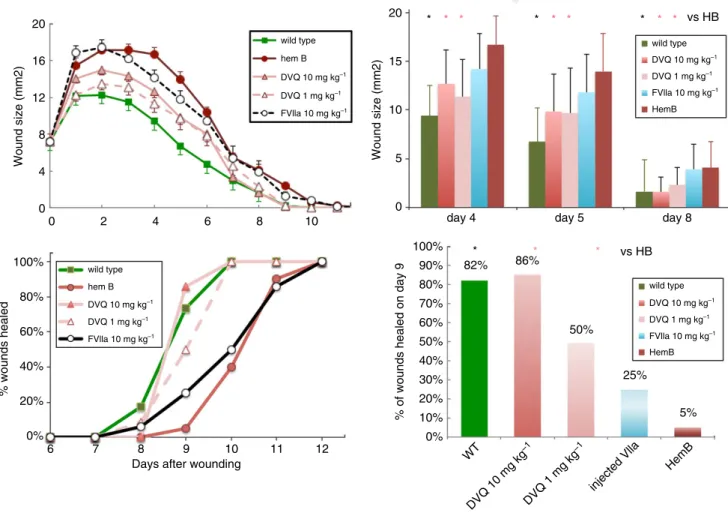
As shown in Fig. 1, a single injection of DVQ at the time of punch biopsy placement normalized wound size (upper left) and time to healing (lower left) in HB mice. HB mice
in our previous study were treated with 10 mg kg 1
rFVIIa [2]. We treated HB mice with the same dose of DVQ, and observed that the wound sizes were smaller than those in untreated HB mice, and, surprisingly, that the time to wound closure was normalized. DVQ is
signif-icantly more potent than rFVIIa in a tail clip bleeding
model in mice, with 1 mg kg 1 having equivalent
hemo-static effectiveness as 10 mg kg 1 rFVIIa [7], so we also
tested the lower dose. Wound sizes in the mice given a
single injection of either 1 mg kg 1 or 10 mg kg 1 DVQ
at the time of wounding were significantly smaller than
those in untreated HB mice (P <0.05; Student’s t-test
with Tukey’s correction for multiple comparisons) at mul-tiple time points, and were not significantly different from those in WT mice. In addition, the time to complete clo-sure of the skin defect in DVQ-treated mice was signifi-cantly improved as compared with untreated mice (by Fisher’s exact test [16]), and not different from that in WT mice. A single injection of rFVIIa, as we have previ-ously reported [2], did not significantly improve wound size or time to healing in these experiments (Fig. 1). Thus,
20
0 2 4 6 8 10
wild type
hem B
DVQ 10 mg kg–1
DVQ 1 mg kg–1
FVlla 10 mg kg–1
wild type
hem B
DVQ 10 mg kg–1
DVQ 1 mg kg–1
FVlla 10 mg kg–1
6 7 8
Days after wounding
9 10 11 12
16
Wound size (mm2)
12
8
4
0
100%
80%
% wounds healed
60%
40%
20%
0%
20
* * * * * * * * * vs HB
*
82% 86%
50%
25%
5%
* * vs HB
wild type
HemB DVQ 10 mg kg–1
DVQ 1 mg kg–1
FVlla 10 mg kg–1
wild type
HemB DVQ 10 mg kg–1
DVQ 1 mg kg–1
FVlla 10 mg kg–1 15
Wound size (mm2)
10
5
0
day 4 day 5 day 8
100%
80%
70% 90%
% of wounds healed on day 9
60%
50%
40%
30%
20%
10%
0%
WT
DVQ 10 mg kg –1
DVQ 1 mg kg –1
injected Vlla
HemB
Fig. 1.Cutaneous wound healing in hemophilia B (HB) mice is normalized by one dose of DVQ. A punch biopsy wound was made on each mouse. Some HB mice received an intravenous injection of DVQ (1 mg kg 1or 10 mg kg 1) or activated factor VII (FVIIa) (10 mg kg 1) at the time of wounding. Each point is the mean of measurements from 17–20 HB mice and 11 wild-type mice. For HB mice treated with 10 mg kg 1DVQ, the numbers of mice were 25 (days 1–4), 23 (days 5–6), 13 (days 7–8), or seven (days 9–12). For 1 mg kg 1DVQ, the num-bers of mice were 15 (days 1–6), 12 (days 7–8), or 10 (days 9–12). For FVIIa, the numbers of mice were 25 (days 1–2), 22 (days 3–4), 19 (days 5–6), 17 (days 7–8), 12 (days 9–10), and seven (days 11–12). The wound areas are plotted as a function of days after wounding in the upper left panel. The bars represent the standard error of the mean. Wound sizes for days 4, 5 and 8 are shown in the upper right panel (*P<0.05 as compared with untreated HB mice; Student’st-test). In this panel, the bars represent the standard deviation. The percentages of wounds healed at each day after wounding are plotted in the lower left panel, and the percentages of wounds healed at day 9 are shown in a bar graph in the lower right panel. A significantly greater proportion of wounds were healed in HB mice treated with 1 mg kg 1or 10 mg kg 1 DVQ at days 9 and 10 than in other HB mice (*P<0.05; Fisher’s exact test). 6;10
Colour
online,
B&W
in
1
2
3
4
5
6
7
8
9
10
11
12
13
14
15
16
17
18
19
20
21
22
23
24
25
26
27
28
29
30
31
32
33
34
35
36
37
38
39
40
41
42
43
44
45
46
47
48
49
50
51
52
53
54
55
a single dose of DVQ, unlike FIX and rFVIIa, could nor-malize the time course of cutaneous healing in HB mice.
A single injection of DVQ does not normalize wound histology in HB mice
One of the histologic abnormalities present in HB mice following wounding is the presence of an excess of angio-genic vessels around the wound bed. These vessels predis-pose the HB mice to episodes of bleeding and hematoma formation for an extended period of time following wounding [17]. Therefore, we tested the hypothesis that a dose of DVQ would also normalize angiogenesis follow-ing woundfollow-ing. However, as shown in Fig. 2, angiogenesis in the wound bed was not normalized by a single injec-tion of DVQ at the time of punch biopsy placement. The
vessel counts in HB mice treated with 10 mg kg 1 DVQ
were not significantly different from those in untreated HB mice at any time point, although both were signifi-cantly greater than those in WT mice at 8 days after wounding.
Topical application of DVQ improves wound healing in HB mice
Intravenous infusion of equally hemostatic doses of DVQ
(1 mg kg 1) and rFVIIa (10 mg kg 1) had different
effects on wound closure; one dose of DVQ normalized wound closure, whereas rFVIIa did not. This could be the result of DVQ producing a more rapid burst of thrombin generation than rFVIIa, leading to more rapid hemostasis [18] and possibly a more favorable fibrin clot structure to support healing. In fact, DVQ was found to
produce more rapid hemostasis in a randomized clinical trial [10]. However, an alternative hypothesis is that DVQ does not enhance epithelial wound closure solely because of its hemostatic effect. Even though treatment with DVQ normalized closure of the epithelial defect, it did
not normalize the underlying tissue histology– even at a
10-fold higher dose than required to restore surface clo-sure. This suggests that DVQ might have a specific effect on the squamous epithelium.
To begin testing this hypothesis, we examined the effect of the topical application of DVQ to the wound surface. As shown in Fig. 3, topical application of DVQ immedi-ately after wounding of HB mice reduced wound sizes to the point that they were not significantly different from
24
20
16
Vessels/hpf
12
8
4
0
6 8
HB *
*
* WT
HB+DVQ
Days after wounding
10
Fig. 2.Angiogenesis in skin wounds is not normalized by one dose of DVQ in hemophilia B (HB) mice. Angiogenesis was assessed by counting vessel profiles in the wound area. Counts are expressed per high-power field (HPF) (940). The scores were averaged for two sections of three to seven wounds per time point. Angiogenesis at day 8 was significantly greater in DVQ-treated or untreated HB mice than in wild-type mice (*P<0.05 as compared with wild-type mice; Student’st-test). Angiogenesis in DVQ-treated HB mice was not sig-nificantly different from that in untreated HB mice at any time point.
24
20
16
Wound size (mm2)
% wounds healed
12
8
4
0
2 4
Days after wounding 6
Wild type
Hem B
Topical DVQ
8 10
0
6 7 8 9
Days after wounding
10 11 12
100%
80%
60%
40%
20%
0%
Fig. 3.Topical application of DVQ improves wound healing in hemophilia B (HB) mice. A single punch biopsy wound was made on HB or wild-type mice. DVQ was applied to wounds on HB mice immediately after wounding. Each point represents the mean of mea-surements from 11 mice for wild-type mice, 17–20 for HB mice, and seven for HB mice treated with DVQ. The wound areas are plotted as a function of days after wounding in the upper panel, with bars showing the standard error of the mean of each group. Wound sizes in HB mice treated with topical DVQ were significantly smaller than those in untreated HB mice at days 8–9 (P<0.05; Student’st-test with Tukey’s correction for multiple comparisons), and not different from those in wild-type mice. The percentage of completely healed wounds is plotted in the lower panel. A significantly greater propor-tion of wounds were closed at days 9–10 in DVQ-treated HB mice than in untreated HB mice (P<0.05; Fisher’s exact test).
Colour
online,
B&W
in
Colour
online,
B&W
in
1
2
3
4
5
6
7
8
9
10
11
12
13
14
15
16
17
18
19
20
21
22
23
24
25
26
27
28
29
30
31
32
33
34
35
36
37
38
39
40
41
42
43
44
45
46
47
48
49
50
51
52
53
54
55
those in WT controls. In addition, all wounds in DVQ-treated HB mice were closed by day 10, which is the same as what was seen in WT mice. Thus, topical DVQ nor-malized wound size and the time to complete wound clo-sure in HB mice.
The efficacy of topical DVQ suggests that it might be
acting through a receptor-mediated mechanism to
enhance squamous epithelial proliferation and/or migra-tion. There are clear precedents for stimulation of cuta-neous healing by components of the coagulation system. Topical thrombin has been reported to enhance wound healing via protease-activated receptor (PAR)-1 [19]. Both activated protein C [20] and FXa [21] have been reported to enhance wound healing via PAR-2. FVIIa in complex with TF can activate PAR-2 [22]. FVIIa in complex with the endothelial protein C receptor can activate PAR-1 [23]. Therefore, we hypothesize that the enhanced prote-olytic activity of the DVQ variant may enable it to directly cleave one or more PAR in the absence of a cofactor, and thereby enhance epithelial healing.
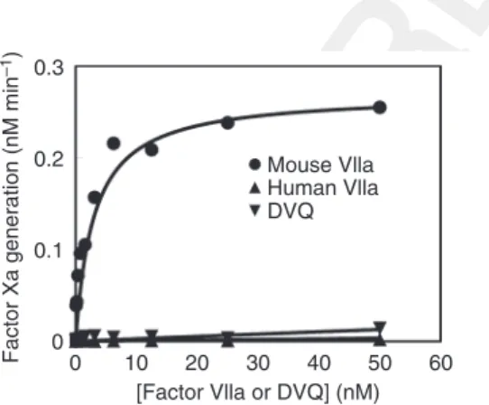
Different effects of FVIIa and DVQ are not attributable to differences in TF binding
It has previously been shown that human rFVIIa binds extremely poorly to mouse TF [13]. It is possible that (hu-man) DVQ has greater affinity for mouse TF than does the wild type. However, as shown in Fig. 4, we found that this was not the case. FVIIa and DVQ had similarly poor affinity for the recombinant extracellular domain of mouse TF (soluble TF). In contrast, mouse FVIIa bound tightly to soluble TF. Thus, the beneficial effects of DVQ
on cutaneous wound healing are not mediated by binding to TF.
Conclusions
We found that a single injection or topical application of DVQ was able to normalize closure of a cutaneous punch biopsy wound in HB mice. Neither a single dose of WT rFVIIa nor a single dose of FIX could similarly normal-ize the time to cutaneous wound closure. Even though surface closure was enhanced by DVQ, the histology of the underlying tissues was not normalized. Because DVQ was more effective in normalizing cutaneous healing than an equally hemostatic dose of FVIIa, we conclude that the ability of DVQ to enhance would healing may not be mediated solely by its hemostatic properties. A number of coagulation proteins modulate other aspects of host defense, such as inflammation, immune responses, and tis-sue repair. This suggests that novel variants of coagula-tion proteins can be designed not only to be more effective hemostatic agents, but also to have additional beneficial effects as biological modulators.
Addendum
M. Hoffman and D. M. Monroe: designed the study, col-lected and analyzed the mouse data, and wrote the manu-script. J.-Y. Chang: cloned and expressed mouse TF, and conducted TF-binding studies. M. Ezban: provided a crit-ical review of the study design, and edited the final manu-script.
Acknowledgements
The authors would like to acknowledge the outstanding technical assistance of J. Brock and A. Crabtree.
Disclosure of Conflict of Interests
M. Hoffman reports receiving grants from Novo Nordisk during the conduct of the study, and grants from CSL-Behring and Boehringer Ingelheim outside the submitted work. D. M. Monroe reports receiving grants, personal fees and non-financial support from Novo Nordisk during the conduct of the study, and grants from Novo Nordisk outside the submitted work. The other authors state that they have no conflict of interest.
References
1 Hoffman M, Harger A, Lenkowski A, Hedner U, Roberts HR, Monroe DM. Cutaneous wound healing is impaired in
hemophil-ia B.Blood2006;108: 3053–60. 7
2 McDonald A, Hoffman M, Hedner U, Roberts HR, Monroe DM. Restoring hemostatic thrombin generation at the time of cutaneous wounding does not normalize healing in hemophilia B.
J Thromb Haemost2007;5: 1577–83.
0.3
0.2
0.1
Factor Xa generation (nM min
–1
)
0
0 10 20
[Factor Vlla or DVQ] (nM) 30
Mouse Vlla Human Vlla DVQ
40 50 60
Fig. 4.Both human activated factor VII (FVIIa) and DVQ bind poorly to mouse tissue factor (TF). The extracellular domain of mouse TF was expressed as described in Materials and methods. FVIIa binding and DVQ binding to mouse TF were assayed as the increase in FX activation when they were bound to TF in the pres-ence of phospholipid vesicles. Although DVQ has much higher activ-ity than FVIIa in the absence of TF, its activactiv-ity is still increased by binding to TF. The activity of human FVIIa and DVQ, with recom-binant mouse FVIIa as a positive control, are plotted as a function of protease concentration. Over the concentration range tested, nei-ther human FVIIa nor DVQ bound significantly to mouse TF. FXa, activated factor X.
1
2
3
4
5
6
7
8
9
10
11
12
13
14
15
16
17
18
19
20
21
22
23
24
25
26
27
28
29
30
31
32
33
34
35
36
37
38
39
40
41
42
43
44
45
46
47
48
49
50
51
52
53
54
55
3 Monroe DM, Hoffman M, Roberts HR, Hedner U. Progressive improvement in wound healing with increased therapy in haemophilia B mice.Haemophilia2013;19: 926–32.
4 Persson E, Kjalke M, Olsen OH. Rational design of coagulation factor VIIa variants with substantially increased intrinsic activity.
Proc Natl Acad Sci USA2001;98: 13583–8.
5 Monroe DM, Hoffman M, Oliver JA, Roberts HR. Platelet activity of high-dose factor VIIa is independent of tissue factor.
Br J Haematol1997;99: 542–7.
6 Allen GA, Persson E, Campbell RA, Ezban M, Hedner U, Wol-berg AS. A variant of recombinant factor VIIa with enhanced procoagulant and antifibrinolytic activities in an in vitro model of hemophilia.Arterioscler Thromb Vasc Biol2007;27: 683–9. 7 Tranholm M, Kristensen K, Kristensen AT, Pyke C, Rojkjaer R,
Persson E. Improved hemostasis with superactive analogs of fac-tor VIIa in a mouse model of hemophilia A.Blood 2003; 102: 3615–20.
8 Brophy DF, Martin EJ, Barrett JC, Nolte ME, Kuhn JG, Hed-ner U, Ezban M. Overcoming delayed in-vitro response to rFVIIa: effects of rFVIIa and rFVIIa analogue (vatreptacog alfa) concentration escalation in whole blood assays. Blood Coagul Fibrinolysis2011.
8
9 de Paula EV, Kavakli K, Mahlangu J, Ayob Y, Lentz SR, Mor-fini M, Nemes L, Salek SZ, Shima M, Windyga J, Ehrenforth S, Chuansumrit A; 1804 (adept(TM)1) Investigators. Recombinant factor VIIa analog (vatreptacog alfa [activated]) for treatment of joint bleeds in hemophilia patients with inhibitors: a randomized controlled trial.J Thromb Haemost2012;10: 81–9.
10 Lentz SR, Ehrenforth S, Karim FA, Matsushita T, Weldingh KN, Windyga J, Mahlangu JN; adeptTM2 investigators.
Recom-binant factor VIIa analog in the management of hemophilia with inhibitors: results from a multicenter, randomized, controlled trial of vatreptacog alfa.J Thromb Haemost2014;12: 1244–53. 11 Lin H, Maeda N, Smithies O, Straight D, Stafford D. A
coagu-lation factor IX-deficient mouse model for human hemophilia B.
Blood1997;90: 3962–6.
12 Petersen LC, Karpf DM, Agerso H, Hermit MB, Pelzer H, Pers-son E, Nichols TC, Ezban M. Intravascular inhibition of fac-tor VIIa and the analogue NN1731 by antithrombin. Br J Haematol2011;152: 99–107.
13 Petersen LC, Norby PL, Branner S, Sorensen BB, Elm T, Sten-nicke HR, Persson E, Bjorn SE. Characterization of recombinant murine factor VIIa and recombinant murine tissue factor: a
human–murine species compatibility study. Thromb Res 2005; 116: 75–85.
14 Hope MJ, Bally MB, Webb G, Cullis PR. Production of large unilamellar vesicles by a rapid extrusion procedure. Characteriza-tion of size distribuCharacteriza-tion, trapped volume and ability to maintain a membrane potential.Biochem Biophys Acta1985;812: 55–65. 15 Yan SCB, Pazzano P, Chao YB, Walls JD, Berg DT, McClure
DB, Grinnell BW. Characterization and novel purification of recombinant human protein C from three mammalian cell lines.
Bio/Technology1990;8: 655–61.
16 Preacher K, Briggs N. Calculation for Fisher’s Exact Test: an interactive calculation tool for Fisher’s exact probability test for 292 tables [Computer software]. http://www.quantpsy.org
2001. 9
17 Hoffman M, Monroe DM. Wound healing in haemophilia – breaking the vicious cycle. Haemophilia2010;16(Suppl. 3): 13– 18.
18 Holmberg HL, Lauritzen B, Tranholm M, Ezban M. Faster onset of effect and greater efficacy of NN1731 compared with rFVIIa, aPCC and FVIII in tail bleeding in hemophilic mice.
J Thromb Haemost2009;7: 1517–22.
19 Carney DH, Mann R, Redin WR, Pernia SD, Berry D, Heggers JP, Hayward PG, Robson MC, Christie J, Annable C. Enhance-ment of incisional wound healing and neovascularization in nor-mal rats by thrombin and synthetic thrombin receptor-activating peptides.J Clin Invest1992;89: 1469–77.
20 Julovi SM, Xue M, Dervish S, Sambrook PN, March L, Jackson CJ. Protease activated receptor-2 mediates activated protein C-induced cutaneous wound healing via inhibition of p38. Am J Pathol2011;179: 2233–42.
21 Borensztajn K, Stiekema J, Nijmeijer S, Reitsma PH, Peppelen-bosch MP, Spek CA. Factor Xa stimulates proinflammatory and profibrotic responses in fibroblasts via protease-activated recep-tor-2 activation.Am J Pathol2008;172: 309–20.
22 Larsen KS, Ostergaard H, Olsen OH, Bjelke JR, Ruf W, Peter-sen LC. Engineering of substrate selectivity for tissue factor.fac-tor VIIa complex signaling through protease-activated receptor 2.J Biol Chem2010;285: 19959–66.
23 Sen P, Gopalakrishnan R, Kothari H, Keshava S, Clark CA, Esmon CT, Pendurthi UR, Rao LV. Factor VIIa bound to endothelial cell protein C receptor activates protease activated receptor-1 and mediates cell signaling and barrier protection.
Blood2011;117: 3199–208. 1
2
3
4
5
6
7
8
9
10
11
12
13
14
15
16
17
18
19
20
21
22
23
24
25
26
27
28
29
30
31
32
33
34
35
36
37
38
39
40
41
42
43
44
45
46
47
48
49
50
51
52
53
54
55