Crystal structure of (
E
)-1-methyl-2-[2-(2-methoxphenyl)ethenyl]-4-nitro-1
H
-imidazole
Hayette Alliouche,aAbdelmalek Bouraiou,aSofiane Bouacida,b,c* Hocine Merazigband Ali Belfaitaha
aLaboratoire des Produits Naturels d’Origine Ve´ge´tale et de Synthe`se Organique, PHYSYNOR, Universite´ Constantine 1, 25000 Constantine, Algeria,bUnite´ de Recherche de Chimie de l’Environnement et Mole´culaire Structurale (CHEMS), Universite´ Constantine 1, 25000 , Algeria, andcDe´partement Sciences de la Matie`re, Faculte´ des Sciences Exactes et Sciences de la Nature et de la Vie, Universite´ Oum El Bouaghi, Algeria. *Correspondence e-mail: [email protected]
Received 19 July 2014; accepted 25 July 2014
Edited by P. C. Healy, Griffith University, Australia
In the asymmetric unit of the title compound, C13H13N3O3, the 2-(2-methoxphenyl)ethenyl unit is connected to the methyl-nitroimidazole 1-methyl-4-nitro-1H-imidazole moiety. The molecule is quasi-planar and the planes of the two rings form a dihedral angle of 0.92 (11). The crystal packing can be described as layers parallel to the (011) plane, stabilized by intermolecular C—H O hydrogen bonding, resulting in the formation of an infinite three-dimensional network linking these layers. Strong –stacking interactions are observed,
viz. benzene–benzene, imidazole–imidazole and benzene– imidazole rings, with centroid–centroid distances of 3.528 (2), 3.457 (2) and 3.544 (2) A˚ , respectively. Intensity statistics indicated twinning by non-merohedry, with refined weighs of the twin components of 0.3687:0.6313.
Keywords:crystal structure; hydrogen bonding;–stacking interactions; nitroimidazoles,.
CCDC reference:1015965
1. Related literature
For the synthesis and applications of this important class of compounds, see: Horiet al.(1997); Bourdin-Trunzet al.(2011). For our previous work on imidazole derivatives, see: Alliouche
et al.(2014); Zamaet al.(2013); Bahnouset al.(2012).
2. Experimental 2.1. Crystal data
C13H13N3O3 Mr= 259.26
Triclinic,P1
a= 7.9339 (18) A˚
b= 8.1994 (19) A˚
c= 10.452 (3) A˚ = 68.877 (17) = 75.037 (17)
= 76.182 (17)
V= 604.7 (2) A˚3 Z= 2
MoKradiation = 0.10 mm1 T= 150 K
0.190.120.08 mm
2.2. Data collection
Bruker APEXII diffractometer Absorption correction: multi-scan
(SADABS; Sheldrick, 2002)
Tmin= 0.754,Tmax= 1.000
5177 measured reflections 5177 independent reflections 3712 reflections withI> 2(I)
2.3. Refinement
R[F2> 2(F2)] = 0.088
wR(F2) = 0.282 S= 1.06 5171 reflections
176 parameters
H-atom parameters constrained max= 0.49 e A˚
3
[image:1.610.314.565.526.582.2]min=0.42 e A˚ 3
Table 1
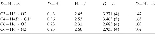
Hydrogen-bond geometry (A˚ ,).
D—H A D—H H A D A D—H A
C3—H3 O2i
0.93 2.45 3.271 (4) 147
C4—H4B O1ii
0.96 2.53 3.465 (5) 165
C6—H6 O3 0.93 2.31 2.685 (4) 103
C6—H6 N2 0.93 2.60 2.935 (4) 102
Symmetry codes: (i)x;yþ1;zþ1; (ii)x;yþ1;z.
Data collection: APEX2 (Bruker, 2006); cell refinement: SAINT (Bruker, 2006); data reduction:SAINT; program(s) used to solve structure: SIR2002 (Burla et al., 2005); program(s) used to refine structure: SHELXL97 (Sheldrick, 2008); molecular graphics: ORTEP-3 for Windows (Farrugia, 2012) and DIAMOND (Bran-denburg & Berndt, 2001); software used to prepare material for publication:WinGX(Farrugia, 2012).
Acknowledgements
We are grateful to the personnel of the PHYSYNOR Laboratory, Universite Constantine 1, Algeria, for their
data reports
o966
Allioucheet al. doi:10.1107/S1600536814017206 Acta Cryst.(2014).E70, o966–o967l’Enseignement Supe´rieur et de la Recherche Scientifique, Alge´rie) for financial support.
Supporting information for this paper is available from the IUCr electronic archives (Reference: HG5400).
References
Alliouche, H., Bouraiou, A., Bouacida, S., Bahnous, M., Roisnel, T. & Belfaitah, A. (2014).Lett. Org. Chem.11, 174–179.
Bahnous, M., Bouraiou, A., Bouacida, S., Roisnel, T. & Belfaitah, A. (2012).
Acta Cryst.E68, o1391.
& Torreele, E. (2011).Eur. J. Med. Chem.46, 1524–1534.
Brandenburg, K. & Berndt, M. (2001).DIAMOND. Crystal Impact, Bonn, Germany.
Bruker (2006).APEX2andSAINT. Bruker AXS Inc., Madison, Wisconsin, USA.
Burla, M. C., Caliandro, R., Camalli, M., Carrozzini, B., Cascarano, G. L., De Caro, L., Giacovazzo, C., Polidori, G. & Spagna, R. (2005).J. Appl. Cryst.38, 381–388.
Farrugia, L. J. (2012).J. Appl. Cryst.45, 849–854.
Hori, H., Jin, C. Z., Kiyono, M., Kasai, S., Shimamura, M. & Inayama, S. (1997).Bioorg. Med. Chem.5, 591–599.
Sheldrick, G. M. (2002).SADABS. University of Go¨ttingen, Germany. Sheldrick, G. M. (2008).Acta Cryst.A64, 112–122.
supporting information
sup-1
Acta Cryst. (2014). E70, o966–o967
supporting information
Acta Cryst. (2014). E70, o966–o967 [doi:10.1107/S1600536814017206]
Crystal structure of (
E
)-1-methyl-2-[2-(2-methoxphenyl)ethenyl]-4-nitro-1
H
-imidazole
Hayette Alliouche, Abdelmalek Bouraiou, Sofiane Bouacida, Hocine Merazig and Ali Belfaitah
S1. Comment
Nitroimidazoles are a class of N-heterocyclic compounds which have a wide range of applications in the drug synthesis
(Hori, et al., 1997) In fact, Metronidazole (Flagyl) and related N-1 substituted 5-nitroimidazoles such as Tinidazole (Fasigyne), Ornidazole (Tiberal) and Secnidazole (Secnol) still commonly used in medicine. Despite their fewer
biological activities compared with 5-nitroimidazoles, a number of 4-nitroimidazoles were reported to exhibit
antileishmanial, antiamebic and anti-parasitic activities (Bourdin-Trunz, et al. 2011). However, only some limited investigations have been carried out using methyl iodide (Alliouche, et al. 2014). In previous work, we have reported the synthesis and structure determination of some new heterocyclic compounds bearing an imidazole unit (Zama, et al., 2013; Bahnous, et al., 2012). Herein, we describe the synthesis and the structure determination of (E )-1-methyl-2-[(2-methoxphenyl)-1-ethenyl]-4-nitroimidazole resulting from the intramolecular transposition reaction of its 5-nitro isomer
in the presence of catalytic amounts of methyl iodide in nitrobenzene. The molecular geometry and the atom-numbering
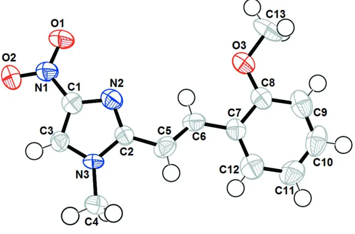
scheme of (I) are shown in Fig. 1. In the asymmetric unit of title compound the methoxphenyl-1-ethenyl unit is linked to
methyl-nitroimidazole moiety. The molecule is quasi-planar and the two rings of phenyl and imidazol form a dihedral
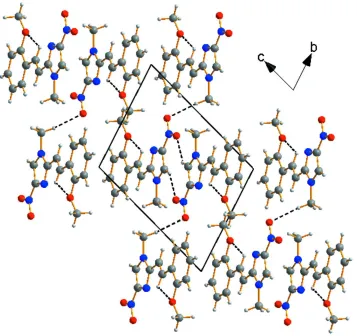
angle of 0.92 (11)°. The crystal packing can be described as layers parallel to (011) plane, along the a axis (Fig. 2). It is stabilized by intermolecular hydrogen bond C—H···O, resulting in the formation of an infinite three-dimensional network
linking these layers together and reinforcing cohesion in the structure (Fig. 2). Hydrogen-bonding parameters are listed in
Table 1. Strong π-π stacking interactions are observed between phenyl-phenyl, imidazol-imidazol and phenyl-imidazol rings, distances centroid-centroid are Cg—Cg = 3.528 (2), 3.457 (2) and 3.544 (2) Å respetively. The crystal used was a non-merohedral twin, the refined ratio of twin components being 0.3687:0.6313.
S2. Experimental
The title compound was obtained as a yellow-green solid in 83% of yield by heating a solution of (E )-1-methyl-2-[(2-methoxphenyl)-1-ethenyl]-5-nitroimidazole in nitrobenzene at 160°C during 24 h in the presence of catalytic amount of
CH3I. Suitable crystal of compound (I) was obtained by slow evaporation from a water/methanol solution at room
temperature, and X-ray crystallographic analysis confirmed the structural assignment (Fig. 1).
S3. Refinement
All non-H atoms were refined with anisotropic atomic displacement parameters. All H atoms were localized on Fourier
maps but introduced in calculated positions and treated as riding on their parent C or N atom. (with C—H = 0.93 and
Figure 1
The structure of the title compound with the atomic labelling scheme. Displacement are drawn at the 50% probability
supporting information
sup-3
[image:5.610.129.486.70.406.2]Acta Cryst. (2014). E70, o966–o967 Figure 2
A diagram of the layered crystal packing of (I) viewed down the a axis and showing hydrogen bond [C—H···O] as dashed line.
1-Methyl-2-[(E)-2-(2-methoxphenyl)ethenyl]-4-nitro-1H-imidazole
Crystal data
C13H13N3O3
Mr = 259.26
Triclinic, P1
a = 7.9339 (18) Å
b = 8.1994 (19) Å
c = 10.452 (3) Å
α = 68.877 (17)°
β = 75.037 (17)°
γ = 76.182 (17)°
V = 604.7 (2) Å3
Z = 2
F(000) = 272
Dx = 1.424 Mg m−3
Mo Kα radiation, λ = 0.71073 Å Cell parameters from 1356 reflections
θ = 2.7–24.6°
µ = 0.10 mm−1
T = 150 K Block, yellow
0.19 × 0.12 × 0.08 mm
Data collection
Bruker APEXII diffractometer
Graphite monochromator
CCD rotation images, thin slices scans Absorption correction: multi-scan
(SADABS; Sheldrick, 2002)
Tmin = 0.754, Tmax = 1.000
5177 measured reflections 5177 independent reflections 3712 reflections with I > 2σ(I)
Rint = 0
k = −9→9
Refinement
Refinement on F2
Least-squares matrix: full
R[F2 > 2σ(F2)] = 0.088
wR(F2) = 0.282
S = 1.06 5171 reflections 176 parameters 0 restraints
Primary atom site location: structure-invariant direct methods
Secondary atom site location: difference Fourier map
Hydrogen site location: inferred from neighbouring sites
H-atom parameters constrained
w = 1/[σ2(F
o2) + (0.1745P)2 + 0.1919P]
where P = (Fo2 + 2Fc2)/3
(Δ/σ)max < 0.001
Δρmax = 0.49 e Å−3
Δρmin = −0.42 e Å−3
Extinction correction: SHELXL97 (Sheldrick, 2008), Fc*=kFc[1+0.001xFc2λ3/sin(2θ)]-1/4
Extinction coefficient: 0.045 (12)
Special details
Geometry. All e.s.d.'s (except the e.s.d. in the dihedral angle between two l.s. planes) are estimated using the full covariance matrix. The cell e.s.d.'s are taken into account individually in the estimation of e.s.d.'s in distances, angles and torsion angles; correlations between e.s.d.'s in cell parameters are only used when they are defined by crystal symmetry. An approximate (isotropic) treatment of cell e.s.d.'s is used for estimating e.s.d.'s involving l.s. planes.
Refinement. Refinement of F2 against ALL reflections. The weighted R-factor wR and goodness of fit S are based on F2,
conventional R-factors R are based on F, with F set to zero for negative F2. The threshold expression of F2 > σ(F2) is used
only for calculating R-factors(gt) etc. and is not relevant to the choice of reflections for refinement. R-factors based on F2
are statistically about twice as large as those based on F, and R- factors based on ALL data will be even larger.
Fractional atomic coordinates and isotropic or equivalent isotropic displacement parameters (Å2)
x y z Uiso*/Ueq
O1 0.4178 (3) 0.2400 (3) 0.2786 (3) 0.0256 (6) O3 1.0969 (3) 0.6148 (3) 0.0846 (2) 0.0219 (6) O2 0.1531 (3) 0.2922 (3) 0.3961 (3) 0.0238 (6) N1 0.3026 (4) 0.3303 (4) 0.3420 (3) 0.0167 (7) N2 0.5059 (3) 0.5331 (4) 0.2979 (3) 0.0162 (7) N3 0.3292 (3) 0.7076 (3) 0.4156 (3) 0.0134 (6) C1 0.3460 (4) 0.4809 (4) 0.3545 (3) 0.0142 (7) C2 0.4934 (4) 0.6726 (4) 0.3362 (3) 0.0125 (7) C5 0.6299 (4) 0.7796 (5) 0.3004 (3) 0.0172 (8)
H5 0.6018 0.8867 0.319 0.021*
C6 0.7973 (4) 0.7279 (4) 0.2408 (3) 0.0151 (7)
H6 0.8198 0.6187 0.2261 0.018*
C7 0.9464 (4) 0.8227 (4) 0.1968 (3) 0.0139 (7) C8 1.1027 (4) 0.7634 (4) 0.1144 (3) 0.0159 (8) C13 1.2438 (4) 0.5580 (5) −0.0106 (4) 0.0273 (9)
H13C 1.2622 0.6542 −0.0961 0.041*
H13A 1.22 0.4604 −0.0296 0.041*
H13B 1.3478 0.5214 0.0299 0.041*
C4 0.2711 (4) 0.8439 (5) 0.4845 (3) 0.0196 (8)
H4A 0.1465 0.8508 0.5215 0.029*
supporting information
sup-5
Acta Cryst. (2014). E70, o966–o967
H4C 0.3339 0.814 0.5591 0.029*
C3 0.2328 (4) 0.5873 (4) 0.4277 (3) 0.0139 (7)
H3 0.117 0.5778 0.4744 0.017*
C9 1.2469 (4) 0.8506 (5) 0.0702 (3) 0.0207 (8)
H9 1.349 0.81 0.0154 0.025*
C10 1.2392 (5) 0.9970 (5) 0.1072 (3) 0.0233 (8)
H10 1.3369 1.0544 0.0781 0.028*
C11 1.0871 (4) 1.0600 (5) 0.1876 (3) 0.0211 (8)
H11 1.0818 1.1602 0.2116 0.025*
C12 0.9434 (4) 0.9723 (5) 0.2317 (3) 0.0199 (8)
H12 0.8418 1.0144 0.2863 0.024*
Atomic displacement parameters (Å2)
U11 U22 U33 U12 U13 U23
O1 0.0232 (13) 0.0238 (14) 0.0331 (14) −0.0046 (11) 0.0055 (11) −0.0199 (12) O3 0.0169 (13) 0.0238 (14) 0.0257 (13) −0.0036 (10) 0.0029 (10) −0.0135 (11) O2 0.0142 (13) 0.0280 (15) 0.0313 (14) −0.0066 (11) 0.0037 (11) −0.0154 (12) N1 0.0173 (15) 0.0150 (15) 0.0183 (14) −0.0021 (12) −0.0013 (12) −0.0077 (12) N2 0.0144 (14) 0.0181 (15) 0.0159 (14) −0.0007 (12) −0.0022 (11) −0.0070 (12) N3 0.0139 (14) 0.0113 (14) 0.0162 (13) −0.0006 (11) −0.0005 (11) −0.0083 (11) C1 0.0135 (16) 0.0166 (17) 0.0122 (16) −0.0007 (13) −0.0053 (13) −0.0032 (13) C2 0.0120 (16) 0.0162 (17) 0.0099 (14) −0.0020 (13) −0.0029 (13) −0.0043 (13) C5 0.0170 (18) 0.0181 (17) 0.0207 (17) −0.0027 (14) −0.0072 (14) −0.0086 (15) C6 0.0155 (17) 0.0144 (17) 0.0177 (16) 0.0010 (14) −0.0055 (13) −0.0084 (14) C7 0.0154 (17) 0.0165 (17) 0.0089 (15) −0.0037 (14) −0.0028 (13) −0.0018 (13) C8 0.0143 (17) 0.0193 (18) 0.0134 (16) −0.0016 (14) −0.0034 (14) −0.0047 (14) C13 0.0200 (19) 0.033 (2) 0.0253 (19) 0.0037 (17) 0.0043 (15) −0.0167 (17) C4 0.0171 (17) 0.0219 (18) 0.0232 (17) −0.0021 (15) −0.0010 (14) −0.0137 (15) C3 0.0126 (17) 0.0178 (18) 0.0129 (15) −0.0037 (14) −0.0026 (13) −0.0058 (13) C9 0.0150 (17) 0.029 (2) 0.0163 (17) −0.0043 (15) −0.0043 (14) −0.0036 (15) C10 0.026 (2) 0.030 (2) 0.0173 (17) −0.0148 (16) −0.0081 (15) −0.0020 (16) C11 0.0237 (19) 0.025 (2) 0.0201 (17) −0.0062 (16) −0.0086 (15) −0.0092 (16) C12 0.0187 (18) 0.025 (2) 0.0176 (17) −0.0039 (15) −0.0067 (14) −0.0068 (15)
Geometric parameters (Å, º)
O1—N1 1.241 (3) C7—C8 1.406 (4)
O3—C8 1.375 (4) C8—C9 1.383 (5)
O3—C13 1.429 (4) C13—H13C 0.96
O2—N1 1.236 (3) C13—H13A 0.96
N1—C1 1.415 (4) C13—H13B 0.96
N2—C2 1.317 (4) C4—H4A 0.96
N2—C1 1.354 (4) C4—H4B 0.96
N3—C3 1.339 (4) C4—H4C 0.96
N3—C2 1.380 (4) C3—H3 0.93
N3—C4 1.464 (4) C9—C10 1.372 (5)
C5—C6 1.351 (5) C10—H10 0.93
C5—H5 0.93 C11—C12 1.382 (5)
C6—C7 1.455 (5) C11—H11 0.93
C6—H6 0.93 C12—H12 0.93
C7—C12 1.394 (5)
C8—O3—C13 117.1 (3) O3—C13—H13A 109.5
O2—N1—O1 122.5 (3) H13C—C13—H13A 109.5
O2—N1—C1 119.0 (3) O3—C13—H13B 109.5
O1—N1—C1 118.4 (3) H13C—C13—H13B 109.5
C2—N2—C1 103.9 (3) H13A—C13—H13B 109.5
C3—N3—C2 108.4 (2) N3—C4—H4A 109.5
C3—N3—C4 124.7 (3) N3—C4—H4B 109.5
C2—N3—C4 126.7 (3) H4A—C4—H4B 109.5
N2—C1—C3 112.9 (3) N3—C4—H4C 109.5
N2—C1—N1 123.0 (3) H4A—C4—H4C 109.5
C3—C1—N1 124.1 (3) H4B—C4—H4C 109.5
N2—C2—N3 111.1 (3) N3—C3—C1 103.7 (3)
N2—C2—C5 125.9 (3) N3—C3—H3 128.1
N3—C2—C5 123.0 (3) C1—C3—H3 128.1
C6—C5—C2 121.7 (3) C10—C9—C8 120.0 (3)
C6—C5—H5 119.2 C10—C9—H9 120
C2—C5—H5 119.2 C8—C9—H9 120
C5—C6—C7 127.7 (3) C9—C10—C11 120.6 (3)
C5—C6—H6 116.1 C9—C10—H10 119.7
C7—C6—H6 116.1 C11—C10—H10 119.7
C12—C7—C8 117.2 (3) C12—C11—C10 119.2 (3)
C12—C7—C6 123.3 (3) C12—C11—H11 120.4
C8—C7—C6 119.5 (3) C10—C11—H11 120.4
O3—C8—C9 124.8 (3) C11—C12—C7 121.9 (3)
O3—C8—C7 114.1 (3) C11—C12—H12 119
C9—C8—C7 121.1 (3) C7—C12—H12 119
O3—C13—H13C 109.5
C2—N2—C1—C3 0.3 (3) C13—O3—C8—C9 −6.8 (5) C2—N2—C1—N1 −178.3 (3) C13—O3—C8—C7 173.6 (3) O2—N1—C1—N2 −179.5 (3) C12—C7—C8—O3 179.6 (3)
O1—N1—C1—N2 1.5 (4) C6—C7—C8—O3 −0.6 (4)
O2—N1—C1—C3 2.0 (5) C12—C7—C8—C9 −0.1 (5) O1—N1—C1—C3 −176.9 (3) C6—C7—C8—C9 179.8 (3)
C1—N2—C2—N3 0.1 (3) C2—N3—C3—C1 0.6 (3)
supporting information
sup-7
Acta Cryst. (2014). E70, o966–o967
N3—C2—C5—C6 170.0 (3) C9—C10—C11—C12 0.8 (5) C2—C5—C6—C7 178.6 (3) C10—C11—C12—C7 −0.5 (5) C5—C6—C7—C12 11.0 (5) C8—C7—C12—C11 0.1 (5) C5—C6—C7—C8 −168.8 (3) C6—C7—C12—C11 −179.7 (3)
Hydrogen-bond geometry (Å, º)
D—H···A D—H H···A D···A D—H···A
C3—H3···O2i 0.93 2.45 3.271 (4) 147
C4—H4B···O1ii 0.96 2.53 3.465 (5) 165
C6—H6···O3 0.93 2.31 2.685 (4) 103
C6—H6···N2 0.93 2.60 2.935 (4) 102