
organic papers
Acta Cryst.(2006). E62, o1565–o1566 doi:10.1107/S1600536806010385 Wardellet al. C
14H10I2N2
o1565
Acta Crystallographica Section E Structure Reports Online
ISSN 1600-5368
(
E
,
E
)-1,4-Bis(2-iodophenyl)-2,3-diaza-1,3-butadiene
James L. Wardell,aJohn N. Lowb and Christopher Glidewellc*
a
Instituto de Quı´mica, Departamento de Quı´mica Inorgaˆnica, Universidade Federal do Rio de Janeiro, CP 68563, 21945-970 Rio de Janeiro-RJ, Brazil,bDepartment of Chemistry,
University of Aberdeen, Meston Walk, Old Aberdeen, AB24 3UE, UK, andcSchool of
Chemistry, University of St Andrews, St Andrews, Fife, KY16 9ST, UK
Correspondence e-mail: [email protected]
Key indicators
Single-crystal X-ray study
T= 120 K
Mean(C–C) = 0.005 A˚
Rfactor = 0.022
wRfactor = 0.075
Data-to-parameter ratio = 19.7
For details of how these key indicators were automatically derived from the article, see http://journals.iucr.org/e.
Received 20 March 2006 Accepted 21 March 2006
#2006 International Union of Crystallography All rights reserved
The molecules of the title compound C14H10I2N2, which are
almost planar, lie across centres of inversion. The structure contains no direction-specific intermolecular interactions of any kind.
Comment
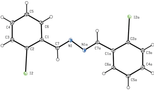
We recently reported that small quantities (ca10%) of the title compound (I) (Fig.1) readily co-crystallized with (E,E )-1-(2-iodophenyl)-4-(2-nitrophenyl)-2,3-diaza-1,3-butadiene (II), and that the presence of (I) probably arose from some minor reorganization of the substituted aryl groups during either the synthesis or the recrystallization of (II) (Glidewellet al., 2005). In order to assess the relationship between the structures of (I) and (II), we now report the structure of (I).
The molecules of (I) lie across inversion centres in space groupP21/n, and they are almost planar, as shown by the key
hydrogen bonds, aromatic–stacking interactions, and short I I interactions are all absent.
The unit-cell dimensions for compounds (I) and (II) are very different, and these compounds also crystallize in different space groups, with molar volumes which differ by some 5%. It is therefore not clear why compound (I) so readily co-crystallizes with compound (II).
Isomeric with (I) is compound (III) where, again, nearly planar molecules lie across centres of inversion (Cho et al., 2005).
Experimental
A mixture of 2-iodobenzaldehyde (1 mmol) and hydrazine hydrate (0.5 mmol) in methanol (20 cm3) was heated under reflux for 30 min.
The mixture was cooled and the solvent was removed under reduced pressure. The product, (I), was purified by chromatography on alumina, using CHCl3as eluent: crystals suitable for single-crystal
X-ray diffraction were grown by slow evaporation of a solution in ethanol.
Crystal data
C14H10I2N2
Mr= 460.04
Monoclinic,P21=n a= 13.2667 (4) A˚
b= 4.1070 (2) A˚
c= 13.8207 (4) A˚
= 110.3140 (15) V= 706.20 (5) A˚3
Z= 2
Dx= 2.163 Mg m
3
MoKradiation Cell parameters from 1818
reflections
= 1.0–27.4 = 4.44 mm1
T= 120 (2) K Lath, yellow
0.640.120.08 mm
Data collection
Bruker–Nonius KappaCCD diffractometer
’and!scans
Absorption correction: multi-scan (SADABS; Sheldrick, 2003)
Tmin= 0.160,Tmax= 0.703
9545 measured reflections
1614 independent reflections 1455 reflections withI> 2(I)
Rint= 0.027 max= 27.5
h=17!15
k=5!5
l=17!17
Refinement
Refinement onF2
R[F2> 2(F2)] = 0.022
wR(F2) = 0.075
S= 1.30 1614 reflections 82 parameters
H-atom parameters constrained
w= 1/[2
(Fo2) + (0.0439P)2]
whereP= (Fo2+ 2Fc2)/3
(/)max= 0.001 max= 0.64 e A˚
3
min=1.16 e A˚ 3
Table 1
Selected bond and torsion angles ().
C3—C2—C1 121.6 (3)
C3—C2—I2 116.2 (2)
C1—C2—I2 122.2 (2)
C6—C1—C7—N1 5.2 (5) C1—C7—N1—N1i
179.9 (3)
Symmetry code: (i)xþ1;yþ1;zþ1.
All H atoms were located in difference maps and they were then treated as riding atoms with C—H distances 0.95 A˚ and Uiso(H) =
1.2Ueq(C). The deepest hole is located 0.72 A˚ from atom I2.
Data collection: COLLECT (Hooft, 1999); cell refinement:
DENZO(Otwinowski & Minor, 1997) &COLLECT); data reduc-tion: DENZO & COLLECT; program(s) used to solve structure:
OSCAIL (McArdle, 2003) and SHELXS97 (Sheldrick, 1997); program(s) used to refine structure: OSCAIL and SHELXL97
(Sheldrick, 1997); molecular graphics:PLATON(Spek, 2003); soft-ware used to prepare material for publication: SHELXL97 and
PRPKAPPA(Ferguson, 1999).
X-Ray data were collected at the EPSRC X-Ray Crystal-lographic Service, University of Southampton, UK; the authors thank the staff of the Service for all their help and advice. JLW thanks CNPq and FAPERJ for financial support.
References
Cho, H. M., Moore, J. S. & Wilson, S. R. (2005).Acta Cryst.E61, o3773–o3774. Ferguson, G. (1999).PRPKAPPA. University of Guelph, Canada.
Glidewell, C., Low, J. N., Skakle, J. M. S. & Wardell, J. L. (2005).Acta Cryst.
C61, o312–o316.
Hooft, R. W. W. (1999).COLLECT. Nonius B. V., Delft, The Netherlands. McArdle, P. (2003). OSCAIL for Windows. Version 10, Crystallography
Centre, Chemistry Department, NUI Galway, Ireland.
Otwinowski, Z. & Minor, W. (1997). Methods in Enzymology, Vol. 276,
Macromolecular Crystallography, Part A, edited by C. W. Carter Jr & R. M. Sweet, pp. 307–326. New York: Academic Press.
Sheldrick, G. M. (1997). SHELXS97 and SHELXL97. University of Go¨ttingen, Germany.
Sheldrick, G. M. (2003).SADABS. Version 2.10. University of Go¨ttingen, Germany.
[image:2.610.314.564.70.216.2]Spek, A. L. (2003).J. Appl. Cryst.36, 7–13. Figure 1
supporting information
sup-1
Acta Cryst. (2006). E62, o1565–o1566
supporting information
Acta Cryst. (2006). E62, o1565–o1566 [https://doi.org/10.1107/S1600536806010385]
(E,E)-1,4-Bis(2-iodophenyl)-2,3-diaza-1,3-butadiene
James L. Wardell, John N. Low and Christopher Glidewell
(E,E)-1,4-Bis(2-iodophenyl)-2,3-diaza-1,3-butadiene
Crystal data
C14H10I2N2
Mr = 460.04
Monoclinic, P21/n
Hall symbol: -P 2yn
a = 13.2667 (4) Å
b = 4.1070 (2) Å
c = 13.8207 (4) Å
β = 110.3140 (15)°
V = 706.20 (5) Å3
Z = 2
F(000) = 428
Dx = 2.163 Mg m−3
Mo Kα radiation, λ = 0.71073 Å Cell parameters from 1818 reflections
θ = 1.0–27.4°
µ = 4.44 mm−1
T = 120 K Lath, yellow
0.64 × 0.12 × 0.08 mm
Data collection
Bruker-Nonius KappaCCD diffractometer
Radiation source: Bruker-Nonius FR591 rotating anode
Graphite monochromator
Detector resolution: 9.091 pixels mm-1
φ & ω scans
Absorption correction: multi-scan (SADABS; Sheldrick, 2003)
Tmin = 0.160, Tmax = 0.703
9545 measured reflections 1614 independent reflections 1455 reflections with I > 2σ(I)
Rint = 0.027
θmax = 27.5°, θmin = 1.8°
h = −17→15
k = −5→5
l = −17→17
Refinement
Refinement on F2
Least-squares matrix: full
R[F2 > 2σ(F2)] = 0.022
wR(F2) = 0.075
S = 1.30 1614 reflections 82 parameters 0 restraints
Primary atom site location: structure-invariant direct methods
Secondary atom site location: difference Fourier map
Hydrogen site location: inferred from neighbouring sites
H-atom parameters constrained
w = 1/[σ2(F
o2) + (0.0439P)2]
where P = (Fo2 + 2Fc2)/3
(Δ/σ)max = 0.001
Δρmax = 0.64 e Å−3
Δρmin = −1.16 e Å−3
Fractional atomic coordinates and isotropic or equivalent isotropic displacement parameters (Å2)
x y z Uiso*/Ueq
C1 0.7374 (3) 0.4440 (7) 0.5557 (2) 0.0150 (6)
C2 0.8164 (2) 0.3184 (8) 0.5201 (2) 0.0149 (6)
C3 0.9252 (3) 0.3381 (8) 0.5790 (3) 0.0204 (7)
C4 0.9564 (3) 0.4865 (10) 0.6753 (3) 0.0243 (7)
C5 0.8802 (3) 0.6146 (7) 0.7125 (3) 0.0217 (8)
C6 0.7720 (3) 0.5935 (7) 0.6531 (3) 0.0183 (7)
C7 0.6223 (3) 0.4242 (7) 0.4961 (3) 0.0173 (7)
N1 0.5529 (2) 0.5194 (8) 0.5345 (2) 0.0220 (6)
H3 0.9776 0.2510 0.5536 0.025*
H4 1.0305 0.5003 0.7160 0.029*
H5 0.9019 0.7163 0.7783 0.026*
H6 0.7202 0.6823 0.6788 0.022*
H7 0.5991 0.3393 0.4279 0.021*
Atomic displacement parameters (Å2)
U11 U22 U33 U12 U13 U23
C1 0.0170 (16) 0.0159 (15) 0.0130 (15) 0.0026 (11) 0.0064 (13) 0.0050 (11) C2 0.0177 (15) 0.0163 (13) 0.0112 (14) −0.0006 (12) 0.0056 (12) 0.0030 (12) I2 0.01818 (16) 0.01792 (16) 0.01565 (16) 0.00262 (6) 0.00751 (11) −0.00004 (6) C3 0.0167 (16) 0.0255 (16) 0.0198 (17) 0.0041 (13) 0.0072 (13) 0.0064 (13) C4 0.0149 (16) 0.0342 (17) 0.0203 (17) −0.0014 (15) 0.0017 (13) 0.0048 (15) C5 0.0231 (19) 0.030 (2) 0.0104 (17) −0.0047 (12) 0.0035 (14) 0.0006 (11) C6 0.0194 (18) 0.0246 (17) 0.0147 (16) 0.0001 (11) 0.0106 (14) −0.0001 (11) C7 0.0148 (16) 0.0243 (17) 0.0116 (16) 0.0018 (11) 0.0033 (13) −0.0003 (11) N1 0.0140 (14) 0.0348 (15) 0.0167 (14) 0.0035 (13) 0.0047 (11) −0.0022 (13)
Geometric parameters (Å, º)
C1—C2 1.402 (4) C4—H4 0.95
C1—C6 1.403 (5) C5—C6 1.387 (5)
C1—C7 1.464 (5) C5—H5 0.95
C2—C3 1.391 (4) C6—H6 0.95
C2—I2 2.111 (3) C7—N1 1.273 (5)
C3—C4 1.391 (5) C7—H7 0.95
C3—H3 0.95 N1—N1i 1.407 (6)
C4—C5 1.387 (6)
C2—C1—C6 117.5 (3) C3—C4—H4 119.7
C2—C1—C7 122.9 (3) C6—C5—C4 119.6 (3)
C6—C1—C7 119.6 (3) C6—C5—H5 120.2
C3—C2—C1 121.6 (3) C4—C5—H5 120.2
C3—C2—I2 116.2 (2) C5—C6—C1 121.5 (3)
C1—C2—I2 122.2 (2) C5—C6—H6 119.2
C4—C3—C2 119.3 (3) C1—C6—H6 119.2
C4—C3—H3 120.4 N1—C7—C1 120.8 (3)
C2—C3—H3 120.4 N1—C7—H7 119.6
C5—C4—C3 120.5 (3) C1—C7—H7 119.6
supporting information
sup-3
Acta Cryst. (2006). E62, o1565–o1566
C6—C1—C2—C3 0.5 (5) C3—C4—C5—C6 0.2 (5)
C7—C1—C2—C3 −179.4 (3) C4—C5—C6—C1 0.2 (5)
C6—C1—C2—I2 −178.3 (2) C2—C1—C6—C5 −0.5 (5)
C7—C1—C2—I2 1.8 (4) C7—C1—C6—C5 179.4 (3)
C1—C2—C3—C4 −0.2 (5) C2—C1—C7—N1 174.7 (3)
I2—C2—C3—C4 178.7 (3) C6—C1—C7—N1 −5.2 (5)
C2—C3—C4—C5 −0.2 (5) C1—C7—N1—N1i −179.9 (3)
![Chlorido(pyridine κN)bis[2 (quinolin 2 yl)phenyl κ2C1,N]iridium(III) monohydrate](data:image/gif;base64,R0lGODlhAQABAIAAAP///wAAACH5BAEAAAAALAAAAAABAAEAAAICRAEAOw==)