Kodoja: A workflow for virus detection in plants using k-mer analysis of RNA-1
sequencing data 2
3
Amanda Baizan-Edge1, Peter Cock2, Stuart MacFarlane3, Wendy McGavin3, Lesley 4
Torrance1,3 and Susan Jones*2 5
1. The School of Biology, University of St Andrews, Biomedical Sciences Research 6
Complex, St Andrews, KY16 9ST, UK 7
2. Information and Computational Sciences Group, The James Hutton Institute, 8
Dundee, DD2 5DA, UK 9
3. Cell and Molecular Sciences Group, The James Hutton Institute, Dundee, DD2 5DA, 10
UK 11
12
* To whom correspondence should be addressed. Tel: +44(0)3449 285428 Email: 13
15 16
Key Words: Plant virus diagnostics, RNA-sequencing, k-mer analysis, Raspberry 17
yellow net virus, Beet ringspot virus, Bioinformatics 18
19
Repositories: The RNA sequences of 2 raspberry plants exhibiting virus-like 20
symptoms have been deposited in the European Nucleotide Archive and assigned 21
Abstract 23
24
Background: RNA-sequencing of plant material allows for hypothesis-free detection 25
of multiple viruses simultaneously. This methodology relies on bioinformatics 26
workflows for virus identification. Most workflows are designed for human clinical data, 27
and few go beyond sequence mapping for virus identification. 28
Methods: We present a new workflow (Kodoja) for the detection of plant virus 29
sequences in RNA-sequence data. Kodoja uses k-mer profiling at the nucleotide level 30
and sequence mapping at the protein level by integrating two existing tools Kraken 31
and Kaiju. 32
Results and Discussion: Kodoja was tested on 3 existing RNA-seq datasets from 33
grapevine, and 2 new RNA-seq datasets from raspberry. For grapevine, Kodoja was 34
shown to be more sensitive than a method based on contig building and Blast 35
alignments (27 viruses detected compared to 19). The application of Kodoja to 36
raspberry, showed that field-grown raspberries were infected by multiple viruses, and 37
that RNA-seq can identify lower amounts of virus material than RT-PCR. This work 38
enabled the design of new PCR-primers for detection of Raspberry yellow net virus 39
and Beet ringspot virus. Kodoja is a sensitive method for plant virus discovery in field 40
samples and enables the design of more accurate primers for detection. Kodoja is 41
available to install through Bioconda and as a tool within Galaxy. 42
1.0 Introduction 48
Virus infection is of specific importance in crops cultivated for food and fuel. Viruses 49
cause significant yield and quality losses, and consequently they have important 50
negative economic impact (1). In the UK, Potato virus Y causes annual potato crop 51
losses of £30-40 million (2), and in Asia viruses infecting rice (such as Rice grassy
52
stunt virus) can cause annual crop losses of $120 million (3). These examples highlight
53
the need for fast and accurate virus detection methods. Viral infection symptoms can 54
include yellowing and stunting, but in many cases symptoms can be absent or masked 55
by other factors. In some cases plant viruses interact synergistically, to cause new or 56
more severe disease symptoms (4). One example is crumbly fruit complex disease of 57
raspberry, which can be caused by the presence of two viruses; Raspberry bushy
58
dwarf virus and Raspberry latent virus (5). As crops are cultivated in new geographical
59
locations and agricultural practices are intensified, there is an increasing risk of new 60
viruses becoming established, and existing ones widening their host range. Hence, 61
plant virus diagnostics is a field of increasing significance in terms of future food 62
security. 63
64
Standard molecular techniques for detection of viruses include methods based on 65
reverse transcriptase polymerase chain reaction (RT-PCR). But such techniques only 66
allow the detection of known viruses, i.e. each test is specific to one virus or a very 67
small number of related viruses (6). Furthermore, viral genomes evolve which can 68
make tests ineffective over time, making disease diagnosis slow and restrictive. Such 69
limitations have recently been overcome through the use of next generation 70
sequencing (NGS) methods for hypothesis-free simultaneous detection of multiple 71
viruses (7). The majority of plant viruses have RNA as their genetic material and those 72
that have DNA genomes produce RNA transcripts. In addition, eukaryote small 73
interfering RNAs (siRNAs) direct antiviral immunity through RNA interference and 74
during this process virus-derived siRNAs are enriched in the host (8). Hence, both 75
RNA and small RNA (sRNA) sequencing are effective methods for virus detection in 76
plants. However, this relies upon two important elements: (a) robust RNA extraction 77
and enrichment protocols, and (b) fast and robust bioinformatics tools for virus 78
identification. 79
80
A range of RNA-extraction and enrichment protocols, and bioinformatics workflows, 81
has previously been developed for human clinical samples (for review see (9). 82
Recently such work has resulted in a viral disease diagnosis and actionable clinical 83
management within 48 hours (10). The workflow used in this clinical work comprised 84
the two main elements required for a virus diagnostic tool: (a) identification and 85
removal of host nucleotide sequences, and (b) identification of virus sequences. 86
However, virus detection in clinical samples presents an easier problem than in plants, 87
as the human genome is well annotated (allowing easy removal of host sequences), 88
and human virus data are more prevalent in sequence databases (allowing for easy 89
genomes are incomplete or poorly annotated, and plant virus sequences are under-91
represented in databases. 92
93
We recently reviewed the bioinformatics tools and workflows currently available for 94
virus detection from NGS data (9). From this we concluded that the majority were 95
optimised for human NGS data, few went beyond sequence identity for virus 96
identification, and many required significant computational knowledge for installation 97
and/or use. Two tools, Taxonmer (11) and VirusDetect (12) are available as web 98
servers and provide the potential for the analysis of RNA-sequence data from plants 99
(2). However, the review highlighted the fact that whilst three published tools had been 100
tested on plant data, projects focused on detecting viruses in plants have not used 101
them. Instead, projects have used standalone mapping and assembly algorithms 102
outside of a workflow, as this approach has generally offered greater flexibility during 103
the analysis. 104
105
Any virus identification workflow needs to be capable of: (a) conducting quality control 106
measures on raw data files, including trimming of poor quality reads and adaptor 107
sequences, (b) identifying host sequences and (c) identifying viral sequences. The 108
identification of known viruses can be done by mapping to a database of existing virus 109
sequences, but the identification of new strains or novel viruses requires expert 110
knowledge and additional analyses beyond a workflow. 111
112
Many of the published virus detection workflows use contig assembly and mapping 113
algorithms to identify viral sequences (9). But, both assembly and mapping can be 114
very computationally intensive, meaning that workflows can have long run times for 115
large datasets. Assembly and mapping methods also result in unassembled reads 116
being left unidentified. One alternative way to identify virus reads in RNA-seq datasets 117
is to use k-mer profiling, which has been successfully implemented in Taxonomer (11). 118
RNA and DNA sequences can be treated as character strings and divided into multiple 119
substrings of length k. In this way a sequence can be represented by k-mer profiles, 120
and these profiles can be compared for taxonomic assignment. K-mer profiles have 121
been used in a range of similarity searches in bioinformatics. In metagenomics it 122
allows alignment-free similarity analyses between sequences (13), and in taxonomic 123
profiling, binning methods use k-mer profiles to cluster sequences and allow draft 124
genome recovery (14). Such methods have also successfully been applied to the 125
identification of viral haplotypes within a population without using a reference genome 126
(15). 127
128
The Kodoja workflow, presented here, combines a set of unique features that make it 129
applicable to a wide range of researchers working with NGS datasets. Our aim was to 130
develop a workflow that went beyond assembly and mapping methods, that was 131
specifically optimised for plant datasets, and was accessible to the non-132
bioinformatician. Kodoja is a workflow that allows virus sequences to be identified from 133
RNA-seq data. Kodoja is unique in that it is (a) specific for plant NGS data, (b) uses 135
k-mer profiling at the nucleotide level and sequence alignment at the protein level for 136
virus classification by integrating the existing tools Kraken (16) and Kaiju (17), (c) is 137
available for local installation through Bioconda (18), and (d) is available as a tool 138
within the Galaxy web-based analytical environment (19). 139
140 141
2.0 Methods 142
143
2.1. The Kodoja workflow 144
The Kodoja workflow combines two existing tools, Kraken (16) for taxonomic 145
classification using k-mers at the nucleotide level and Kaiju (17) for sequence 146
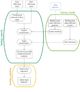
matching at the protein level. Kodoja has three main components, summarized in 147
Figure 1: (a) kodoja_build for database generation for Kraken and Kaiju (b) 148
kodoja_search for the taxonomic classification of RNA-seq reads, and (c) 149
kodoja_retrieve for extraction of viral sequences by species for downstream analysis. 150
151
2.1.1. Kodoja_build: Database generation 152
For virus classification, the main Kodoja components (Kraken (16) and Kaiju (17)) 153
each require a database generated from the genome or proteome of known plant 154
viruses, and (if available) the genome or proteome of the plant host. Data download 155
and database generation are achieved using the kodoja_build module. This module 156
downloads genomes and protein sequence files from RefSeq (20), and then 157
implements code from Kraken and Kaiju to generate tool-specific databases. The user 158
can specify if all viruses or only plant viruses are included in the databases. If a host 159
genome is available (either provided by the user or in RefSeq (20)), this can also be 160
added to the database for host sequence classification. 161
162
To make Kodoja easy to use, ready-made plant-specific viral databases for Kraken 163
and Kaiju are provided for download at https://doi.org/10.5281/zenodo.1406071. 164
These were generated by downloading all complete virus and viroid genomes and 165
protein sequence files in NCBI RefSeq (Release 89)(20) and selecting plant viruses 166
using information from the Virus-Host DB (21). For Kraken, k-mer size is specified 167
when building the database, and a k-mer size of 31 was used for the RNA-seq 168
datasets. 169
170
2.1.2. Kodoja_search: Taxonomic classification of virus reads 171
Kodoja_search is the main Kodoja component. RNA-seq reads are first quality 172
checked using Trimmomatic (22) which trims and discards low-quality reads. FastQC 173
(https://www.bioinformatics.babraham.ac.uk/projects/fastqc/) is used for summarizing 174
the read quality after trimming, and the FASTQC report forms part of Kodoja’s results. 175
Kraken (16) is then used for the nucleotide-level classification. Kraken is a sequence 176
classification algorithm for assigning taxonomic labels to short sequences (16). It does 177
this through dividing each sequence into k-mers and querying each against a k-mer
database. K-mers which are shared between organisms are mapped to the lowest 179
common ancestor, and this information is then used to build a subtree of the general 180
taxonomy tree for the classification of the sequences. In the tree, each node has a 181
weight equal to the number of k-mers in the sequence associated with the node’s 182
taxon. Each root-to-leaf path in the tree is scored by adding all the weights in the path. 183
The leaf of the path with the largest score is the classification used for the sequence 184
(16). The use of the k-mer database makes the classification algorithm very fast 185
compared to alignment based methods (11). 186
187
In the next step full length sequence reads are translated and classified at the protein-188
level using Kaiju (17). Kaiju translates the sequences into six frames and splits the 189
resulting translations into fragments using translation termination codons (UAG, UAA, 190
UGA). Kaiju balances precision and sensitivity by using a minimum fragment length 191
parameter. We used a minimum fragment length of 15 and the number of mismatches 192
permitted was one. Fragments are queried against a protein database using a 193
modified version of the backwards search algorithm in the Burrows–Wheeler transform 194
(23). A key component of sequence classification for both Kraken and Kaiju is the tool-195
specific database. We have provided pre-computed plant virus databases that can be 196
used directly with the Kodoja workflow, but custom databases can also be made using 197
kodoja_build (see section 2.1.1). 198
199
Implementation of the kodoja_search module results in reads being assigned to 200
taxonomic classes by both Kraken and Kaiju. Reads assigned to the same virus class 201
by both tools (set intersection) are designated as stringent assignments; and reads 202
assigned to a virus class by either Kraken or Kaiju (set union) are assigned as non-203
stringent assignments. The assignments are given in a results summary, which 204
includes the reads counts for each type of assignment. Full results from Kraken and 205
Kaiju are also provided so that users can analyse these data further, outside of the 206
Kodoja workflow. 207
208
2.1.3 Kodoja_retrieve: Extraction of viral reads 209
This module can be used to extract species-specific sequences for downstream 210
analysis outside of the Kodoja workflow. The user can specify retrieval of reads 211
classified to a species, and/or genus, using either stringent or non-stringent 212
assignments. The ability to retrieve and download all reads assigned to a specific virus 213
gives the user the potential to assemble complete viral genomes for further analysis. 214
215
2.1.4 Kodoja workflow availability 216
Kodoja is available for direct installation and use at the command line in Linux through 217
Bioconda (18) (https://anaconda.org/bioconda/kodoja). Alternatively, the code can be 218
downloaded from github (https://github.com/abaizan/kodoja). Kodoja is also provided 219
as a package in Galaxy, an open source web-based analytical environment for data 220
analysis (19). This is available on GitHub (https://github.com/abaizan/kodoja_galaxy) 221
Developing Kodoja as a package within Galaxy makes it available to researchers with 223
a local installation of Galaxy, and allows analysis to be completed with no command 224
line input. By using an open source workflow platform in this way, the tool can also 225
potentially be used on a cloud-based Galaxy server. 226
227
2.2 Benchmarking Kodoja using existing datasets 228
Kodoja was tested on three publicly available RNA-seq grapevine datasets (24) 229
analysed for the presence of viral sequences (25). In the original work sequencing 230
data for 11 grapevine samples was obtained, including multiple samples from skin, 231
grain, and seed (24). In the analysis work viral sequences were identified using contig 232
building and subsequence Blast alignment of contigs to a reference viral database 233
(25). For the Kodoja benchmarking, we selected one library from grain (G1R1) 234
(Sequence Read Archive (SRA) identifier SRR866540), skin (S3R1) (SRA: 235
SRR866571) and seed (S3R3) (SRA:SRR866576); representative of those datasets 236
with the largest and most diverse viromes. These datasets are denoted GV1, GV2 and 237
GV3 respectively in the current analysis. 238
239
2.2.1. Assembly and alignment for confirmation 240
To confirm the viruses predicted by Kodoja, kodoja_retrieve was used to extract reads 241
assigned to each virus. Reads for each virus were then assembled using Trinity (26) 242
with minimum contig length of 200 nucleotides. The longest contig for each virus was 243
then aligned against the NCBI non-redundant nucleotide database using Blastn, and 244
the match with lowest e-value selected for taxonomic comparison. Where too few 245
reads were available for contig assembly, all reads assigned to a virus species by 246
Kodoja were aligned. 247
248
2.3. Applying Kodoja to virus detection in Raspberry (Rubus idaeus) 249
Kodoja was then applied to RNA-seq libraries generated from two raspberry plants of 250
variety Glen Dee (denoted D5 and D6) collected from a commercial raspberry 251
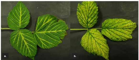
plantation in Angus, Scotland, UK. Both plants showed viral infection symptoms: D5 252
showed vein yellowing and D6 showed leaf blade yellowing (Figure 2). 253
254
2.3.1 RNA-sequencing 255
Symptomatic leaves were collected from each plant (D5 and D6) and frozen at -80ºC 256
for long-term storage (15 months). Two samples of leaf were placed in a clean, 257
autoclaved 2 ml Eppendorf tube together with a sterile 3 mm glass bead, frozen with 258
liquid nitrogen and then powdered using a bead beater (Qiagen TissueLyser). Then 259
100 mg of powdered leaf was resuspended in a mixture of 450 µl Qiagen RNeasy 260
Plant Mini Kit buffer RLT, 45 µl Ambion Plant RNA isolation aid and 4.5 µl 2-261
mercaptoethanol. Thereafter the RNA extraction followed the manufacturer’s 262
instructions to the RNeasy kit, and the RNA was eluted in RNAse-free water. The RNA 263
was supplied to the Glasgow Polyomics facility (UK) for quality control, ribosomal RNA 264
depletion, library preparation (paired-end 200 bp) and high-throughput sequencing 265
sample D5 and D6 comprised 64 M and 62 M reads respectively (available from the 267
European Nucleotide Archive (27) under accessions ERR2784286 and ERR2784287 268
respectively). 269
270
2.3.2 Kodoja analysis of raspberry RNA-seq datasets 271
The Kodoja workflow was run on the two raspberry RNA-seq datasets, using the 272
draft genome of black raspberry (Rubus occidentalis)(28) as the host in the Kraken 273
database build. 274
275 276
2.3.3 Assembly and alignment for confirmation 277
To confirm the predicted viruses, kodoja_retrieve was used to extract reads assigned 278
to each virus species; and contigs were assembled and aligned to a reference 279
database as described in section 2.2.1. 280
281
2.3.4 PCR confirmation of virus sequences 282
To confirm that the viruses identified by Kodoja were present, new samples of total 283
RNA were extracted from the frozen leaves of sample D5 and D6 using the Thompson 284
buffer method as described previously (29) and eluted in RNAse-free water. For 285
detection of Raspberry leaf mottle virus (RLMV) the plant RNA was converted to cDNA 286
using SuperScript III (Invitrogen) reverse transcriptase and random hexamer primer 287
following the manufacturer’s instructions. For other RNA viruses (Raspberry leaf
288
blotch virus (RLBV) and Beet ringspot virus (BRSV)) the extracted plant RNA was
289
added directly to a 25 µl illustra Ready-to-Go RT-PCR bead (GE Healthcare) reaction 290
together with virus-specific PCR primers (Table 1). To detect the DNA plant virus 291
Rubus yellow net virus (RYNV), six 1 cm diameter frozen D5 and D6 leaf discs were
292
extracted using the DNeasy Plant Mini Kit (Qiagen) according to the manufacturer’s 293
instructions. RYNV was detected in the eluted DNA by amplification in a 25 µl illustra 294
Ready-to-Go PCR bead (GE Healthcare) reaction with virus-specific primers (Table 295
1). Positive controls for virus-detection were RNAs extracted from raspberry plants 296
previously demonstrated to carry specific viruses. 297
298
3.0 Results 299
300
3.1 Benchmarking of Kodoja workflow on RNA-seq from grapevine 301
The Kodoja workflow was applied to three publicly available RNA-seq libraries 302
generated from grapevine (24) and analysed for virus sequences (25). The viral 303
sequences detected [with stringent level assignments for viruses and non-stringent for 304
viroids (as viroids do not have protein assignments in RefSeq)] in each sample are 305
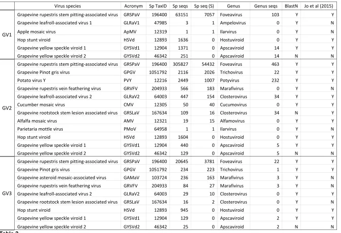
summarized in Table 2. Kodoja identified 6, 12 and 9 virus sequences in samples GV1, 306
GV2 and GV3 respectively. For each sample, Kodoja identified all the viral sequences 307
reported in the previous study (25), and in addition, identified 8 viral sequences not 308
reported in the previous study; Grapevine leafroll-associated virus 1 (GLRaV1), Apple
309
mosaic virus (ApMV), Grapevine yellow speckle viroid 2 (GYSVd2), Grapevine rupetris
vein feathering virus (GRVFV), Parietaria mottle virus (PMoV), Grapevine asteroid
311
mosaic-associated virus (GAMaV) and Grapevine rootstock stem lesion associated
312
virus (GRSLaV) (Table 2). One explanation for the identification of additional virus
313
sequences, could be their submission to GenBank after the date of the previous study 314
(2015). However, 6 of the additional sequences have GenBank submission dates prior 315
to 2011 and only GRVFV and GAMaV have submission dates after 2014 (GAMaV: 316
2016 and GRVFV: 2017). GAMaV was identified by Kodoja in GV3, and GRVFV was 317
identified in GV2 and GV3. Only two viruses reported in the previous study were not 318
identified by Kodoja: Grapevine Pinot Gris virus (GPGV) in GV1, and the Oat blue
319
dwarf virus (OBDV) in GV3.
320 321
Overall, 85.2% (23/27) of virus species identified by Kodoja were confirmed by the 322
contig assembly and Blast alignment process (Table 2). This included viruses that had 323
not been identified in the previous study (including GRVFV in GV2 and GV3, and 324
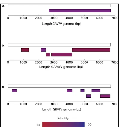
GAMaV in GV3). Contig mapping to reference genomes for these two viruses, showed 325
that multiple and extensive regions of the virus genomes were present in the dataset 326
(Figure 3). However, two viruses identified by Kodoja were classified as different 327
species by contig assembly and Blast alignment. GYSVd2 was a viroid identified in all 328
three samples by Kodoja (Table 2), but was classified as Grapevine yellow speckle
329
viroid 1 (GYSVd1) by the confirmation process. GRSLaV sequences were identified
330
in GV2 by both Kodoja and Jo et al., 2015 but the sequences were classified as 331
Grapevine leafroll-associated virus 2 (GLRaV2) by the confirmation process.
332 333
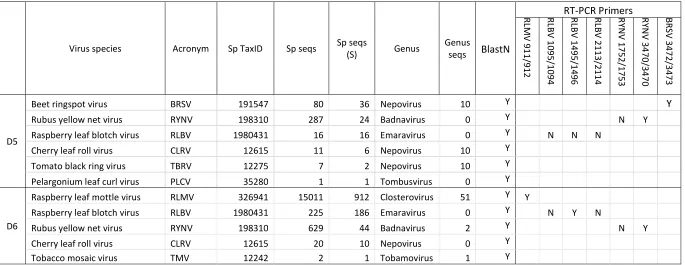
3.2 Application of Kodoja for the detection of viruses in Raspberry 334
Kodoja was then applied to the identification of virus sequences in two field-grown 335
raspberry plants with virus-like symptoms (Figure 2). Classifying reads with stringent 336
assignments only, Kodoja identified six viruses in D5 and five viruses in D6 (Table 3). 337
This included Raspberry leaf blotch virus (RLBV), Rubus yellow net virus (RYNV) and 338
Cherry leaf roll virus (CLRV) detected in both samples; and Beet ringspot virus (BRSV)
339
detected in D5 only and Raspberry leaf mottle virus (RLMV) detected in D6 only. The 340
contig assembly and Blast confirmation process showed that all the assembled contigs 341
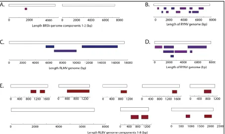
corresponded to the viruses identified by Kodoja (Table 3). Contig mapping to 342
reference genomes for selected viruses, showed that multiple and extensive regions 343
of RYNV, RLMV and RLBV genomes were present in the datasets, but only a very 344
short region of the BRSV genome was detected (Figure 4). 345
346
In a further confirmation step, RT-PCR was done with a previously used virus-specific 347
primer pair for each of RLMV, RLBV and RYNV (Table 1; primers designed using 348
previously published sequences). These primers detected RLMV in D6 only as 349
predicted by Kodoja (Table 3). However, these primers did not detect RLBV or RYNV 350
in either D5 or D6 as predicted by Kodoja (Table 3). Hence, samples D5 and D6 were 351
tested with three additional RLBV primer pairs [1491/1492, 1495/1496, 2113/2114 352
(Table 1)] that target three different RLBV RNAs (RLBV has eight viral RNAs in total) 353
obtained with primer pair 1491/1492, suggesting a low level of RLBV RNA was present 355
in this sample. However, none of the other RLBV-specific primer pairs produced a 356
positive result for RLBV in D6. None of the 4 RLBV primer pairs gave amplification 357
bands for D5, despite Kodoja predicting the virus was present and despite these 358
primers producing a strong amplification of RLBV from a positive control plant. It 359
should be mentioned that contamination of material submitted for deep sequencing 360
can occur, particularly when preparation work is done in laboratories lacking 361
designated clean rooms. This could be an alternative explanation for the failure to 362
confirm the presence of RLBV by RT-PCR from the D5 and D6 samples. 363
364
An additional primer pair was then designed for RYNV [3470/3471(Table 1)] based on 365
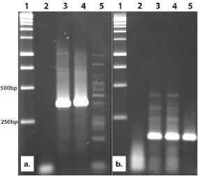
the sequence assembled from D6 and was tested on samples D5 and D6. This RT-366
PCR gave an amplification band for both D5 and D6 but produced non-specific 367
amplification with a RYNV positive control plant (Figure 5A). In an additional test, a 368
new BRSV RNA2-specific primer pair [3472/3473 (Table 1)] was designed based on 369
the sequences assembled from D5. This primer pair detected BRSV in both D5 and 370
D6, even though Kodoja only predicted the presence of BRSV in D5 (Figure 5B). 371
372
Discussion 373
We have developed and applied a new computational workflow (Kodoja) for the 374
identification of plant virus sequences in RNA-seq data. The testing of Kodoja on 3 375
existing RNA-seq datasets from grapevine showed it had increased sensitivity 376
compared to an analysis comprising the traditional tools of contig building and Blast 377
alignment. The previous analysis identified a total of 19 (non-unique) viruses across 378
the 3 samples (25), but Kodoja identified 27. This increased sensitivity comes from the 379
use of k-mer profiling, rather than contig assembly. The ability of Kodoja, to identify 380
virus sequences present at lower levels than are detectable using contig building 381
methods, means that viruses could be detected in plants before symptoms appear. 382
This sensitivity was also exemplified when Kodoja was applied to raspberry. RYNV 383
was reported with just 44 reads meeting the stringent classification criteria, and the 384
presence of this virus sequence was confirmed by the RT-PCR. 385
386
The work to benchmark Kodoja using existing datasets gave insights into the difficulty 387
of viral sequence identification in mixed (comprising both plant and potentially viral, 388
bacterial and fungal nucleotides) RNA-seq datasets. One key complexity, when a 389
workflow does not include contig building, is the miss-classification of viruses, which 390
arises due to the small evolutionary distances existing between some viral taxa. This 391
was the case when Kodoja identified GYSVd1 as GYSVd2 and GLRaV2 as GRSLaV. 392
GYSVd1 and GYSVda are viroids, that have a single stranded circular RNA genome 393
that does not code for protein. Hence, the Kodoja assignment for this viroid was made 394
only at the nucleotide level, and this could explain its incorrect classification. In 395
addition, GLRaV2 was incorrectly classified as GRSLaV. GLRaV2 is known to be the 396
closest related virus to GRSLaV within the Closteroviridae family (30), with between 397
71-79% sequence identify across 9 ORFs and this could explain why the k-mer
analysis made an incorrect classification. The raspberry analysis showed that Kodoja 399
reports viruses even if they are present at reads at low levels. 400
401
The detection of known viruses using Kodoja is dependent upon the virus dataset used 402
to generate the k-mer databases for Kraken (16) and Kaiju (17). The size of the 403
databases will greatly influence both the sensitivity and the speed of the workflow. We 404
have used a dataset derived from RefSeq (v89)(20) which comprises 7946 non-405
redundant viral genome sequences. This means that one virus is represented by a 406
single reference sequence and variants are excluded. Hence, the workflow is in some 407
ways restrictive, and could potentially leave some sequences unclassified or miss-408
classified if they are derived from diverse sequence variants. An alternative to RefSeq 409
would be Genbank (31), which comprises 2.7 million redundant viral sequences 410
(v228) and includes virus variants. However, creating k-mer databases for such a large 411
dataset would be prohibitively expensive in terms of time taken for the database build 412
and running the kodoja_search module. A trade-off between run time and sensitivity 413
could potentially be achieved by using a new database Reference Viral database 414
(RVDB)(32). This database includes a clustered set of virus sequences, extracted 415
from Genbank (31) which comprises 561,676 representatives. This clustered 416
database was designed to retain viral diversity and reduce redundancy (32). It would 417
be possible to use this dataset to generate k-mer databases for Kodoja that would 418
increase sensitivity further, without completely compromising speed. 419
420
The application of the Kodoja workflow to RNA-seq data from raspberry demonstrated 421
that field-grown raspberry plants can frequently be infected with multiple viruses, and 422
that relying on visual symptoms to identify viruses is often not possible. In addition, 423
this work clearly demonstrated the limitations of primer-based methods for virus 424
detection (RT-PCR and PCR). The innate variability in the nucleotide sequence of 425
plant viruses means that it is very difficult/impossible to design diagnostic primers that 426
can detect many/all isolates of the same virus. For RYNV, the primer pair 1752/1753 427
gave strong amplification of the isolate carried within our positive control plant but 428
could not amplify the virus in D5 and D6. 429
430
The prediction of BRSV, a nepovirus, in D5 and the creation of a new PCR-primer pair 431
based on the D5 sequences, represents a step forward in virus testing for raspberry. 432
Nepoviruses are soil-borne, nematode-transmitted, viruses that are recognized as 433
important pathogens of many crops, including raspberries (33). Historically, when 434
serological reactions and host ranges were used to characterise viruses, BRSV was 435
thought to be an isolate of Tomato black ring virus (TBRV) (34). However, it is now 436
clear that BRSV is a different virus to TBRV (35), and the BRSV test we have designed 437
here will now become part of the battery of molecular tests we use for virus testing of 438
raspberry. 439
440
The sequencing and analysis of small RNAs (sRNA-seq) has also proved successful 441
has been developed for this purpose (12). Whilst Kodoja is optimized for RNA-seq 443
datasets, we did apply Kodoja to a previously published sRNA-seq dataset from 444
Grapevine (37) (unpublished data), however the success of Kodoja was less clear 445
than for the RNA-seq datasets. Using Kodoja, we detected all viruses reported in the 446
original study, but in addition a further 16 viruses were detected. However, these 447
additional viruses could not be validated as the read counts were low and made contig 448
building impossible. Further optimization and benchmarking would be required for 449
Kodoja to be used effectively on sRNA-seq datasets. 450
451
The testing and application of Kodoja, has exemplified its ability to be used 452
successfully for virus identification in RNA-seq datasets. Kodoja is the first workflow 453
to apply a k-mers analysis method for virus detection specifically in plants, and in 454
addition it is the first plant virus detection workflow to be made available through 455
BioConda and as a Galaxy application. This accessibility will make it available to a 456
wide range of researchers, working on diverse plant species. Our application of the 457
workflow to raspberry has highlighted its potential to develop new primers to enhance 458
serological testing and such advances will also be possible with other crops. 459 460 Author Statements 461 462 Funding 463
This work was supported by the Biotechnology and Biological Sciences Research 464
Council [BB/N023293/1]. The work of LT, SJ, SM and PC was additionally supported 465
by the Scottish Government’s Rural and Environment Science and Analytical Services 466
division (RESAS). 467
468
Conflict of Interest 469
No conflicts of interest are declared. 470
471
Acknowledgments 472
We would like to thank the Bioinformatics Unit, School of Medicine, University of St 473
Andrews, North Haugh, St Andrews, UK, KY16 9TF, for helpful discussions. 474
475 476
References 477
1. Nicaise V. Crop immunity against viruses: outcomes and future challenges. 478
Front Plant Sci. 2014;5:1–18. 479
2. Valkonen JPT. Viruses: Economical Losses and Biotechnological Potential. In: 480
Potato Biology and Biotechnology: Advances and Perspectives. Elsevier; 481
2007. p. 619–42. 482
3. Sasaya T, Nakazono-Nagaoka E, Saika H, Aoki H, Hiraguri A, Netsu O, et al. 483
Transgenic strategies to confer resistance against viruses in rice plants. Front 484
Microbiol. 2013;4:1–11. 485
4. Lamichhane JR, Venturi V. Synergisms between microbial pathogens in plant 486
5. Martin RR, MacFarlane S, Sabanadzovic S, Quito D, Poudel B, Tzanetakis IE. 488
Blackberry Yellow Vein Disease ( BYVD ) Complex and Associated Viruses. 489
Plant Dis. 2013;97(2):168–82. 490
6. Mumford R, Boonham N, Tomlinson J, Barker I. Advances in molecular 491
phytodiagnostics - New solutions for old problems. Eur J Plant Pathol. 492
2006;116(1):1–19. 493
7. Boonham N, Kreuze J, Winter S, van der Vlugt R, Bergervoet J, Tomlinson J, 494
et al. Methods in virus diagnostics: From ELISA to next generation 495
sequencing. Virus Res . 2014;186:20–31. 496
8. Wu Q, Luo Y, Lu R, Lau N, Lai EC, Li W-X, et al. Virus discovery by deep 497
sequencing and assembly of virus-derived small silencing RNAs. Proc Natl 498
Acad Sci U S A. 2010;107(4):1606–11. 499
9. Jones S, Baizan-Edge A, MacFarlane S, Torrance L. Viral diagnostics in plants 500
using next generation sequencing: Computational analysis in practice. Front 501
Plant Sci. 2017;8:1–10. 502
10. Wilson MR, Naccache SN, Samayoa E, Biagtan M, Bashir H, Yu G, et al. 503
Actionable diagnosis of neuroleptospirosis by next-generation sequencing. N 504
Engl J Med . 2014;370(25):2408–17. 505
11. Flygare S, Simmon K, Miller C, Qiao Y, Kennedy B, Di Sera T, et al. 506
Taxonomer: an interactive metagenomics analysis portal for universal 507
pathogen detection and host mRNA expression profiling. Genome Biol . 508
2016;17(1):111. 509
12. Zheng Y, Gao S, Padmanabhan C, Li R, Galvez M, Gutierrez D, et al. 510
VirusDetect: An automated pipeline for efficient virus discovery using deep 511
sequencing of small RNAs. Virology . 2017;500:130–8. 512
13. Trifonov V, Rabadan R. Frequency Analysis Techniques for Identification of 513
Viral Genetic Data. MBio. 2010;1(3):1–8. 514
14. Dröge J, Gregor I, Mchardy A. Taxator- tk: Precise taxonomic assignment of 515
aetagenomes by fast approximation of evolutionary neighbourhoods. 516
Bioinformatics. 2014;31(6):817–24. 517
15. Malhotra S, Sowdhamini R. Genome-wide survey of DNA-binding proteins in 518
Arabidopsis thaliana: analysis of distribution and functions. Nucleic Acids Res . 519
2013 Aug;41(15):7212–9. 520
16. Wood DE, Salzberg SL. Kraken: ultrafast metagenomic sequence 521
classification using exact alignments. Genome Biol . 2014;15(3):R46. 522
17. Menzel P, Ng KL, Krogh A. Fast and sensitive taxonomic classification for 523
metagenomics with Kaiju. Nat Commun . 2016;7:1–9. 524
18. Dale R, Grüning B, Sjödin A, Rowe J, Chapman BA, Tomkins-Tinch CH, et al. 525
Bioconda: A sustainable and comprehensive software distribution for the life 526
sciences. Nat Methods. 2018;15:475–6. 527
19. Afgan E, Baker D, van den Beek M, Blankenberg D, Bouvier D, Čech M, et al. 528
The Galaxy platform for accessible, reproducible and collaborative biomedical 529
analyses: 2016 update. Nucleic Acids Res . 2016;44:gkw343. 530
20. O’Leary NA, Wright MW, Brister JR, Ciufo S, Haddad D, McVeigh R, et al. 531
Reference sequence (RefSeq) database at NCBI: Current status, taxonomic 532
expansion, and functional annotation. Nucleic Acids Res. 2016;44(D1):D733– 533
45. 534
21. Mihara T, Nishimura Y, Shimizu Y, Nishiyama H, Yoshikawa G, Uehara H, et 535
al. Linking virus genomes with host taxonomy. Viruses. 2016;8(3):10–5. 536
sequence data. Bioinformatics . 2014 May 28;1–7. 538
23. Li H, Durbin R. Fast and accurate short read alignment with Burrows-Wheeler 539
transform. Bioinformatics. 2009;25(14):1754–60. 540
24. Da Silva C, Zamperin G, Ferrarini A, Minio A, Dal Molin A, Venturini L, et al. 541
The High Polyphenol Content of Grapevine Cultivar Tannat Berries Is 542
Conferred Primarily by Genes That Are Not Shared with the Reference 543
Genome. Plant Cell . 2013;25(12):4777–88. 544
25. Jo Y, Choi H, Kyong Cho J, Yoon J-Y, Choi S-K, Kyong Cho W. In silico 545
approach to reveal viral populations in grapevine cultivar Tannat using 546
transcriptome data. Sci Rep . 2015;5(1):15841. 547
26. Grabherr MG, Haas BJ, Yassour M, Levin JZ, Thompson D a, Amit I, et al. 548
Full-length transcriptome assembly from RNA-Seq data without a reference 549
genome. Nat Biotechnol . 2011;29(7):644–52. 550
27. Silvester N, Alako B, Amid C, Cerdeño-Tarrága A, Clarke L, Cleland I, et al. 551
The European Nucleotide Archive in 2017. Nucleic Acids Res. 552
2018;46(D1):D36–40. 553
28. VanBuren R, Bryant D, Bushakra JM, Vining KJ, Edger PP, Rowley ER, et al. 554
The genome of black raspberry (Rubus occidentalis). Plant J. 2016;87(6):535– 555
47. 556
29. Macfarlane S, Mcgavin W, Tzanetakis I. Virus Testing by PCR and RT-PCR 557
Amplifi cation in Berry Fruit. In: Lacomme C, editor. Plant Pathology: 558
Techniques and Protocols . Springer; 2015. p. 227–48. 559
30. Alkowni R, Zhang YP, Rowhani A, Uyemoto JK, Minafra A. Biological, 560
molecular, and serological studies of a novel strain of grapevine leafroll-561
associated virus 2. Virus Genes. 2011;43(1):102–10. 562
31. Benson DA, Cavanaugh M, Clark K, Karsch-Mizrachi I, Lipman DJ, Ostell J, et 563
al. GenBank. Nucleic Acids Res. 2013;41(D1):36–42. 564
32. Goodacre N, Aljanahi A, Nandakumar S, Mikailov M, Khan AS. A Reference 565
Viral Database ( RVDB ) To Enhance Bioinformatics Analysis of High-566
Throughput Sequencing for Novel Virus. mSphere. 2018;3(2):1–18. 567
33. Martin RR, Polashock JJ, Tzanetakis IE. New and emerging viruses of 568
blueberry and cranberry. Viruses. 2012;4(11):2831–52. 569
34. Harrison B. Relationship between Beet Ringspot, Potato Bouquet and Tomato 570
Black Ring Viruses. J Gen Microbiol. 1958;18:450–60. 571
35. Kis S, Salamon P, Kis V, Szittya G. Molecular characterization of a beet 572
ringspot nepovirus isolated from Begonia ricinifolia in Hungary. Arch Virol. 573
2017;162(11):3559–62. 574
36. Niu X, Sun Y, Chen Z, Li R, Padmanabhan C, Ruan J, et al. Using small RNA-575
seq data to detect siRNA duplexes induced by plant viruses. Genes (Basel). 576
2017;8(6):1–8. 577
37. Barrero RA, Napier KR, Cunnington J, Liefting L, Keenan S, Frampton RA, et 578
al. An internet-based bioinformatics toolkit for plant biosecurity diagnosis and 579
surveillance of viruses and viroids. BMC Bioinformatics . 2017;18(1):26. 580
38. Federhen S. The NCBI Taxonomy Database. Nucleic Acids Res. 581
2012;40(D1):D136–43. 582
Table and Figure Legends 585
586
Table 1. 587
Information on the RT-PCR primer pairs for the confirmation of four raspberry viruses; 588
Raspberry leaf mottle virus (RLMV), Raspberry leaf blotch virus (RLBV), Rubus yellow 589
net virus (RYNV) and Beet ringspot virus (BRSV) predicted to be present in raspberry 590
samples D5 and D6 by Kodoja. 591
592
Table 2. 593
Kodoja results for the three RNA-sequence datasets from grapevine. The species 594
taxonomic identify from the NCBI Taxonomy database (38) is shown in column 4 (Sp 595
TaxID), the number total number of reads that were classified to each virus species is 596
shown in column 5 (Sp seq), the number of reads classified by both Kaiju and Kraken 597
(stringent for viruses) (Sp seq (S)) is shown in column 6. The results of the contig 598
building and BlastN confirmation process are indicated in column 9. Y indicates that 599
the BlastN alignment assigned the sequences to the same species as Kodoja. N 600
indicates that BlastN assigned the sequences to a different species to Kodoja. The 601
detection of the viruses in the original work is indicated in Column 10. 602
603
Table 3. Kodoja results for the 2 RNA-seq datasets from raspberry. The column 604
headers are as described for Table 2. The results of the RT-PCR confirmation 605
experiments are indicated in the last 7 columns, with Y indicating the virus was 606
detected with the specified primer pair and N indicating the virus was not detected. 607
608
Figure 1. 609
Flow diagram summarizing the 3 modules of the Kodoja workflow: kodoja_build, 610
kodoja_search and kodoja_retrieve. 611
612
Figure 2. 613
Images of leaves taken from two Glen Dee raspberry plants grown on a commercial 614
farm in Angus, Scotland, UK. (A) Plant D5 showing major vein yellowing and (B) 615
Plant D6 showing leaf blade yellowing. 616
617 618 619
Figure 3. 620
Diagrammatic alignments of selected virus contigs to their reference genomes. (A) 621
Alignment for Grapevine rupestris vein feathering virus (GRVFV) from dataset GV2, 622
(B) Alignment for Grapevine asteroid mosaic-associated virus (GAMaV) from GV3, (C) 623
Alignment for GRVFV from dataset GV3. 624
625
Figure 4. 626
Diagrammatic alignments of the selected virus contigs to their reference genomes. (A) 627
Raspberry leaf mottle virus (RLMV) from D6, (D) RYNV from D6 and (E) Raspberry 629
leaf blotch virus from D6. 630
631
Figure 5. Virus detection by RT-PCR in raspberry. (A) Raspberry yellow net virus 632
(RYNV) amplified with primers 3470/3471. (B) BRSV amplified with primers 633
3472/3473. Within each panel, lane 1 is kilobase DNA markers (500bp and 250bp 634
markers are indicated), lane 2 is water only amplification, lane 3 is sample D5 RNA, 635
lane 4 is sample D6 RNA, lane 5 is RNA extracted from a known RYNV-infected (A) 636
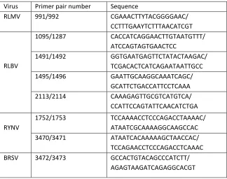
Virus Primer pair number Sequence
RLMV 991/992 CGAAACTTYTACGGGGAAC/ CCTTTGAAYTCTTTAACATCGT
RLBV
1095/1287 CACCATCAGGAACTTGTAATGTTT/ ATCCAGTAGTGAACTCC
1491/1492 GGTGAATGAGTTCTATACTAAGAC/ TCGACACTCATCAGAATAATTGCC 1495/1496 GAATTGCAAGGCAAATCAGC/
GCATTCTGACCATTCCTCAAA 2113/2114 CAAAGAGTTGCGTCATGTCA/
CCATTCCAGTATTCAACATCTGA
RYNV
1752/1753 TCCAAAACCTCCCAGACCTAAAAC/ ATAATCGCAAAAGGCAAGCCAC 3470/3471 ATAATCACAAAAAGCTAACCAC/ TCCAGAACCTCCCAGACCTCAAAC BRSV 3472/3473 GCCACTGTACAGCCCATCTT/
[image:17.595.73.397.97.353.2]AGAGTAAGATCAGAGGCACGT
18
Virus species Acronym Sp TaxID Sp seqs Sp seq (S) Genus Genus seqs BlastN Jo et al (2015)
GV1
Grapevine rupestris stem pitting-associated virus GRSPaV 196400 63151 7057 Foveavirus 103 Y Y Grapevine leafroll-associated virus 1 GLRaV1 47985 3 1 Ampelovirus 0 Y N
Apple mosaic virus ApMV 12319 1 1 Ilarvirus 0 Y N
Hop stunt viroid HSVd 12893 1636 0 Hostuviroid 0 Y Y
Grapevine yellow speckle viroid 1 GYSVd1 12904 1371 0 Apscaviroid 14 Y Y Grapevine yellow speckle viroid 2 GYSVd2 46342 251 0 Apscaviroid 14 N N
GV2
Grapevine rupestris stem pitting-associated virus GRSPaV 196400 305827 54432 Foveavirus 463 Y Y Grapevine Pinot gris virus GPGV 1051792 2116 2026 Trichovirus 22 Y Y
Potato virus Y PVY 12216 2449 1007 Potyvirus 232 Y Y
Grapevine rupestris vein feathering virus GRVFV 204933 566 183 Marafivirus 0 Y N Grapevine leafroll-associated virus 2 GLRaV2 64003 447 154 Closterovirus 34 Y Y Cucumber mosaic virus CMV 12305 50 40 Cucumovirus 0 Y Y Grapevine rootstock stem lesion associated virus GRSLaV 167634 109 16 Closterovirus 34 N Y Alfalfa mosaic virus AMV 12321 19 15 Alfamovirus 0 Y Y Parietaria mottle virus PMoV 64958 1 1 Ilarvirus 0 Y N
Hop stunt viroid HSVd 12893 1604 0 Hostuviroid 0 Y Y
Grapevine yellow speckle viroid 1 GYSVd1 12904 440 0 Apscaviroid 5 Y Y Grapevine yellow speckle viroid 2 GYSVd2 46342 129 0 Apscaviroid 5 N N
GV3
Grapevine rupestris stem pitting-associated virus GRSPaV 196400 20645 3781 Foveavirus 22 Y Y Grapevine Pinot gris virus GPGV 1051792 234 223 Trichovirus 1 Y Y Grapevine asteroid mosaic-associated virus GAMaV 103724 236 163 Marafivirus 3 Y N Grapevine rupestris vein feathering virus GRVFV 204933 84 27 Marafivirus 3 Y N Grapevine leafroll-associated virus 2 GLRaV2 64003 29 10 Closterovirus 0 Y Y Grapevine rootstock stem lesion associated virus GRSLaV 167634 16 2 Closterovirus 0 Y N
Hop stunt viroid HSVd 12893 945 0 Hostuviroid 0 Y Y
Grapevine yellow speckle viroid 1 GYSVd1 12904 129 0 Apscaviroid 2 Y Y
[image:18.842.74.747.48.508.2]Grapevine yellow speckle viroid 2 GYSVd2 46342 25 0 Apscaviroid 2 N N
19
Virus species Acronym Sp TaxID Sp seqs Sp seqs
(S) Genus
Genus
seqs BlastN
RT-PCR Primers
RLMV
911
/912
RLBV 109
5/10
94
RLBV 149
5/14
96
RLBV 211
3/21
14
RY
N
V 17
52/1
7
53
RY
N
V 34
70/3
4
70
BRS
V 34
72/3
473
D5
Beet ringspot virus BRSV 191547 80 36 Nepovirus 10 Y Y
Rubus yellow net virus RYNV 198310 287 24 Badnavirus 0 Y N Y
Raspberry leaf blotch virus RLBV 1980431 16 16 Emaravirus 0 Y N N N
Cherry leaf roll virus CLRV 12615 11 6 Nepovirus 10 Y
Tomato black ring virus TBRV 12275 7 2 Nepovirus 10 Y
Pelargonium leaf curl virus PLCV 35280 1 1 Tombusvirus 0 Y
D6
Raspberry leaf mottle virus RLMV 326941 15011 912 Closterovirus 51 Y Y
Raspberry leaf blotch virus RLBV 1980431 225 186 Emaravirus 0 Y N Y N
Rubus yellow net virus RYNV 198310 629 44 Badnavirus 2 Y N Y
Cherry leaf roll virus CLRV 12615 20 10 Nepovirus 0 Y
[image:19.842.74.759.104.369.2]20
21
22
23
24