Dr. Abbas Taher
Dept of OMF Surgery, Faculty of Dentistry, University of Kufa, Iraq.
E-mail: [email protected]
Address for correspondence
Access this article online
www.japer.in
Amelioration of Cerebral Ischemia reperfusion injury by
Rosuvastatin via interference with inflammatory response and
apoptosis
Introduction
The term ischemia reperfusion injury (IRI) describes the experimentally and clinically prevalent finding of tissue ischemia with inadequate oxygen supply followed by successful reperfusion initiates a wide and complex array of inflammatory responses that may both aggravate local injury as well as induce impairment of remote organ function 1.
Ischemia-reperfusion injury contributes to the pathophysiology of many conditions, include the different forms of acute vascular occlusions such as stroke, myocardial infarction, peripheral vascular insufficiency and hypovolemic shock With the relevant reperfusion
strategies like thrombolytic therapy, coronary angioplasty, cardiopulmonary bypass and operative revascularization 2. Cerebral ischemia leads to energy
depletion and cell death, which can stimulate immune responses, leading to inflammatory cells activation and infiltration. Reperfusion of the occluded vessel, either spontaneously or by the collateral circulation or by therapeutic recanalization, leads to the generation of reactive oxygen species (ROS) that are delivered with the reperfused oxygenated blood or produced within brain and immune cells. ROS can then stimulate ischemic cells, even ischemic neurons, to secrete inflammatory chemokines and cytokines that enhance the biosynthesis of adhesion molecule in the cerebral vasculature and also lead to peripheral leukocyte recruitment 3. As inflammatory cells become activated,
they can release a variety of cytotoxic molecules including more cytokines, matrix metalloproteinases (MMPs), nitric oxide (NO) and more ROS. These Objectives: The objective of this study is to investigate the possible cerebroprotective potential of rosuvastatin in brain ischemia reperfusion injuryvia interfering with inflammation and oxidative pathway and apoptosis.
Materials and Methods: Adult albino rats were randomized into four groups as follow: Group (1) sham group: the rats were subjected to the same surgical procedures as other groups but the common carotid arteries were not occluded; Group (2) control (ischemic-reperfused) group: the rats were subjected to the same surgical procedures as other groups with bilateral common carotid artery occlusion (BCCAO) for 30 min. followed by reperfusion for 1 hr but without drug; Group (3) control vehicle group: three days before surgery, rats received daily the vehicle of rosuvastatin drug, normal saline (0.9% Nacl) (1 ml/kg/day) intraperitoneally, then anesthesia and surgery with BCCAO for 30 min. followed by reperfusion for 1 hr were done and Group(4) rosuvastatin treated group: rats received daily rosuvastatin intraperitoneally. The dose of rosuvastatin was (10 mg/kg /day) for three days before the surgery, then anesthesia and surgery with BCCAO for 30 min. followed by reperfusion for 1 hr.
Results: At the end of the experiment, the levels of cerebral TNF-α, IL-6,
IL-10,Caspase-3,Bax,MDA,CD4 and MPO significantly (p < 0.05) increased in control group as compared with the sham group and the level of cerebral GSH significantly (p < 0.05) decreased in control group as compared with the sham group, while there was insignificant difference in cerebral levels of CD8 between the four experimental groups. Histopothological analysis showed that rats in control group showed significant cerebral injury. Treatment with rosuvastatin significantly counteracted the increase in the cerebral levels of TNF-α, IL-6, IL-10,Caspase-3,Bax,MDA,CD4 and MPO and
the decrease of GSH. Histopathological analysis revealed that rosuvastatin significantly (P < 0.05) reduced the severity of cerebral injury in the rats underwent BCCAO.
Conclusions: The results of the study revealed that inflammatory cytokines, apoptosis pathways and oxidative stress mediators are involved in global cerebral ischemia induced by bilateral common carotid artery occlusion. Cerebral ischemia reperfusion injury can be modified by rosuvastatin via its anti-inflammatory, anti-apoptosis and anti-oxidant effects.
Keywords: Cerebral ischemia reperfusion injury, inflammation, apoptosis, oxidative stress, rosuvastatin.
ABSTRACT ABSTRACT ABSTRACT ABSTRACT
Najah R Hadi*
Hashim T Raheem1
Karrar J Kareem2 Kahlaa A Kadhim3 Abbas Taher4
*Najah R Hadi, Dept of Pharmacology and Therapeutics, Faculty of Medicine, University of Kufa, Iraq
1Hashim R Taresh, Dept of
Microbiology Faculty of Medicine, University of Kufa, Iraq
2Khalaa Kadim, Dept of Microbiology
Faculty of Medicine, University of Kufa, Iraq
3Karrar K, Dept of Pharmacology and
Therapeutics, Faculty of Medicine, University of Kufa, Iraq
4Abbas Taher, Dept of OMF Surgery,
Faculty of Dentistry, University of Kufa, Iraq
molecules may provoke more cell damage as well as disruption of the extracellular matrix and blood-brain barrier (BBB) 4,5. Secondary ischemic brain damage
occurs as a consequence of brain edema and post-ischemic microvascular stasis leading to hypoperfusion and post-ischemic inflammation 6,7.
The recruitment of peripheral circulating leukocytes into the brain parenchyma can produce an augmentation of inflammatory signal cascades, which will enhance the damage. These processes are mainly prominent during reperfusion which is often associated with microvascular injury, particularly due to increased permeability of capillaries and arterioles that lead to an increase of fluid filtration across the tissues. These activated endothelial cells produce more ROS following reperfusion, and results in a subsequent inflammatory response. White blood cells, carried to the area by the newly returning blood, release a mass of inflammatory factors such as interleukins (ILs) as well as free radicals in response to tissue damage. The restored blood flow reintroduces oxygen within cells that damages cellular proteins, DNA, and the plasma membrane. Damage to the cell's membrane may in turn causes the release of more free radicals. ROS may also act indirectly in redox signaling to turn on apoptosis. White blood cells may also bind to the endothelium of small capillaries, obstructing them and leading to more ischemia 8,9.
Early restoration of blood flow remains the treatment of choice for limiting brain injury following ischemic stroke. Improved educational efforts that emphasize the early signs and symptoms of stroke, coupled with the widespread application of thrombolytic therapy to patients with acute ischemic stroke have increased the number of patients benefiting from reperfusion 10.
While reperfusion of the ischemic brain is desirable, tissue damage may result from reperfusion only. Reperfusion appears to enhance the inflammatory response and causes additional injury to adjacent brain tissue 11. From experimental stroke, blocking
various aspects of the inflammatory cascade has shown to improve injury 8.Rosuvastatin belongs to a
new generation of statins which are 3-hydrox-3-methylglutaryl-coenzyme A (HMG-CoA) reductase. The study of Ma et al,2013 12 showed that
pretreatment with rosuvastatin extensively protects against reperfusion injury after middle cerebral artery occlusion, as shown by inhibition of neuronal cell death, and these helpful effects were associated with suppression of oxidative stress or inflammation-related pathways, such as reduction of an increase in cerebral superoxide and NADPH oxidase subunits, suppression of activation of microglia and macrophage, and inhibition of upregulation of NF-KB, iNOS and COX-2, so pretreatment with rosuvastatin reduced neuronal cell apoptosis in reperfusion injury through inhibition of inflammation 12 .
Materials and methods
Animals
The study was performed using 24 Adult albino rats weighting (200-250 g), provided by the animal house of high institute of infertility diagnosis and assisted reproductive technologies / Al-nahrain University. The rats were housed in the animal house of College of pharmacy/ Kufa University, in a room in which lighting was controlled (12 hr on, 12 hr off),
temperature was kept at (25±1°C) and humidity was kept at (60–65%) with unlimited access to food and water until the start of experiments. The Animal Investigation Committee (AIC) office of Kufa university approved the experimental protocol.
Preparation of rosuvastatin
Rosuvastatin was supplied by (Pioneer co. Sulaymanieyah/Kurdistan Iraq), and was prepared immediately before use by dissolving it in normal saline.
Experimental groups
group: The rats were subjected to the same surgical procedures as other groups with bilateral common carotid artery occlusion (BCCAO) for 30 min. followed by reperfusion for 1 hr but without drug; Group(3)control vehicle group:
Three days before surgery , rats received daily the vehicle of rosuvastatin drug, normal saline (0.9% Nacl) (1 ml/kg/day), intraperitoneally (iP) 13,14 then
anesthesia and surgery with BCCAO for 30 min. followed by reperfusion for 1 hr were done; Group(4) rosuvastatin treated group: The rats received daily rosuvastatin intraperitoneally (iP). The dose of rosuvastatin was (10 mg/kg /day) for three days before the surgery 13,14, then anesthesia and surgery
with BCCAO for 30 min. followed by reperfusion for 1 hr were done.
Induction of global brain ischemia
Each rat was anesthetized by intraperitoneal (iP) injection of 100 mg/kg of ketamine and 5 mg/kg of xylazine 15. Within few min, the rat became
unconscious, then placed in supine position and exposed to light source to keep it worm. After that a midline ventral small skin incision in the neck was made and the paratracheal muscles and fascia were splitted and pulled by stay sutures to expose the trachea, carotid arteries and vagal nerves. Both common carotid arteries were exposed, with special attention paid to separate and preserve the vagus nerve fibers and global cerebral ischemia was induced by BCCAO 16 by using vascular clamps for 30 min.
After 30 min of global cerebral ischemia, the clamps were removed to allow the reflow of blood through carotid arteries (reperfusion) for 1 hr 17.
Preparation of samples
Tissue preparation for TNF-α, IL-6, IL-10, Caspase-3
,Bax ,CD4,CD8, MPO-ANCA IgG, ,MDA and GSH
measurement
After decapitation, the brain was removed and washed in cold normal saline (0.9% Nacl) to remove any blood or debris and subsequently blotted on filter paper. Afterward, brain tissues were homogenized in
ice-cold 1:10 (w/v) 0.1 M phosphate-buffered saline (PBS) (pH 7.4), containing 1× protease inhibitor cocktail and 0.2% Triton X-100 for 30 seconds, using a high intensity ultrasonic liquid processor. The resulting homogenates were centrifuged at 15,000 g for 30 min, at 4°C, and supernatants were stored at
−80°C until analysis was done18.
Tissue preparation for histopathological analysis
and Scoring of brain damage
After 30 min. ischemia and 60 min.
reperfusion, decapitation was done and the brain was
removed and fixed with 10% formalin and embedded in paraffin wax and cut into coronal sections of 4-8μm thickness. The sections were stained with haematoxylin and eosin (H&E) dye for histopathological examination that was done by pathologist. The scoring system for the pathological changes in ischemia reperfusion injury isas follows19
: 0:(normal) = no morphological signs of damage; 1:(slight) = edema or eosinophilic or dark neurons (pyknotic) or dark/ shrunk cerebellar Purkinje cells; 2:(moderate)= at least two small hemorrhages; 3:(severe) = clearly infarctive foci (local necrosis).
Statistical analysis
All data are expressed as mean ± SEM. The difference between various groups were analyzed by one-way analysis of variance (ANOVA) followed by multiple comparison tests as Post Hoc. LSD. Non- parametric tests were used to assess the statistical significance of histopathological parameter. Cerebral lesions is a non-normally distributed variable. The Fisher exact test is used when members of two independent groups can fall into one of two mutually exclusive categories. The test is used to determine whether the proportions of those falling into each category differ by group. In all tests P< 0.05 was considered to be statistically significant.
Results
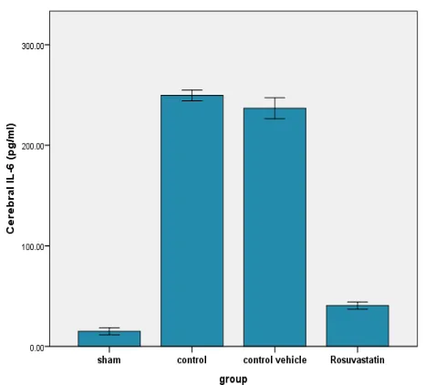
At the end of the experiment, the levels of cerebral TNF-α, IL-6, IL-10 significantly (P<0.05) increased in control group as compared with sham group. The levels of cerebral TNF-α and IL-6 of rosuvastatin treated group were significantly (p<0.05) lower than that of control-vehicle group. The levels of cerebral IL-10 of rosuvastatin treated group were significantly (p<0.05) higher than that of control-vehicle group. The values of cerebral TNF-α, IL-6, IL-10 are shown in figures (1, 2 and 3).
Figure (1): Error bar chart shows the difference in
mean± SEM values of cerebral TNF-α level (pg/mg) in the four experimental groups at the end of the experiment (No. of animals = 6 in each group). * P <
0.05 vs. sham group, ** P < 0.05 vs. control-vehicle group.
Figure (2): Error bar chart shows the difference in mean± SEM values of cerebral IL-6 level (pg/mg) in
the four experimental groups at the end of the
experiment (No. of animals = 6 in each group). * P < 0.05 vs. sham group, ** P < 0.05 vs. control-vehicle
group.
Figure (3): Error bar chart shows the difference in mean± SEM values of cerebral IL-10 level (pg/mg) in
the four experimental groups at the end of the experiment (No. of animals = 6 in each group). * P <
0.05 vs. sham group, ** P < 0.05 vs. control-vehicle group.
Effect on apoptptic markers (caspase-3 and Bax)
At the end of the experiment, the levels of cerebral caspase-3 and Bax significantly (P<0.05) increased in control group as compared with sham group. The levels of cerebral caspase-3 and Bax of rosuvastatin treated group were significantly (p<0.05) lower than that of control-vehicle group. The values of cerebral caspase-3 and Bax are shown in figures (4 and 5).
Figure (4): Error bar chart shows the difference in mean± SEM values of cerebral caspase-3 level (pg/mg) in the four experimental groups at the end of the experiment (No. of animals = 6 in each group). * P < 0.05 vs. sham group, ** P < 0.05 vs. control-vehicle
Figure (5): Error bar chart shows the difference in mean± SEM values of cerebral Bax level (pg/mg) in
the four experimental groups at the end of the experiment (No. of animals = 6 in each group). * P <
0.05 vs. sham group, ** P < 0.05 vs. control-vehicle group.
Effect on oxidative stress markers (MDA and GSH)
At the end of the experiment, the level of cerebral MDA significantly (P<0.05) increased in control group as compared with sham group, while the level of cerebral GSH significantly (P<0.05) decreased in control group as compared with sham group. The level of cerebral MDA of rosuvastatin treated group was significantly (p<0.05) lower than that of control-vehicle group, while the level of cerebral GSH of rosuvastatin treated group was significantly (p<0.05) higher than that of control-vehicle group. The values of cerebral MDA and GSH are shown in figures (6 and 7).
Figure (6): Error bar chart shows the difference in mean± SEM values of cerebral MDA level (pg/mg) in
the four experimental groups at the end of the experiment (No. of animals = 6 in each group). * P <
0.05 vs. sham group, ** P < 0.05 vs. control-vehicle group.
Figure (7): Error bar chart shows the difference in mean± SEM values of cerebral GSH level (pg/mg) in
the four experimental groups at the end of the experiment (No. of animals = 6 in each group). * P <
0.05 vs. sham group, ** P < 0.05 vs. control-vehicle group.
Effect on CD4+T-Lymphocytes
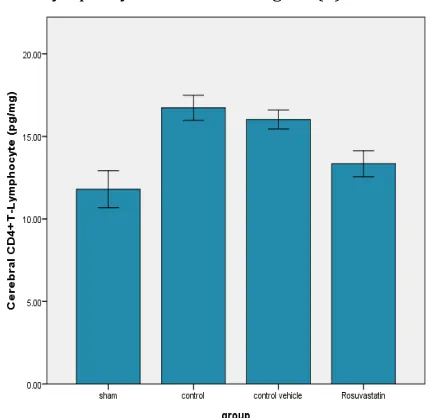
At the end of the experiment, the level of cerebral CD4+T-Lymphocytes significantly (P<0.05) increased in control group as compared with sham group. The level of cerebral CD4+T-Lymphocytes of rosuvastatin treated group was significantly (p<0.05) lower than that of control-vehicle group. The value of cerebral CD4+T-Lymphocytes is shown in figure (8).
Figure (8): Error bar chart shows the difference in mean± SEM values of cerebral CD4+T-Lymphocytes level (pg/mg) in the four experimental groups at the end of the experiment (No. of animals = 6 in each
Effect on CD8+T-Lymphocytes
At the end of the experiment, there was insignificant difference in cerebral level of CD8+T-Lymphocytes between the four experimental groups. The value of cerebral CD8+T-Lymphocytes is shown in figure (9).
Figure (9): Error bar chart shows the difference in
mean± SEM values of cerebral CD8+T-Lymphocytes level (pg/mg) in the four experimental groups at the end of the experiment (No. of animals = 6 in each
group).
Effect on Myeloperoxidas-Antineutrophil
Cytoplasmic Antibody IgG ( MPO -ANCA IgG)
At the end of the experiment, the level of cerebral MPO significantly (P<0.05) increased in control group as compared with sham group. The level of cerebral MPO of rosuvastatin treated group was significantly (p<0.05) lower than that of control-vehicle group. The changes in cerebral MPO is shown in table (1).
Table 1: Relation between rosuvastatin and control vehicle groups regarding MPO.
MPO +ve MPO -ve Total
Rosuvastatin 2 4 6
Control vehicle 6 0 6
Total 8 4 12
The fisher exact statistic value is significant at p<0.05.
Histopathological findings
The cerebral injury was assessed in the rat's brain of the four experimental groups according to 19
and the results were as follow: In the sham group, a cross sections of rat's brain showed normal
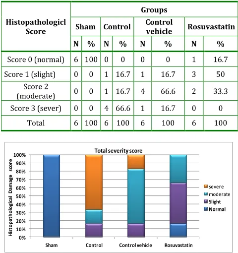
appearance (100%) as shown in table (2) and figure (10). Statistically, there was significant difference between control group and sham group, and the score of the control group showed that (66.6%) of the group had severe cerebral injury , (16.7%) had moderate cerebral injury and (16.7%) had slight cerebral injury as shown in table (2) and figure (10). Statistically, there was insignificant difference between control group and vehicle control group , and the score of the control vehicle group showed that (16.7%) had severe cerebral injury, (66.6%) had moderate cerebral injury and (16.7%) had slight cerebral injury as shown in table (2) and figure (10). pretreatment of rats with rosuvastatin improved cerebral injury score significantly as compared with control vehicle group and the score of this group showed that (16.7%) had normal histopathological appearance, (50%) had slight cerebral injury and (33.3%) had moderate injury as shown in table (2) and figure (10 and 11). The histopathological cerebral changes are shown in figures (11-16).
Table 2:
The differences in histopathological
grading of abnormal cerebral changes among the
four experimental groups.
Histopathologicl Score
Groups
Sham Control Control
vehicle Rosuvastatin
N % N % N % N %
Score 0 (normal) 6 100 0 0 0 0 1 16.7
Score 1 (slight) 0 0 1 16.7 1 16.7 3 50
Score 2
(moderate) 0 0 1 16.7 4 66.6 2 33.3
Score 3 (sever) 0 0 4 66.6 1 16.7 0 0
Total 6 100 6 100 6 100 6 100
Figure 10: Component bar chart shows the relative frequency of different histopathological grading of
abnormal cerebral changes among the four experimental groups.
0% 10% 20% 30% 40% 50% 60% 70% 80% 90% 100%
Sham Control Control vehicle Rosuvastatin
H
is
to
p
a
th
o
lo
g
ic
a
l
D
a
m
a
g
e
sc
o
re
Total severity score
severe
moderate
Figure 11: A Photomicrograph of normal rat's brain section shows normal tissue and the histopathological
score =0. The section stained with H&E (X 40).
Figure 12: Photomicrograph of rat's brain section after global cerebral ischemia shows edema(black arrow) and the histopathological score = 1 (Slight
injury). The section stained with H&E (X 40).
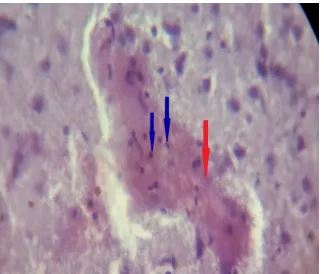
Figure 13: Photomicrograph of rat's brain section after global cerebral ischemia shows edema(black
arrow) and hemorrhage(red arrow). The histopathological score = 2 (Moderate injury). The
section stained with H&E (X 40).
Figure 14: Photomicrograph of rat's brain section after global cerebral ischemia shows edema(black arrow) and neutrophil infiltration(blue arrow). The
histopathological score = 2 (Moderate injury). The section stained with H&E (X 40).
Figure 15: Photomicrograph of rat's brain section after global cerebral ischemia shows edema(black arrow), hemorrhage(red arrow) and area of necrosis
and destructed neuron(green arrow). The histopathological score = 3 (Moderate injury). The
section stained with H&E (X 40).
Figure 16:Photomicrograph for brain tissue of rats treated with rosuvastatin drug shows mild edema (slight injury). The histopathological score =1 .The
section stained with H&E (X 40).
Discussion
Effect of global cerebral ischemia reperfusion
injury on inflammatory mediators (TNF-α, 6,
IL-10)
In the present study, a significant increase (P< 0.05) in the inflammatory cytokine (TNF-α , IL-6 and IL-10) level was found in the control group as compared with the sham group. Chu et al. (2012) 20
been detected in the peripheral blood of patients with acute cerebral ischemia than in control subjects22,23.
Increased plasma and cerebrospinal fluid IL-6 levels are correlated with a larger infarct size 22.Zingarelli et
al.(2001) 24 established that the anti-inflammatory
properties of endogenous IL-10 include negative modulation of secretion of proinflammatory TNF-α and IL-6, endothelial expression of P-selectin and ICAM-1, with consequent reduction of neutrophil infiltration and related oxidative and nitrosative stress. Zhai et al.(1997) 25 demonstrated that IL-10
and TNF-α gene expression is induced early following MCAO, where TNF-α induces 10, subsequently IL-10 inhibits TNF-α expression.
Effect of global cerebral ischemia reperfusion
injury on apoptotic markers (caspase-3 and Bax)
In the present study, a significant increase (P< 0.05) in cerebral levels of Caspase-3 and Bax were found in the control group as compared with the sham group. Caspase-3 has been identified as a key mediator of apoptosis in animal models of ischemic stroke. Asahi et al.(1997) 26 demonstrated
upregulation of caspase-3 mRNA in rat brain 1 hr after the onset of focal ischemia. In addition, Namura et al.(1998) 27 detected caspase-3 and its cleavage
products in mouse brain during early reperfusion after 2 hr MCAO. Importantly, comparable observations have been extended to ischemic human brain tissue in that caspase-3 was upregulated after ischemia 28. Liu et al.(2013)29 concluded that cerebral
IRI may cause neurological impairment and causes neuron apoptosis that may be associated with the activation of caspase-3 and Bax and the down regulation of Bcl-2. Bax has been demonstrated to promote apoptosis, whereas Bcl-2 is important for cell survival and antiapoptotic effects 30. Liu and Yang
.(2004) 31 established that the expression of Bax
mRNA in ischemic group was increased significantly as compared with sham group. Yin et al.(2013) 32
showed that the pro-apoptotic protein Bax content in brain tissues of ischemic reperfused group was
markedly elevated as compared with sham-operated animals.
Effect of global cerebral ischemia reperfusion
injury onoxidative stress markers (MDA and GSH)
In the current study, a significant increase (P< 0.05) in cerebral level of MDA was found in the control group as compared with the sham group; while there is a significant decrease (P< 0.05) in cerebral level of GSH for the control group as compared with sham group. It is well documented that transient global cerebral ischemia results in neurological abnormality and BCCAO for 30 min followed by 45 min of reperfusion was associated with increase generation of ROS and free radicals (Raghvendra et al., 2009) 33. In agreement with our
observation,Vekaria et al.(2012) 34 found that BCCAO
for 30 min, followed by reperfusion for 45 min showed increase in MDA in brain affected by ischemic-reperfusion injury in controlled group which suggested enhanced lipid peroxidation. Cosar et al.(2014) 35 demonstrated that when cerebral
ischemia was performed via the occlusion of bilateral internal carotid artery for 45 minutes and continued with reperfusion process ,the MDA levels increased from sham group to IR group and the GSH levels decreased from sham to IR group. In the study of Vekaria et al.(2012)34, they observed that GSH levels
decreased in hippocampus of ischemic rats (BCCAO control group) as compared to sham operated group. It has been shown that depletion in GSH levels in IRI can be attributed to several factors such as cleavage GSH levels to cysteine, decrease in synthesis of GSH and formation of mixed disulfides, causing their cellular stores to be depleted 36. Also Mukherjee et
al.(2007)37andCosar et al.(2014) 35 observed that the GSH levels decrease due to cerebral IRI.
Effect of global cerebral ischemia reperfusion
injury on T-Lymphocytes
cerebral level of CD8+ T- lymphocyte in the control group as compared with the sham group. T lymphocytes are central to the development of a sustained inflammatory response and there is now good evidence that these cells accumulate in the post ischemic brain within a few hr of reperfusion 38. Liesz et al. (2011b) 39 also reported significant reduced
infarct volumes in mice depleted of T helper and T cytotoxic cells following permanent ischemia. Thus, these findings suggest that both T helper and T cytotoxic cells contribute to the development of brain injury following stroke. Alison.(2006) 40 study
strongly implicated both T lymphocytes and IFN-γ as key participants in the microvascular dysfunction and tissue injury that result from transient focal ischemia and reperfusion of mouse brain. Lai et al.(2007)41 and
Winerdal et al.(2012)42 demonstrated that the initial
influx of T-lymphocytes was dominated by CD4+
T-helper cells, followed one week later by CD8+ cytotoxic
T-cells. These are mainly in agreement with our
results, where a significant increase in cerebral level
of CD4+ T- lymphocyte was found in the control group, but there was insignificant changes in cerebral level of CD8+T- lymphocyte. The differences seen could be explained by the time of ischemia and reperfusion, where in our experiment, the time of ischemia was 30 min and the time of reperfusion was 1 hr.
Effect of global cerebral ischemia reperfusion
injury on MPO-ANCA IgG
In the current study, a significant increase (P< 0.05) in cerebral level of MPO-ANCA IgG was found in the control group as compared with the sham group. MPO activity, which is an essential enzyme for normal neutrophil function that is released as a response to various stimulations 43, was evaluated by Cosar et
al.(2014)35who found that cerebral ischemia via the
occlusion of bilateral internal carotid artery for 45 minutes and followed by reperfusion process, caused the MPO levels to increase from sham group to ischemic reperfused group.After MCAO for 2 hr, Chen et al.(2012) 44 measured MPO activity at 6 and 24 hr
of reperfusion, and found that neutrophil infiltration
was significantly higher in the ischemic reperfused group than in the sham group.Annapurna et al.(2013)
45 demonstrated that MPO activity was increased
significantly in control vehicle group when compared to sham group and was correlated positively with infarct size.
Effect of global cerebral ischemia reperfusion
injury on cerebral histopathology
There was a statistically significant difference between control group and sham group. The score of the control group shows slight and moderate cerebral injury. From the histopathological study of Prakash et al.(2011) 46, it was observed that sections of brain
tissue showed swollen neurons, dilated blood vessels with neuronal loss occurred in brain regions of ischemic reperfused rats induced by BCCAO for 30 min followed by 1 hr and 4 hr reperfusion in ischemic control group . While no apparent morphological changes in sham and brain section showing normal structure. Chandrashekhar et al.(2010) 17
demonstrated that global cerebral ischemia on rats by BCCAO for 30 min followed by 1 hr reperfusion caused marked congestion of blood vessels and neutrophil infiltration and neuronal necrosis. Shah et al . (2005)
47 found that in BCCAO for 30 min, caused marked
congestion of blood vessels and these effects were further augmented following reperfusion for 1hr i.e. lymphocytic proliferation and neuronal necrosis.
Effect of rosuvastatin on study parameters
Effect of rosuvastatin on inflammatory
markers(TNF α , IL-6 and IL-10)
The present study showed that rosuvastatin administration before the induction of cerebral ischemia caused a significant lowering (P<0.05) in cerebral level of TNF-α,IL-6 and a significant increase (P<0.05) in IL-10 as compared with control and control vehicle groups. So our results indicated that rosuvastatin can prevent cerebral inflammation and decrease ischemic brain damage. This finding is in agreement with Sironi et al.(2005)48 who found that
Sharif.(2010) 49 showed that rosuvastatin
pretreatment appeared to protect the liver, lung, kidney, intestine, and heart tissues after hepatic IRI through the reduction of proinflammatory cytokines (TNF-α, IL-6, and MCP-1) and stimulation of anti-inflammatory cytokines (IL-10) production. So this data suggested a therapeutic potential for rosuvastatin in attenuating inflammation and modulating immune response independent of lipid lowering effect. Li et al. (2005) 50 demonstrated that
treatment with rosuvastatin has acute anti-inflammatory actions that likely participate in its beneficial actions during atherogenesis. Liu et al.(2014) 51 demonstrated that in hypertensive
patients with carotid atherosclerosis, there was a significant effect of rosuvastatin and ARBs on reducing carotid intima-media thickness (IMT), 17, 6, 23 and TNF-α, and increasing Treg cells frequency, IL-10 and transforming growth factor(TGF)-β1.
Effect of rosuvastatin on apoptic markers
(caspase-3 and Bax)
The current study showed that rosuvastatin administration before the induction of cerebral ischemia caused a significant decrease (P<0.05) in cerebral level of caspase-3and Bax as compared with control and control vehicle groups. So our results indicated that rosuvastatin can reduce cerebral apoptosis and decrease ischemic brain injury. This result is in accordance with Guoqian et al.(2011) 52 who showed that rosuvastatin was significantly related to the down regulation of Bax expression, the upregulation of Bcl-2 expression and the increase of the ratio of Bcl-2/Bax in focal cerebral ischemia reperfusion, which suggested that rosuvastatin could be related with the inhibitory effects on ischemic neurocyte apoptosis. Kilic et al.(2005)53 established
that rosuvastatin administration reduced infarct volume and reduced activated caspase-3 levels in ischemic brain areas. Also they found that rosuvastatin significantly diminished expression levels of iNOS in the ischemic brain. So their results indicated that rosuvastatin may have utility not only
as stroke prophylaxis but also as acute therapy inhibiting executive cell death pathways. Xing et al.(2006) 54 demonstrated that the expression of
activated caspase-3 increased after ischemia, and rosuvastatin significantly diminished it. Ma et al.(2013) 12 demonstrated that pretreatment with
rosuvastatin reduced neuronal cell apoptosis and improved neurological deficit in cerebral IRI.
Effect of rosuvastatin on oxidative stress markers
(MDA and GSH)
The current study showed that rosuvastatin administration before the induction of cerebral ischemia caused a significant decrease (P<0.05) in cerebral level of MDA and a significant increase (P<0.05) in GSH as compared with control and control vehicle groups. So our results indicated that rosuvastatin can attenuate oxidative damage of the brain. Rosuvastatin is reported to have direct neuroprotective actions, which may be more effective than other statins 55. It is reported that rosuvastatin
ameliorates ischemic brain injury via mainly eNOS activation 56. Ma et al.(2013) 12 showed that
pretreatment with rosuvastatin significantly protected against reperfusion injury after MCAO, as shown by suppression of neuronal cell death and neurological deficit, and these beneficial effects were associated with inhibition of oxidative stress or inflammation-related pathways, such as reduction of an increase in cerebral superoxide and NADPH oxidase subunits, inhibition of microglia and macrophage activation, and suppression of upregulation of NF-kB, COX-2,and iNOS. Moreover, rosuvastatin decreased the superoxide levels in the peri-infarct area. Quidgley et al.(2014) 57 found that statin decreased perivascular
fibrosis and media thickness, and the markers of oxidative stress (MDA) in aortic homogenates from diabetic rats.
Effect of rosuvastatin on T-Lymphocytes
changes (P>0.05) in cerebral level of CD8+ T-Lymphocytes as compared with control and control vehicle groups. Statin interfere with the interaction between MHC (class I/class II) and CD8/CD4 that required to get efficient T-cell activation 58. All the
statins are able to block interferon-γ (IFN-γ)-induced MHC-II expression on endothelial cells, macrophages, and microglia 59,60. To the best of our knowledge,
there is no data available about the effect of RSV on CD4+ and CD8+ T-Lymphocytes in global cerebral IRI .
Effect of rosuvastatin on MPO-ANCA IgG
The present study showed that rosuvastatin administration before the induction of global cerebral ischemia, significantly decrease (P<0.05) the cerebral level of MPO as compared with control and control vehicle groups. Naito et al.(2006)61 concluded that
rosuvastatin inhibits rat's intestinal injury and inflammation induced by ischemia reperfusion, and its protection is associated with inhibition of MPO activity. Kahveci et al.(2014) 62 revealed that rosuvastatin exhibits meaningful neuroprotective effects against spinal cord injury by decreasing the tissue MPO activity, caspase-3 activity, TNF-α levels, MDA levels and nitric oxide levels. Statin administration inhibits up-regulation of adhesion molecules (ICAM-1, VCAM-1 and P-selectin); and so reduce neutrophil rolling, adherence and influx(Ozacmak et al.,2007) 63.This decreased
expression of adhesion molecules and neutrophil infiltration, is thought to be regulated through NO release from the endothelium 64. To the best of our
knowledge, there is no data available about effect of rosuvastatin on MPO in global cerebral IRI.
Effect of rosuvastatin on brain histopathology
In the current study, pretreatment with rosuvastatin for 3 days before cerebral ischemia, ameliorated the brain injury significantly as compared with control group. Xing et al.(2006) 54 demonstrated that
rosuvastatin could remarkably decrease infarct volume and cerebral edema after MCAO. This effect could be attributed to the pleotropic effect of
rosuvastatin as anti-inflammatory, anti-oxidant and anti-apoptotic agent.
Acknowledgement
We are grateful for everyone help us in our work.
References
1. Neary P, Redmond HP. (1999) Ischaemia–
reperfusion injury and the systemic inflammatory
response syndrome. Ischaemia–Reperfusion Injury,
Oxford: Blackwell Science; 123–136.
2. Bernard SA, Gray TW, Buist MD, et al. (2002)
Treatment of comatose survivors of out-of hospital
cardiac arrest with induced hypothermia. N Engl J
Med; 346 :557–63.
3. Carden DL and Granger DN. (2000) Pathophysiology
of ischemia-reperfusion injury. Louisiana State
University Health Sciences Center; 190(3) :255-66.
4. Danton GH & Dietrich WD (2003) Inflammatory
mechanisms after ischemia and stroke. J
Neuropathol Exp Neurol 62, 127–136.
5. Emsley HC, Tyrrell PJ.(2002) Inflammation and
infection in clinical stroke. J Cereb Blood Flow
Metab; 22 :1399–1419.
6. Dirnagl U, Iadecola C, Moskowitz MA.(1999)
Pathobiology of ischaemic stroke: an integrated
view. Trends Neurosci; 22 :391–397.
7. Siesjo BK, Siesjo P. (1996) Mechanisms of secondary
brain injury. Eur J Anaesthesiol; 13 :247–268.
8. Han HS, Yenari MA.(2003) Cellular targets of brain
inflammation in stroke. Curr Opin Investig Drugs; 4:
522–529.
9. Adhiyaman V, Alexander S.(2007) Cerebral
hyperperfusion syndrome following carotid
endarterectomy. QJM ; 100(4) :239-44.
10. Davis SM, Hand PJ, Donnan GA.(2007) Tissue
plasminogen activator for ischaemic stroke: highly
effective, reasonably safe and grossly underused.
Med J Aust; 187 :548–549.
11. Schaller B, Graf R. (2004) Cerebral ischemia and
reperfusion: the pathophysiologic concept as a basis
for clinical therapy. J Cereb Blood Flow Metab; 24
:351–371.
12. Ma M, Uekawa K, Hasegawa Y, Nakagawa T,
Katayama T, Sueta D, Toyama K, Kataoka K, Koibuchi
with rosuvastatin protects against focal cerebral
ischemia/reperfusion injury in rats through
attenuation of oxidative stress and inflammation.
Brain Res; 1519 :87-94.
13. Laufs U, Gertz K, Dirnagl U, Bo¨hm M, Nickenig G,
Endres M. (2002) Rosuvastatin, a new HMG-CoA
reductase inhibitor, upregulates endothelial nitric
oxide synthase and protects from ischemic stroke in
mice. Brain Res; 942 :23–30.
14. Li W, Asagami T, Matsushita H, Lee KH, Tsao PS.
(2005) Rosuvastatin attenuates
monocyte-endothelial cell interactions and vascular free radical
production in hypercholesterolemic mice. J
Pharmacol Exp Ther; 313 :557–62.
15. Terao S, Yilmaz G, Stokes KY, Russell J, Ishikawa M,
Kawase T, Granger DN: Blood cell-derived RANTES
mediates cerebral microvascular dysfunction,
inflammation, and tissue injury after focal
ischemia-reperfusion. Stroke 2008, 39:2560-70.
16. Tang H, Tang Y, Li N, Shi Q, Guo J, Shang E, Duan JA.
(2014) Neuroprotective effects of scutellarin and
scutellarein on repeatedly cerebral
ischemia-reperfusion in rats. Pharmacol Biochem Behav; 118
:51-9.
17. Chandrashekhar VM, Ranpariya VL, Ganapaty S,
Parashar A, Muchandi AA. (2010) Neuroprotective
activity of Matricaria recutita Linn against global
model of ischemia in rats. J Ethnopharmacol;
127(3):645-51.
18. Famakin B, Mou Y, Spatz M, Lawal M, Hallenbeck J.
(2012) Downstream Toll-like receptor signaling
mediates adaptor-specific cytokine expression
following focal cerebral ischemia. J
Neuroinflammation; 9 :174.
19. Pokela.M. (2003) Predictors of brain injury after
experimental hypothermic circulatory arrest .An
experimental study using a chronic porcine
model.Department of Surgery, University of
Oulu:47-49.
20. Chu K , Yin B,Wang J ,Peng G ,Liang H ,Xu Z,Du Y,
Fang M,Xia Q and Luo B.(2012) Inhibition of P2X7
receptor ameliorates transient global cerebral
ischemia/ reperfusion injury via modulating
inflammatory responses in the rat hippocampus. Journal of Neuroinflammation; 9:69.
21. Jing YH, Hou YP, Song YF and Yin J.(2012)
Methylprednisolone improves the survival of new
neurons following transient cerebral ischemia in
rats. Acta Neurobiol Exp ; 72: 240–252.
22. Tarkowski E, Rosengren L, Blomstrand C, Wikkelsö
C, Jensen C,Ekholm S, Tarkowski A. (1997)
Intrathecal release of pro- and anti-inflammatory
cytokines during stroke. Clin Exp Immunol;
110:492-499.
23. Kim, Y.H., Kim, E.Y., Gwag, B.J., Sohn, S., and Koh,
J.Y.(1999) Zincinducedcortical neuronal death with
features of apoptosis and necrosis:mediation by free
radicals. Neuroscience; 89 :175–182.
24. Zingarelli B, Yang Z, Hake P W, Denenberg A, Wong H
R. (2001) Absence of endogenous interleukin 10
enhances early stress response during
post-ischaemic injury in mice intestine Gut; 48 :610–622.
25. Zhai QH, Futrell N, Chen FJ. (1997) Gene expression
of IL-10 in relationship to TNF-alpha, IL-1beta and
IL-2 in the rat brain following middle cerebral artery
occlusion. J Neurol Sci; 152 :119 –124.
26. Asahi M, Hoshimaru M, Uemura Y, Tokime T, Kojima
M, Ohtsuka T, Matsuura N, Aoki T, Shibahara K,
Kikuchi H. (1997) Expression of interleukin-1 beta
converting enzyme gene family and bcl-2 gene family
in the rat brain following permanent occlusion of the
middle cerebral artery. J Cereb Blood Flow Metab;
17: 11–18.
27. Namura S, Zhu J, Fink K, Endres M, Srinivasan A,
Tomaselli KJ, YuanJ, Moskowitz MA.(1998)
Activation and cleavage of caspase-3 in apoptosis
induced by experimental cerebral ischemia. J
Neurosci1; 8 :3659–3668.
28. Rami A, Sims J, Botez G, Winckler J. (2003) Spatial
resolution of phospholipid scramblase 1 (PLSCR1),
caspase-3 activation and DNA-fragmentation in the
human hippocampus after cerebral ischemia.
Neurochem Int; 43 : 79–87.
29. Liu G, Wang T, Wang T, Song J, Zhou Z. (2013) Effects
of apoptosis-related proteins caspase-3, Bax and
Bcl-2 on cerebral ischemia rats. Biomedical Reports;(6)
:861-867.
30. Wang P, Fang H, Chen J, Lin S, Liu Y, Xiong X,
Wang Y, Xiong R, lv F, Wang
Jhttp://www.jimmunol.org/content/192/10/4783.f
ull - aff-6 and Yang Q. (2014)
Polyinosinic-Polycytidylic Acid Has Therapeutic Effects against
Downregulation of TLR4 Signaling via TLR3. The
Journal of Immunology; 192 : 4783-4794.
31. Liu B, Yang G.(2004) Effects of
L-tetrahydropalmatine on the expressions of bcl-2 and
bax in rat after acute global cerebral ischemia and
reperfusion. J Huazhong Univ Sci Technolog Med Sci;
5 :445-8.
32. Yin J, Tu C, Zhao J, Ou D, Chen G, Liu Y, Xiao X. (2013).
Exogenous hydrogen sulfide protects against global
cerebral ischemia/reperfusion injury via its
anti-oxidative, anti-inflammatory and anti-apoptotic
effects in rats.Central South University; 1491
:188-96.
33. Raghavendra M, Maiti R, Kumar S, Trigunayat A,
Mitra S, Acharya SB. (2009) Role of Centella asiatica
on cerebral post-ischemic reperfusion and long-term
hypoperfusion in rats. Int J Green Pharm; 3 : 88-96.
34. Vekaria RH, Patel MN, Bhalodiya PN, Patel V, Desai
TR,Tirgar PR. (2012) Evaluation of neuroprotective
effect of coriandrum sativum linn. against
ischemic-reperfusion insult in brain. International Journal of
Phytopharmacology; 2 :186-193.
35. Cosar M, Kaner T, Sahin O, Topaloglu N, Guven M,
Aras AB, Akman T, Ozkan A, Sen HM, Memi G, Deniz
M .(2014) The neuroprotective effect of Sulindac
after ischemia-reperfusion injury in rats. Acta Cir
Bras ; 4 : 268-73.
36. Nagini, Subapriya.(2003) Antioxidant approach to
disease management and the role of 'Rasayana'
herbs of Ayurveda. J Ethnopharmacology; 99 :
165-78.
37. Mukherjee PK, Ahamed KF, Kumar V, Mukherjee K,
Houghton PJ.(2007) Protective effect of biflavones
from Araucaria bidwillii Hook in rat cerebral
ischemia/reperfusion induced oxidative stress.
Behav Brain Res;178 :221-8.
38. Brait VH, Jackman KA, Walduck AK, Selemidis S, Diep
H, Mast AE, Guida E, Broughton BRS, Drummond GR,
Sobey CG.(2010)Mechanisms contributing to
cerebral infarct size after stroke: gender,
reperfusion, T lymphocytes, and Nox2-derived
superoxide. J Cereb Blood Flow Metab; 30:1306–
1317.
39. Liesz A, Zhou W, Mracsko E, Karcher S, Bauer H,
Schwarting S, Sun L, Bruder D, Stegemann S,
Cerwenka A, Sommer C, Dalpke AH, Veltkamp R.
Inhibition of lymphocyte trafficking shields the brain
against deleterious neuroinflammation after stroke.
Brain. 2011b; 134:704–720.
40. Alison E B. ( 2006) The Forgotten Lymphocyte:
Immunity and Stroke. American Heart Association;
113:2035-2036.
41. Lai LW, Yong KC, Igarashi S, Lien YH. (2007) A
sphingosine-1 phosphate type 1 receptor agonist
inhibits the early T-cell transient following renal
ischemia-reperfusion injury; 71(12) :1223-31.
42. Winerdal M, Winerdal ME, Kinn J, Urmaliya V,
Winqvist O, et al. (2012) Long Lasting Local and
Systemic Inflammation after Cerebral Hypoxic
ischemia in Newborn Mice . PLoS ONE; 7(5) : 36422.
43. Winterbourn CC, Kettle AJ. (1988) Reactions of
myeloperoxidase with superoxide and hydrogen
peroxide: significance for its function in the
neutrophil. Basic Life Sci; 49 :823-7.
44. Chen Y, Wu X, Yu S, Lin X, Wu J, Li L, Zhao J, Zhao
Y.(2012) Neuroprotection of tanshinone IIA against
cerebral ischemia/reperfusion injury through
inhibition of macrophage migration inhibitory factor
in rats. PLoS One ; 7(6) :40165.
45. Annapurna A, Ansari MA, Manjunath PM. (2013)
Partial role of multiple pathways in infarct size
limiting effect of quercetin and rutin against cerebral
ischemia-reperfusion injury in rats. European
Review for Medical and Pharmacological Sciences;
17: 491-500.
46. Prakash T, Kotresha D, and Nedendla R R. (2011)
Neuroprotective activity of Wedelia calendulacea
on cerebral ischemia/reperfusion induced oxidative
stress in rats. Indian J Pharmacol; 2011; 43(6) : 676–
682.
47. Shah ZA, Gilani RA, Sharma P, Vohora SB. (2005)
Cerebroprotective effect of Korean ginseng tea
against global and focal models of ischemia in rats. J
Ethnopharmacol; 101(1-3) :299-307.
48. Sironi L., Gianazza E., Gelosa P., Guerrini U., Nobili E.,
Gianella A., Cremonesi B., Paoletti R., Tremoli E.
(2005) Rosuvastatin, but not simvastatin, provides
end-organ protection in stroke-prone rats by
antiinflammatory effects. Arterioscler. Thromb. Vasc.
Biol; 25:598–603.
49. Awad AS, El Sharif A.(2010) Immunomodulatory
effects of rosuvastatin on hepatic
ischemia/reperfusion induced injury.
50. Li W, Asagami T, Matsushita H, Lee KH, Tsao PS.
(2005) Rosuvastatin attenuates
monocyte-endothelial cell interactions and vascular free radical
production in hypercholesterolemic mice. J
Pharmacol Exp Ther; 313 :557–62.
51. Liu Z, Zhao Y, Wei F, Ye L, Lu F, Zhang H, Diao Y, Song
H, Qi Z.(2014) Treatment with
telmisartan/rosuvastatin combination has a
beneficial synergistic effect on ameliorating
Th17/Treg functional imbalance in hypertensive
patients with carotid atherosclerosis.
Atherosclerosis; 233(1) :291-9.
52. Guoqian L, Jiehua W, Xiaoxia Y, Zhuquan H.(2011)
Effects of rosuvastatin preconditioning on the
expression of Bcl-2 and Bax in rats with focal
cerebral ischemia-reperfusion. Chinese Journal of Neuroanatomy.
53. Kilic U, Bassetti CL, Kilic E, Xing H, Wang Z, Hermann
DM.(2005) Post-ischemic delivery of the
3-hydroxy-3-methylglutaryl coenzyme A reductase inhibitor
rosuvastatin protects against focal cerebral ischemia
in mice via inhibition of extracellular-regulated
kinase-1/-2. Neuroscience ;134(3) :901-6.
54. Xing H, Sun S, Mei Y, Dirk H.(2006) The protective
effect of rosuvastatin on ischemic brain injury and its
mechanism; Journal of Huazhong University of
Science and Technology ; 26(6) : 667-669.
55. Zacco A, Togo J, Spence K, Ellis A, Lloyd D, Furlong S,
Piser T. (2003) 3-hydroxy-3-methylglutaryl
coenzyme A reductase inhibitors protect cortical
neurons from excitotoxicity. J Neurosci; 23 :11104–
11.
56. Laufs U, Gertz K, Dirnagl U, Bo¨hm M, Nickenig G,
Endres M. (2002) Rosuvastatin, a new HMG-CoA
reductase inhibitor, upregulates endothelial nitric
oxide synthase and protects from ischemic stroke in
mice. Brain Res; 942 :23–30.
57. Quidgley J, Cruz N, Crespo MJ. (2014)Atorvastatin
improves systolic function, but does not prevent the
development of dilated cardiomyopathy in
streptozotocin-induced diabetic rats. Ther Adv
Cardiovasc Dis ; 8(4) :133-144.
58. Ganesan A, Crum-Cianflone N, Higgins J, Qin J, Rehm
C, Metcalf J, Brandt C, Vita J, Decker CF, Sklar P, et al.
(2011) High dose atorvastatin decreases cellular
markers of immune activation without affecting
HIV-1 RNA levels: results of a double-blind randomized
placebo controlled clinical trial. J Infect Dis 203:756–
764.
59. Youssef S, Stüve O, Patarroyo JC, Ruiz PJ, Radosevich
JL, Hur EM, Bravo M, Mitchell DJ, Sobel RA, Steinman
L, et al. (2002) The HMG-CoA reductase inhibitor,
atorvastatin, promotes a Th2 bias and reverses
paralysis in central nervous system autoimmune
disease. Nature 420:78–84.
60. Lee SJ, Qin H, Benveniste EN. (2008) The
IFN-gamma-induced transcriptional program of the
CIITA gene is inhibited by statins. Eur J Immunol
38:2325–2336.
61. Naito Y, Katada K, Takagi T, Tsuboi H, Kuroda M,
Handa O, Kokura S, Yoshida N, Ichikawa H,
Yoshikawa T.(2006) Rosuvastatin reduces rat
intestinal ischemia-reperfusion injury associated
with the preservation of endothelial nitric oxide
synthase protein. World J Gastroenterol;
12(13):2024-30.
62. Kahveci R, Gökçe EC, Gürer B, Gökçe A, Kisa U, Cemil
DB, Sargon MF, Kahveci FO, Aksoy N, Erdoğan
B.(2014) Neuroprotective effects of rosuvastatin
against traumatic spinal cord injury in rats.
European Journal of Pharmacology; 741: 45–54.
Khatri R, Mckinney AM, Swenson B and Janardhan V.
(2012)Basic Science , Neurology ; 79 : 52-S57.
63. Ozacmak VH, Sayan H, Igdem AA, Cetin A, Ozacmak
ID. (2007)Attenuation of contractile dysfunction by
atorvastatin after intestinal ischemia reperfusion
injury in rats.Eur J Pharmacol; 562 (1-2) : 138.
64. Birnbaum Y, Ashitkov T, Uretsky BF, Ballinger S,
Motamedi M. (2003) Reduction of infarct size by
short-term pretreatment with atorvastatin.
Cardiovasc Drugs Ther; 17 (1): 25.
How to cite this article: Najah R Hadi*, Hashim T Raheem, Karrar J Kareem, Kahlaa A Kadhim, Abbas Taher; Amelioration of Cerebral Ischemia reperfusion injury by Rosuvastatin via interference with inflammatory response and apoptosis; J. Adv. Pharm. Edu. & Res. 2015: 5(1): 50-63.