632 | P a g e
Synthesis and Characterization of Silver-oxide
Nanoparticles by Sol-Gel Method
Harish Kumar
1, Manisha
1* 1Material Science & Electrochemistry Lab., Dept. of Chemistry,
Chaudhary Devi Lal University, Sirsa (Haryana) (India)
ABSTRACT
Silver oxide (Ag2O) nanoparticles were synthesized via modified Sol-gel method. Sol-gel technique is very simple and low cost synthesis technique. In this modified method, oxalic acid was replaced with citric acid which was mixed with ethylene glycol to form a homogenous gel. Here we replaced ethyl alcohol with ethylene glycol. Ethylene glycol here acted as a sol stabilizer. The metal nanoparticles were characterized by XRD, UV-Visible and FT-IR spectroscopy. The results obtained from XRD, UV-UV-Visible and FT-IR spectral analysis confirms the formation of silver oxide nanoparticles. Thermal and optical study of metal nanoparticles was also carried out using TGA/DTA and Tauc plot methods.
Keywords: Ag2O nanoparticles, Citric acid, Ethylene glycol, FTIR, Sol-gel method, XRD.
I.INTRODUCTION
Nanoparticles have attracted much interest as they exhibit unique properties that differ significantly from their
bulk counterparts. Metal nanoparticles have many potential applications, including use in biomedical [1],
optoelectronic [2] and catalysis systems [3] which relate to their size-dependent properties. Silver and its salts as
nanoparticles are of particular interest due to their potential applications in fuel cells [4], heterogeneous catalysts
[5, 6], coating of medical devices [7], sterilization of sanitary napkins [8], anti-tumour [9] and antimicrobial
chemotherapeutic agents [10], etc. Silver nanoparticles possess unique physical, chemical and biological
properties and so are the most extensively used nanoparticles in wound dressings, antimicrobial coatings,
anti-cancer chemotherapy and cosmetics [11]. Silver exhibits multiple modes of inhibitory action against
microorganisms. Silver nanoparticles are common antimicrobial agents because their production costs are low
[12].
Noble nanomaterials have been synthesized by various methods such as hard template, bio-reduction and
solution phase synthesis. Some commonly used chemical approaches used for the synthesis of silver
nanoparticles (NPs), includes chemical reduction using a variety of organic and inorganic reducing agents,
electrochemical techniques, physicochemical reduction, and radiolysis. But most of these methods are still in
development stage and the experienced problems are the stability and aggregation of NPs, control of crystal
growth, morphology, size and size distribution. Moreover, extraction and purification of synthesized NPs for
633 | P a g e
In continuation to our earlier study [16-24], present study is focussed on developing a novel synthesis technique
to synthesize Ag2O nanoparticles using modified sol-gel technique by using AgNO3 as the inorganic precursor.
This method has many advantages also, which includes operational simplicity, high purity and high yield of
product and no special equipment required. Samples were characterized by FTIR, UV-VIS and XRD.Thermal
and optical study of metal nanoparticles was also carried out using TGA/DTA and Tauc plot methods.
II.EXPERIMENTAL
2.1. Chemicals and Apparatus
All chemicals used in experiment were of analytical grade. The chemicals used in the synthesis wereAgNO3,
citric acid and ethylene glycol. All the solutions were prepared in double distilled water.
2.2. Synthesis of Ag2O Nanoparticles
Metal salt solution was mixed in double distilled water. In another beaker citric acid solution was prepared in
double distilled water and then two solutions prepared separately were mixed and continuously stirred for 15
minutes. Ethylene glycol was then mixed into it drop wise with continuous stirring for three hours. The resultant
precipitates thus obtained were washed with double distilled water and then dried at 100.0 °C in oven for 2.0
hours. Finally, these metal nanoparticles were put into the muffle furnace maintained at a constant temperature
of 400.0°C for 3.0 hours. Silver coloured Ag2O nanoparticles were thus obtained.
2.3. Characterization techniques
The size, structure and morphology of metal oxide nanoparticles were characterised by FTIR (Thermo-USA,
FTIR-380) in the wavelength range 400-4000 cm-1, UV-Visible spectroscopy (Shimadzu 1800) in the
wavelength range 200-1000 cm-1 and XRD (Rikagu mini-2 using Cuα1, λ=0.15406 nm radiations).Thermal and
optical study of metal nanoparticles was also carried out using TGA/DTA and Tauc plot methods.
III.RESULTS AND DISCUSSIONS
3.1 X-Ray Diffraction Study
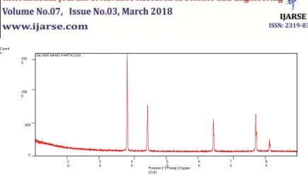
“Fig.1” represents X-ray diffraction pattern of metal oxide nanoparticles. The X-ray diffraction pattern revealed
major peaks at 2 values of 38.08 (111), 44.26 (200), 64.39 (220), 77.36 (311), 81.52 (222), respectively. All diffraction peaks of sample correspond to the characteristic face centered cubic structure of silver oxide which
634 | P a g e
Figure 1. XRD spectra of Silver oxide nanoparticles.
3.2 FTIR Spectroscopy
“Fig 2” presents the FTIR spectra in the range 4000-400 cm-1. The characteristic absorption bands are 935 and
677 cm-1 correspond to the Ag-O stretching and bending vibration mode respectively. The peaks observed
around 3500 cm–1 and 1740 cm−1 corresponds to stretching and bending vibration of –OH bond.
Figure 2. FTIR Spectra of Silver oxide nanoparticles.
3.3 UV-Visible Spectroscopy
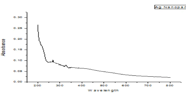
“Fig.3” represents the absorbance spectra of the Ag2O nanoparticles. The UV absorption spectra of Ag2O
nanoparticles show a characteristics peak at a wavelength of 340 nm. The peak corresponds to the formation of
Silver oxide nanoparticles. “Fig. 3(a)” is the Tauc plot showing variation of (αhν)½ vs. hν and “Fig. 3(b)” is the
Tauc plot showing variation of (αhν)² vs. hν. These plots were drawn from the data of UV-Visible absorption
Position [°2Theta] (Copper (Cu))
2 0
3 0
4 0
5 0
6 0
7 0
8 0 Count
s
0 500 100 0 150 0
635 | P a g e
spectra of silver oxide nanoparticles. The direct and indirect band gap (Eg) of silver oxide nanoparticles
corresponding to absorption peak at 340 nm was found to be 2.4 and 2.4 eV, respectively. It is clear from band
gap that synthesized nanoparticles are of semiconductor type.
Figure 3. UV-Visible spectra of Silver-oxide nanoparticles.
Figure 3(a) Tauc plot of (αhν)1/2 vs. hν Figure 3(b) Tauc plot of (αhν)2 vs. hν
3.4TGA and DTA Analysis:
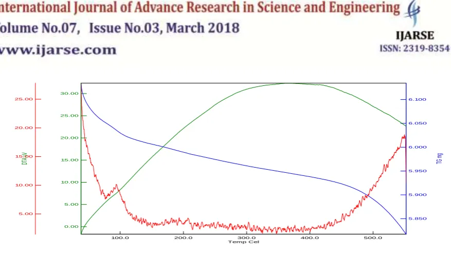
“Fig.4” represents the TGA and DTA spectra of the Ag2O nanoparticles. Thermogravimetric studies were
carried out under nitrogen atmosphere with a heating rate of 10 ºC/ min, to check the stability of Ag2O
nanoparticles. Weight takes place first around 100 ºC (due to loss of moisture) and then in the temperature
region 400–600 °C (due to loss of CO2 and H2O).
2.4 2.6 2.8 3.0 3.2 3.4 3.6 3.8 4.0 0.0005
0.0010 0.0015 0.0020 0.0025 0.0030 0.0035 0.0040 0.0045
(
h
)
2
h
2.4 2.6 2.8 3.0 3.2 3.4 3.6 3.8 4.0 0.0005
0.0010 0.0015 0.0020 0.0025 0.0030 0.0035 0.0040 0.0045
(
h
)
2
636 | P a g e
Temp Cel300.0 400.0 500.0
200.0 100.0
DTA
uV
30.00
25.00
20.00
15.00
10.00
5.00
0.00
TG
mg
6.100
6.050
6.000
5.950
5.900
5.850
DTG
ug
/m
in
25.00
20.00
15.00
10.00
5.00
Figure 4 TGA/DTA thermal analysis curves of Silver oxide nanoparticles.
IV.CONCLUSIONS
Ag2O metal nanoparticles synthesized by modified sol-gel technique are typical fcc in structure. The results of
UV-VIS, FTIR and XRD, indicates the formation of Ag2O nanoparticles and TGA/DTA results indicate the
stability of silver oxide nanoparticles. The optical absorption spectrum of Silver oxide nanoparticles was studied
by UV-Visible spectroscopy. The Tauc Plots shows the good conductivity of synthesized nanoparticles. The
absorption band in finger print region of FTIR spectra i.e. 935 and 677 cm-1 indicated metal oxide stretching
vibrations. This modified sol-gel method is one of the simplest, environment friendly, cost effective and less
time consuming method of synthesis of nanoparticles.
REFERENCES
[1] Y.S. Tarahovsky, Y.A. Kim and G.R. Ivanitsky, Dokl. Biochem. Biophys., 422, 2008, 265.
[2] M. Zhang, S. Fang, A.A. Zakhidov, S.B. Lee, A.E. Aliev, C.D. Williams, K.R. Atkinson and R.H.
Baughman, Science, 309, 2005, 1215.
[3] A.R. Mayer and J.E. Mark, Colloid Polym. Sci., 275, 1997, 333.
[4] M.H. Pishbin, A.R. Mohammadi and M. Nasri, Fuel Cells (Weinheim,Germany), 7, 2007, 291. [5] J.H. Lee, S.K. Hong and W.B. Ko, Asian J. Chem., 23, 2011, 5447.
[6] K.I. Shimizu, K. Sugino, K. Sawabe and A. Satsuma, Chem. Eur. J., 15, 2009, 2341. [7] J.M. Schierholz, L.J. Lucas, A. Rump and G. Pulverer, J. Hosp. Infect., 40, 1998, 257. [8] S.H. Shin, M.K. Ye, H.S. Kim and H.S. Kang, Int. J. Immunopharmacol., 7, 2007, 1813.
[9] C. Baker, A. Pradhan, L. Pakstis, D.J. Pochan and S.I. Shah, J. Nanosci.Nanotechnol., 5, 2005, 244. [10] J. Chen, C.M. Han, X.W. Lin, Z.J. Tang and S.J. Su, Zhonghua Wai Ke Za Zhi, 44, 2006, 50.
[11]S. W. Kim, J. H. Jung, K. Lamsal, Y. S. Kim, J. S. Min, Y. S. Lee.Antifungal effects of silver nanoparticles
637 | P a g e
[12]K. Lamsal , S. W. Kim, J. H. Jung, Y. S. Kim, K. S. Kim, Y. S. Lee. Application of silver nanoparticles for
the control of Colletotrichum species in vitro and pepper anthracnose disease in field. Mycobiology, 39, 2011, 194-199.
[13]M. Sastry, A. Ahmad, M. I. Khan, R. Kumar. Biosynthesis of metal nanoparticles using fungi and
actinomycete. Curr Sci., 85, 2003, 162–170.
[14]S. Iravani. Green synthesis of metal nanoparticles using plants. Green Chem., 13, 2011, 2638–2650. [15]H. Korbekandi,S. Iravani, S. Abbasi. Production of nanoparticles using organisms. Crit Rev Biotech., 29,
2009, 279-306.
[16]R.Rani, H. Kumar, R. K. Salar, S. S. Purewal, Antibacterial activity of copper oxide nanoparticles against
gram-negative bacterial strain synthesized by reverse micelle route, Int. J. Pharmac. Res. Develop., 06 (01), 2014, 72-78.
[17] H. Kumar, Manisha and P. Sangwan, Synthesis & Characterization of MnO2 nanoparticles using
co-precipitation technique, Int. J. Chemistry & Chem. Engg., 3 (3), 2013,155-160.
[18] H. Kumar and R. Rani, Structural Characterization of Silver Nanoparticles Synthesized by Microemulsion
Route, Int. J. Engg. & Innovative Tech., 3 (3), 2013, 344-348.
[19] H. Kumar and R. Rani, Structural and optical Characterization of ZnO Nanoparticles Synthesized by
Microemulsion Route. Int. Letters of Chem., Phys. & Astronomy, 14, 2013, 26-36.
[20] H. Kumar, R. Rani and R. K. Salar, Synthesis of nickel hydroxide nanoparticles by reverse micelle
method & its antimicrobial activity, Res. J. of Chem. Sci., 1 (9), 2011, 1-7.
[21]Manisha and Neetu. Synthesis and Characterization of Nickle oxide Nanoparticles, Internat. J. of Advance Res. in Sci. & Engg. (ISSN 2319-8354), 5 (8) 2016.
[22]Manisha and H. Kumar. Synthesis and Characterization of Iron oxide Nanoparticles by Sol-Gel
Method.nanochemistry (Published by Anu Books, Meerut, 2018) ISBN: 978-93-82166-87-0.
[23]H. Kumar and M. Kumari. Synthesis, Characterization and Antibacterial study of Zinc oxide- graphene
Nanocomposites, Asian J. of Pharmaceutical & Clinical Res., 10 ( 9), 2017, 206-209.
[24]H. Kumar and Manisha. Synthesis, Characterization and Antibacterial study of Copper oxide- graphene