A Method of Automatic Silkworm Eggs
Counting System
Sanaha Pathan, Avinash Harale
P.G Student, Dept. of Electronics, SPSPM SKN College of Engineering, Korti, Pandhrpur, India Professor, Dept. of Electronics, SPSPM SKN College of Engineering, Korti, Pandhrpur, India
ABSTRACT: Silk is the most elegant textile in the world. Sericulture is an agro industry involved in production of
cocoons. Silkworm eggs are the sheet anchor of silk industry. Counting of silkworm eggs from each DFL is very important variability of eggs quantities laid on sheet. Accurate counting is essential to avoid economic loss either sericulturists or to grainages. Current method is not suitable for counting because it is manual, waste of time also required extra man power. So A method of automatic silkworm eggs counting system developed for counting number of silkworm eggs with better accuracy and avoiding loss to sericulturists as well as farmers. For segmentation process thresholding is used, for removing noise again thresholding method is used based on physical properties of object. Counting number of eggs is done by using matlab command.
KEYWORDS:Object counting; area detection; segmentation; color transformation.
I. INTRODUCTION
Since ages, silk and silk fabrics have attracted mankind and have found their place among the most valued and elegant human fabrics. Sericulture is both an art and science of raising silkworms for silk production [1]. Sericulture and silk production have an enormous potential in India provided it is made available to rural people, especially women and its marketing is organized independently. It can serve as an excellent mode for employment generation and augmentation of income. This requires not only providing fresh technological inputs to primary producers but more importantly, evolving and establishing new systems of organizing of silkworm eggs accurately in order to generate statistics related to hatching percentage of different silkworm species. Centers can forecast seasonal demands and plan egg production by knowing hatching capability of specific silkworm species. Good planning of infrastructure in the grainages is required to increase the production efficiency. Farmers look to grainages for supply of silkworm seeds. The count of silkworm seeds is required for plantation of mulberry trees. Variation in the supply of silkworm seed quantity leads to economic losses. It is necessary to count silkworm eggs accurately for selling the silkworm eggs to farmers. Thus this research work attempts to introduce automation in Sericulture industry and thus to increase the total production.
In silk production, the number of silkworms required for a particular plantation of mulberry trees, should be approximate one for good yield of silk, so that mulberry leaves will not be wasted. For this farmers must purchase approximate number of eggs from grainages. Disease Free Layings (DFLs) are prepared in grainages and supplied to the farmers for rearing [9]. Variability of egg quantities laid on sheets during production can cause economic losses. In addition quality control measures to monitor the egg numbers is tedious and laborious. While selling the silkworm eggs for rearing it is necessary to count the silkworm eggs accurately, so that farmers can pay accordingly and they should not suffer a loss. This counting determines the fecundity (Laying capability of female moth) and hatching percentage required for silkworm rearing.
sections, II section describes literature survey based on silkworm egg counting system and other object and cell counting system. Section III describes proposed methods related to automated silkworm egg counting system. Section IV describes results and conclusion is included in section V.
II. LITERATURE REVIEW
Md. Sharifur Rehman et al. [4] presented a method for object counting using marker controlled watershed segmentation and thresholding technique. Image segmentation is the most fundamental operation required for object counting. Watershed segmentation includes discontinuity detection, thresholding and region processing. Marker Controlled Watershed Segmentation follows five steps: Execute segmentation function to get an image whose dark regions are the objects that are segmented. Compute foreground markers for connecting drops of pixels within each of the objects. Compute background markers i.e. pixels that are not part of object. Modify the segmentation function so that it only has minima at the foreground and background marker locations. Execute watershed transformation of the modified segmentation function. Therefore it is considered to be most effective and stable over other segmentation techniques. Use of thresholding method along with marker controlled Watershed Segmentation gives satisfactory results.
J. G. A. Barbedo et al [5] presented a method for counting of microorganisms in microscopic images. This method uses morphological operations to isolate and count the objects of interest. In the first step, RGB image is converted to grayscale. Noise and other artifacts are eliminated using two-dimensional median-filter. Ideal size of the neighborhood over which filter should be applied depends on three main factors: size of objects of interest, size of spurious artifacts and resolution of the image. There are two approaches for deciding neighborhood. The first approach has user entered estimate of diameter of objects and artifacts. Here, the neighborhood will be square region with side length equal to the average of diameters. In the second approach, the estimation is done using multiple counts and the program is forced to estimate the number of objects considering neighborhood sizes from zero to N/50 pixels, where N is width of image in pixels. Next, contrast enhancement is done so as it is possible to distinguish between background and objects of interest. In following, the algorithm checks for illumination of background. A complement operation is performed if the background is brighter. The image is then submitted to top-hat morphological filtering. Image is binarized with thresholding technique. After that object counting is done.
Marjan Ramin et al. [6] used image analysis technique for counting number of cells in Immunocytochemical (ICC) images . The proposed system contains four major steps: Pre-processing, Classification, Separating Bound Nucleus and Cell Counting. Pre-processing consists of removal of random noise by smoothening spatial filter. Morphological open operator is utilized to eliminate images background. Banding noise is removed by subtracting median of the red channel from all channels. In order to separate nucleus from antigens, nearest neighbor classification method with Euclidean distance metric is used in L*a*b color space. The bound nucleus is separated by local thresholding algorithm. For this purpose, statistical analysis is done and optimal threshold is found with the help of genetic algorithm. Finally, cell counting is done by tracing the boundaries. From the results, the error Ratio and standard.
III.PROPOSED ALGORITHM
Fig.1. Block diagram of proposed method.
A. BLOCK DIAGRAM DISCRIPTION:
The input images that is database used for this project is DFL sheet photo captured by camera from BAIF industry, Uralikanchan (India).
TABLE I. DATABASE COLLECTED WITH DIFFERENT CAMERAS
Type of camera Resolution Number of Images Taken
Nokia XL(8 MP) 2592X1944 20
Samsung duos(8 MP)
2592X1944 20
Sony digital camera(12 MP)
2592X1944 20
1) Image acquisition
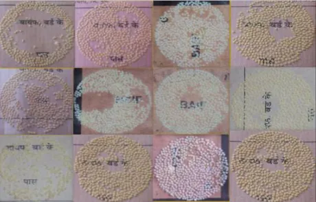
Image acquisition can be done for offline eggs counting; images from database (captured by various digital cameras) are processed. Figure 2 shows different database images
2) Image preprocessing
In image pre-processing part of image processing basic image resize, HSV transformations, binary image conversion etc. is carried out.
Resizng
An RGB color image is an MxNx3 array of color pixels is a triplet corresponding to the red, green and blue components of an RGB image at a specific spatial location. Resizing returns image that has the number of rows and columns specified by [256 256]. Either numrows or numcols may be same, in which case imresize computes the number of rows or columns automatically to preserve the image aspect ratio.
Color Conversion
An RGB color image is an MxNx3 array of color pixels is a triplet corresponding to the red, green and blue components of an RGB image at a specific spatial location [8]. The data class of the component images determines their range of values. If an RGB image is of class double, the range of values is [0, 1]. Similarly, the range of values is [0,255] or [0, 65535] for RGB images of class uint8 or uint16 respectively. The number of bits used to represent the pixel values of component images determines the bit depth of an RGB image. As RGB color models are not well suited for describing the colors in terms that are not well suited for human interpretation. Therefore RGB image is converted into HSV.
B. DESCRIPTION OF THE PROPOSED ALGORITHM:
We have followed following steps for implementing it:
Step 1: Created Image database for silkworm eggs counting system in the form of different DFL sample images.
Step 2: After creating the database, the second task is image resizing, HSV color transformation for retrieving useful
information from an image.
Step 3: after resizing and color conversion, same resized color image is converted in to binary image for finding pixel
position.
Step 4: After binary conversion of color image, one value pixel represent information about white pixel and zero value
pixel represent information about black pixel.
Step 5: After finding position of white pixel, access information of HSV image for white pixel and converted it into
matrix form.
Step 6: After representing information in matrix form, mean value is calculated from matrix. Mean value is obtained by
µ= ∑ ……….(1) Where, variable vector A made up of N scalar observations.
Step 7: After calculating mean value, image segmentation is performed by using thresholding. Threshold value is
decided by trying different mean value for better result. In segmentation process non egg portion is removed and only egg portion is keep as it is. Thresholding is obtained by
Step 8: After eliminating non egg portion, identify individual eggs and overlapping eggs by applying thresholding
based on distance between HSV image pixel value and mean value. Distance is calculated by using formula
= ( 2− 1) + ( 2− 1) …………...(2)
Step 9: After final segmentation, final segmented image is converted in to binary image because final segmented image
is not suitable for count.
Step 10: After identifying individual eggs and overlapping eggs and converted it into binary , still there are some barriers between eggs. All barriers eliminated by using again thresholding technique and threshold value is based on area. Physical information of object is obtained by using regionprops function.
IV.RESULTS AND DISCUSSION
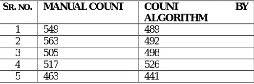
The proposed method is developed in MATLAB 2014 programming software. GUI of proposed method is also developed in same MATLAB programming software 2014. Figure 3 and 4 shows result of counting number of eggs in GUI format. In GUI result, load image contain original color database image. Color conversion shows HSV transform image. Segmentation process shows final segmented image and area based thresholding image. Total number of silkworm eggs displayed total count of eggs. Table II Shows result for five different DFL sample image.
Fig.3. GUI result for DFL sample 1
TABLE II. RESULTS OF PROPOSED METHOD
SR. NO. MANUALCOUNT COUNT BY
ALGORITHM
1 549 489 2 563 492 3 505 498 4 517 526 5 463 441
After analyzing results, it is observed that in some images, eggs were having very small size and it is difficult to identify difference between egg and non egg particles, so sometimes they were considered as non egg particles. Some eggs have area smaller so they were also eliminated from area based thresholding and not considered for counting .In some images, eggs were tightly overlapped so that in segmentation process it is difficult to identifying overlapping eggs. Some eggs have low intensity level so it is below threshold level and it was considered as non egg portion and so it has not counted while counting the number of eggs.
V. CONCLUSION AND FUTURE WORK
This paper presents an image processing algorithm for automatic silkworm eggs counting system. This algorithm also useful in other application fields for counting small objects. Segmentation method is very useful for separating egg portion from non egg portion. Object counting is based on matlab count command which gives better result.
ACKNOWLEDGMENT
I would like to thanks BAIF research foundation Uralikanchan for supported regarding database of images. I express deep sense of gratitude and sincere thanks for valuable assistance that I have received from my guide Prof. Avinash D. Harale, I also express thanks to HOD and all staff of E&TC dept, who guided me, supported me during this work.
REFERENCES
[1] Dr.M.N.Narasimhanna,“Manual on Silkworm Egg Production,” Central Silk board,1st ed.Jan 1988.
[2] Hemant Tulsani, Saransh Saxena, Naveen Yadav, “Segmentation using Morphological Watershed Transformation for Counting Blood Cells” , International Journal of Computer Applications and Information Theory, Vol. 2, Issue III, Apr-May 2013.
[3] Chomtip Pornpanomchai and Fuangchat Stheitsthienchai Sorawat Rattanachuen, “Object Detection and Counting System ”,IEEE, Congress on Image and Signal Processing, pp. 61-65, Thailand, May 2008.
[4] Md. Sharifur Rahman and Md. Rafiqul Islam, “Counting Objects in an Image by Marker Controlled Watershed Segmentation and Thresholding,” IEEE 3rd International Conference on Advanced Computing, Feb. 2013.
[5] Jayme Garcia Arnal Barbedo, “Method for Counting Microorganisms and Colonies in Microscopic Images”, 12th IEEE International Conference on Computer Science and Its Applications, pp. 84-87, June 2012.
[6] Ramin M., Ahmadvand P., Sepas-Moghaddam, M.Dehshibi and A.Moghaddam. ,“Counting Number of Cells in Immunocytochemical Images using Genetic Algorithm,”12th International Conference on Hybrid Intelligent Systems, pp. 185-190,Dec. 2012.
[7] Carlos A. B. Mello, Wellington P. dos Santos and Marco A. B. Rodrigues and Ana Lcia B.Candeias and Cristine M. G. Gusmao, “Image Segmentation of Ovitraps for Automatic Counting of Aedes Aegypti Eggs,”30th Annual International IEEE EMBS Conference Vancouver, British Columbia, Canada, pp. 3103-3106. Aug. 2008.
[8] Julián Calderón González and Òscar Daniel Carmona Salazar,“ Image enhancement with matlb algorithm,” Blekinge Institute of Technology,Karlskrona Sweden.
[9] [Online].Available: http://www.tnsericulture.gov.in [viewed on Aug.8,2013]
BIOGRAPHY
Pathan Sanaha Shabbekadir is a ME Student in the Electronics Department, SPSPM, SKN College of Engineering,
korti, Pandhrpur, Solapur University. She received Bachelor of Electronics and telecommunication (BE) degree in 2014 from BMIT, Solapur, India.
Harale Avinash D. is a Professor in the Electronics Department, SPSPM, SKN College of Engineering, korti,