© 2019 by the Serbian Biological Society How to cite this article: Choi JW, Lee MH, Fujii T. Relationship between 621 neutrophil gelatinase-associated lipocalin, cardiac biomarkers, inflammation index and renal parameters in cardiovascular disease. Arch Biol Sci. 2019;71(4):621-8.
Relationship between neutrophil gelatinase-associated lipocalin, cardiac biomarkers,
inflammation index and renal parameters in cardiovascular disease
Jong Weon Choi1,*, Moon Hee Lee2 and Tatsuyoshi Fujii3
1Department of Laboratory Medicine, College of Medicine, Inha University, Incheon, Republic of Korea
2Department of Internal Medicine, College of Medicine, Inha University, Incheon, Republic of Korea
3Department of General Medicine, Tsukuba University Hospital Mito Clinical Education and Training Center, Mito Kyodo
General Hospital, Mito, Ibaraki, Japan
*Corresponding author: [email protected]
Received: June 4, 2019; Revised: July 15, 2019; Accepted: July 23, 2019; Published online:July 29, 2019
Abstract: Theplasma neutrophil gelatinase-associated lipocalin (NGAL) level is elevated in myocardial infarction (MI) and affected by inflammation and kidney function. The aim of this study was to determine which of these conditions more critically affects the plasma NGAL level in MI. Patients with MI were evaluated by measuring the NGAL concentration and its corrected values. No significant association was observed between plasma NGAL concentration and cardiac biomarkers. However, the NGAL/inflammation index ratio (NGAL/Inf ratio) was positively correlated with troponin-I (r=0.289, p<0.001), and the NGAL/serum creatinine ratio (NGAL/sCr ratio) was significantly correlated with creatine kinase-MB (r=0.251, p<0.001). After adjusting for inflammation and kidney function, increased NGAL concentrations returned to baseline levels, which were not different from those of healthy individuals. The percent difference between NGAL and the NGAL/Inf ratio was 35.6%, significantly higher than that between NGAL and the NGAL/sCr ratio (15.4%; p<0.001). The severity of inflam-mation seems to play a more crucial role than renal and myocardial dysfunction in affecting plasma NGAL levels in MI. Plasma NGAL levels need to be corrected using the inflammation index and sCr levels for exactly evaluating patients with MI.
Keywords: neutrophil gelatinase-associated lipocalin; inflammation index; myocardial infarction; renal dysfunction; car-diac biomarkers
INTRODUCTION
Neutrophil gelatinase-associated lipocalin (NGAL) is a glycosylated protein belonging to the lipocalin fam-ily, expressed by activated granulocytes and a variety of epithelial cells [1]. As NGAL increases within two hours of renal damage, particularly before an elevation in serum creatinine (sCr), it has been used as an early predictor of acute kidney injury [2]. In addition, recent studies have reported that NGAL is involved in vascular inflammation and affects plaque instability and vascular remodeling after coronary heart disease [3,4]. Evidence supports the role of NGAL in the pathophysiology of cardiovascular diseases [5-7]. Despite the emerging role of NGAL in cardiovascular diseases, the significance of elevated NGAL levels remains unclear. There have been inconsistent results on its predictive value. One study demonstrated that the expression of NGAL is increased
in atherosclerosis and myocardial infarction (MI) [8]. In contrast, other studies reported that underlying renal insufficiency is a stronger determinant than myocardial dysfunction [9,10].
coro-nary heart disease and concurrent renal dysfunction, particularly in cases of systemic inflammation.
Few studies have closely examined the contribu-tion of myocardial injury, inflammacontribu-tion severity or impaired renal function to augmented plasma NGAL levels in acute MI. In the present study, we calculated the corrected values of NGAL, including the NGAL/ inflammation index ratio (NGAL/Inf ratio), the NGAL/sCr ratio and the NGAL/inflammation index and sCr ratio (NGAL/Inf-sCr ratio). Based on these parameters, we estimated the effect of myocardial damage on plasma NGAL levels and investigated the efficacy of the parameters for predicting renal dys-function in patients with MI.
MATERIALS AND METHODS
Ethics statement
The study protocol was reviewed and approved by the institutional review board, and written informed con-sents were obtained from all subjects. This study was conducted in accordance with the guidelines of the Helsinki Declaration. All blood samples were collected after a sufficient explanation of the study procedure.
Study population
A total of 117 patients with acute MI were evaluated. Their ages ranged from 53 to 78 years (median age, 65 years), and 79 patients were males (67.5%). Basic demographic data and medical history are summarized in Supplementary Table S1. Age-matched healthy sub-jects (n=35), who had no evidence of heart diseases, in-flammation and renal dysfunction, were enrolled as the control group. Diagnosis of MI was performed based on the criteria for acute MI [15]. Heart failure was de-fined as plasma N-terminal pro-brain natriuretic pep-tide (NT-proBNP) concentration ≥400 pmol/L, which was based on European guidelines for the diagnosis and treatment of acute and chronic heart failure [16]. Subjects with infectious disease (n=2), sepsis (n=1), and multiple trauma (n=1) were excluded from the study. Patients who had incomplete data (n=3), a recent op-eration (n=1), or a medication history of antiinflamma-tory drugs (n=6) were also excluded from the analysis.
Measurement of laboratory parameters
On admission, blood samples were collected to es-timate troponin-I, creatine kinase-MB (CK-MB), NT-proBNP, sCr and complete blood cell counts. The blood samples were obtained prior to treatment, such as primary percutaneous coronary intervention, lipid-lowering medications and the administration of antithrombotic and antihypertensive drugs. Plasma NGAL levels were measured by fluorescence immu-noassay using the Triage NGAL Test (Alere, Inc., San Diego, CA, USA). The upper normal limit of plasma NGAL was set at 150 ng/mL [17]. All assays for car-diac biomarkers were performed using an Elecsys 2010 analyzer (Roche Diagnostics GmbH, Mannheim, Germany). The cardiac biomarkers were measured by an electrochemiluminescence immunoassay using the Elecsys CK-MB, cardiac troponin-I and anti-NT-proBNP antibodies (Roche Diagnostics GmbH). High-sensitivity C-reactive protein (hsCRP) was ana-lyzed by an immunonephelometry assay (Dade Behri-ing, Inc, Deerfield, IL, USA). An increased level of hsCRP was defined as >0.3 mg/dL, which was based on the cutoff point with a 95% confidence interval (CI) for the hsCRP of healthy individuals. The es-timated glomerular filtration rate (eGFR) was com-puted using the Modification of Diet in Renal Disease formula. A renal impairment was defined as having an eGFR of lower than 60 mL/min/1.73 m2 [18].
Corrected values of NGAL
The NGAL/Inf ratio was calculated using the follow-ing equation:
NGAL/Inf ratio = plasma NGAL concentration/ inflammation index.
of 0.5 is given to each of the patients with a hsCRP concentration <0.3 mg/dL and with a cESR level <15.0 mm/h, the sum of the scores in the patients who were within the reference interval in hsCRP and cESR be-comes 1 (0.5 plus 0.5). Thus, in patients without evi-dence of inflammation, the corrected NGAL levels are the same values as the uncorrected NGAL levels.
The cutoff limit of the NGAL/Inf ratio for assess-ing the risk of renal dysfunction in patients with MI was defined as 145 ng/mL. The cutoff was based on the highest sensitivity and specificity value for iden-tifying renal dysfunction in receiver operating char-acteristic (ROC) curve analysis. The NGAL/sCr ratio was computed by the following formula:
NGAL/sCr ratio=plasma NGAL level/sCr con-centration.
For subjects with sCr<1.0 mg/dL, 1.0 mg/dL of sCr was used to avoid the overestimation due to the decimal point. The NGAL/Inf-sCr ratio was deter-mined by the following equation:
NGAL/Inf-sCr ratio=(NGAL/Inf ratio)/sCr con-centration.
Percent difference for NGAL was calculated by the following equation:
[(NGAL-corrected NGAL)/NGAL]×100.
Categorization of subjects
The subjects were classified into two groups: patients with MI (n=117) and healthy controls (n=35). To in-vestigate the effects of renal dysfunction and inflam-mation severity on plasma NGAL level, the patients were compared to healthy controls after alternately excluding patients with eGFR<60 mL/min/1.73 m2
(n=38) and hsCRP>0.3 mg/dL (n=83) from the study population.
Statistical analysis
Continuous variables were expressed as the mean± standard deviation, and asymmetrically distributed data were presented as the median and interquartile range. Categorical variables were described using frequencies and percentage. The Student’s t-test and
Mann-Whitney U test were used. The ROC curve was analyzed to determine the diagnostic efficacy of NGAL and the NGAL/Inf ratio for identifying im-paired renal function in patients. Multivariate linear regression analysis was performed to determine the association between cardiac biomarkers and lipocalin levels after adjusting for potential confounders. The association between the NGAL/Inf ratio and the pres-ence of renal dysfunction was assessed by multivariate logistic regression analysis. The data were analyzed using SPSS software (version 19.0; SPSS Inc., Armonk, NY, USA). P values<0.05 were considered as statisti-cally significant.
RESULTS
NGAL and its corrected values
Plasma NGAL levels were significantly higher in pa-tients with MI than in healthy controls (148.5 ng/mL versus 67.4 ng/mL; p<0.001). Of the 117 patients, 38 (32.4%) patients had impaired renal function, 34 (29.1%) had heart failure and 83 (70.1%) had elevated hsCRP. The percent difference between NGAL and the NGAL/Inf ratio was 35.6%, whereas the percent difference between NGAL and the NGAL/sCr ratio was 15.4% (p<0.001). The NGAL/Inf-sCr ratio of the patients did not differ from that of healthy individuals (72.3 ng/mL vs. 65.9 ng/mL; p=0.527), and the percent difference between the two median values was 9.7% (Supplementary Table S1).
NGAL according to hsCRP and eGFR
The effects of impaired renal function (eGFR<60 mL/ min/1.73 m2) and active inflammation (hsCRP>0.3
mg/dL) on the plasma NGAL concentration of pa-tients with MI were assessed. After excluding subjects with eGFR<60 mL/min/1.73 m2 from the study
Linear regression analysis
In multivariate linear regression analy-sis adjusted for potential confounders, no significant association was observed between plasma NGAL concentration and the levels of troponin-I and CK-MB. However, the NGAL/Inf ratio was positively correlated with troponin-I (r=0.289, p<0.001) and the NGAL/sCr ratio was significantly correlated with CK-MB (r=0.251, p<0.001) (Table 2). Correlation between the NGAL/Inf ra-tio and troponin-I is illustrated in Fig. 1.
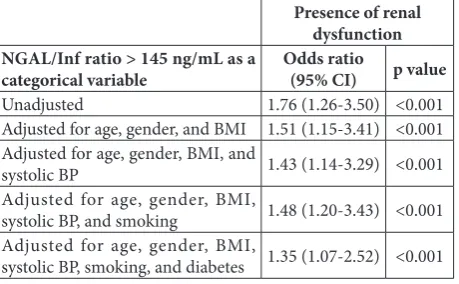
Multivariate logistic regression analysis
In logistic regression analysis, an elevat-ed NGAL/Inf ratio (>145 ng/mL) was significantly associated with the presence of renal dysfunction in patients with MI following adjustment for potential con-founders, such as age, gender, BMI, sys-tolic blood pressure, smoking habit and diabetes (odds ratio, 1.35; 95% CI, 1.07-2.52; p<0.001) (Table 3).
ROC curve analysis
The diagnostic ability of the NGAL/Inf ratio for identifying impaired renal function in patients with MI was investigated by ROC curve analysis. The area under the curve (AUC) of the NGAL/Inf ratio (0.811; 95% CI, 0.729-0.893) for identifying re-nal dysfunction was significantly larger than that of plasma NGAL concentration (0.694; 95% CI, 0.599-0.790; p<0.001) (Fig. 2).
DISCUSSION
In the present study, the relationship between plasma NGAL level, cardiac biomarkers, kidney function and the severity of inflammation in MI was investi-gated. The median plasma NGAL concentration of patients with MI was significantly higher than that of healthy individuals. Our results are in agreement
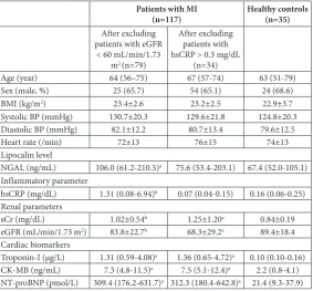
Table 1. Plasma NGAL levels after excluding patients with renal dysfunction and active inflammation.
Patients with MI
(n=117) Healthy controls(n=35)
After excluding patients with eGFR
< 60 mL/min/1.73 m2 (n=79)
After excluding patients with hsCRP > 0.3 mg/dL
(n=34)
Age (year) 64 (56–75) 67 (57-74) 63 (51-79)
Sex (male, %) 25 (65.7) 54 (65.1) 24 (68.6)
BMI (kg/m2) 23.4±2.6 23.2±2.5 22.9±3.7
Systolic BP (mmHg) 130.7±20.3 129.6±21.8 124.8±20.3 Diastolic BP (mmHg) 82.1±12.2 80.7±13.4 79.6±12.5
Heart rate (/min) 72±13 76±15 74±13
Lipocalin level
NGAL (ng/mL) 106.0 (61.2-210.5)a 75.6 (53.4-203.1) 67.4 (52.0-105.1)
Inflammatory parameter
hsCRP (mg/dL) 1.31 (0.08-6.94)b 0.07 (0.04-0.15) 0.16 (0.06-0.25)
Renal parameters
sCr (mg/dL) 1.02±0.54b 1.25±1.20a 0.84±0.19
eGFR (mL/min/1.73 m2) 83.8±22.7b 68.3±29.2a 89.4±18.4
Cardiac biomarkers
Troponin-I (μg/L) 1.31 (0.59-4.08)a 1.36 (0.65-4.72)a 0.10 (0.10-0.16)
CK-MB (ng/mL) 7.3 (4.8-11.5)a 7.5 (5.1-12.4)a 2.2 (0.8-4.1)
NT-proBNP (pmol/L) 309.4 (176.2-631.7)a 312.3 (180.4-642.8)a 21.4 (9.3-37.9)
Data are expressed as the mean±SD, median (interquartile range) or frequency (percentage).
a Significant (p<0.05), compared to healthy controls.
b Significant (p<0.05), compared to the patients after excluding patients with
hsCRP>0.3 mg/dL.
MI – myocardial infarction; BMI – body mass index; BP – blood pressure; NGAL – neutrophil gelatinase-associated lipocalin; sCr – serum creatinine;
hsCRP – high-sensitivity C-reactive protein; eGFR – estimated glomerular filtration rate; CK-MB – creatine kinase-MB; NT-proBNP – N-terminal pro-brain natriuretic peptide.
Table 2. Univariate and multivariate linear regression analysis of NGAL and its corrected values in relation to cardiac biomarkers.
Univariate Multivariate*
Standard β p value Standard β p value
NGAL
Troponin-I (μg/L) 0.163 0.142 0.131 0.352
CK-MB (ng/mL) 0.148 0.259 0.122 0.316
NT-proBNP (pmol/L) 0.173 0.139 0.140 0.238
NGAL/Inf ratio
Troponin-I (μg/L) 0.325 <0.001 0.289 <0.001
CK-MB (ng/mL) 0.165 0.139 0.127 0.307
NT-proBNP (pmol/L) 0.294 <0.001 0.253 <0.001
NGAL/sCr ratio
Troponin-I (μg/L) 0.305 <0.001 0.256 <0.001
CK-MB (ng/mL) 0.293 <0.001 0.251 <0.001
NT-proBNP (pmol/L) 0.178 0.142 0.139 0.258
with those of a previous study, which demonstrated the enhancement of NGAL expression in patients with acute MI and the increase of serum NGAL level in cases of coronary heart disease [20].
NGAL is an acute phase reactant that participates in various antibacterial immune responses [21]. In normal populations, plasma NGAL levels are largely determined by the granulocyte count and hsCRP con-centration, whereas in patients with impaired renal function, plasma NGAL levels are mainly determined by the eGFR [22]. The activation of granulocytes
and the subsequent release of NGAL are crucial in the development of inflammatory reactions in the course of cardiovascular diseases [23]. A previous study reported that elevated NGAL concentrations re-flect the inflammatory status in various stages of coro-nary artery disease [24]. In our study, 32.4%, 29.1%, and 70.1% of the patients had renal dysfunction, heart failure and elevated hsCRP, respectively. Therefore, the elevated plasma NGAL levels of our patients may reflect the overall effect of several adverse conditions, such as myocardial injury, activated neutrophils due to systemic inflammation and impaired renal function associated with MI.
A study has proposed that elevated NGAL level in cardiovascular diseases is caused by the enhanced NGAL expression in the cardiomyocytes of the in-jured myocardium in response to proinflammatory cytokines [25]. In our study, to adjust the effect of inflammation and kidney function on plasma NGAL levels, corrected values of plasma NGAL were calcu-lated. The NGAL/Inf-sCr ratio of patients with MI exhibited an increasing trend compared to that of healthy individuals; however, the differences were not
Table 3. Multivariate logistic regression analysis for the presence of renal dysfunction in relation to the NGAL/Inf ratio.
Presence of renal dysfunction NGAL/Inf ratio > 145 ng/mL as a
categorical variable Odds ratio(95% CI) p value
Unadjusted 1.76 (1.26-3.50) <0.001
Adjusted for age, gender, and BMI 1.51 (1.15-3.41) <0.001 Adjusted for age, gender, BMI, and
systolic BP 1.43 (1.14-3.29) <0.001
Adjusted for age, gender, BMI,
systolic BP, and smoking 1.48 (1.20-3.43) <0.001
Adjusted for age, gender, BMI,
systolic BP, smoking, and diabetes 1.35 (1.07-2.52) <0.001
NGAL/Inf ratio – ratio of NGAL to the inflammation index; BMI – body mass index; BP – blood pressure; CI – confidence interval.
Fig. 1. Correlation between the NGAL/Inf ratio and troponin-I in patients with MI. The NGAL/Inf ratio correlates positively with
troponin-I level (y=0.002x+0.977; r2=0.105; p<0.001). NGAL/Inf
ratio – ratio of NGAL to the inflammation index; MI – myocardial infarction.
statistically significant. After adjusting for inflamma-tion and kidney funcinflamma-tion, the elevated NGAL levels returned to baseline levels, which were not significant-ly different from those of healthy individuals, suggest-ing that enhanced NGAL production in MI may be caused by inflammation and renal dysfunction rather than failing myocardium.
A study reported that augmented NGAL expres-sion in acute MI may be considered as a mediator of postischemic inflammation [26]. In contrast, another study demonstrated that an increased plasma NGAL level, which was observed in coronary artery disease, may be largely attributed to impaired renal func-tion [27]. In the current study, we examined which of the conditions (inflammation or kidney function) would more critically affect plasma NGAL levels in MI. When patients with impaired renal function were excluded from the subject populations, the plasma NGAL level remained significantly higher than that of the healthy individuals. However, when patients with elevated hsCRP were excluded from the study population, the plasma NGAL level of the patients did not differ from that of the healthy controls. Addition-ally, the percent difference between NGAL and the NGAL/Inf ratio of patients with MI was 35.6%, which was significantly different from that (15.4%) between NGAL and the NGAL/sCr ratio. These results suggest that inflammation and renal dysfunction contributed to approximately 35.6% and 15.4% of the elevated NGAL levels, respectively, in patients with MI. The results also suggest that the severity of inflammation could play a more crucial role than impaired renal function in affecting plasma NGAL levels, at least for the population in this study.
As MI is frequently accompanied by systemic in-flammation and renal dysfunction, it is difficult to assess to what extent myocardial damage contributes to NGAL production. In our study, after adjusting for inflammation and kidney function, the median per-cent difference of the NGAL/Inf-sCr ratio between patients and healthy controls was 9.7%. Based on these results, it can be estimated that myocardial injury ac-counted for approximately 9.7% of the increase in the total plasma NGAL level of patients with MI. Overall, our results indicated that the effect of myocardial in-jury on plasma NGAL levels was smaller than that of inflammation and impaired renal function.
Renal insufficiency is an important independent predictor of poor prognosis in patients with cardio-vascular diseases [28]. A group of researchers report-ed that measurement of plasma NGAL is useful for evaluating renal dysfunction during hospitalization in cases of cardiac diseases [29]. In the present study, the diagnostic efficacy of NGAL, which identifies re-nal dysfunction in MI, was compared to that of the NGAL/Inf ratio. The NGAL/Inf ratio demonstrated better results in ROC curve analysis. Moreover, in logistic regression analysis, compared to a decreased NGAL/Inf ratio, an elevated NGAL/Inf ratio resulted in a 1.35-fold increase in the risk of renal dysfunction. The findings of our study indicated that the NGAL/ Inf ratio was superior to NGAL as a predictor of renal insufficiency in MI. The corrected value of calculated NGAL ratio seems to reduce the impact of the inflam-matory condition on the plasma NGAL concentration in patients with renal dysfunction in conjunction with inflammatory diseases.
In a previous study, there were no significant re-lationships between plasma NGAL concentration and cardiac function in patients with MI [25]. Similarly, in our study, the plasma NGAL level was not signifi-cantly associated with the levels of troponin-I and CK-MB. However, the NGAL/Inf ratio was closely associated with troponin-I, and the NGAL/sCr ratio was significantly associated with CK-MB. A possi-ble explanation for these findings is that the overes-timated plasma NGAL level, attributed to concomitant inflammation and renal dysfunction, was corrected by adjusting for the inflammation index and sCr levels. Our results suggest that plasma NGAL levels need to be amended using the inflammation index and sCr levels of patients with MI, particularly when systemic inflammation is presented with renal impairment.
particularly in patients with concurrent inflammation and renal dysfunction. However, further validation is needed in larger, randomized prospective studies.
CONCLUSION
This study demonstrates that the severity of inflam-mation plays a more crucial role than myocardial in-jury and impaired renal function in affecting plasma NGAL levels in patients with MI. The diagnostic ef-ficacy of the NGAL/Inf ratio showed a better perfor-mance than that of plasma NGAL, suggesting that the measurement of the NGAL/Inf ratio may be an addi-tional benefit in predicting worsening renal function in patients with MI.
Funding: This study was supported by a research grant from Inha University Hospital.
Author contributions: Jong Weon Choi designed the study,
or-ganized the research, analyzed the data, and wrote the manuscript. Moon Hee Lee analyzed the data, prepared the tables and reviewed the drafts of the manuscript. Tatsuyoshi Fujii performed the sta-tistical analyses, analyzed the data, searched the literature data and edited the manuscript.
Conflict of interest disclosure: The authors declare that they have no conflict of interest.
REFERENCES
1. Md Ralib A, Mat Nor MB, Pickering JW. Plasma neutrophil gelatinase-associated lipocalin diagnosed acute kidney injury in patients with systemic inflammatory disease and sepsis. Nephrology (Carlton). 2017;22(5):412-9.
2. Constantin JM, Futier E, Perbet S, Roszyk L, Lautrette A, Gil-lart T, Guerin R, Jabaudon M, Souweine B, Bazin JE, Sapin V. Plasma neutrophil gelatinase-associated lipocalin is an early marker of acute kidney injury in adult critically ill patients: a prospective study. J Crit Care. 2010;25(1):176.e1-6.
3. te Boekhorst BC, Bovens SM, Hellings WE, van der Kraak PH, van de Kolk KW, Vink A, Moll FL, van Oosterhout MF, de Vries JP, Doevendans PA, Goumans MJ, de Kleijn DP, van Echteld CJ, Pasterkamp G, Sluijter JP. Molecular MRI of murine atherosclerotic plaque targeting NGAL: a protein associated with unstable human plaque characteristics. Car-diovasc Res. 2011;89(3):680-8.
4. Sahinarslan A, Kocaman SA, Bas D, Akyel A, Ercin U, Zengin O, Timurkaynak T. Plasma neutrophil gelatinase-associated lipocalin levels in acute myocardial infarction and stable coronary artery disease. Coron Artery Dis. 2011;22(5):333-8. 5. Cruz DN, Gaiao S, Maisel A, Ronco C, Devarajan P. Neutro-phil gelatinase-associated lipocalin as a biomarker of
cardio-vascular disease: a systematic review. Clin Chem Lab Med. 2012;50(9):1533-45.
6. Zografos T, Haliassos A, Korovesis S, Giazitzoglou E, Voridis E, Katritsis D. Association of neutrophil gelatinase-associated lipocalin with the severity of coronary artery disease. Am J Cardiol. 2009;104(7):917-20.
7. Lindberg S, Pedersen SH, Mogelvang R, Jensen JS, Flyvbjerg A, Galatius S, Magnusson NE. Prognostic utility of neutrophil gelatinase-associated lipocalin in predicting mortality and cardiovascular events in patients with ST-segment elevation myocardial infarction treated with primary percutaneous coronary intervention. J Am Coll Cardiol. 2012;60(4):339-45. 8. Hemdahl AL, Gabrielsen A, Zhu C, Eriksson P, Hedin U, Kas-trup J, Thorén P, Hansson GK. Expression of neutrophil gela-tinase-associated lipocalin in atherosclerosis and myocardial infarction. Arterioscler Thromb Vasc Biol. 2006;26(1):136-42. 9. Shrestha K, Borowski AG, Troughton RW, Thomas JD, Klein AL, Tang WH. Renal dysfunction is a stronger determinant of systemic neutrophil gelatinase-associated lipocalin levels than myocardial dysfunction in systolic heart failure. J Card Fail. 2011;17(6):472-8.
10. Maisel AS, Wettersten N, van Veldhuisen DJ, Mueller C, Filip-patos G, Nowak R, Hogan C, Kontos MC, Cannon CM, Mül-ler GA, Birkhahn R, Clopton P, Taub P, Vilke GM, McDonald K, Mahon N, Nuñez J, Briguori C, Passino C, Murray PT. Neutrophil gelatinase-associated lipocalin for acute kidney injury during acute heart failure hospitalizations: The AKI-NESIS Study. J Am Coll Cardiol. 2016;68(13):1420-31. 11. Ross R. Atherosclerosis-an inflammatory disease. N Engl J
Med. 1999;340(2):115-26.
12. Finlay S, Bray B, Lewington AJ, Hunter-Rowe CT, Banerjee A, Atkinson JM, Jones MC. Identification of risk factors associ-ated with acute kidney injury in patients admitted to acute medical units. Clin Med. 2013;13(3):233-8.
13. Hatamizadeh P, Fonarow GC, Budoff MJ, Darabian S, Kovesdy CP, Kalantar-Zadeh K. Cardiorenal syndrome: pathophysiology and potential targets for clinical manage-ment. Nat Rev Nephrol. 2013;9(2):99-111.
14. Giasson J, Li GH, Chen Y. Neutrophil gelatinase-associated lipocalin (NGAL) as a new biomarker for non-acute kid-ney injury (AKI) diseases. Inflamm Allergy Drug Targets. 2011;10(4):272-82.
15. Thygesen K, Alpert JS, Jaffe AS, Chaitman BR, Bax JJ, Morrow DA, White HD; Executive Group on behalf of the Joint Euro-pean Society of Cardiology (ESC)/American College of Cardiol-ogy (ACC)/American Heart Association (AHA)/World Heart Federation (WHF) Task Force for the Universal Definition of Myocardial Infarction. Fourth universal definition of myocar-dial infarction (2018). Circulation. 2018;138(20):e618-51. 16. Mahmood SS, Levy D, Vasan RS, Wang TJ. The Framingham
Heart Study and the epidemiology of cardiovascular disease: a historical perspective. Lancet. 2014;383(9921):999-1008. 17. Kim H, Hur M, Lee S, Marino R, Magrini L, Cardelli P, Struck
J, Bergmann A, Hartmann O, Di Somma S; GREAT Network. Proenkephalin, neutrophil gelatinase-associated lipocalin, and estimated glomerular filtration rates in patients with sepsis. Ann Lab Med. 2017;37(5):388-97.
Collaboration. Expressing the Modification of Diet in Renal Disease Study equation for estimating glomerular filtration rate with standardized serum creatinine values. Clin Chem. 2007;53(4):766-72.
19. Choi JW, Fujii T, Fujii N. Corrected neutrophil gelatinase-associated lipocalin (NGAL) level adjusted by the scoring sys-tem of an inflammation index for screening renal dysfunction in patients with systemic inflammation. Ann Clin Lab Sci. 2015;45(3):248-55.
20. Paulsson J, Dadfar E, Held C, Jacobson SH, Lundahl J. Activa-tion of peripheral and in vivo transmigrated neutrophils in patients with stable coronary artery disease. Atherosclerosis. 2007;192(2):328-34.
21. Xu SY, Carlson M, Engström A, Garcia R, Peterson CG, Venge P. Purification and characterization of a human neutrophil lipocalin (HNL) from the secondary granules of human neu-trophils. Scand J Clin Lab Invest. 1994;54(5):365-76. 22. Lindberg S, Jensen JS, Mogelvang R, Pedersen SH, Galatius S,
Flyvbjerg A, Magnusson NE. Plasma neutrophil gelatinase-associated lipocalinin in the general population: association with inflammation and prognosis. Arterioscler Thromb Vasc Biol. 2014;34(9):2135-42.
23. Furuya F, Shimura H, Yokomichi H, Takahashi K, Akiyama D, Asakawa C, Okamura A, Motosugi A, Haraguchi K, Yamagata Z, Kobayashi T. Neutrophil gelatinase-associated lipocalin levels associated with cardiovascular disease in chronic kid-ney disease patients. Clin Exp Nephrol. 2014;18(5):778-83. 24. Kafkas N, Demponeras C, Zoubouloglou F, Spanou L, Babalis
D, Makris K. Serum levels of gelatinase associated lipocalin
as indicator of the inflammatory status in coronary artery disease. Int J Inflam. 2012;2012:189797.
25. Yndestad A, Landrø L, Ueland T, Dahl CP, Flo TH, Vinge LE, Espevik T, Frøland SS, Husberg C, Christensen G, Dick-stein K, Kjekshus J, Øie E, Gullestad L, Aukrust P. Increased systemic and myocardial expression of neutrophil gelatinase-associated lipocalin in clinical and experimental heart failure. Eur Heart J. 2009;30(10):1229-36.
26. Helanova K, Spinar J, Parenica J. Diagnostic and prognostic utility of neutrophil gelatinase-associated lipocalin (NGAL) in patients with cardiovascular diseases. Kidney Blood Press Res. 2014;39(6):623-9.
27. Malyszko J, Bachorzewska-Gajewska H, Malyszko JS, Paw-lak K, Dobrzycki S. Serum neutrophil gelatinase-associated lipocalin as a marker of renal function in hypertensive and normotensive patients with coronary artery disease. Nephrol Carlton Vic. 2008;13(2):153-6.
28. Lekawanvijit S, Krum H. Cardiorenal syndrome: acute kidney injury secondary to cardiovascular disease and role of pro-tein-bound uraemic toxins. J Physiol. 2014;592(18):3969-83. 29. Palazzuoli A, Ruocco G, Beltrami M, Franci B, Pellegrini M,
Lucani B, Nuti R, Ronco C. Admission plasma neutrophil gelatinase associated lipocalin (NGAL) predicts worsening renal function during hospitalization and post discharge out-come in patients with acute heart failure. Acute Card Care. 2014;16(3): 93-101.
Supplementary Material