Source Imaging
Timothy P. L. Roberts and Howard A. Rowley
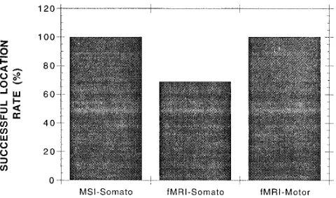
PURPOSE: To assess the reliability and comparability of functional MR imaging and magnetic source imaging for mapping the somatosensory cortex. METHODS: Parallel studies were per-formed in eight volunteer subjects in whom both hemispheres were measured with the use of painless tactile stimulation of the tip of each index finger. Magnetic source imaging was performed using a 37-channel biomagnetometer; evoked magnetic fields were analyzed using the single-equivalent dipole representation to ascertain the neuronal source. Functional MR imaging was performed on a 1.5-T MR unit. Blocks of images during periods of rest and activation were acquired using gradient-echo echo-planar imaging. Correlation analysis identified pixels in which signal intensity correlated with the stimulus function. A subsequent requirement for spatial connectivity of activation was imposed to reduce the random occurrence of pixels satisfying the correlation criteria. RESULTS: Using temporal and spatial statistical criteria for activation, we found that functional MR imaging showed activation in 11 of 16 hemispheres. In three cases, this was accompanied by activity either frontally or ipsilateral to the stimulus. Magnetic source imaging showed parietal contralateral location in all 16 cases. Where successful localization was achieved with both methods, the separation between sources appeared to be between 1 and 4 cm. Func-tional MR imaging localizations tended to lie more superficially than the magnetic source imaging localizations. Performance of a simple motor task, rather than use of somatosensory stimulation, resulted in a cortical signal change detectable with a similar functional MR imaging approach in all cases, suggesting the more robust nature of this stimulus. CONCLUSIONS: Functional mapping of the somatosensory cortex can be achieved with functional MR imaging or magnetic source imaging. Functional MR imaging yields more spurious locations and fails to show localization more often. If neuronal signal propagation pathways are of interest, the temporal resolution of functional MR imaging alone may be inadequate. A combination of magnetic source imaging and functional MR imaging may allow improved sensitivity, fewer false-positive results, and high spatial and temporal resolution.
Index terms: Magnetic resonance, functional; Magnetic resonance, technique
AJNR Am J Neuroradiol18:871–880, May 1997
Clinical implementation of echo-planar imag-ing (1, 2) combined with appreciation of the blood oxygen level– dependent (BOLD) (3–5) contrast mechanism has given rise to a rapid
explosion in functional magnetic resonance (MR) imaging of the cortical response to periph-eral stimulation and cognitive task performance (6). These studies tend to involve stimulation paradigms lasting from seconds to minutes, with high-speed MR images being acquired at a frame rate on the order of one per second. The BOLD contrast mechanism relies on the fact that an increase in regional cortical blood flow occurs in response to task performance or stim-ulation but that this is not accompanied by a concomitant increase in local tissue oxygen ex-traction (6 – 8). This consequence can be de-tected with magnetic susceptibility (T2* )–sensi-tive imaging sequences such as gradient-echo Received July 2, 1996; accepted after revision November 22.
Supported in part by Biomagnetic Technologies Inc.
Presented in part at the annual meeting of the Radiological Society of North America, Chicago, Ill, November 1995.
From the Biomagnetic Imaging Laboratory, Department of Radiology, Box 0628, University of California, San Francisco, 513 Parnassus Ave, San Francisco, CA 94143. Address reprint requests to Timothy P. L. Roberts, PhD.
AJNR 18:871–880, May 1997 0195-6108/97/1805–0871 ©American Society of Neuroradiology
and echo-planar imaging (9 –13). The hemody-namic response to stimulation is not instanta-neous but rather has a time constant on the order of a few seconds and thus essentially lim-its the meaningful temporal resolution achiev-able in such studies.
However, studies of the electrical activity as-sociated with brain function have demonstrated that significant events occur within tens of mil-liseconds of stimulus presentation (14 –24). Particularly if the goal of a study is to track the propagation of signal from one brain area to another, higher temporal resolution is required. Electroencephalographic studies with scalp-placed electrodes, while providing such tempo-ral resolution, may not be adequate for locating the neuronal current source.
Magnetoencephalography shares the high temporal resolution of electroencephalography, being limited only by analog-to-digital conver-sion rates (typically, 1 to 4 kHz). However, since it detects the magnetic rather than the electric component of the extracranial field, it can form the basis of a more robust method of neuronal source localization, provided an array of magnetic field detectors can be used to cover sufficient spatial extent to allow adequate mod-eling of the current source. When source local-izations modeled from the
magnetoencephalo-graphic signal are registered with
high-resolution MR imaging, the resulting magnetic source images display functional information in an anatomic context (15, 21, 25).
One of the important clinical applications of functional brain imaging is presurgical mapping to allow definition of eloquent cortex in relation to mass lesions that may be treated by resection or by alternative nonsurgical approaches, such as focused irradiation (gamma knife) or chemo-therapy/radiation therapy, according to the functional nature of nearby brain tissue.
The purpose of our study was to compare functional MR imaging and magnetic source im-aging for functional mapping of the sensorimo-tor cortex. The methods were assessed in healthy volunteers for the rate at which localiza-tions were detected using comparable stimuli.
Materials and Methods
All studies were performed with the approval of our Institutional Human Studies Committee. Eight healthy vol-unteers (three women and five men; mean age, 30 years; range, 25 to 36 years) were studied with both MR imaging
and magnetic source imaging techniques. Functional MR imaging and magnetic source imaging examinations were performed on separate occasions and analyzed by a single reviewer. For four subjects, magnetic source imaging data were analyzed before functional MR imaging and in the other four, analysis order was reversed. Analysis of func-tional MR imaging and magnetic source imaging data for any individual subject was not performed on the same day.
Stimulation
Painless tactile stimulation of the fingertips was used as the mode of activation in all studies. In both MR and mag-netic source imaging environments, a compressed air– driven balloon diaphragm was clipped to the tip of the index finger of the subjects’ left and subsequently right hands. The diaphragm was driven with bursts of com-pressed air (15 to 30 psi) lasting approximately 30 milli-seconds and repeated at an interval of 0.5 to 1 second. Additionally, to test the adequacy of the functional MR imaging and analysis strategy, functional MR imaging was used to observe the cortical activity associated with the performance of a simple motor task, involving the flexion of the index finger, reported by many groups as providing a reliably reproducible response.
Functional MR Imaging
Functional MR imaging was performed on a 1.5-T sys-tem equipped with gradient coils that can produce620 mT/m with a rise time of 230 mT/m per millisecond, which allows echo-planar imaging capability.
For functional MR imaging studies, periods of activation were interleaved with similar periods of rest. Each period, or block, was of 20 seconds’ duration. Throughout the entire protocol, multisection echo-planar images (1283 128 matrix, 40320-cm field of view [FOV]) were acquired with a period of 2.5 seconds between successive images of the same section. Thus, eight multisection image sets were acquired during each 20-second block according to the paradigm below (R represents a multisection image set acquired during rest and A during activation).
RRRRRRRRAAAAAAAARRRRRRRRAAAAAAAARRRRRRRR
In each multisection set, five sections with a thickness of 7 mm and an intersection gap of 3 mm were acquired in an axial plane covering an area from the corpus callosum to the vertex. In addition to gradient echo-planar imaging (2500/60/1 [repetition time/echo time/excitations]), spin-echo (2500/100/1) spin-echo-planar imaging sequences were obtained in three subjects.
Functional MR Imaging Postprocessing
were correlated with the stimulus function (26) to deter-mine ther, correlation coefficient, and the correspondingt
statistics. A t test was then performed to determine the significance of the correlation. Correlations were regarded as significant if P was less than .01. Further, a spatial constraint was imposed. To eliminate spurious random activation noise, it was required that at least five contigu-ous pixels (with no geometric constraints) be similarly correlated (27). This effectively placed a requirement for the cortical extent to exhibit BOLD-related signal enhance-ment to exceed 530.730.3130.1650.17 cm3.
Magnetic Source Imaging
Magnetoencephalography was performed using a 37-channel biomagnetometer positioned over the parietal portion of each subject’s head, contralateral to the stimu-lation site. Data were collected in 300-millisecond epochs, centered on the stimulation trigger, with a sampling rate of 297.8 Hz per channel. Two hundred fifty-six to 512 ep-ochs, collected with identical stimuli and pseudorandom interstimulus intervals in the range of 500 milliseconds to 1 second, were averaged to improve signal-to-noise ratio. Latencies within the range of 30 to 70 milliseconds after stimulation onset were examined. Extracranial magnetic fields were modeled by using the single-equivalent dipole method to obtain the spatial coordinates of the neuronal current source (21). The anatomic location of the activity source was found by coregistration with high-resolution three-dimensional gradient-echo MR imaging, using ana-tomic landmarks (nasion, left and right preauricular points) marked during magnetic source imaging examina-tion, and used to define the magnetic source imaging spatial reference frame. These landmarks were subse-quently identified on the high-resolution MR image, and the appropriate spatial transformation matrix was calcu-lated and applied to magnetic source imaging source lo-calizations (Fig 1).
Results
With a total of 16 stimulation sites (eight sub-jects, left and right index finger independently stimulated), functional MR imaging did show pericentral sulcal localization in 11 cases; of the stimulations that failed to elicit a detectable functional MR imaging response, one case was bilateral and the remaining three were unilat-eral. Magnetic source imaging showed satisfac-tory localization in all cases (Fig 2), satisfying standard clinical criteria for model-data agree-ment (correlation coefficient, . .98; statistical confidence volume,,1 cm3; latency range, 30 to 70 milliseconds). Furthermore, all magnetic source imaging localizations were found to lie close to the central sulcus (the anterior bank of the postcentral gyrus being the presumed origin
of this type of early component of the somato-sensory evoked neuromagnetic field). Errone-ous ipsilateral and frontal/premotor activation was detected with functional MR imaging in three cases; this was inherently avoided with magnetic source imaging by the spatial position of the detector probe and its restricted FOV. It is clear from Figures 3 through 6 that functional MR imaging and magnetic source imaging lo-calizations appear in accordant gyri and at sim-ilar axial levels. There was an observed ten-dency for the functional MR imaging localization to lie more superficially than the magnetic source imaging location, attributable to the sig-nal contribution from sulcal veins. Figures 3 through 6 show examples of functional MR im-aging and magnetic source imim-aging localiza-tions corresponding to similar, tactile stimula-tion of the left and right index fingers. A typical time course of signal intensity changes from pixels, identified on such overlay maps as acti-vated, is illustrated in Figure 7. The tendency toward spurious artifactual localization with functional MR imaging is illustrated in Figure 8A, which shows apparent significant activation of anterior areas in response to stimulation of the right index finger; the corresponding post-central localization identified with magnetic source imaging in response to a similar stimulus is shown in Figure 8B. Figure 9 illustrates the potential for false-negative results with func-tional MR imaging. In that subject, there was no clearly identifiable activation with functional MR imaging; however, magnetic source imaging provided a reliable postcentral localization with a similar stimulus (Fig 9B). In the three subjects in whom spin-echo echo-planar imaging was performed with a similar stimulus protocol, no significant activation was detected using the above statistical approach. This observation is in accordance with the reduced signal-to-noise ratio expected from the spin-echo experiment, associated with its (desired) insensitivity to sig-nal from larger (venous) structures (28 –30). Further investigations probing the experimental paradigm (number of activation/rest images) required to elicit robustly detectable signal changes on spin-echo echo-planar images are underway.
Discussion
regard-ing the functional organization of cortical tissue, particularly in the planning of a surgical ap-proach route for the resection of mass lesions, epileptogenic tissues, and other intracranial anomalies (24, 25, 31–33). Both functional MR imaging and magnetic source imaging offer a potential for noninvasive preoperative cortical mapping. The purpose of this study was to com-pare the two techniques for the reliability with which they allowed description of the sensorim-otor cortical organization.
It is worth considering the scale of the neuro-surgical requirement: it is rarely required to de-fine sensory or motor homuncular organization precisely, but rather to identify areas of somato-sensory and motor control in general (24, 25, 31–33). In many cases this reduces to a
[image:4.587.50.549.91.442.2]“func-tional” identification of the central sulcus, sep-arating the precentral from postcentral gyri. Even in healthy volunteers and with high-reso-lution anatomic MR imaging, the central sulcus is not always unequivocally identifiable; it is certainly the case that in the presence of mass lesions, sulcal and gyral definition is commonly degraded (34). It is particularly in these cases that identification of functional organization as-sumes such importance, and thus anatomic-based morphologic methods alone are unlikely to be adequate for presurgical mapping (33, 34). Functional MR imaging offers attractive ben-efits for clinical cortical mapping. Most neuro-surgery candidates undergo a preoperative MR examination, and therefore a functional map-ping protocol could simply be incorporated into
the standard, avoiding the need for a separate study and associated errors of image coregis-tration (as well as penalties associated with time and cost). Furthermore, the sensitivity of func-tional MR imaging does not vary with the depth or geometric extent or orientation of the acti-vated source and so it is appropriate for use with a wide variety of stimuli to map a range of primary and associated functional activities. Some regions (eg, brain stem and perisinus frontal lobe) may be less successfully imaged with functional MR imaging, since the technique is inherently sensitive to magnetic susceptibility (this is indeed the basis of the signal increase observed in response to stimulation, the BOLD effect). In these anatomic areas, however, this sensitivity leads to image distortion and signal-voiding magnetic susceptibility artifacts, asso-ciated with interfaces between media with dif-ferent magnetic susceptibilities (specifically, air, bone, and tissue).
To achieve clinical utility, it is necessary to have functional MR imaging coverage of more than a single anatomic plane or section. This is particularly important in cases in which func-tional areas might be displaced from their ex-pected anatomic site, either by mass lesions or adaptation. To this end, multisection or 3-D ap-proaches should be advocated. To achieve this and maintain adequate temporal resolution (on the order of 1 to 2 seconds), echo-planar imag-ing sequences are required. Conventional gra-dient-echo imaging, while providing appropri-ate contrast, cannot satisfy such multisection
capabilities without incurring a loss of temporal resolution.
A number of statistical approaches for the analysis of functional MR imaging data have been proposed (9, 12, 26). All share the com-mon goal of identifying pixels within the image that respond to stimulation or task perfor-mance. Since the observed response is only on the order of a few percent and image signal-to-noise ratio itself may be poor, these methods must attempt to provide a rigorous basis for the unequivocal identification of cortical activation. Simple subtraction of “resting state” from “ac-tivated state” images generally suffers from in-adequate signal intensity difference compared with random image noise. To counter this, sev-eral acquisitions in each state may be averaged (9). However, this necessarily reduces temporal resolution. If a large number of images is ac-quired while a periodic stimulus is applied, a Fourier transform of the image series may be performed to identify pixels with signal intensity variations of a similar periodicity. Following the method of Bandettini et al (26), we performed a correlation analysis in this study, which allows identification of pixels whose signal intensity variations correlate significantly with the stimu-lus presentation. However, a simple temporal correlation is insufficient (with a threshold of
P , .01, an image matrix of 128 3 128 may
contain many random or false-positive correla-tions). Thus, we invoked a requirement for spa-tial connectivity, reducing the frequency of such correlation noise. This spatial connectivity or clustering algorithm has been modeled recently by Forman et al (27), who concluded that iso-lated false-positive pixels could be effectively eliminated with this approach.
[image:5.587.50.290.83.225.2]Locations of neuronal activity based on mag-netoencephalographic data were analyzed us-ing the sus-ingle-equivalent dipole approach. This simplification of the neuronal environment, al-though not a faithful description of human brain activation, has been widely used because of its practical implementation (21, 35, 36). Further-more, several studies have indicated that in the case of simple cortical processes, such as the primary response to somatosensory stimula-tion, the model provides adequate accuracy of location compared with alternative standards, such as invasive electrocorticography (24, 34). Additionally, when anatomic landmarks are used to define precentral and postcentral gyri, magnetic source imaging was able to identify
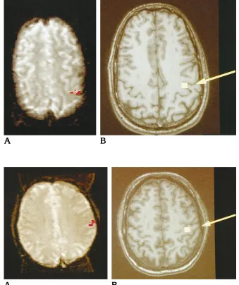
Fig 3. Left index finger stimulation: functional MR imaging (A) and magnetic source imaging (B). Pixels identified as activated by functional MR imaging are overlaid on acquired gradient-echo echo-planar image (echo time, 60; flip angle, 90°; FOV, 403 20 cm; matrix, 1283 128). Magnetic source imaging locations are overlaid on a spoiled gradient-echo MR image (35/5) (flip angle, 30°; section thickness, 1.5 mm).
Fig 4. Right index finger stimulation (same subject as in Figure 3): functional MR imaging (A) and magnetic source im-aging (B). Pixels identified as activated by functional MR imaging are overlaid on an acquired gradient-echo echo-planar im-age (echo time, 60; flip angle, 90°; FOV, 40320 cm; matrix, 1283128). Magnetic source imaging locations are overlaid on a spoiled gradient-echo MR image (35/5) (flip angle, 30°; section thickness, 1.5 mm).
[image:6.587.212.551.320.724.2]the central sulcus by the postcentral location of the single-equivalent dipole representing the re-sponse to somatosensory stimulation (33). The single-equivalent dipole model provides an es-timate of a single current source that might give rise to the measured extracranial fields. The modeling process involves the nonlinear least-squares fitting of dipole strength, location, and orientation to minimize the difference between observed and predicted extracranial fields (based on Biot Savart law computation). The fitting process provides a measure of data-model correlation and goodness-of-fit and 95% confidence volume. In accord with previous in-vestigators, we chose to accept dipole locations
that satisfied the observed data with a
confi-dence volume of less than 1 cm3 and with a
correlation coefficient ofrgreater than .98 (30, 37–39). Clearly, changing such dipole selection criteria influences the rate of localization suc-cess.
In our study, somatosensory stimulation with painless pneumatic tapping to the fingertips proved inadequate for routine use with func-tional MR imaging (69% acceptable locations), although in the 11 of 16 instances in which localization was shown, good correspondence with magnetic source imaging and anatomic expectation was found. Thus, it seems that fail-ure to find localization results from poor signal-to-noise ratio and statistical power rather than to an inherent inability of the technique. Fur-thermore, incomplete spatial coverage (five sections with a thickness of 7 mm and a 3-mm separation were used) and weakness of the stimulus itself might account for some of the failures. Stronger, but still painless, somatosen-sory stimulation methods are under develop-ment. Also, although attempts were made to provide identical somatosensory stimuli for functional MR imaging and magnetic source im-aging studies, the pressure and duration of the stimulus pulse, as well as the interstimulus in-terval, may have varied in the different operat-ing environments (the magnetic source imagoperat-ing stimulus was computer-generated and auto-matically regulated; however, to operate in the strong fringe fields of the 1.5-T magnet during functional MR imaging, the stimulus generation system was manually driven). This issue
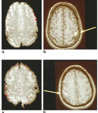
con-Fig 6. Left index finger stimulation: functional MR imaging (A) and magnetic source imaging (B). Pixels identified as activated by functional MR imaging are overlaid on an acquired gradient-echo echo-planar image (echo time, 60; flip an-gle, 90°; FOV, 40320 cm, matrix, 1283 128). Magnetic source imaging locations are overlaid on a spoiled gradient-echo MR image (35/5) (flip, 30°; section thickness, 1.5 mm). Both techniques show agree-ment on the superior level of somatosen-sory digit representation in this subject.
[image:7.587.47.372.79.485.2]cerning stimuli also should be considered in comparisons between other functional imaging techniques. Alternatively, simple motor task performance seems more reliable, having pro-vided 16 of 16 successful locations with func-tional MR imaging.
The general agreement between the two techniques is encouraging, particularly when considering the different physiological aspects they probe (hemodynamic versus electrical ac-tivity). The lack of precise colocalization might be anticipated, especially because to achieve detectable functional MR imaging we adopted the gradient-echo echo-planar imaging se-quence with its dominant venous contribution. It seems likely that the early poststimulus nega-tive BOLD effect, seen as signal loss caused by increased tissue oxygen uptake immediately before increased cerebral blood flow (40), might compare more closely with the locations
on the magnetic source images. However, this effect was not studied with our 1.5-T system. Furthermore, the simplistic magnetic source imaging modeling adopted (single-equivalent dipole) limits the confidence of magnetic source imaging location to a few millimeters (38).
[image:8.587.212.550.85.474.2]Improved magnetic source imaging modeling strategies with more complex current source descriptions might become feasible if limited in range by some prior spatial knowledge, pro-vided, for example, by functional MR imaging (41). However, clearly strict spatial constraints imposed by functional MR imaging should be avoided, because of underlying physiological differences. Although not specifically required in the application of presurgical mapping of the sensorimotor strip, it also appears that the tem-poral resolution of magnetic source imaging might allow the elucidation of temporal se-quences of multiple sites of activation identified
Fig 8. Functional MR imaging has a tendency toward spurious locations (over-laid on a gradient-echo echo-planar im-age) (echo time, 60; flip angle, 90°; FOV, 40320 cm; matrix, 1283128). This case shows anterior activation in response to right-hand index finger stimulation (A). This apparent location is presumed attrib-utable to signal in draining veins remote from the neuronal activation site. The cor-responding magnetic source imaging loca-tion (B) appears as predicted in the post-central gyrus (seen coregistered with a spoiled gradient-echo image (35/5) (flip angle, 30°; section thickness, 1.5 mm).
by functional MR imaging (and inherently time-averaged and thus unresolvable in time and ap-parently simultaneous with this technique). Thus, the combination of techniques, even with-out the requirement for strict spatial colocaliza-tion, might provide valuable information about neural signal propagation, unobtainable from either method alone.
Acknowledgments
We are grateful to Dan Vigneron for his advice regarding the echo-planar imaging implementation, Gary Cicirello for excellent technical assistance, and William P. Dillon and David Poeppel for invaluable discussions. We also thank Biomagnetic Technologies Inc for technical support of the magnetic source imaging component of the study.
References
1. Mansfield P. Multiplanar image formation using NMR spin echoes.
J Physiol1977;C10:L55–L58
2. Edelman RR, Wielopolski P, Schmitt F. Echo planar MR imaging.
Radiology1994;192:600 – 612
3. Ogawa S, Lee TM, Kay AR, Tank DW. Brain magnetic resonance imaging with contrast dependent on blood oxygenation.Proc Natl Acad Sci U S A1990;87:9868 –9872
4. Ogawa S, Lee TM. Magnetic resonance imaging of blood vessels at high fields: in vivo and in vitro measurements and image sim-ulation.Magn Reson Med1990;16:9 –18
5. Ogawa S, Lee TM, Nayak AS, Glynn P. Oxygenation sensitive contrast in magnetic resonance image of rodent brain at high magnetic fields.Magn Reson Med1990;14:68 –78
6. Kwong KK. Functional magnetic resonance imaging with echo planar imaging.Magn Reson Q1995;11:1–20
7. Fox P, Raichle M. Focal physiological uncoupling of cerebral blood flow and oxidative metabolism during somatosensory stim-ulation in human subjects.Proc Natl Acad Sci U S A1986;83: 1140 –1144
8. Fox PT, Raichle ME, Mintun MA, Dence C. Nonoxidative glucose consumption during focal physiologic neural activity. Science
1988;241:462– 464
9. Frahm J, Bruhn H, Merboldt K-D, He`nicke W. Dynamic MR im-aging of human brain activation during rest and photic stimula-tion.J Magn Reson Imaging1992;2:501–505
10. Bandettini PA, Wong EC, Hinks RS, Tikofsky RS, Hyde JS. Time course EPI of human brain function during task activation.Magn Reson Med1992;25:390 –397
11. Hammeke TA, Yetkin FZ, Mueller WM, et al. Functional magnetic resonance imaging of somatosensory stimulation.Neurosurgery
1994;35:677– 681
12. Kwong KK, Belliveau JW, Chesler DA, et al. Dynamic magnetic resonance imaging of human brain activity during primary sen-sory stimulation.Proc Natl Acad Sci U S A1992;89:5675–5679 13. McCarthy G, Blamire AM, Rothman DL, Gruetter R, Shulman RG.
Echo planar magnetic resonance imaging studies of frontal cortex activation during word generation in humans.Proc Natl Acad Sci U S A1993;90:4952– 4956
14. Baumgartner C, Doppelbauer A, Deecke L, et al. Neuromagnetic investigation of somatotopy of human hand somatosensory cor-tex.Exp Brain Res1991;87:641– 648
15. Gallen CC, Sobel DF, Schwartz B, Copeland B, Waltz T, Aung M. Magnetic source imaging: present and future.Invest Radiol1993; 28(Suppl)3:S153–S157
16. Gevins A, Cutillo B, DuRousseau D, et al. Imaging the spatiotem-poral dynamics of cognition with high-resolution evoked potential methods.Hum Brain Mapping1994;1:101–116
17. Harding GF, Armstrong RA, Janday B. Visual evoked electrical and magnetic response to half-field stimulation using pattern re-versal stimulation.Ophthalmic Physiol Opt1992;12:171–174 18. Hari R. On brain’s magnetic responses to sensory stimuli.J Clin
Neurophysiol1991;8:157–169
19. Hari R. Magnetic evoked fields of the human brain: basic princi-ples and applications. Electroencephalogr Clin Neurophysiol Suppl1990;41:3–12
20. Nuwer MR. Electroencephalographic brain mapping.West J Med
1991;155:67
21. Darcey TM, Ary JP, Fender DH. Methods for location of electrical sources in the human brain.Prog Brain Res1980;54:128 –134 22. Rose DF. Magnetic evoked responses: comparison with electrical
evoked responses.Adv Neurol1990;54:89 –94
23. Williamson SJ, Kaufman L. Evolution of neuromagnetic topo-graphic mapping.Brain Topogr1990;3:113–127
24. Wood C, Spencer DD, Allison T, McCarthy G, Williamson PD, Goff WR. Localization of human sensorimotor cortex during surgery by cortical surface recording of somatosensory evoked potentials.
J Neurosurg1988;68:99 –111
25. Roberts TPL, Rowley HA, Kucharczyk J. Applications of magnetic source imaging to presurgical brain mapping. In: Kucharczyk J, Roberts TPL, Moseley ME, Orrison W, eds.Neuroimaging Clinics of North America “Functional Neuroimaging.”Philadelphia, Pa: Saunders; 1995;5:251–266
26. Bandettini PA, Jesmanowicz A, Wong EC, Hyde JS. Processing strategies for timecourse data sets in functional MRI of the brain.
Magn Reson Med1993;30:161–173
27. Forman SD, Cohen JD, Fitzgerald M, Eddy WF. Improved assess-ment of significant activation in functional magnetic resonance imaging (fMRI): use of a cluster-size threshold.Magn Reson Med
1995;33:636 – 647
28. Weisskoff RM, Zuo CS, Boxerman JL, Rosen BR. Microscopic susceptibility variation and transverse relaxation: theory and ex-periment.Magn Reson Med1994;31:601– 610
29. Kennan RP, Zhong J, Gore JC. Intravascular susceptibility con-trast mechanisms in tissues.Magn Reson Med1994;31:9 –21 30. Lai S, Hopkins AL, Haacke EM, et al. Identification of vascular
structures as a major source of signal contrast in high resolution 2D and 3D functional activation imaging of the motor cortex at 1.5T: preliminary results.Magn Reson Med1993;30:387–392 31. Jack CR, Thompson RM, Butts RK, et al. Sensory motor cortex:
correlation of presurgical mapping with functional MR imaging and invasive cortical mapping.Radiology1994;190:85–92 32. Ojemann G, Sutherling W, Lesser R, Dinner D, Jayakar P,
Saint-Hilaire J-M. Cortical stimulation. In: Engel J, ed.Surgical Treat-ment of the Epilepsies. 2nd ed. New York, NY: Raven Press; 1993:399 – 414
33. Sobel DF, Gallen CC, Schwartz BJ, et al. Locating the central sulcus: comparison of MR anatomic and magnetoencephalo-graphic functional methods.AJNR Am J Neuroradiol1993;14: 915–925
34. Roberts TPL, Zusman E, McDermott M, Barbaro N, Rowley HA. Correlation of functional magnetic source imaging with intra-operative cortical stimulation in neurosurgical patients. J Imag Guided Surg1996;1:339 –347
36. Pantev C, Gallen C, Hampson S, Buchanan S, Sobel D. Repro-ducibility and validity of neuromagnetic source localization using a large array biomagnetometer.Am J EEG Technol1991;31:83– 101
37. Gallen CC, Sobel DF, Waltz T, et al. Noninvasive presurgical neuromagnetic mapping of somatosensory cortex.Neurosurgery
1993;33:260 –268
38. Gallen CC, Schwartz B, Rieke K, et al. Intrasubject reliability and validity of somatosensory source localization using a large array
biomagnetometer. Electroencephalogr Clin Neurophysiol1994; 90:145–156
39. Gallen C, Schwartz BJ, Bucholz RD, et al. Presurgical localization of functional cortex using magnetic source imaging.J Neurosurg
1995;82:988 –994
40. Ernst T, Hennig J. Observation of a fast response in functional MR.
Magn Reson Med1994;32:146 –149
41. Robinson SE.Magnetoencephalography by Lead Field Synthesis.