Copyright © 2003, American Society for Microbiology. All Rights Reserved.
Phospholipid-Binding Protein Cts1 Controls Septation and Functions
Coordinately with Calcineurin in
Cryptococcus neoformans
Deborah S. Fox,
1,2Gary M. Cox,
1,2and Joseph Heitman
1,2,3,4*
Departments of Molecular Genetics and Microbiology,1Pharmacology and Cancer Biology,3and Medicine,2and
Howard Hughes Medical Institute,4Duke University Medical Center, Durham, North Carolina
Received 16 June 2003/Accepted 6 August 2003
Cryptococcus neoformansis an opportunistic fungal pathogen that causes life-threatening
meningoencepha-litis in immunocompromised patients. The Ca2ⴙ-calmodulin-activated protein phosphatase calcineurin is
necessary for virulence ofC.neoformans. Mutants lacking the calcineurin catalytic (Cna1) or regulatory (Cnb1) subunit fail to grow at elevated temperature and are defective in virulence and hyphal elongation. Here we isolated a multicopy suppressor gene,CTS1, which restores growth of a calcineurin mutant strain at 37°C. The
CTS1gene (for calcineurin temperature suppressor 1) encodes a protein containing a C2 domain and a leucine zipper motif that may function as an effector of calcineurin. The CTS1gene was disrupted by homologous recombination, and cts1 mutants were viable but exhibited defects in cell separation, growth, mating, and haploid fruiting. In addition,cts1mutants were inviable when calcineurin was mutated or inhibited. Taken together, these findings suggest that calcineurin and Cts1 function in parallel pathways that regulate growth, cell separation, and hyphal elongation.
Signal transduction cascades are utilized by all organisms to sense signals at the cell surface and to transmit this informa-tion to effectors within the cell (17, 50). The primary compo-nents of signal transduction cascades are protein kinases and phosphatases, which act on protein targets via a mechanism of reversible phosphorylation. Many pathogenic fungi utilize sig-nal transduction pathways to sense the host and rapidly adapt to changing environmental conditions (24). Our studies ad-dress the signaling cascades that enable pathogenic fungi to sense and interact with the host.
The human fungal pathogenCryptococcus neoformansis a leading cause of disease among immunosuppressed individu-als, resulting in an often-fatal form of meningoencephalitis. However, even healthy individuals are at increased risk due to the ubiquitous environmental presence of this fungus. A recent outbreak of infection withCryptococcus neoformansvar.gattii
among residents and visitors to Vancouver Island has resulted in more than 66 cases of cryptococcal disease, including several deaths, since 1999 (44). In addition to the incidence of cryp-tococcal infections among apparently healthy persons, isolates have been reported that are resistant to currently available antifungal drugs (36). Thus, further research into the patho-genic mechanisms of this medically important fungus and the discovery of novel drug targets are needed.
Calcineurin is a highly conserved Ca2⫹-calmodulin-activated
serine/threonine protein phosphatase that is necessary for the pathogenesis ofC.neoformansand other medically important fungi (reviewed in reference 14). In other organisms, cal-cineurin regulates many physiological processes necessary for life, including morphogenesis, cell wall biosynthesis, septation, and cytokinesis (16, 26, 31, 33, 51, 52, 54). Calcineurin is the
target of the immunosuppressive drugs FK506 and cyclosporin, which inhibit calcineurin activity (25). InC. neoformans, cal-cineurin is essential for growth at 37°C and for virulence in animal models of cryptococcosis (9, 13, 35). In addition to a role in growth at 37°C, calcineurin is also required for mor-phogenic events involving hyphal elongation inC.neoformans, a process central to the development of infectious spores (7). In this study we examined the hypothesis that calcineurin-dependent protein effectors are present inC.neoformansand that their overexpression will suppress the temperature-sensi-tive growth defect conferred by a calcineurin mutation. To identify and characterize components of the calcineurin signal-ing pathway inC.neoformans, we isolated multicopy suppres-sors of the temperature-sensitive defect of a calcineurin-defi-cient strain. Plasmid-dependent transformants were isolated which restored growth at high temperature, revealing a novel gene, named CTS1 for calcineurin temperature suppressor.
CTS1encodes a protein containing a phospholipid-binding C2 domain and a leucine zipper motif. Overexpression of Cts1 conferred resistance to FK506 and cyclosporin in wild-type organisms. Disruption of the CTS1 gene by homologous re-combination resulted in temperature sensitivity, a cell separa-tion and septal posisepara-tioning defect, a reducsepara-tion in growth, inhi-bition of hyphal elongation and virulence, synthetic lethality with calcineurin mutations, and enhanced sensitivity to FK506 and cyclosporin. In addition, domain analysis revealed that, while the C-terminal leucine zipper motif is important for full Cts1 function, the C2 domain is absolutely required for high-temperature growth, cell separation, phospholipid binding, and suppression of calcineurin mutation. Our findings suggest that calcineurin and Cts1 function in parallel pathways that control growth, cell separation, and hyphal elongation.
MATERIALS AND METHODS
Strains, media, and reagents.AllC.neoformansstrains used in this study are derived from the congenic serotype D (Cryptococcus neoformansvar.
neofor-* Corresponding author. Mailing address: Department of Molecular Genetics and Microbiology, 322 CARL Bldg., Box 3546, Duke Uni-versity Medical Center, Research Drive, Durham, NC 27710. Phone: (919) 684-2824. Fax: (919) 684-5458. E-mail: [email protected].
1025
on September 8, 2020 by guest
http://ec.asm.org/
mans) strains JEC21 (MAT␣) and JEC20 (MATa). The genotype of strain MCC3 isMATacna1::ADE2 ura5. Strain JEC43 (MAT␣ura5) is a 5-fluoroorotic acid-resistant derivative of JEC21. The genotypes of thects1deletion or truncation strains derived from JEC20, JEC43, or JEC21 are as follows: DSF22 (MAT␣ cts1-570::NAT), DSF20 (MATa cts1-570::NAT), DSF42 (MATa cts1⌬::NAT), DSF45 (MAT␣ cts1⌬::NAT), DSF50 (MAT␣ cts1⌬::NAT ura5), and DSF11 (MAT␣cnb1::NAT).C.neoformansstrains were grown on standard yeast me-dium except where otherwise indicated.
Oligonucleotide primers and sequencing.Oligonucleotide primers for PCR and sequencing were synthesized by Integrated DNA Technologies, Inc. Se-quencing was performed by the Duke University DNA analysis facility with the ABI 377 sequencer, version 3.3 (PE Applied Biosystems). Sequence data were analyzed with MacVector, version 7.0, and the DNASTAR software suite, ver-sion 4.0. Primers used include JOHE6289 (5⬘-CATACAACGCACTGCAAGT GCCC), JOHE6297 (5⬘-TACATTACTCTTCTCATCTCC), JOHE8912 (5⬘-GC ACCCCTATAGATTATAAGGATGATGATGATAAGGAACCCAAAGA GC), JOHE8913 (5⬘-CTCTTTGGGTTCCTTATCATCATCATCCTTATAAT CTATAGGGGTGC), JOHE8914 (5⬘-GGCACGTTGATCGATTATAAGGAT GATGATGATAAGGTCGTTCAACGG), JOHE8915 (5⬘-CCGTTGAACGAC CTTATCATCATCATCCTTATAATCGATCAACGTGCC), JOHE8916 (5⬘-G CGCAACATGGCGATTATAAGGATGATGATGATAAGCCGATGTCGT CG), and JOHE8917 (5⬘-CGACGACATGCCCTTATCATCATCATCCTTAT AATCGCCATGTTGCGC).
Multicopy suppressor library.The multicopy suppressor library was con-structed as previously described (7). Briefly, genomic DNA from the serotype D MAT␣strain C21F2 was partially digested withSau3AI, size selected to yield fragments from 6 to 12 kb, and ligated into theBamHI site of theC.neoformans shuttle vector pPm8 as described previously (32). The resulting genomic library was linearized with I-SceI and introduced by electroporation into MCC3. Trans-formants were grown on medium lacking uracil to maintain the pPm8 plasmid. Disruption of theC.neoformans CTS1gene.TheCTS1gene was disrupted by homologous recombination with a cassette containing the nourseothricin domi-nant drug resistance gene fromStreptomyces noursei,nat1, fused to theC. neo-formans ACT1promoter andTRP1terminator and flanked byCTS1gene se-quence (29). The cassette was introduced as a 1.7-kbEcoRV fragment into the bluntedNdeI sites of theCTS1gene. Disruption was confirmed by Southern blot analysis ofHindIII-digested genomic DNA from wild type andcts1-570::natand cts1⌬::natdisruptants with a 300-bp probe corresponding to theCTS1gene. The 1.7-kbEcoRV fragment containing the gene for resistance to the aminoglycoside antibiotic nourseothricin,nat1, fused to theC.neoformansactin promoter, was generated as previously described (13, 29).
Virulence assay.The pathogenicity of thects1⌬andcts1-570mutant strains was determined in the murine inhalation model of cryptococcosis. Strains to be analyzed were grown to mid-log phase in yeast extract-peptone-dextrose (YPD) at 25°C for 24 h, washed, and resuspended in phosphate-buffered saline (PBS) to a concentration of 5 ⫻104 organisms per 0.1 ml. Organisms (wild type or mutant) were introduced by intranasal inoculation of complement (C5)-deficient DBA/1 mice with 5⫻104organisms introduced per mouse with 10 mice per strain tested. Mice were monitored for survival over a period of 105 days. Surviving mice were analyzed for the presence of viableC.neoformansorganisms as previously described (13).
Mating and haploid fruiting assays.Mating assays were performed by growing strains of opposite mating types on YPD solid medium for 48 to 72 h at 25°C prior to coculturing them on V8 medium at 25°C for 7 days in the dark. Haploid fruiting assays were performed similarly, with strains being grown on YPD solid medium prior to being spotted onto filamentation agar followed by incubation at 25°C for 14 days in the dark. Filamentation for both mating and haploid fruiting assays was scored microscopically, as previously described (7).
FLAG tagging and site-directed mutagenesis of Cts1.The FLAG epitope (DYKDDDDK) was introduced into theCTS1coding region by using a PCR overlap approach to generate an N-terminal FLAG-Cts1 fusion. Briefly, PCR products for the left end of theCTS1coding region (JOHE6289 and JOHE8913) and the right end (JOHE6297 and JOHE8912) were generated, purified, and combined to generate the full-length 3-kb overlap product with the flanking primer set (JOHE6289 and JOHE6297). Domain deletions of Cts1 were gener-ated by replacement of the predicted phospholipid-binding C2 domain or a region bearing limited homology to a calmodulin binding domain (CMD) with the FLAG epitope. Briefly, the C2 domain (amino acids 16 to 161) and putative CMD (amino acids 284 to 305) of Cts1 were replaced by an in-frame substitution with the FLAG epitope (DYKDDDDK) by the use of a PCR overlap method as described above with primers JOHE8914 and JOHE8915 being paired with the flanking primer set to generate the C2 domain deletion product and primers JOHE9816 and JOHE8917 being paired with the flanking primer set to generate
the CMD deletion product. All overlap products were then ligated into the TA cloning vector pCR2.1 (Invitrogen) and introduced intoEscherichia coliTOP10 cells (Invitrogen).
Expression and purification of Cts1 fusion proteins.To generate the FLAG-Cts1, Cts1⌬C2-FLAG, and Cts1⌬CMD-FLAG protein fusions, the FLAG:: CTS1,CTS1⌬C2::FLAG, andCTS1⌬CMD::FLAGcoding regions were isolated asBamHI-XbaI fragments from the pCR2.1 constructs described above and ligated into theBamHI andXbaI sites of the pPm8 shuttle vector. The 1.7-kb CTS1promoter region was then introduced as aBglII fragment into the BamHI-BglII sites of each pPm8 construct in both forward and reverse orientations for FLAG::CTS1and in the sense direction forCTS1⌬C2::FLAGandCTS1⌬CMD:: FLAG. The resulting constructs were linearized and introduced by electropora-tion into DSF50. Transformants were grown on medium lacking uracil to main-tain the pPm8 plasmid. Total protein was isolated from transformants grown at 25°C with complete yeast extraction buffer (Calbiochem), purified with anti-FLAG M2 affinity agarose (Sigma), eluted with 0.1 M glycine-HCl (pH 3.5), precipitated with acetone, and resuspended in PBS with protease inhibitors.
Phospholipid binding assays.The binding of FLAG-Cts1 or the Cts1⌬C2-FLAG fusion proteins was evaluated by a dot blot assay utilizing phosphoinosi-tide (PIP) strips (Molecular Probes). Purified fusion protein in PBS containing 0.1% Tween 20 (PBS-T) with 5% bovine serum albumin was used to probe the PIP membrane strips overnight at 4°C. Bound protein was visualized by Western blot analysis with a primary mouse anti-FLAG M2 antibody (Sigma) and a secondary sheep anti-mouse antibody conjugated to horseradish peroxidase (Amersham Biosciences) followed by detection with SuperSignal West Femto maximum-sensitivity substrate (Pierce). All antibody incubations were per-formed in PBS-T with 3% bovine serum albumin at room temperature, and washes were performed in PBS-T at room temperature.
Chitinase assays.C.neoformansstrains were grown to log phase in liquid YPD medium at 25°C, washed with PBS, and treated with chitinase (Sigma, 2.5 U) or PBS alone for 2 h at 25°C. Cells were then examined by differential interference contrast (DIC) microscopy with a Zeiss AxioPhot 2 fluorescence microscope with a Zeiss AxioCam MR digital camera and image capture software.
Fluorescence microscopy.For fluorescence microscopic analysisC. neofor-mansstrains were grown to log phase in liquid YPD medium at 25°C prior to fixation and permeabilization. Cells were fixed by incubation in PBS containing 7.7% formaldehyde for 1 h at room temperature, washed in PBS, and fixed in PBS containing 1% Triton X-100. Fixed and permeabilized cells were stained with Alexa Fluor 488-conjugated wheat germ agglutinin (WGA; Molecular Probes) to visualize chitin and Alexa Fluor 594-conjugated phalloidin (Molecular Probes) to visualize F-actin. Fluorescence and DIC microscopy were performed on a Zeiss AxioPhot 2 fluorescence microscope equipped with a Zeiss AxioCam MR digital camera and software for image capture and analysis.
Electron microscopy.Conventional electron microscopy was performed as previously described with the following modifications (19). Cells were grown to mid-log phase in YPD at 25°C, rinsed in 0.1 M cacodylate buffer (pH 7.4), and fixed in 1% osmium tetroxide in 0.1 M cacodylate buffer containing 0.15% ruthenium red for 1 h at 25°C. Following three washes in cacodylate buffer, cells were dehydrated through a graded series of ethanol from 30 to 100% and subjected to an extended infiltration series in propylene oxide-Spurr resin prior to being embedded in 100% Spurr resin. Embedded samples were cut on an RMC MT-7 ultramicrotome, and sections of 50 to 80 nm were collected on Formvar-coated grids and stained with uranyl acetate and Reynold’s lead citrate. All sections were viewed on a JEOL 1200EX transmission electron microscope. Nucleotide sequence accession numbers.The cDNA and genomic sequences for CTS1 have been deposited in GenBank under the accession numbers AY163383 (cDNA) and AY163382 (genomic).
RESULTS
Identification of a suppressor of calcineurin mutant tem-perature sensitivity.To identify genes whose products function downstream of calcineurin, or in parallel pathways, a multicopy suppressor screen was conducted to discover genes that restore growth at 37°C of a calcineurin mutant strain. The multicopy genomic library, containing genomic fragments of 6 to 12 kb of the serotype D strain C21F2 cloned in the shuttle vector pPm8, was linearized with I-SceI and introduced into the calcineurin A (cna1) mutant strain MCC3 by electroporation (7). Trans-formants were selected on medium lacking uracil at 25°C and
on September 8, 2020 by guest
http://ec.asm.org/
screened for growth at 35°C by replica plating. Four suppres-sors were isolated from a screen of 2,000 transformants. We verified that growth at 35°C was plasmid dependent by plasmid loss on 5-fluoroorotic acid medium to counterselect theURA5
marker. Plasmids were rescued from suppressor strains, and the inserts were sequenced. One of the four suppressors was found to contain the CNA1 gene, which encodes the cal-cineurin catalytic subunit. The three remaining suppressors contained genes of unknown function.
Sequence analysis of the open reading frame (ORF) of one of the three remaining suppressors revealed a predicted pro-tein of 834 amino acid residues that possessed homology within the amino terminus to a putative C2 domain-containing pro-tein of unknown function inNeurospora crassaencoded by the NCU09847.1 locus. In addition to the phospholipid-binding (C2) domain at the amino terminus, two additional functional domains were identified, including a predicted transmembrane domain adjacent to the C2 domain and a putative leucine
zipper region at the carboxy terminus. Because phospholipid-associated proteins, such as Its3 in Schizosaccharomyces
pombe, have been shown to function coordinately with
calci-neurin, we chose to examine this novel gene in greater detail, designating the gene CTS1 for calcineurin temperature sup-pressor 1 (54).
To confirm that the overexpression ofCTS1sufficed to re-store growth at 37°C in calcineurin mutants, the rescued plas-mid containing theCTS1 gene under the control of its own promoter (pCTS1) or the plasmid pPm8 was introduced into isogenic wild-type andcna1mutant strains (JEC34 and MCC3, respectively). Overexpression ofCTS1restored growth at 37°C in calcineurin mutants (Fig. 1). However, overexpression of
CTS1did not restore hyphal elongation during mating or hap-loid fruiting in calcineurin mutant strains (data not shown) (7). Taken together, these results suggest that Cts1 and calcineurin share an essential function in the regulation of growth at ele-vated temperature. As this functional overlap is specific to the regulation of growth at 37°C, Cts1 is probably not involved in all aspects of calcineurin function. However, the possibility exists that Cts1 could have the potential to suppress the addi-tional calcineurin mutant phenotypes if expressed at higher levels.
Disruption of theCTS1gene by homologous recombination.
TheCTS1gene was disrupted by homologous recombination with a cassette containing the nourseothricin resistance gene
nat1. The nat1 selectable marker cassette was either intro-duced into a singleNdeI site at position 5146 (corresponding to amino acid 570) ofCTS1to generate the C-terminal truncation insertion allelects1-570::nat1or introduced between theNdeI sites at positions 2685 and 5146 within the coding region of FIG. 1. Overexpression of theCTS1gene restores growth of
cal-cineurin mutant strains at 37°C. Isogenic wild-type JEC34 (ura5) and calcineurin MCC3 (cna1 ura5) mutant strains were transformed with the rescued suppressor plasmid pCTS1 or the control plasmid pPm8 and grown on synthetic dextrose medium lacking uracil for 3 days at 25 or 37°C in the presence or absence of 1g of FK506/ml.
FIG. 2. Disruption of theCTS1gene by homologous recombination. (A) TheCTS1gene was disrupted by the insertion of the dominant selectable markernat1(nourseothricin resistance) between twoNdeI sites in theCTS1gene, generating thects1⌬::natallele, while thects1-570
truncation mutation was generated by introducing thenatcassette into the 3⬘NdeI site corresponding to amino acid 570 of Cts1. The ORF ofCTS1
is a solid box, the 5⬘and 3⬘untranslated regions are indicated by diagonal lines, and the transcription start site is marked by a bent arrow. The 300-bp bar denotes the region used as a probe in panel B. Restriction sites are HindIII (H) and NdeI (D). (B) Southern blot analysis of
HindIII-digested genomic DNA from wild-type (WT; JEC21),cts1-570(DSF22), andcts1⌬(DSF45) strains. Sizes in kilobases are indicated at right.
on September 8, 2020 by guest
http://ec.asm.org/
CTS1to generate thects1⌬::nat1deletion allele (Fig. 2A). The resulting truncation and deletion cassettes were introduced into the isogenic serotype D wild-type strains JEC20 (MATa) and JEC21 (MAT␣). Nourseothricin-resistant transformants were screened by colony PCR for the absence of the wild-type
CTS1gene, and 8MAT␣cts1⌬(8 of 16, 50%), 11MATacts1⌬ (11 of 22, 50%), 8 MAT␣ cts1-570 (8 of 20, 40%), and four
MATacts1-570(4 of 20, 20%) mutants were identified. Muta-tion of theCTS1gene was verified by Southern blotting (Fig. 2B). In addition, each confirmed transformant was subjected to Northern analysis to verify that theCTS1transcript was absent in cts1⌬ mutant strains or truncated in the cts1-570 mutant strain (data not shown and Fig. 10).
CTS1is required for growth at elevated temperature.Both the catalytic (Cna1) and the regulatory (Cnb1) subunits of calcineurin are necessary for the growth ofC.neoformansat 37°C (9, 13, 35). Because the multicopy expression ofCTS1was sufficient to suppress the temperature-sensitive growth defect of a calcineurin mutant, we reasoned that Cts1 might also be required for growth at 37°C. In addition, as leucine zipper motifs have been shown to provide essential functions in many membrane-associated proteins, we also sought to examine the importance of the leucine zipper region for Cts1 protein func-tion (Fig. 3A). To address these quesfunc-tions, we examined the influence of the complete absence of functional Cts1, as well as the loss of the leucine zipper motif of Cts1, by testing the viability of both thects1⌬andcts1-570mutant strains at 25 and 37°C. The viability of the cts1⌬ mutant was significantly re-duced compared to that of the wild type at 37°C and moder-ately reduced for viability at 25°C (Fig. 3B). However, the
cts1-570truncation mutant lacking the C-terminal leucine zip-per region was indistinguishable from wild type at either tem-perature (Fig. 3B). The inability of thects1⌬mutant to grow at 37°C is the result of the loss of functional Cts1 because trans-formation of thects1⌬strain with an episomal shuttle plasmid containing theCTS1gene under the control of its endogenous
promoter restored growth at 37°C (Fig. 8B and 9A). Thus, full-length Cts1, but not the leucine zipper region, is required for growth at 37°C.
CTS1is necessary for virulence.The ability to grow at ele-vated temperatures is essential for virulence ofC.neoformans
(13, 22, 35). Because the cts1⌬ mutant is unable to grow at 37°C, we anticipated that Cts1 would be required for virulence. In contrast, because thects1-570mutant is viable at 37°C, this allele allowed us to test whether Cts1 plays a role in virulence beyond growth at 37°C. Wild-type,cts1⌬, andcts1-570mutant strains were introduced by intranasal inoculation into mice lacking the C5 component of complement (DBA), and survival was monitored over a period of 105 days. Survival of mice infected with the wild-type strain was significantly reduced compared with that of mice inoculated with either thects1⌬or
cts1-570mutant strain (Fig. 4). Theaand␣wild-type strains resulted in similar mortality values, with 50% mortality by day 40 and 100% mortality by day 42 to 46 (Fig. 4). In contrast, 100% of mice infected with either the cts1⌬ mutant or the
cts1-570truncation mutant strain survived to day 105 irrespec-tive of mating type, indicating that Cts1 and its leucine zipper region are required for virulence (Fig. 4). The ability to pro-duce both melanin and a polysaccharide capsule is an impor-tance virulence determinant forC.neoformans(15, 22, 23, 37). To determine whether the unanticipated loss of virulence in thects1-570mutant strain was due to a defect in either melanin or capsule production, thects1⌬deletion andcts1-570 trunca-tion mutant strains were examined. Interestingly, both strains were able to produce melanin and capsule at levels comparable to those of wild-type cells, suggesting that, in addition to cal-cineurin, Cts1 also plays an essential role in virulence that is independent of the ability to synthesize melanin or capsule components.
Cts1 is required for hyphal elongation.In addition to growth at 37°C, calcineurin is also required for hyphal elongation during mating and haploid fruiting in C. neoformans(7). To FIG. 3. Growth ofcts1⌬mutants is temperature sensitive. (A) Diagram of the domain structure of Cts1 showing the amino-terminal C2 domain (C2); the serine, proline, or glutamine repeat regions (hatched); and the carboxy-terminal leucine zipper region (LZ). The amino acid positions for each repeat or domain are indicated. Thects1⌬allele completely disrupts the ORF ofCTS1, resulting in the absence of Cts1 protein production, while thects1-570::natallele truncates 264 amino acids from the C terminus. (B)cts1⌬confers a growth defect. The isogenic wild-type (JEC21) andcts1⌬(DSF45) andcts1-570(DSF22) mutant strains were serially diluted and grown on YPD medium for 3 days at 25 or 37°C.
on September 8, 2020 by guest
http://ec.asm.org/
examine whether Cts1 also plays a role in mating,cts1⌬and
cts1-570mutant strains were analyzed for the ability to produce filaments. There were no defects in mating whenMAT␣cts1⌬ orMATacts1⌬mutant strains were mixed with wild-type tester strains in a unilateral cross on mating (V8) medium, as numer-ous filaments and basidiospores were produced, typical of a wild-type mating reaction (data not shown). In contrast, when
MAT␣cts1⌬andMATacts1⌬mutant strains were cocultured in a bilateral cross, no filamentation was observed and no basidia or basidiospores were produced (Fig. 5A). These find-ings indicate that, in addition to calcineurin, Cts1 is also re-quired for mating, as loss of Cts1 confers a bilateral mating defect. However, the carboxyl-terminal portion of Cts1, which contains the leucine zipper region, is not absolutely essential for mating, as filamentation and basidiospore production arise from a bilateral cross of cts1-570mutants, although the fila-ments generated from this cross are abnormal (data not
shown). We note that the leucine zipper region does become necessary for mating when the opposite mating partner lacks Cts1 entirely (Fig. 5A).
In addition to a defect in mating, ␣ cts1⌬ mutant strains were also unable to undergo haploid fruiting, a differentiation process that occurs in␣cells in response to nitrogen limitation and desiccation (Fig. 5B) (49). Haploid fruiting of thects1-570
mutant strains lacking the leucine zipper region was reduced compared to that of wild type, but not abolished (Fig. 5B). These results demonstrate that Cts1 is involved in hyphal elon-gation, a process required for both mating and haploid fruiting. Thus, the carboxyl-terminal portion of Cts1 containing the leucine zipper region is important but not essential for hyphal elongation during mating and haploid fruiting.
Cts1 mutation confers a septation defect. For the fission yeastS.pombe, calcineurin has been shown to play an impor-tant role in morphogenic processes including cytokinesis, sep-tation, polarity acquisition, and mating (52). As mutations in eitherCTS1or calcineurin confer defects in growth, virulence, and hyphal elongation inC.neoformans, we sought to examine whether Cts1 plays a role in morphogenesis. The wild-type,
cts1⌬mutant, calcineurin (cnb1⌬) mutant, andcts1-570mutant strains were grown to mid-log phase in YPD at 25°C and fixed and permeabilized. Fixed cells were then stained with phalloi-din or WGA to visualize actin or chitin, respectively.
Wild-type cells treated with phalloidin revealed a pattern typical for C. neoformans, with punctate staining of cortical actin in the cytoplasm, as well as staining at the mother-bud neck and at the growing end of the emerging bud (data not shown) (18). Although there were minor differences between the actin staining pattern for the cnb1⌬, cts1⌬, or cts1-570
mutant cells and that for the wild type, the most striking dif-ference was the increased frequency among the cts1⌬ and
cts1-570mutant strains of cells that failed to complete separa-tion, resulting in long chains of cells with some branching (data not shown).
Analysis of chitin staining with WGA revealed that, in con-trast to wild-type cells or calcineurin-deficient cells, thects1⌬ FIG. 4. Cts1 is required for virulence ofC.neoformans.
Comple-ment (C5)-deficient DBA mice were infected with 5⫻104cells of the prototrophic wild-typeastrain JEC20 (■) or the congenicacts1⌬(Œ)
oracts1-570(F) mutant (DSF42 or DSF20, respectively) (A) and the
prototrophic wild-type␣strain JEC21 (■) or the congenic␣cts1⌬(Œ)
or␣cts1-570(F) mutant (DSF45 or DSF22, respectively) (B). Mice
were monitored daily for survival over 105 days, and the percentage of surviving mice was plotted versus time.
FIG. 5. Cts1 is required for hyphal elongation. (A) Deletion of the
CTS1gene results in bilateral sterility. Isogenicaand␣cts1⌬(DSF42 and DSF45),cts1-570(DSF20 and DSF22), and wild-type (JEC20 and JEC21) strains were coincubated on nitrogen-limiting agar (V8) and photographed at 100⫻after incubation at 25°C for 7 days in the dark. (B) Cts1 is required for haploid fruiting. Isogenic wild-type (JEC21),
cts1-570 (DSF22), andcts1⌬ (DSF45) ␣ strains were incubated on filamentation agar at 25°C for 14 days in the dark. WT, wild type.
on September 8, 2020 by guest
http://ec.asm.org/
andcts1-570mutant cells fail to complete cell separation. Sep-tal material that stained with WGA could be found at the mother-bud junction and at bud scars in wild-type cells (Fig. 6A). However, compared to wild-type cells, the accumulation of chitin at the mother-bud neck in both cts1⌬and cts1-570
mutant cells was quite striking (Fig. 6A). To determine the role of chitin accumulation in the failure of cts1 mutant cells to separate, we exposed cells to chitinase and examined the ef-fects of chitin digestion on the morphology of cts1⌬ mutant cells and found that exposure ofcts1⌬mutant cells to chitinase resulted in the separation of cells at the mother-bud neck (Fig. 6B).
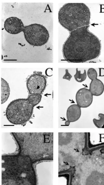
To examine the cell separation defect of thects1⌬andcts1
-570 mutants in greater detail, transmission electron micros-copy was performed. Ultrastructural studies revealed that both
the cts1⌬andcts1-570mutant cells had thickened septa with frequent defects in septal formation and positioning (Fig. 7B to D). A more detailed examination of early septal formation in both wild-type andcts1⌬cells revealed that, while completed primary septa were observed in wild-type cells (Fig. 7E), the
cts1⌬ mutant cells did not form typical primary septa and instead appeared to close the neck by an abnormal advance-ment of septal material and gradual thickening (Fig. 7F). Nei-ther the calcineurin-deficient cells nor wild-type cells exhibited any septal defects (Fig. 7A and data not shown). These obser-vations indicate that Cts1, but not calcineurin, is required for proper septal positioning and formation.
The C2 domain is essential for Cts1 function in vivo.We next sought to examine the function of the C2 domain of the Cts1 protein, a candidate calcineurin effector. FLAG-tagged FIG. 6. cts1⌬mutation confers a separation defect. (A) Isogenic wild-type (JEC21),cts1⌬mutant (DSF45), andcts1-570mutant (DSF22) strains were grown to logarithmic phase at 25°C in YPD, fixed and permeabilized, stained with Alexa Fluor-conjugated WGA, and visualized by fluorescence and DIC optics. (B) Isogenic wild-type (JEC21) andcts1⌬mutant (DSF45) strains were grown to logarithmic phase at 25°C in YPD, washed, and incubated in PBS in the presence or absence of chitinase for 2 h prior to visualization by DIC optics. Magnification,⫻1,000.
on September 8, 2020 by guest
http://ec.asm.org/
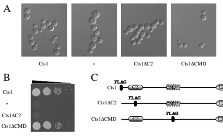
versions of the wild-type allele (FLAG::CTS1) and alleles in which the C2 domain (CTS1⌬C2::FLAG) or the putative CMD (CTS1⌬CMD::FLAG) was replaced with the FLAG epitope-encoding sequence were expressed under the control of the
CTS1promoter in the pPm8 shuttle plasmid in the tempera-ture-sensitive cts1⌬ and cnb1⌬ strains (Fig. 8C). While the wild-type CTS1 and CTS1⌬CMD alleles restored growth at 37°C in the presence of the calcineurin inhibitor FK506 in both
cts1⌬andcnb1⌬strains, neither theCTS1⌬C2allele nor the antisenseCTS1promoter control construct restored growth in either cts1⌬ orcnb1⌬strains (Fig. 8B and 9A). In addition,
cts1⌬ cells expressing the wild-type CTS1 and CTS1⌬CMD
alleles were restored for normal septation, whereascts1⌬cells expressing the CTS1⌬C2 allele or the antisense CTS1 pro-moter control were septation defective (Fig. 8A).
C2 domains have been shown to confer phospholipid-, ino-sitol polyphosphate-, and protein-binding functions on many proteins involved in diverse cellular processes, including cy-toskeletal organization, cell growth and gene expression, pro-tein transport, signal transduction, and vesicular trafficking. To address the role of Cts1 in phospholipid binding, we charac-terized the PIP-binding capacity of the C2 domain of Cts1 by
a protein-lipid overlay assay. FLAG-tagged Cts1 and Cts1⌬C2 fusion proteins were expressed inC. neoformans, purified by immunoaffinity anti-FLAG resin fromC.neoformansprotein extracts, and eluted from the resin. The FLAG-Cts1 and Cts1⌬C2-FLAG fusion proteins were each applied to a mem-brane-fixed PIP array in a protein-lipid overlay assay. The FLAG-Cts1 fusion protein bound phosphatidylinositol-5-phos-phate [PI(5)P] and PI(4)P, whereas the Cts1⌬C2-FLAG fusion did not show significant binding, demonstrating that the C2 domain is essential for the phospholipid-binding activity of Cts1 (Fig. 9B). The binding of Cts1 was specific to PI(5)P and PI(4)P, and no binding to other phospholipids, including PI(3)P, PI(4,5)P2, and PI(3,4)P2was detected (data not shown).
Taken together, these data provide compelling evidence that the C2 domain is important for the in vivo function of Cts1, as the C2 domain of Cts1 is required for growth at high temper-ature, septation, and phospholipid binding. In addition, dele-tion of the putative CMD of Cts1 had no effect on growth or septation.
Cts1 and calcineurin function in parallel pathways.We next examined whether Cts1 and calcineurin function in the same or parallel pathways by genetic epistasis tests. TheMAT␣cts1-570
mutant strain was crossed with a MATa cna1mutant strain, and basidiospores were isolated. Although 13 meiotic recom-binants were obtained from this cross, no viablects1-570 cna1
double mutants were obtained, indicating that thects1 muta-tion is synthetically lethal in combinamuta-tion with a calcineurin mutation (data not shown). Thus, calcineurin becomes essen-tial at all growth temperatures in a strain lacking full-length Cts1, providing evidence that the two function in either parallel pathways or a branched pathway. In support of this conclusion, overexpression of Cts1 conferred resistance to FK506 in wild-type cells (Fig. 1 and 8B). Together, these results demonstrate that Cts1 can compensate for the loss of calcineurin function to permit growth at 37°C and that Cts1 and calcineurin interact genetically to maintain viability upon exposure to elevated temperatures. Additionally, these data indicate that the car-boxyl terminus of Cts1, which contains the leucine zipper re-gion, is not normally required for growth at elevated temper-ature but becomes necessary for growth in the absence of functional calcineurin.
To further study the functional relationship between cal-cineurin and Cts1, we examined the influence of calcal-cineurin on the transcriptional regulation of theCTS1gene. By Northern analysis, the CTS1gene was transcribed at elevated levels in bothcna1andcnb1mutant strains and in thects1-570 trunca-tion mutant at both 25 and 37°C but was transcribed at barely detectable levels in the wild type (Fig. 10). These results indi-cate that in the absence of calcineurin function, or in the absence of fully functional Cts1, the expression of theCTS1
mRNA is elevated.
We further addressed the functional relationship between calcineurin and Cts1 by in vitro protein binding assay analysis. Resin-immobilized FLAG-Cts1 was incubated with purified bovine calcineurin. No binding of Cts1 to calcineurin was de-tected in the presence or absence of Ca2⫹-calmodulin or Ca2⫹
(data not shown), suggesting that Cts1 and calcineurin do not physically interact. In addition, no interaction between Cts1 and either the catalytic (Cna1) or the regulatory (Cnb1) sub-unit of calcineurin fromC.neoformans could be detected by FIG. 7. Defective septal positioning and formation in cts1⌬and
cts1-570 mutants. Transmission electron micrograph images of iso-genic wild-type (JEC21) (A and E),cts1-570(DSF22) (B and C), and
cts1⌬(DSF45) (D and F) cells grown to logarithmic phase at 25°C in YPD and fixed in osmium tetroxide. Arrows indicate abnormally po-sitioned and aberrant septa in thects1⌬andcts1-570mutant strains. Bars, 2 (A to D) and 0.1 (E and F)m.
on September 8, 2020 by guest
http://ec.asm.org/
yeast two-hybrid analysis or FLAG-Cts1 pull-down experi-ments with C. neoformansprotein extracts (data not shown). Although the methods employed to detect possible interac-tions between Cts1 and calcineurin are suitable for the detec-tion of a majority of protein-protein interacdetec-tions, it is possible that these methods would not detect an interaction involving a bridging protein or protein complex. Therefore, we cannot definitively rule out the possibility that Cts1 and calcineurin physically interact.
DISCUSSION
The goal of this study was to identify calcineurin-dependent protein effectors through a multicopy suppressor approach. As a result of this screen, we isolated the calcineurin temperature suppressor gene, CTS1, which encodes a novel protein con-taining both a phospholipid-binding C2 domain and a leucine zipper motif. Overexpression of the CTS1 gene restored growth at elevated temperatures in a calcineurin-deficient strain and conferred resistance to the calcineurin inhibitor FK506 when the gene was overexpressed in wild-type cells. Disruption of theCTS1gene conferred temperature-sensitive growth and inhibited virulence in the murine model of crypto-coccosis, similar to the calcineurin mutant strains. In addition, the cts1⌬ mutation conferred hypersensitivity to FK506, syn-thetic lethality with a calcineurin mutation, and a septation defect. Taken together, these data demonstrate that the novel C2 domain-containing protein Cts1 is essential for virulence of
C. neoformansand performs essential functions that overlap
with those of the Ca2⫹-calmodulin-activated serine/threonine
phosphatase calcineurin in vivo.
Interestingly, we found thatCTS1expression is responsive to
calcineurin activity, as the expression of CTS1 is induced in calcineurin-deficient strains. Although this result is suggestive of a potential derepression of CTS1in the absence of func-tional calcineurin, a recent study involving a genome-wide analysis of calcineurin-dependent gene expression has found that calcineurin primarily directs the activation of gene expres-sion rather than represexpres-sion in Saccharomyces cerevisiae(53). However, this does not preclude the possibility that calcineurin could direct gene repression inC.neoformans. Therefore, al-though the induction ofCTS1expression could be the result of derepression due to a lack of functional calcineurin, a more plausible explanation fitting with our observations is thatCTS1
expression is induced as part of a compensation mechanism in response to the lack of functional calcineurin.
Calcineurin plays a central role in many physiological pro-cesses in fungi and has been shown to be necessary for viru-lence in bothC.neoformansandCandida albicans(reviewed in references 3, 4, 8, and 14). The functions of calcineurin have been extensively studied in two model fungi, the budding yeast,
S.cerevisiae, and the fission yeast,S. pombe(1, 45–47). In S.
cerevisiae, calcineurin controls gene expression necessary for
cell wall biosynthesis, cation homeostasis, and morphogenesis via the regulation of the activity and localization of the cal-cineurin-responsive transcription factor Crz1 (5, 28, 30, 42, 43, 53). However, no Crz1 homolog is apparent in theC. neofor-mans10⫻genome coverage of the serotype A or DC. neofor-mansstrains.
In addition, several proteins that function coordinately with calcineurin to regulate cytokinesis, ion homeostasis, and sep-tation inS.pombehave been identified (16, 26, 51, 54). Among these are two proteins, Its3 and Its10, which function in the FIG. 8. The C2 domain of Cts1 is necessary for proper septation. (A)cts1⌬mutant strains overexpressing FLAG-Cts1 under the control of the
CTS1promoter in either sense or antisense orientation, Cts1⌬C2-FLAG, or Cts1⌬CMD-FLAG were grown to log phase at 25°C in YPD and visualized by DIC optics. (B)cts1⌬mutant strains overexpressing FLAG-CTS1 under the control of theCTS1promoter in either sense or antisense orientation, Cts1⌬C2-FLAG, or Cts1⌬CMD-FLAG were serially diluted and grown on YPD medium for 3 days at 37°C in the presence of 1g of FK506/ml. (C) Domain structure of Cts1 showing the amino-terminal C2 domain, the putative CMD (hatched box), and the carboxy-terminal leucine zipper region. The position of the FLAG epitope is indicated by a filled octagon.
on September 8, 2020 by guest
http://ec.asm.org/
regulation of cytokinesis and septation. Its3 is a homolog of the
S. cerevisiae PI(4)P 5-kinase Mss4, which is localized to the
plasma membrane and concentrated at the septum of dividing cells (10, 54). Theits10gene encodes a putative novel allele of thecdc7gene, which encodes a serine/threonine protein kinase involved in the initiation of septum formation, and Its10 has been shown to function in a calcineurin-dependent manner (11, 26). Taken together, these findings support a central role for calcineurin in the regulation of cytokinesis and suggest that calcineurin may act to regulate multiple steps in cytokinesis, including formation of the actin ring at the mother-bud neck, septum formation, and cell separation.
We have identified Cts1, a suppressor of calcineurin tem-perature sensitivity that functions in septal positioning and septation inC.neoformans. By functional domain analysis, we have shown that the carboxyl-terminal leucine zipper region is not necessary for growth at 37°C but is required for virulence in the murine model of cryptococcosis, proper hyphal elonga-tion, and viability in the absence of calcineurin function. Loss of this region also results in a defect in septal positioning and cell separation. Thus, the leucine zipper region is essential for several functions of Cts1. Although no obvious homologs of Cts1 have been identified, analysis of functional domains among proteins with similar functions identified a leucine-zipper-containing protein in S. pombe, Cdc14, which is also required for septum formation, likely via the mediation of proteprotein interactions with other septation proteins, in-cluding Cdc7 (12).
In addition to the leucine zipper motif, Cts1 also possesses an amino-terminal C2 domain that binds PI(4)P and PI(5)P. The Cts1 C2 domain shares homology with nonclassical Ca2⫹
-independent protein kinase C isoforms that contain amino-terminal C2 domains (34, 38). C2 domains have been identified in many proteins involved in signal transduction, gene expres-sion, and cytoskeletal organization and function to bind phos-pholipids in either a Ca2⫹-dependent or an independent
man-ner. C2 domains facilitate protein-protein interactions and signal transduction membrane association (6, 21, 40). FIG. 9. The C2 domain of Cts1 is necessary for growth at 37°C and binds phosphatidylinositol-derived phospholipids. (A) Isogenic wild-type (JEC21) andcts1⌬(DSF45) andcnb1⌬(DSF11) mutant strains overexpressing FLAG-Cts1 under the control of theCTS1promoter in either sense or antisense orientation or Cts1⌬C2-FLAG were serially diluted and grown on YPD medium for 3 days at 37°C in the presence or absence of 1 g of FK506/ml. (B) Purified FLAG-Cts1 and Cts1⌬C2-FLAG fusion proteins and the extract control were overlaid on the PIP array membrane. Bound protein was detected with a monoclonal anti-FLAG antibody. The input material for each binding assay was separated by sodium dodecyl sulfate-polyacrylamide gel electrophoresis, transferred to polyvinylidene difluoride, and detected with anti-FLAG antibody. At the bottom of panel B is a table showing the relative density of input sample binding to either PI(4)P or PI(5)P expressed as the percent density of each sample relative to the FLAG-Cts1 sample.
FIG. 10. CTS1mRNA expression is upregulated in calcineurin and
cts1⌬570mutants. Shown are results of Northern blot analysis of total RNA isolated from isogenic wild-type (JEC21) and cna1 (MCC3),
cnb1(DSF11), andcts1-570(DSF22) mutant strains grown at 25 or 37°C for 4 h. Following hybridization with theCTS1300-bp probe, the blot was stripped and reprobed withCNB1, followed byGPD1as a control for loading and hybridization conditions. WT, wild type.
on September 8, 2020 by guest
http://ec.asm.org/
We have shown that Cts1 and calcineurin function coordi-nately to regulate morphogenic events necessary for growth, cytokinesis, and filamentation. Cts1 and calcineurin are com-ponents of separate pathways with shared functions essential for growth at 37°C, virulence, and hyphal elongation. In addi-tion, Cts1 is required for proper septal positioning and forma-tion, and chains of cts1⌬ cells can be separated by chitinase treatment, suggesting that Cts1 may promote the localization or assembly of protein complexes that carry out polarized chitin deposition or dissolution. Polarized chitin deposition is essential for proper septum biogenesis and septation in yeast and involves the actions of several proteins, including chitin synthases, chitinases, endoglucanases, and components of the contractile ring (39, 41, 48). Although Cts1 does not share homology with chitinases or endoglucanases known to be nec-essary for septum dissolution, recent studies have identified a role for lipid signaling in chitin deposition during septum bio-genesis in S. cerevisiae(2, 27, 48). In these studies, the PIP phosphatase Sac1, an integral membrane protein that acts on PI(4)P, was found to be necessary for the proper sorting of the Chs3 chitin synthase from the septum to the trans-Golgi net-work (48). Therefore, Cts1 may regulate the localization of chitinases or endoglucanases to the septum to promote septal dissolution via interactions with PIPs and other proteins. Al-ternatively, Cts1 may be necessary for the proper localization of chitin synthesis machinery, wherein the lack of Cts1 leads to excessive accumulation of chitin at the septum. The ability of Cts1 to suppress the calcineurin defect when overexpressed supports a model in which Cts1 promotes the proper regula-tion of chitin synthase localizaregula-tion, as calcineurin mutants have severe cell wall and membrane integrity defects that could be suppressed by excess chitin deposition at the cell periphery (20). Future studies will address the role of Cts1 in the regu-lation of chitin biogenesis at the septum and cell periphery, as well as the role of Cts1 in chitin dissolution at the septum. In conclusion, the identification of the novel calcineurin temper-ature suppressor Cts1 has provided insights into the functions of the calcineurin signal transduction pathway in C. neofor-mans and may further our understanding of the roles of cal-cineurin in virulence and differentiation.
ACKNOWLEDGMENTS
We thank Wandy Beatty at Washington University (St. Louis, Mo.) for performing the transmission electron microscopy experiments, James Fraser and Connie Nichols for comments and discussions, Fu-jisawa, Inc., for providing FK506, and members of theC.neoformans
genome project at the Stanford Genome Technology Center and Na-gasaki University for sequence data supported by the NIH (U01 AI47087).
This work was supported by NIAID (AIDS training grant) postdoc-toral fellowship AI07392-10 (to D. Fox); NIAID R01 grants AI39115, AI42159, and AI50438 (to J. Heitman); and P01 award AI44975 from NIAID to the Duke University Mycology Research Unit. Gary Cox was supported by a Burroughs Wellcome New Investigator Award in Molecular Pathogenic Mycology. Joseph Heitman is a Burroughs Well-come Scholar in Molecular Pathogenic Mycology and an associate investigator of the Howard Hughes Medical Institute.
REFERENCES
1. Aramburu, J., A. Rao, and C. B. Klee.2000. Calcineurin: from structure to function. Curr. Top. Cell Regul.36:237–295.
2. Baladron, V., S. Ufano, E. Duenas, A. B. Martin-Cuadrado, F. del Rey, and C. R. Vazquez de Aldana.2002. Eng1p, an endo-1,3--glucanase localized at
the daughter side of the septum, is involved in cell separation in Saccharo-myces cerevisiae. Eukaryot. Cell1:774–786.
3. Blankenship, J. R., W. J. Steinbach, J. R. Perfect, and J. Heitman.2003. Teaching old drugs new tricks: reincarnating immunosuppressants as anti-fungal drugs. Curr. Opin. Investig. Drugs4:192–199.
4. Blankenship, J. R., F. L. Wormley, M. K. Boyce, W. A. Schell, S. G. Filler, J. R. Perfect, and J. Heitman.2003. Calcineurin is essential forCandida albicanssurvival in serum and virulence. Eukaryot. Cell2:422–430. 5. Boustany, L. M., and M. S. Cyert.2002. Calcineurin-dependent regulation of
Crz1p nuclear export requires Msn5p and a conserved calcineurin docking site. Genes Dev.16:608–619.
6. Catz, S. D., J. L. Johnson, and B. M. Babior.2002. The C2A domain of JFC1 binds to 3⬘-phosphorylated phosphoinositides and directs plasma membrane association in living cells. Proc. Natl. Acad. Sci. USA99:11652–11657. 7. Cruz, M. C., D. S. Fox, and J. Heitman.2001. Calcineurin is required for
hyphal elongation during mating and haploid fruiting inCryptococcus neo-formans. EMBO J.20:1020–1032.
8. Cruz, M. C., A. L. Goldstein, J. R. Blankenship, M. Del Poeta, D. Davis, M. E. Cardenas, J. R. Perfect, J. H. McCusker, and J. Heitman.2002. Calcineurin is essential for survival during membrane stress inCandida albicans. EMBO J.21:546–559.
9. Cruz, M. C., R. A. Sia, M. Olson, G. M. Cox, and J. Heitman.2000. Com-parison of the roles of calcineurin in physiology and virulence in serotype D and serotype A strains ofCryptococcus neoformans. Infect Immun.68:982– 985.
10. Desrivieres, S., F. T. Cooke, P. J. Parker, and M. N. Hall.1998. MSS4, a phosphatidylinositol-4-phosphate 5-kinase required for organization of the actin cytoskeleton inSaccharomyces cerevisiae. J. Biol. Chem.273:15787– 15793.
11. Fankhauser, C., and V. Simanis.1994. The cdc7 protein kinase is a dosage dependent regulator of septum formation in fission yeast. EMBO J.13:3011– 3019.
12. Fankhauser, C., and V. Simanis.1993. TheSchizosaccharomyces pombe cdc14gene is required for septum formation and can also inhibit nuclear division. Mol. Biol. Cell4:531–539.
13. Fox, D. S., M. C. Cruz, R. A. Sia, H. Ke, G. M. Cox, M. E. Cardenas, and J. Heitman.2001. Calcineurin regulatory subunit is essential for virulence and mediates interactions with FKBP12-FK506 inCryptococcus neoformans. Mol. Microbiol.39:835–849.
14. Fox, D. S., and J. Heitman.2002. Good fungi gone bad: the corruption of calcineurin. Bioessays24:894–903.
15. Fromtling, R. A., H. J. Shadomy, and E. S. Jacobson.1982. Decreased virulence in stable, acapsular mutants ofCryptococcus neoformans. Myco-pathologia79:23–29.
16. Fujita, M., R. Sugiura, Y. Lu, L. Xu, Y. Xia, H. Shuntoh, and T. Kuno.2002. Genetic interaction between calcineurin and type 2 myosin and their involve-ment in the regulation of cytokinesis and chloride ion homeostasis in fission yeast. Genetics161:971–981.
17. Gustin, M. C., J. Albertyn, M. Alexander, and K. Davenport.1998. MAP kinase pathways in the yeastSaccharomyces cerevisiae. Microbiol. Mol. Biol. Rev.62:1264–1300.
18. Kopecka, M., M. Gabriel, K. Takeo, M. Yamaguchi, A. Svoboda, M. Ohkusu, K. Hata, and S. Yoshida.2001. Microtubules and actin cytoskeleton in Cryptococcus neoformanscompared with ascomycetous budding and fission yeasts. Eur. J. Cell Biol.80:303–311.
19. Kopecka, M., M. Yamaguchi, M. Gabriel, K. Takeo, and A. Svoboda.2000. Morphological transitions during the cell division cycle ofCryptococcus neo-formansas revealed by transmission electron microscopy of ultrathin sections and freeze substitution. Scr. Med.73:369–380.
20. Kraus, P. R., D. S. Fox, G. M. Cox, and J. Heitman.2003. TheCryptococcus neoformansMAP kinase Mpk1 regulates cell integrity in response to anti-fungal drugs and loss of calcineurin function. Mol. Microbiol.48:1377–1387. 21. Kulkarni, S., S. Das, C. D. Funk, D. Murray, and W. Cho.2002. Molecular basis of the specific subcellular localization of the C2-like domain of 5-li-poxygenase. J. Biol. Chem.277:13167–13174.
22. Kwon-Chung, K. J., I. Polacheck, and T. J. Popkin.1982. Melanin-lacking mutants ofCryptococcus neoformansand their virulence for mice. J. Bacte-riol.150:1414–1421.
23. Kwon-Chung, K. J., and J. C. Rhodes.1986. Encapsulation and melanin formation as indicators of virulence in Cryptococcus neoformans. Infect. Immun.51:218–223.
24. Lengeler, K. B., R. C. Davidson, C. D’Souza, T. Harashima, W. C. Shen, P. Wang, X. Pan, M. Waugh, and J. Heitman.2000. Signal transduction cas-cades regulating fungal development and virulence. Microbiol. Mol. Biol. Rev.64:746–785.
25. Liu, J., J. D. Farmer, Jr., W. S. Lane, J. Friedman, I. Weissman, and S. L. Schreiber.1991. Calcineurin is a common target of cyclophilin-cyclosporin A and FKBP-FK506 complexes. Cell66:807–815.
26. Lu, Y., R. Sugiura, T. Yada, H. Cheng, S. O. Sio, H. Shuntoh, and T. Kuno. 2002. Calcineurin is implicated in the regulation of the septation initiation network in fission yeast. Genes Cells7:1009–1019.
27. Martin-Cuadrado, A. B., E. Duenas, M. Sipiczki, C. R. De Aldana, and F.
on September 8, 2020 by guest
http://ec.asm.org/
Del Rey.2003. The endo-beta-1,3-glucanase eng1p is required for dissolution of the primary septum during cell separation inSchizosaccharomyces pombe. J. Cell Sci.116:1689–1698.
28. Matheos, D. P., T. J. Kingsbury, U. S. Ahsan, and K. W. Cunningham.1997. Tcn1p/Crz1p, a calcineurin-dependent transcription factor that differentially regulates gene expression inSaccharomyces cerevisiae. Genes Dev.11:3445– 3458.
29. McDade, H. C., and G. M. Cox.2001. A new dominant selectable marker for use inCryptococcus neoformans. Med. Mycol.39:151–154.
30. Mendizabal, I., A. Pascual-Ahuir, R. Serrano, and I. F. de Larrinoa.2001. Promoter sequences regulated by the calcineurin-activated transcription fac-tor Crz1 in the yeast ENA1 gene. Mol. Genet. Genomics265:801–811. 31. Mendoza, I., F. J. Quintero, R. A. Bressan, P. M. Hasegawa, and J. M.
Pardo.1996. Activated calcineurin confers high tolerance to ion stress and alters the budding pattern and cell morphology of yeast cells. J. Biol. Chem. 271:23061–23067.
32. Mondon, P., Y. C. Chang, A. Varma, and K. J. Kwon-Chung.2000. A novel episomal shuttle vector for transformation ofCryptococcus neoformanswith theccdBgene as a positive selection marker in bacteria. FEMS Microbiol. Lett.187:41–45.
33. Nakamura, T., T. Ohmoto, D. Hirata, E. Tsuchiya, and T. Miyakawa.1996. Genetic evidence for the functional redundancy of the calcineurin- and Mpk1-mediated pathways in the regulation of cellular events important for growth inSaccharomyces cerevisiae. Mol. Gen. Genet.251:211–219. 34. Nalefski, E. A., and J. J. Falke.1996. The C2 domain calcium-binding motif:
structural and functional diversity. Protein Sci.5:2375–2390.
35. Odom, A., S. Muir, E. Lim, D. L. Toffaletti, J. Perfect, and J. Heitman.1997. Calcineurin is required for virulence ofCryptococcus neoformans. EMBO J. 16:2576–2589.
36. Perfect, J. R., and G. M. Cox.1999. Drug resistance inCryptococcus neofor-mans. Drug Resist. Updates2:259–269.
37. Rhodes, J. C., I. Polacheck, and K. J. Kwon-Chung.1982. Phenoloxidase activity and virulence in isogenic strains ofCryptococcus neoformans. Infect. Immun.36:1175–1184.
38. Rizo, J., and T. C. Sudhof.1998. C2-domains, structure and function of a universal Ca2⫹-binding domain. J. Biol. Chem.273:15879–15882.
39. Roncero, C.2002. The genetic complexity of chitin synthesis in fungi. Curr. Genet.41:367–378.
40. Scheid, M. P., M. Huber, J. E. Damen, M. Hughes, V. Kang, P. Neilsen, G. D. Prestwich, G. Krystal, and V. Duronio.2002. Phosphatidylinositol (3,4,5)P3 is essential but not sufficient for protein kinase B (PKB) activation; phos-phatidylinositol (3,4)P2is required for PKB phosphorylation at Ser-473: studies using cells from SH2-containing inositol-5-phosphatase knockout mice. J. Biol. Chem.277:9027–9035.
41. Schmidt, M., B. Bowers, A. Varma, D. H. Roh, and E. Cabib.2002. In
budding yeast, contraction of the actomyosin ring and formation of the primary septum at cytokinesis depend on each other. J. Cell Sci.115:293– 302.
42. Stathopoulos, A. M., and M. S. Cyert.1997. Calcineurin acts through the CRZ1/TCN1-encoded transcription factor to regulate gene expression in yeast. Genes Dev.11:3432–3444.
43. Stathopoulos-Gerontides, A., J. J. Guo, and M. S. Cyert.1999. Yeast cal-cineurin regulates nuclear localization of the Crz1p transcription factor through dephosphorylation. Genes Dev.13:798–803.
44. Stephen, C., S. Lester, W. Black, M. Fyfe, and S. Raverty.2002. Multispecies outbreak of cryptococcosis on southern Vancouver Island, British Columbia. Can. Vet. J.43:792–794.
45. Sugiura, R. 2002. Functional analysis of calcineurin-mediated signalling pathway using fission yeast as a model system. Nippon Yakurigaku Zasshi 119:155–161.
46. Sugiura, R., T. Kuno, and H. Shuntoh.1998. Calcineurin-mediated signal transduction pathways in yeast. Tanpakushitsu Kakusan Koso43:1021–1028. 47. Sugiura, R., S. O. Sio, H. Shuntoh, and T. Kuno.2002. Calcineurin phos-phatase in signal transduction: lessons from fission yeast. Genes Cells7:619– 627.
48. Tahirovic, S., M. Schorr, A. Then, J. Berger, H. Schwarz, and P. Mayinger. 2003. Role for lipid signaling and the cell integrity MAP kinase cascade in yeast septum biogenesis. Curr. Genet.43:71–78.
49. Wickes, B. L., M. E. Mayorga, U. Edman, and J. C. Edman.1996. Dimor-phism and haploid fruiting inCryptococcus neoformans: association with the alpha-mating type. Proc. Natl. Acad. Sci. USA93:7327–7331.
50. Widmann, C., S. Gibson, M. B. Jarpe, and G. L. Johnson.1999. Mitogen-activated protein kinase: conservation of a three-kinase module from yeast to human. Physiol. Rev.79:143–180.
51. Yada, T., R. Sugiura, A. Kita, Y. Itoh, Y. Lu, Y. Hong, T. Kinoshita, H. Shuntoh, and T. Kuno.2001. Its8, a fission yeast homolog of Mcd4 and Pig-n, is involved in GPI anchor synthesis and shares an essential function with calcineurin in cytokinesis. J. Biol. Chem.276:13579–13586.
52. Yoshida, T., T. Toda, and M. Yanagida.1994. A calcineurin-like gene ppb1⫹ in fission yeast: mutant defects in cytokinesis, cell polarity, mating and spindle pole body positioning. J. Cell Sci.107:1725–1735.
53. Yoshimoto, H., K. Saltsman, A. P. Gasch, H. X. Li, N. Ogawa, D. Botstein, P. O. Brown, and M. S. Cyert.2002. Genome-wide analysis of gene expres-sion regulated by the calcineurin/Crz1p signaling pathway inSaccharomyces cerevisiae. J. Biol. Chem.277:31079–31088.
54. Zhang, Y., R. Sugiura, Y. Lu, M. Asami, T. Maeda, T. Itoh, T. Takenawa, H. Shuntoh, and T. Kuno.2000. Phosphatidylinositol 4-phosphate 5-kinase Its3 and calcineurin Ppb1 coordinately regulate cytokinesis in fission yeast. J. Biol. Chem.275:35600–35606.