Biology Dissertations Department of Biology
12-16-2019
IDENTIFICATION OF NOVEL DRUGGABLE TARGETS IN TWO
IDENTIFICATION OF NOVEL DRUGGABLE TARGETS IN TWO
MEMBERS OF MONONEGAVIRALES
MEMBERS OF MONONEGAVIRALES
Venice DuPont
Follow this and additional works at: https://scholarworks.gsu.edu/biology_diss
Recommended Citation Recommended Citation
DuPont, Venice, "IDENTIFICATION OF NOVEL DRUGGABLE TARGETS IN TWO MEMBERS OF MONONEGAVIRALES." Dissertation, Georgia State University, 2019.
https://scholarworks.gsu.edu/biology_diss/226
MONONEGAVIRALES
by
VENICE DU PONT
Under the Direction of Richard K. Plemper, PhD
ABSTRACT
Measles virus (MeV) is considered one of the most contagious human viruses
and has recently been declared endemic again in several countries despite a highly
efficacious vaccine. The viral RNA dependent RNA polymerase (RdRP) is a
heterologous complex comprised of the large protein (L), which provides all catalytic
domains for RNA synthesis, and the phosphoprotein (P), which provides chaperone
support for L and mediates the interaction between L and the ribonucleoprotein complex
(RNP). Though essential for polymerase function, the interface between P and L
remains poorly characterized, as well as the role of P in RdRP advancement along the
conserved helical motif upstream of the oligomerization domain (OD), and the second is
a face of the P X-domain (PXD). Using stoichiometrically-controlled
trans-complementation studies and applied mathematical modeling, we also determined the
PXD:L interaction to be mutually exclusive to the PXD:N interaction. These findings
suggest a model that centers PXD as a master regulator of RdRP advancement.
Rabies virus (RABV) causes a severe and 100% fatal neurological disease that
is vaccine preventable and treatable prior to the onset of clinical symptoms. The
post-exposure prophylaxis (PEP) for RABV treatment is prohibitively expensive, especially in
developing countries where the majority of cases occur and does not confer
cross-protection against the newly emerging phylogroup II lyssaviruses. To address the unmet
clinical need for cross-protective anti-RABV therapeutics, we developed and
implemented an innovative high-throughput screening approach utilizing a novel single
cycle RABV reporter strain maintained in BSL-2 laboratory conditions. From our
extensive screening library, we have identified the first direct-acting multi-strain RABV
entry inhibitor, GRP-60367. Resistance profiling of GRP-60367 revealed escape
mutations that accelerate the fusion kinetic of the RABV glycoprotein (G). We have
solved two of the feasibility issues with current RABV antiviral drug discovery: i) BSL-2+
RABV containment restraints and ii) reliable drug efficacy determination, thus paving the
way for future drug discovery campaigns to alleviate the deficit and cost of current
therapeutic options against lyssaviruses.
MONONEGAVIRALES
by
VENICE DU PONT
A Dissertation Submitted in Partial Fulfillment of the Requirements for the Degree of Doctor of Philosophy
in the College of Arts and Sciences Georgia State University
Copyright by
MONONEGAVIRALES
by
VENICE DU PONT
Committee Chair: Richard Plemper
Committee: Margo Brinton Sang-Moo Kang
Electronic Version Approved:
DEDICATION
This dissertation is dedicated to my mother and husband, for their incredible support
ACKNOWLEDGEMENTS
First and foremost, I would like to express my sincerest gratitude to my advisor
Dr. Richard Plemper for the invaluable guidance and support he has given to me during
this incredible journey. Many thanks to my committee members Dr. Margo Brinton and
Dr. Sang-Moo Kang, who have provided invaluable and perceptive feedback for my
dissertation projects. I would also like to thank the Plemper lab members and GSU
TABLE OF CONTENTS
ACKNOWLEDGEMENTS ... V
LIST OF TABLES ... VIII
LIST OF FIGURES ... IX
1 LIST OF ABBREVIATIONS ... XI
1 INTRODUCTION ... 1
1.1 Mononegavirales Overview ... 1
1.2 Chapter 1: Bipartite interface of the measles virus phosphoprotein X domain with the large polymerase protein regulates viral polymerase dynamics ... 3
1.2.1 Measles Virus Disease and Treatment... 3
1.2.2 MeV Lifecycle ... 6
1.2.3 MeV Transcription and Replication ... 8
1.2.4 Viral Proteins involved in MeV RNA synthesis ... 9
1.2.5 Aim of Dissertation Chapter 1 ... 19
1.2.6 Chapter 1 Materials and Methods ... 20
1.2.7 Chapter 1 Results ... 27
1.2.8 Chapter 1 Discussion... 52
1.3 Chapter 2: Identification of a novel RABV entry inhibitor ... 58
1.3.1 RABV Disease and Current Treatment ... 58
1.3.3 Current RABV Treatment ... 62
1.3.4 RABV Drug Discovery ... 63
1.3.5 Aim of Dissertation Chapter 2 ... 70
1.3.6 Chapter 2 Materials and Methods ... 71
1.3.7 Chapter 2 Results ... 76
1.3.8 Chapter 2 Discussion... 84
2 CONCLUSIONS... 88
2.1 Chapter 1 Conclusions ... 88
2.2 Chapter 2 Conclusions ... 88
3 REFERENCES ... 90
4 APPENDICES ... 104
4.1 Appendix A Chapter 1 Results and Discussion Source ... 104
LIST OF TABLES
LIST OF FIGURES
Figure 1. Measles virus lifecycle ... 6
Figure 2. The Measles Nucleoprotein ... 9
Figure 3. The Measles Large Protein. ... 12
Figure 4. The Measles Phosphoprotein ... 15
Figure 5. P OD C-terminal microdomain is required for P-to-L binding. ... 28
Figure 6. Full-length L and truncated L1708 interact with P with equal efficiency ... 29
Figure 7. P (361-364)Ala acts on RdRP bioactivity in a dominant-negative manner. ... 31
Figure 8. Native PAGE analysis of P (361-364)Ala ... 33
Figure 9. RdRP minireplicon activity profile describing the effect of different relative amounts of wild type P-encoding plasmid DNA transfected ... 33
Figure 10. P-XD is essential for efficient P:L interaction, but P-XD deletion mutants lack cooperative negative impact on RdRP activity ... 35
Figure 11. Minireplicon analysis of bioactivity of the C-terminally truncated P mutants depicted in 10A. ... 36
Figure 12. Identification of a specific L-binding face of the triangular prism fold of P-XD with regulatory effect on RdRP bioactivity ... 39
Figure 13. Effect of P (361-364)Ala and P-V463R mutations on P binding to full-length L. ... 40
Figure 14. Multi-sequence alignments of P proteins of selected paramyxoviruses. ... 42
Figure 15. L-binding null-mutants in P OD C-terminal microdomain and P-XD efficiently trans-complement ... 43
Figure 17. Homo- and hetero-typic interaction of MeV L with P derived from MeV or CDV,
respectively ... 46
Figure 18. Residues in the connector region between P position 377 and 458 trans-complement P-XD mutations but are functionally linked to the OD C-terminal microdomain ... 47
Figure 19. Minireplicon analysis of relative bioactivity of P mutants with alanine substitutions in conserved patches in the connector domain. ... 49
Figure 20. 2-way and 3-way trans-complementations to probe stoichiometric requirements of the different P-XD functionalities in bioactive RdRP complexes ... 51
Figure 21. Schematic diagram representing the current post-exposure prophylaxis treatment.... 59
Figure 22. RABV Genome, Virion and Lifecycle ... 60
Figure 23. Rabies virus glycoprotein ... 67
Figure 24. Rabies virus large protein ... 69
Figure 25. Flow chart detailing HTS protocol ... 70
Figure 26. Identification of a small-molecule hit specific against RABV ... 76
Figure 27. Mechanistic characterization of GRP-60367 ... 79
Figure 28. Adaptation of recVSV-GFP-RABVG to GRP-60367 ... 80
1 LIST OF ABBREVIATIONS
MeV Measles Virus
RABV Rabies Virus
MuV Mumps Virus
PIVs Parainfluenza Viruses
HeV Hendra Virus
NiV Nipah Virus
SeV Sendai Virus
RPV Rinderpest Virus
PPRV Pestes-des-Petites Ruminants Virus
MOKV Mokola Virus
N Nucleoprotein
P Phosphoprotein
M Matrix Protein
G Glycoprotein
H Attachment Protein
F Fusion Protein
L Large Protein
RNP Ribonucleoprotein Complex
RdRP Viral RNA-dependent RNA Polymerase
IGS Intergenic Junctions
ADEM Acute Disseminated Encephalomyelitis
MIBE Measles Inclusion Body Encephalitis
SSPE Subacute Sclerosing Panencephalitis
IG Immunoglobulin
PPI Protein-Protein Interfaces
SLAM Signal Lymphocyte Activation Molecule
N-4 Nectin-4
PXD Extreme C-terminal Domain of P
MoRE P Molecular Recognition Element
Ncore N-terminal Core Domain
Ntail C-terminal Intrinsically Disordered Tail Domain
NNTD MeV N N-terminal Domain
NCTD MeV N C-terminal Domain
CRI-VI MeV L Conserved Regions
LR MeV L Long Region
HR MeV L Hinge Region
PNBD MeV P N0 Binding Domain
OD MeV P Oligomerization Domain
KD Dissociation Constant
coIP Co-Immunoprecipitation
Y2H Yeast-2-Hybrid
HSP90 Heatshock Protein 90
hRIG Human-Derived Anti-RABV Immunoglobin
MP Milwaukee Protocol
IFN-a Interferon-a
BBB Blood-Brain Barrier
DAA Direct-Acting Antiviral
HTA Host-Targeting Antiviral
EC50 50% Effective Dose Concentration
CC50 50% Cytotoxic Concentration
SI Selectivity Index
1 INTRODUCTION
1.1 Mononegavirales Overview
The viruses of the order Mononegavirales are characterized by enveloped virions of variable morphologies that shelter a linear non-segmented, negative sense RNA genome. The viral genome is encapsulated by nucleoprotein (N) which forms the ribonucleoprotein complex (RNP) [1]. The genome encodes a linear sequence of genes, with limited overlaps, and each ORF is divided by noncoding intergenic junctions (IGS). Each gene is expressed as an individual transcription unit that is bordered by short transcription start and termination sequences
recognized by the viral RdRP. The 3¢ end of the genome contains the leader sequence that directs the RdRP for transcription initiation and mRNA is transcribed by sequential interrupted synthesis [2]. Transcription for members of Mononegavirales occurs with a gradient that results in
decreasing expression of proteins further down the genome [3]. The viral RdRP contains an enzymatic capping domain that is conserved throughout the order called the GDP
polyribonucleotidyltransferase (PRNTase). This domain caps 5¢ viral mRNA using unique capping mechanism that co-transcriptionally incorporates a GDP to the cap structure, which is then methylated at the G-N7 using the viral RdRP methyltransferase (MTase) domain [4, 5]. This
method is in contrast to the traditional capping strategy employed by eukaryotic cells and viruses with positive sense RNA genomes, which utilizes the enzyme RNA 5¢ -triphosphatase (RTPase) for phosphate removal that results in GDP followed by transfer of a GMP moiety to the cap structure by guanylytransferase (GTase) [6]. The RdRP punctuates mRNA synthesis by
The order of major structural proteins is highly conserved amongst members of
Mononegavirales and include N, phosphoprotein (P), matrix protein (M), envelope proteins, and the large protein (L) [1]. The RNP comes pre-loaded with the viral RdRP, which consists of a dimeric heterocomplex of L and P. To ensure particle assembly and budding, vital contacts are made by the RNP with M coating the luminal side of the viral envelope bilayer. M in turn interacts with the cytosolic tail of surface glycoproteins (the attachment (H), and fusion (F) proteins, for measles virus (MeV) or G, for rabies virus (RABV). N, P, M, envelope proteins, and L proteins are standard for all members of the order, though there are several viral genomes that encode additional proteins. For example, several viruses encode for virulence factors such as MeV C and V proteins, which are transcribed by leaky scanning and mRNA editing,
respectively, RdRP processivity factors, or separation of function for attachment and fusion, e.g. the MeV receptor protein, H, and its fusion protein F. Of the eight families that comprise the viral order Mononegavirales, two of which harbor the most contagious virus, MeV, within
Paramyxoviridae, and the most lethal virus, RABV, within in Rhabdoviridae, both of which continue to cause significant morbidity and mortality for humans worldwide [7, 8].
Although efficacious vaccines exist for MeV and RABV, there are currently no drugs licensed for therapeutic intervention. It was the goal of the first chapter of this dissertation to determine the critical contacts between MeV polymerase core components (L and P). We determined that the L:P interaction is bipartite. Based on this discovery, we also present a new model of polymerase advancement along the viral genomic template due to a dynamic
interaction between extreme C-terminal domain of P (PXD) and L. Furthermore, due to
In the second chapter, this dissertation details our development and implementation of a novel high-throughput screening (HTS) protocol for small-molecule drug discovery against RABV. From our screening library of over 150,000 compounds, we identified GRP-60367 as a potent entry inhibitor of RABV. Discovery of this compound not only demonstrates the
feasibility of a large-scale RABV drug discovery campaign, but further resistance profiling of GRP-60367 shed new light into RABV fusion kinetics.
1.2 Chapter 1: Bipartite interface of the measles virus phosphoprotein X domain with the
large polymerase protein regulates viral polymerase dynamics
1.2.1 Measles Virus Disease and Treatment
With over 100,000 deaths annually, MeV continues to be a leading cause of vaccine-preventable morbidity and mortality for children worldwide. MeV is identified as one of the most infectious respiratory viruses with a basic reproduction number (R0) ranging between 12 to
acute disseminated encephalomyelitis (ADEM), measles inclusion body encephalitis (MIBE), or subacute sclerosing panencephalitis (SSPE) [12-15]. The late-onset SSPE occurs within 7-10 years. A tri-residue motif within the M protein has been suggested to serve as a molecular determinant for SPPE because it promotes trans-synaptic migration of viral particles, though the exact mechanism involved remains undetermined [16]. MeV also induces immunosuppression up to 2-3 years post-infection, resulting in a predisposition to bacterial superinfections and increased risk of mortality [17]. Although there is a highly efficacious live-attenuated MeV vaccine readily available, there has been a global resurgence of MeV cases and within the United States, with over 1000 cases reported this year alone. Post-exposure prophylaxis of MeV
infection involves vaccination, if exposure is under 72 hours, or a dose of human
immunoglobulin (IG) within six days of exposure [18, 19]. Administration of vaccine or IG within the limited time window presents a challenge while cost and requirement of cold-chain for treatment with IG are also barriers for treatment. Another consideration is that treatment with IG also does not confer immunity, and, additionally, interferes with the immune response to
vaccination [20]. For cases involving neurological complications there are no standard treatment protocols and typically broad-spectrum antivirals, such as IFN-a and/or ribavirin, were
administered. However, these treatments have questionable efficacy and SSPE continues to have a 95% fatality rate [21-23]. The increase in cases as well as the chance of progression toward life-threatening neurological disease has highlighted the unmet need for MeV specific antivirals.
therapeutic option against MeV. Outwardly, designer peptides are a promising antiviral solution due to their low cytotoxicity, and high specificity to their target. Despite promising in vitro
efficacy, advancement of peptides as a therapeutic solution remains hindered by poor oral bioavailability, relatively short half-life, poor membrane permeability, and the high cost of production [21, 33]. Natural compounds have also been extensively explored as antiviral options, yet they are considered to be poor candidates due to inherent cytotoxicity, high efficacious dose requirements, and unknown mechanisms of action [34].
Due to its critical role in the viral life cycle, its highly conserved nature, lack of cellular orthologs and its multifunctional catalytic domains, the RdRP complex presents an enticing target for anti-viral therapy. The RdRP also engages in important PPI such as the L-P binding domain [35]. The only approved nucleoside analog against MeV to date remains ribavirin, a broad-spectrum RdRP inhibitor with reportedly poor efficacy and a plethora of adverse side-effects [21]. Recently, a highly encouraging non-nucleoside inhibitor of the MeV RdRP, ERDRP-0519, has been identified and characterized as both orally bioavailable and highly efficacious against multiple MeV genotypes. Furthermore, drug treatment resulted in the generation of attenuated escape mutants. The compound showed in vivo efficacy in the closely related canine distemper virus (CDV)/ferret model when given prophylactically (50 mg/kg twice daily) with a substantial decrease in viral load and delayed lymphopenia [32]. Although,
antiviral impact of targeting the viral polymerase as well as demonstrated that the delicate balance of drug administration and immune stimulation must also be considered.
[image:22.612.75.526.155.421.2]1.2.2 MeV Lifecycle
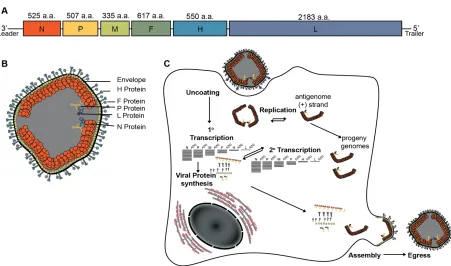
Figure 1. Measles virus lifecycleA) Schematic of genomic organization of MeV B)
Illustrative depiction of MeV virion including Envelope (black), Attachment protein (H, light blue), Fusion protein (F, green), Phosphoprotein (P, yellow), Large protein (L, lavender) and Nucleoprotein encapsidated genome (N, orange). C) Graphic depiction of MeV replication.
and the trimeric type-1 fusion protein (F) [37]. The MeV H tetramer has a globular head domain containing the receptor binding sites, a stalk domain that is arranged in a four-helix bundle conformation, and four transmembrane domains [38]. F trimerization is thought to occur within the endoplasmic reticulum, where it is also predicted to hetero-oligomerize with H [39]. F is first synthesized as an F0 precursor, which is then cleaved into two distinct polypeptides, F1 and F2,
RNA-free N (N0)protein has been suggested as a trigger [48]. As nascent viral genomes are
synthesized, they are concurrently encapsidated by N. Virion assembly is orchestrated by direct interaction of N with M proteins, which in turn interact with the surface glycoproteins. Fully assembled infectious MeV virions egress in an ESCRT-independent manner [49-51] (Figure 1C).
1.2.3 MeV Transcription and Replication
Replication and transcription of the MeV genome is accomplished through the concerted cooperation between three viral proteins, N, P, and L. The bioactive RdRP is a
hetero-oligomeric complex comprised of L and P. L provides the enzymatic domains required for RNA synthesis, capping, and cap methylation, while P provides L-chaperoning support and bridges the interaction of L to the RNP through direct interaction with N. The RdRP can function both as transcriptase and replicase, though the exact mechanism for the switch between functions remains unknown. It has been shown, however, that an accumulation of N0P complexes
promotes replication over transcription [48]. The viral genome contains cis-acting elements that provide transcription cues for the RdRP such as those for initiation, polyadenylation, and
termination of viral mRNA synthesis. RdRP begins all transcription events at the leader sequence at the 3¢ end of the antigenome and progresses through the template in a continuous cycle of N-RNA disassociation, mN-RNA capping, mN-RNA synthesis, non-templated polyadenylation followed by scanning through IGS for the next gene initiation sequence [52]. Re-initiation of mRNA synthesis is only partially efficient, resulting in an attenuation gradient that varies between IGS for each successive gene [48, 53]. Access of L to the template involves a dynamic interplay of attachment and release between the PXD and a highly conserved region that acts as a P
remains fixed, while PXD proceeds in a dynamic cartwheel fashion to engage adjacent MoRE domains of neighboring Ns [55]. What is also unknown is the trigger resolving PXD binding to MoRE, since PXD and MoRE form a highly stable four-helix bundle.
1.2.4 Viral Proteins involved in MeV RNA synthesis
[image:25.612.76.427.210.449.2]1.2.4.1 The Nucleoprotein (N)
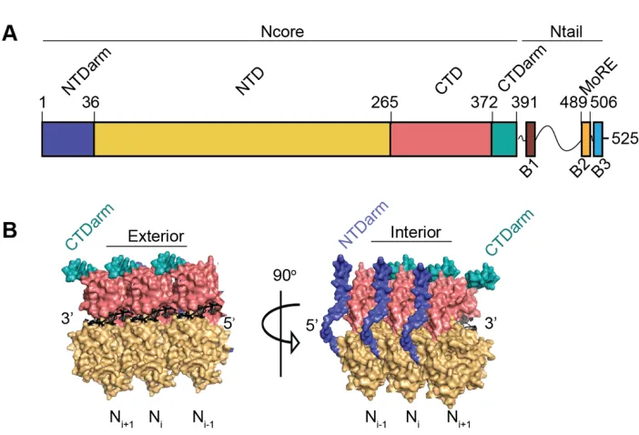
Figure 2. The Measles NucleoproteinA) Schematic representation of MeV N organization B) Structure of oligomeric interactions between individual N monomers. The CTDarm is aqua, the NTDarm is purple and RNA is black. The CTD is in pink, and the NTD is yellow (PDB: 4UFT).
The N protein of MeV consists of 525 residues and is divided into an N-terminal core domain (Ncore) (residues 1-391) and a C-terminal intrinsically disordered tail domain (Ntail) (residues 392-525) (Figure 2A). Ncore is further divided into two globular domains, the N-terminal domain (NNTD) (residues 37-265) and the C-terminal domain (NCTD) (residues 266-372),
NNTDarm (1-36) and the NCTDarm (373-391) [60]. Recent cryo-EM structure determination of RNP
revealed that N oligomerization occurs through the binding of an NNTDarm protomer into a groove
of the NCTDarm of the neighboring N monomer (Figure 2B) [57, 61]. The highly disordered Ntail
contains three highly conserved regions, called boxes 1 (400-420), 2 (489-506), and 3 (517-525) [59]. Ntail is also critical in the spatial organization of the Ncore protomers by ioccupying the space between each successive rung of the N:RNA helix. This organization causes
approximately the first 50 residues of Ntail to be buried within the RNP helical capsid [58, 62]. It has also been shown that removal of Ntail increases rigidity of the N:RNA helix, causing
decreased diameter and pitch of the total nucleocapsid structure [63]. The box 2 of Ntail is responsible for interacting with the PXD through an intrinsically disordered region, called MoRE. Prior to PXD binding, MoRE adopts an equilibrium between partially a-helical and fully unfolded [64]. Mutagenesis studies performed to investigate the binding kinetics between PXD and MoRE have revealed that the binding affinity of the complex is finely tuned to ensure efficient transcription and replication [65]. Of course, mutations that completely disrupt the interaction abolish polymerase activity. However, those mutations that resulted in an increased binding affinity, such as PXDF497A, are associated with decreased transcription elongation rates
[66, 67]. This is further supported by the observation that circulating MeV strains contain only substitutions of residues with the least disruptive impact on PXD-MoRE binding affinity [68]. Similar mutagenesis studies have determined that MoRE did not evolve to undergo
of PXD with MoRE, though the exact mechanism of resolution remains unclear [70]. Although a popular fly-catching model has been proposed in which Ntails are required for polymerase recruitment and subsequent transcription, Ntail truncation studies have shown that boxes 2 and 3 are dispensable for loading RdRP onto the template [71]. Despite activity in minigenome assays, an Ntail truncated recombinant virus was not able to be recovered, thus further supporting the hypothesis that Ntail is critical for preventing premature polymerase termination[72].
Furthermore, internal deletion of residues within Ntail produced viable, albeit attenuated,
recombinant MeV and CDV viruses. This study that demonstrated that the region plays more of a regulatory role in transcription rather than polymerase recruitment [72]. Relocation of MoRE from Box 2 to Ncore resulted in restored bioactivity and recovery of recombinant virus [73]. The recovered virus was proficient in promoting replication and mRNA editing, yet the mutant was temperature sensitive and had a flattened transcription gradient. The translocation of MoRE into Ncore demonstrated that Ntail is required for regulating the transcription gradient, thus acting as a vital modulator of gene expression [73]. Not all viral families within the Order
1.2.4.2 The Large Protein (L)
Figure 3. The Measles Large Protein A) Schematic representation of L protein organization. Long regions (LR), Conserved regions (CRI, pink; CRII, orange; CRIII,yellow; CRIV, green; CRV, blue; CRVI, purple), Hinge regions (HR) and catalytic GDNQ motif are denoted. Numbers represent amino acid numbering N-terminal to C-terminal orientation. B)
MeV homology model based on vesicular stomatitis virus (VSV) L cryoEM structure (PDB: 5A22), colored regions depict CRI-VI and GDNQ site (magenta) surface model and ribbon form in 20Å resolution.
sense RNA viruses, including MeV, contain a PRNTase-like domain suggesting that they share an unconventional capping mechanism as shown for vesicular stomatitis virus (VSV), discussed in Chapter 2 [4, 5]. This is in contrast to previously published data that suggest the
paramyxovirus L protein uses GTase and RTPase activities for capping [77, 78]. Without an equivalent trans-capping assay to the one used to establish VSV L’s unique capping mechanism, we cannot definitively assign a specific capping activity to the paramyxovirus L. The L sequence is divided into 3 conserved long regions (LR) with variable sequences between, called hinge regions (HR). HRI (residues 607-650) is less tolerant of epitope tag insertion than HRII (residues 1695-1717) suggesting less flexibility in HRI [79]. Based on these data along with in silico
protein folding and sequence analyses, it was determined that the first 1,708 N-terminal residues (consisting of LR1 and LR2), which include the catalytic center along with the P and putative L interaction sites of the L protein, form an independent folding domain [80]. Currently, there are no high-resolution structural data available for any of the Paramyxoviridae L proteins. Recently, the structure of the L protein for respiratory syncytial virus (RSV) has been solved by cryoEM to a resolution of 3.2Å. Within this structure the phosphoprotein wraps around L with the
1.2.4.3 The Phosphoprotein (P)
Figure 4. The Measles Phosphoprotein A) Schematic representation of P protein organization. The N-terminal region of P (PNTD, yellow), C-terminal region of P (PCTD),
oligomerization domain (OD, green), and the extreme C-terminal domain (XD, blue) are annotated by residue numbering. Residues 361-377 within the OD are dark green. B) ODs of closely related paramyxoviruses: MeV (green, PDB: 4ZDO), MuV (purple, PDB: 4EIJ), Sendai virus (pink, SeV, PDB: 1EZJ), NiV (yellow, PDB: 6EB9) C) Diagram depicting critical residue interactions with PXD, either with MoRE (left), or the hydrophobic contacts within the PXD bundle (right). The MoRE helix is depicted as yellow, and the PXD is colored blue. (PDB: 1T60)
D) Summary depiction of current knowledge of the L binding site for several members of Mononegavirales.
MeV P, though catalytically inactive, is an essential RdRP cofactor that provides a diverse repertoire of functions critical for viral replication and transcription. These functions include sequestering soluble monomeric RNA-free N (N0)to prevent unintentional encapsidation
long disordered regions: the N0 binding domain (P
NBD, a.a. 1-40), the oligomerization domain
(OD, a.a. 303-373), and PXD (a.a. 459-507) (Figure 4A) [85-87]. The N-terminal residues 1-48 have been previously mapped as the N0 binding region found to be highly conserved among
members of the Mononegavirales [88]. Recent high-resolution crystallographic data has unveiled that the molecular mechanism of P interaction with the Ncore occurs through hydrophobic coiled-coil interactions with two adjacent N protomer arms. The resulting steric hindrance is predicted to prevent N protomer oligomerization [56]. These interactions are also presumed to induce a conformational change that lowers RNA binding affinity. For the
paramyxovirus, Nipah virus (NiV), a similar mechanism of NTarm interaction was uncovered. However, it was proposed to interfere with the molecular opening and closing switch of N0, thus
rigidifying the NiV P in an open conformation [89]. This mechanism contrasts with that of VSV P, however, since part of the N-terminal region of VSV P extends into the N protomer arm binding cavity and a small helical region binds the hinge region between NCTD and NNTD, thereby
extending into the RNA-binding groove [90]. Although the crystal structure of the MeV N0P
complex provides insight into the chaperoning function of P, the mechanism of release of N0 and
subsequent encapsidation of newly synthesized viral genomes still remains poorly understood. Self-association of P is highly conserved among all members of the Mononegavirales.
several other paramyxoviruses, e.g. NiV and mumps virus (MuV), exhibit a central kink in the three-dimensional structure due to a break in the heptad repeat (Figure 4B) [92]. In the case of MeV, this break is due to a stammer of leucine repeats that induce the coiled-coils to adopt a 310
a-helical conformation. The role of the kink has recently been identified as a regulatory element
for viral gene expression [93]. Interestingly, a short disordered section within the C-terminal region of the P OD consisting of residues 361-375 was identified through comparison of two contrasting solved crystal structures of P [94]. Recent studies have further implicated these residues as crucial for L structural stabilization in addition to promoting RdRP bioactivity [93]
Bridging of the polymerase complex to the viral genomic template is orchestrated by the direct interaction of PXD with MoRE. Recent structural characterization of PXD revealed it to be a globular domain organized into three antiparallel a-helices that are held together primarily by hydrophobic intermolecular interactions [87]. The association of MoRE with PXD
implements an association-induced folding by the induction of a local a-helical fold of MoRE to form a hetero-four-helix bundle that principally relies on hydrophobic contacts within the large hydrophobic cleft between the a-2/a-3 face of the triangular PXD prism [65] (Figure 4C). The PXD-MoRE complex is assembled in a 1:1 stoichiometry and has an equilibrium dissociation constant (KD) of about 3.1 µM [67]. A complex hydrogen bonding network governs the
characterized and shows varying levels of compaction, presumably reflecting a necessity to negotiate association and dissociation with MoRE.
P interacts directly with L to stabilize its native conformation and to mediate physical interaction of the RdRP with the viral genome. Though the L:P interaction is essential for viral transcription and replication, the mode of contact between the two proteins is poorly
characterized. Early MeV P mapping studies using co-immunoprecipitation (coIP) and yeast-2-hybrid (Y2H) analyses of truncated P fragments determined that PNT does not harbor the L binding domain [99]. The rinderpest virus (RPV) L binding domain on P was investigated with C-terminally truncated P fragments using Y2H assays. This study resulted in the proposal that the interface comprises the entire P C-terminus (PCT) including the OD [100]. This finding was bolstered by data obtained using the construction of chimeras consisting of swapped
found to be essential for L stability, hinting at a role in binding of L, either directly or indirectly [93]. P is furthermore critical for recruitment of host heatshock protein 90 (HSP90) chaperones that mediate proper folding and solubilization of functional L proteins and the subsequent
formation of mature and stable L-P complexes [102]. Though required for maturation of the core RdRP complex, HSP90 is dispensable for bioactivity [102]. The recruitment of HSP90 for chaperoning support has been confirmed for various mononegaviruses including, besides MeV, VSV and NiV. However, the location and purpose of the HSP90 interaction with P has yet to be determined [102-105] .
1.2.5 Aim of Dissertation Chapter 1
The overarching aim of Chapter 1 is to disclose critical elements involved in the L:P interaction, as well as to disclose how the L:P heterocomplex negotiates the viral template through its interaction with MoRE.
The first aim of this study was to characterize and identify critical molecular determinants for L:P interaction. This was accomplished via mutational analysis, alanine
scanning, terminal and internal truncations in combination with co-immunoprecipitation studies. Critical residues for L:P binding were confirmed by minireplicon bioactivity studies, and
biological relevance was tested by recovery of recombinant viruses.
1.2.6 Chapter 1 Materials and Methods
Cell culture: Baby hamster kidney cells (C-13; ATCC) stably expressing T7 polymerase (BSR-T7/5), African green monkey kidney epithelial (Vero) cells (CCK-81; ATCC), and Vero cells stably expressing human signaling lymphocytic activation molecule (Vero-hSLAM) were maintained in Dulbecco’s modified Eagle’s medium (DMEM) supplemented with 7.5% fetal bovine serum at 37°C and 5% CO2 . The stable cell lines were incubated in the presence of G418
(Thermo-Fisher) (100 µg/ml) at every fifth passage. Cells were transiently transfected using GeneJuice (Novagen) according to the manufacturer’s instructions.
Molecular biology: Codon-optimized open reading frames encoding MeV IC-B-derived L, L1708, and Pwere synthesized in vitro (GeneWiz). The PDOD variant lacking residues 303-360
was generated through PCR amplification and re-ligation at an added HindIII site. Yeast-derived GCN4 tetramerization domain was PCR amplified from a previously generated template [47] and inserted using the HindIII site. All alanine substitutions and amino acid changes were performed by site-directed PCR mutagenesis using the QuikChange protocol (Stratagene). C-terminal truncations were performed by PCR mutagenesis and subsequent religation at an added AgeI site. Plasmids encoding the MeV minireplicon luciferase reporter, non-optimized MeV IC-B N, IC-B P, and IC-B L under T7 promoter control, and a full-length cDNA copy of the MeV IC-B genome were previously described [106]. A full-length cDNA of recMeV IC-B expressing P-V463R and P (361-364)Ala in tandem was generated based on non-codon optimized mutant
versions of IC-B P using the NEBuilder® HiFi DNA Assembly kit, introducing an artificial
and P-M intergenic junctions using existing XbaI and SalI restriction sites. Mutagenesis success and the integrity of all PCR-amplified nucleic acids was confirmed through Sanger sequencing.
SDS-PAGE, immunoblotting and densitometric quantitations: Cells were transfected in a 6-well plate format (5 x 10⁵ cells/well) with 1 µg of plasmid DNA encoding
codon-optimized MeV L1708 with a C-terminal FLAG epitope tag and 1.8 µg of plasmid DNA encoding
MeV P or P mutants, each with a C-terminal HA epitope tag. After 36 hours, cells were washed two times with phosphate buffered saline (PBS) and lysed chemically (50 mM HEPES (pH 7.2), 300 mM NaCl, 1.0 mM EDTA, 1% Triton X-100, protease inhibitors (Roche)). Cleared (10-minute centrifugation at 12,000 rpm, 4°C) lysates were mixed with 5´urea buffer (200 mM Tris/Cl [pH 6.8], 8 M urea, 5% SDS, 0.1 mM EDTA, 0.03% bromophenol blue, 1.5%
dithiothreitol). Samples were incubated for 30 minutes at 50°C and separated on 8% SDS-PAGE gels, tank-blotted on polyvinylidene difluoride (PVDF) membranes (Millipore), and subjected to chemiluminescence detection using specific antibodies directed against the FLAG (M2; Sigma) or HA (16B12; Abcam) epitopes, MeV N (clone 83KKII; Millipore Sigma), or against cellular glyceraldehyde-3-phosphate dehydrogenase (GAPDH; 6C; Ambion) as specified. Immunoblots were developed using a ChemiDoc digital imaging system (Bio-Rad) for image visualization. Densitometry was carried out on non-saturated images with global background correction. A full set of positive (wild type P) and negative (equivalent amount of vector DNA replacing
P-encoding plasmid DNA) controls were included on each immunoblot, and no normalizations across different blots were conducted.
fractionation was carried out using 3-12% Bis-Tris gradient gels (Invitrogen) and NativePAGE running buffer. Immunoblotting and detection were performed as outlined above.
Co-immunoprecipitation: Cells (5 x 10⁵ cells/well) were transfected with MeV L1708
and P or P mutant-encoding plasmid DNA as detailed above. Cells were lysed 24 hours after transfection and cleared lysates incubated with specific antibodies directed against HA or HIS epitopes (HIS.H8; Invitrogen; only for immunodetection after trans-complementation; Figure 5A), or against MeV N (only for immunoprecipitations in Figure 4D) at 4°C, followed by precipitation of immunocomplexes with immobilized protein G (Pierce) in 50 µl bed volume at 4°C. G-protein bound protein samples were washed twice each in cold lysis buffer and PBS, each wash with 20 bed volume equivalents (1 ml), followed by resuspension in 5x urea buffer. Denatured samples were subjected to SDS-PAGE analysis using 8% homogenous gels followed by immunoblotting and detection using specific antibodies directed respectively against the FLAG, HA, or HIS epitopes as described. To calculate relative co-immunoprecipitation
efficiencies, densitometric quantitations of precipitated L were normalized for those of L co-precipitated by standard P. This approach is based on the rationale that although L turnover rates are increased in the absence of P or presence of L binding-incompetent P mutants, synthesis rates of plasmid-encoded L is independent of P and standard and mutant P have therefore equal initial opportunity to productively interact with nascent L polypeptides.
Minireplicon reporter assay: BSR-T7/5 cells (5,000 cells/well in a 96-well plate format) were transfected in nine technical replicates per condition assessed with plasmids
P-encoding plasmid DNA ratios, empty vector (pUC-19) DNA was added in the appropriate amounts to ensure that all transfection reactions received the same total amount of DNA. Firefly luciferase activities were determined 24 hours post-transfection in a Synergy H1 microplate reader (BioTek), using Bright-Glo luciferase substrate (Promega) directly added to the wells and signal detection after a 1 to 2-minute stabilization period. Relative RdRP activities, expressed as percentages of that observed in the presence of wild type P, were determined according to the formula % rel. activity = (signalsample-signalmin)/(signalmax-signalmin) ´ 100, with signalmax
corresponding to cells having received wild type P and signalmin corresponding to cells having
received equal amounts of pUC-19 in place of P-encoding plasmid. Results calculated for each biological repeat represent the means of the nine technical repeats, and each condition (P mutant, competition or trans-complementation setting) was assessed in at least three biological repeats.
Recovery of recombinant MeV: recMeV were recovered by transfection of BSR-T7/5 cells with full-length antigenomic plasmid (1.25 µg) and the plasmids encoding IC-B N (0.42 µg), IC-B-P (0.54 µg), and IC-B-L (0.55 µg). Transfected cells were overlaid after 48 hours onto Vero-hSLAM cells. Emerging infectious centers were individually transferred to and then passaged twice on Vero-hSLAM cells, followed by whole RNA extraction from infected cells (RNeasy kit; Qiagen), first strand synthesis using random hexamer primers and Superscript III reverse transcriptase (Invitrogen), PCR amplification of synthesized cDNAs using appropriate primers, and Sanger sequencing. Titers of MeV stocks were determined through TCID50 titration
on Vero-hSLAM cells as described [107].
Viral growth kinetics: Vero-hSLAM cells were plated in 12-well format (1.0´105 cells/
by replacing the inoculum with growth media. To ensure accurate inoculum concentrations, virus stocks were pre-diluted to approximately 5,000 TCID50/ml and inoculum titers validated through
TCID50 titration. Cell-associated viral particles were harvested in 12-hours intervals through
scraping of cells in serum-free DMEM, two consecutive freeze/thaw cycles, and clearance centrifugation (2,000 rpm, 4°C). Viral titers in cleared samples were determined through TCID50
titration.
Photometric CPE quantitation: Vero-hSLAM cells were plated in a 12-well format (1.0´105 cells/well) and infected with either recMeV IC-B or recMeV IC-B P-V463R-P
(361-364)Ala at a MOI of 0.01 TCID50/cell for one hour, then inoculum was replaced with growth
media. Infected cells were imaged in 2-hour increments over a period of 36 hours post-infection with a Cytation5 automated high content imager with phase contrast microscopy capacity (BioTek). The increase in µm2 area size covered by individual infectious centers first detected at
24 hours after infection was quantified over time using the Gen5 microplate software program (Ver. 3.05.11) (BioTek). A maximum of four syncytia/well were followed, quantitations are based on 10 distinct syncytia/virus analyzed. CPE kinetics are expressed as fold-change of area size relative to the 24-hour after infection timepoint.
Mathematical models of bioactivity: Based on behavior in native-PAGE, mutant and wild type
P co-expressed in the same cell were given equal probability to tetramerize and the mutant
occupation of one P monomer was treated independent of the other three monomers. The ratio
between mutant (Mx) and wild type (wt) P was denoted as ρ. Adapting a previous approach to a
comparable problem [108], the probabilities of the formation of tetramers with different
compositions were calculated as follows:
𝑃,#$'- = 4 &'()' *,&'()) *',
𝑃/#$/- = 6 &'()' * /
&'()) */,
𝑃'#$,- = 4 &'()' *'&'()) *,,
𝑃"- = &'()) *".
A number of interactions of host proteins with MeV P and N have been proposed [109], but very limited
insight into this complex interactome precludes integration into rational modeling. We therefore applied a
deconstruction approach, concentrating on individual point mutations affecting specific interactions
between P-XD and L or N-MoRE, respectively. At any given mutant to wild type P ratio ρ, all five
different P tetramer compositions will be present in the system (illustrated in Figure 7D). Two hypotheses
regarding the bioactivities of these tetramer species were examined. In both models, the a-values reflect
the individual contribution of each of the different P tetramer assemblies to overall bioactivity, with
consideration of the relative abundance of each assembly in the system.
i) Linear combination: all tetramers with at least one wild type P monomer is bioactive and their
contributions to RdRP bioactivity (A) are linearly additive:
𝐴 = 2" 𝛼4𝑃4#$
45' = 𝛼6𝑃6#$"- + 𝛼'𝑃'#$,-+ 𝛼/𝑃/#$/- + 𝛼,𝑃,#$'-+ 𝛼"𝑃
"#$6-Assumptions made: all-mutant P tetramers (P0wt4M) are bio-inactive (Figures 7C, 10,
12E), 𝛼6 = 0. Therefore, only four weights needed to be fitted; positive weights reflect positive
and negative weights negative contributions to overall bioactivity; tetramer species with a weight
of 0 are bio-inactive.
ii) Non-linear pairwise P-XD competition: in addition to each tetramer species potentially
𝐴 = 𝑋𝐷 𝑏𝑖𝑛𝑑𝑖𝑛𝑔 𝑡𝑜 𝐿 + 𝑐𝑜𝑛𝑠𝑡. 𝑋𝐷 𝑏𝑖𝑛𝑑𝑖𝑛𝑔 𝑡𝑜 𝑀𝑜𝑅𝐸 −
𝑋𝐷𝑠 𝑐𝑜𝑚𝑝𝑒𝑡𝑖𝑛𝑔 𝑓𝑜𝑟 𝑏𝑖𝑛𝑑𝑖𝑛𝑔 𝑡𝑜 𝐿
as in:
𝐴 = 𝛼'P𝑃'#$,- +𝑃
/#$/-2 +
𝑃
,#$'-3 +
𝑃
"#$6-4 S + 𝛼/ 𝑃-TUV
− 𝛼,WP𝑃
/#$/-2 S
/
+ P𝑃
,#$'-3 S
/
+ P𝑃
"#$6-4 S
/
X
Assumptions made:all-mutant P tetramers (P0wt4M) are bio-inactive (Figures 8E, 12A,
12C); all four P-XDs within a P tetramer function independently of each other; bioactivity requires XD binding to L and MoRE; both wild type and mutant XDs are equally MoRE binding competent (Figure 8B); the V463R mutation selectively impairs XD interaction with L only; and a single L binding-competent XD in the P tetramer is necessary and sufficient for bioactivity of the RdRP complex (Figures 12B-3 and 12D-1).
Data fitting: Experimental measurements of relative RdRP bioactivities were fitted to the above
models using lsqcurvefit, a nonlinear regression model in Optimication Toolbox of MATLAB
R2017a (MathWorks). Goodness of fit was calculated using R2 values, defined as the proportion
of the variance in the dependent variable that is predicable from the independent variables.
Activity data sets for P (361-364)Ala and P-V463R were assigned a default bioactivity data point
A = 1 when ρ = 0.
graphical representations of experimental results show both the means of all biological replicates ± standard deviations (SDs) and individual biological repeats. All experiments were carried out in at least three biological repeats.
1.2.7 Chapter 1 Results
To dissect the contribution of individual MeV PCT domains to L binding, we first focused on OD. In MeV P expression plasmids, we either deleted this domain by removing residues 303-360 (OD structure up to residue 303-360 was resolved in [110]) or replaced this section up to residue 360 or 377 (end of defined heptad repeat motif), respectively, with the tetrameric mutant of the yeast general control protein 4 (GCN4) coiled-coil [111] (Figure 5B). The effect of these modifications on the physical interaction of P with L was assessed through
co-immunoprecipitation (co-IP) of the proteins from the lysates of transiently transfected cells. All co-IPs were carried out with a C-terminally truncated form of L, L1708 lacking the L
methyltransferase domain, that we have previously shown to be fully folding competent and capable of forming bioactive polymerase complexes when post-translationally combined with the corresponding C-terminal L fragment [80]. Co-IP efficiencies of L1708 or full-length L with P are
indistinguishable (Figure 6), but L1708 facilitates biochemical analysis of P-to-L binding due to a
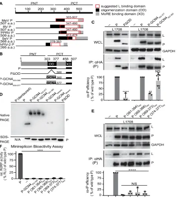
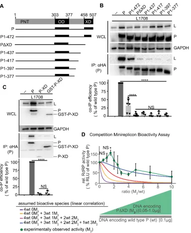
Figure 5. P OD C-terminal microdomain is required for P-to-L binding A) Overview of proposed L binding domains on P for different Paramyxoviridae. Numbering refers to MeV P. B)
Schematic representation of P with OD deletion or exchange with yeast GCN4. C) Immunoblots of whole cell lysates (WCL) and immunoprecipitates (IP) after co-transfection with L1708 (L) and
P variants shown in (B). P was detected with anti-HA antibodies, L with anti-FLAG antibodies. GAPDH served as loading control. Graph shows relative co-IP efficiency of L with P; columns are means ± SD, symbols show individual biological repeats (n = 3). D) Native PAGE analysis of P constructs shown in (B). Trimeric and dimeric variants of yeast GCN4 (P-GCN4Trimeric and
P-GCN4Dimeric, respectively) were included for mobility reference. SDS-PAGE shows
Figure 6. Full-length L and truncated L1708 interact with P with equal efficiency. Cartoons provide a schematic overview of the L constructs. Numbers refer to amino acids. Immunoblots of input and co-precipitated material assessing P interaction with full-length and truncated L. Detection and quantitative analysis as in Figure 1C. Columns show means ± SD, symbols represent individual biological repeats (n = 3). Statistical analysis through unpaired T-test (NS, not significant).
1.2.7.1 MeV P residues proximal to the structurally defined OD are critical for L interaction
When subjected to this assay, the PDOD mutant lacking the oligomerization domain was unable to co-precipitate L (Figure 5C). In contrast, efficient wild type P-equivalent L binding was maintained when P OD up to residue 360 was replaced with yeast GCN4 (P-GCN4303-360).
Further extension of the GCN4-substituted area up to P residue 377 eliminated any appreciable interaction with L. Native-PAGE analysis confirmed that both GCN4-substituted P mutants tetramerized efficiently, whereas PDOD predictably remained monomeric (Figure 5D). In contrast to previous theories [86, 92, 99], residues in the MeV P OD core do not, therefore, form a physical interface with L but contribute to the interaction only indirectly through the initiation of mandatory P tetramerization. However, residues in the short stretch at the OD C-terminus from position 361 to 377 are candidates for direct L binding.
were statistically equivalent, we noted the lowest relative interaction with L when P residues 361-364 were substituted with alanines, creating P (361-364)Ala. Employing a mono-cistronic
firefly luciferase MeV minireplicon reporter system that we have previously described [73], we assessed bioactivity of the different alanine mutants after co-expression with unmodified, homotypic MeV N and L proteins. Consistent with impaired physical interaction of these P mutants with L, all four constructs abolished RdRP bioactivity (Figure 5F). Despite its unaltered physical interaction with L, P-GCN4303-360 also lacked bioactivity in minireplicon assays,
indicating that the P OD has a role in viral RdRP activity distinct from L binding and mediating P tetramerization.
1.2.7.2 Substitutions in the P OD C-terminal microdomain are dominant-negative to RdRP
bioactivity
Residues in the P OD section 361-377 follow a 3-4 heptad repeat pattern that is
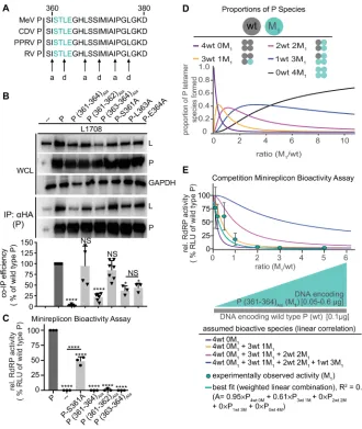
Figure 7. P (361-364)Ala acts on RdRP bioactivity in a dominant-negative manner A)
Alignment of P residues 361-377 of selected morbilliviruses. Predicted a and d positions of heptad repeat motif are highlighted. B) P:L interaction analysis of P mutants with alanine substitutions at positions 361-364. Detection and quantitative analysis as in Figure 1C (n ³ 3).
C) Minireplicon assay with P alanine substitutions at positions 361-344 (n = 3). D) Schematic of mixed P tetramer species present after co-expression of P (361-364)Ala (M1) and wild type P (wt).
Equations specify the probability of formation for each tetramer species, and the graph shows the relative proportion of these species in co-transfected cells as a function of wild type and mutant input plasmid ratio, graphically represented below the x-axis. E) Observed RdRP activity in minireplicon assays in the presence of different P (361-364)Ala and wt P ratios as depicted in
(D). Symbols show means of experimentally observed biological repeats ± SD (n = 3). Solid lines represent activity curve predictions according to a linear combination model and the relative distributions of the P tetramer species shown in (D). The dotted line (red) represents the best fit curve of the experimental data with weight assignments specified in the equation (A, relative RdRP activity), goodness of fit (R2) is indicated. All statistical analyses and symbols as detailed
Structural analysis has revealed that some helices of the P-OD coiled-coil extend to residue 373 [86]. Combined with an intact heptad repeat pattern up to residue 377, assembly of the entire 361-377 stretch into an extended coiled-coil and interaction with L as a tetrameric assembly appears likely. To test whether the P (361-364)Ala substitution has a cooperative effect on P
tetramer activity, we generated competition profiles of mixed P tetramers by gradually increasing the relative amount of mutant P in minireplicon assays in the presence of a constant, low level of wild type P. Under these conditions, five distinct P tetramer species will form in all
co-transfected cells, with the relative species distribution determined by the input ratio of mutant and wild type P-encoding plasmid DNA (Figure 7D), since native-PAGE analysis confirmed that the 361-364 alanine substitution did not affect the ability of P to tetramerize (Figure 8). Minireplicon-based assessment of RdRP bioactivity in the different cell populations revealed a steep decline in minireplicon expression in the presence of increasing amounts of the P (361-364)Ala mutant (Figure 7E). This decline was not simply due to increasingly unfavorable ratios
Figure 8. Native PAGE analysis of P (361-364)Ala and respectively denatured and native wild type P. SDS-PAGE shows immunoblots of the identical samples after denaturation and reduction.
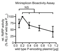
Figure 9. RdRP minireplicon activity profile describing the effect of different relative amounts of wild type P-encoding plasmid DNA transfected. Amounts of plasmids encoding L and N were kept constant. Symbols show means of experimentally observed biological repeats ± SD (n = 3). Statistical analyses through one-way ANOVA with Dunnett’s multiple comparison test, relative to starting conditions (0.1 µg); (NS, not significant).
We tested a series of mathematical models for the best description of the experimental competition data. Assigning all mutant and wild type P proteins co-expressed in the same cell equal probability to tetramerize, we considered the mutant occupation of one P monomer to be
independent of the other three monomers. Goodness of fit was excellent (R2 = 0.96) for a linear
combination model in which only tetramers consisting of four wild type (4´wt) or three wild type and one mutant (3´wt/1´P (361-364)Ala) P monomers contribute to RdRP bioactivity, while
[image:49.612.72.187.309.411.2]most one P (361-364)Ala monomer can be present in a partially bioactive P tetramer, revealed a
dominant-negative effect of the P (361-364)Ala mutation that we consider to be due to
interference with coiled-coil extension.
1.2.7.3 P-XD is an essential contributor to efficient P interaction with L
The C-terminal 50 residues of P form the XD, which has been shown biochemically and crystallographically to mediate binding of the polymerase complex to the RNP through
interaction with the MoREs located near the end of each N-tail [96, 98, 112]. To confirm that P-XD has a direct role in L binding, we generated a series of mutants with C-terminal truncations of gradually increasing length (Figure 10A). Co-IP analyses identified an essential function of P-XD in mediating efficient interaction with L (Figure 10B), since even partial shortening of P-XD was sufficient to cause significant reduction in co-IP efficiency and larger truncations abolished all appreciable interaction between P and L. Consistent with a dual role of P-XD in both MoRE and L binding, all C-terminal P truncation mutants lacked bioactivity in minireplicon assays (Figure 11). When we co-expressed isolated P-XDs – either in the form of individual polypeptides or as fusion proteins with GST for stabilization – with L, however, no biochemical interaction could be detected (Figure 10C). We conclude that P-XD is an essential contributor to efficient P-to-L binding. Unlike the strong interaction of P-XD with MoRE affording efficient co-immunoprecipitation [106], the affinity of isolated P-XD polypeptides for L is low,
Figure 10. P-XD is essential for efficient P:L interaction, but P-XD deletion mutants lack cooperative negative impact on RdRP activity .A) Schematic of P mutants generated with C-terminal truncations. B, C) P:L interaction analysis of P mutants shown in (A) and P-XD expressed in isolation (P-XD) or as a GST fusion protein (GST-P-XD). Detection and
quantitative analysis as in Figure 1C (n ³4 (B) and n = 3 (C)). D) Observed RdRP activity in minireplicon assays in the presence of different PDXD (M2) and wt P ratios as graphically
depicted below the graph. Symbols with connecting line represent means of experimentally observed biological repeats ± SD (n = 3). Solid lines represent activity curve predictions
Figure 11. Minireplicon analysis of bioactivity of the C-terminally truncated P mutants depicted in Figure 3A. Columns represent mean relative RdRP activities, symbols show
individual biological repeats ± SD (n = 3). Statistical analyses through one-way ANOVA and Tukey’s multiple comparison test (NS, not significant; ****, p ≤ 0.0001).
To explore the minimal stoichiometry of intact XDs per P tetramer that is required for bioactivity, we again co-expressed increasing relative amounts of the PDXD mutant with wild type P in minireplicon competition assays. The resulting activity profile was notably distinct from that obtained with the P (361-364)Ala substitution (Figure 10D). Even at the highest relative
ratios (up to 10) of the PDXD mutant, RdRP activity remained significantly higher than
background, indicating that PDXD has no dominant-negative effect, and lower relative ratios of the PDXD unexpectedly resulted in a significant boost in RdRP activity compared to that observed in the presence of wild type P only.
1.2.7.4 Distinct faces of the P-XD triangular prism mediate binding to MoRE and L
In co-crystals with MoRE, MeV P-XD assumed a helix-turn-helix fold of three a-helices approximately arranged as a triangular prism (Figure 12A). One side of this prism forms the interface with MoRE. To map individual residues mediating P-XD interaction with L, we targeted amino acids located at the surface of the other two sides of the prism through charge-reversal or charge-introducing substitutions, respectively. All of the resulting mutants were expressed efficiently (Figures 12B and C). Substitutions in the prism face between P-XD helices a1 and a2 did not affect L binding (Figure 12B), but three residues located in the face between a1 and a3 (V463 and S466 on a1, and H498 on a3) either abolished, or significantly reduced, P interaction with L (Figure 12C). Together these residues form a continuous
microdomain covering the lower quadrant of the a1/a3 P-XD prism surface (Figure 12A). To test whether the absence of the C-terminal methyltransferase domain in the L1708 fragment
impacted co-IP results, we re-examined two P mutations abolishing interaction, P (361-364)Ala
and P-V463R, against full-length L. Neither P mutant precipitated L efficiently, validating the L1708-based results (figure 13).
We next asked whether the P-V463R substitution in a1 that caused the strongest reduction in co-IP efficiency specifically impairs P-to-L binding or globally alters the P-XD conformation. To address this question, we examined its impact on the P-XD interaction with MoRE. Since N-terminal residues of P directly bind to Ncore [56], we generated and employed P C-N-terminal fragments (PCTs) starting upstream of the tetramerization domain at P residue 231 for this analysis. Co-IPs of wild type PCT and the PCT-V463R mutant with standard N revealed
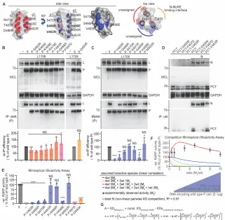
Figure 12. Identification of a specific L-binding face of the triangular prism fold of P-XD with regulatory effect on RdRP bioactivity A) Structural representations of MeV P-XD (PDB: 1T6O; only the P-XD component is represented), shown in side and top views. Areas between helices a1/a2 (red) and a1/a3 (blue) form faces of the prism without known interaction partners or bioactivities. Specific residues on each face are specified. Top view shows the known MoRE-binding face of P-XD between helices a2/a3 (yellow), residues confirmed (F497) or predicted (D493) to respectively impair P-XD fold or MoRE binding when mutated are highlighted. Grey background represents space-fill surface model. B, C) P:L interaction analysis of P mutants with individual substitutions of residues specified in (A), forming the “red” and “blue” prism
surfaces or implicated in MoRE binding (yellow). Detection and quantitative analysis as in Figure 1C (n = 4 (B); n = 3 (C)). D) PCT:N interaction after mutation of residue D493 on the MoRE interaction face of P-XD. Anti-N antibodies were used for IPs and anti-HA to detect PCT variants. E) Minireplicon analysis of RdRP activity in the presence of the specified P mutants. Columns represent means of experimentally observed values ± SD, symbols show individual biological repeats (n ³ 3). F) Observed RdRP activity in minireplicon assays in the presence of different P-V463R (M3) and wt P ratios as graphically depicted below the graph. Symbols show
means of experimentally observed biological repeats ± SD (n = 3). Solid lines represent activity curve predictions according to a linear combination model as in Figure 2E. The dotted line (red) represents the best fit curve of the experimental data based on a non-linear pairwise P-XD competition model, goodness of fit (R2) is indicated. G) Mathematical description of the model
Figure 13. Effect of P (361-364)Ala and P-V463R mutations on P binding to full-length L. Interaction analysis was carried out as specified in Figure 5C, using equally Flag epitope-tagged L1708 and full-length L as co-IP targets.
Bioactivity testing of all P-XD mutants in minireplicon assays demonstrated a direct correlation between the effect of substitutions in the a1/a3 P-XD prism face on L binding and RdRP bioactivity (Figure 12E). a1 substitutions V463R and S466R and the a3 mutation H498R in particular eliminated all polymerase activity, as did the F497D and D493K changes
suppressing P-XD binding to MoRE. Competition profiles of the P-V463R mutant with wild type P revealed remarkable RdRP hyper-activity at higher relative amounts of the V463R mutant, more than double that seen in the presence of wild type P alone (Figure 12F). Also, this experimental data set was incompatible with a linear combination function.
We, therefore, considered again non-linear models, based on the following assumptions: all four P-XDs within a P tetramer function independently of each other; bioactivity requires XD binding to L and MoRE; both wild type and mutant XDs are equally MoRE binding competent; and the V463R mutation selectively impairs XD interaction with L only. Most notably, the best fit (R2 = 0.80) mathematical description of the experimental data critically depends on the
tetramer is necessary and sufficient for bioactivity of the RdRP complex. Corroborating RdRP hyperactivity seen in the earlier PDXD competition profiles, these results for the MoRE binding-competent but L binding-defective P-V463R mutant revealed that assignment of L-binding competence to only one XD per P tetramer creates conditions most favorable for overall RdRP activity.
1.2.7.5 Trans-complementation of P mutants with distinct L binding deficiencies in
minireplicon and recMeV
Having identified two discrete P microdomains that are required for interaction with L and highly conserved across major human pathogens in the paramyxovirus family (Figure 14), we explored whether these domains are functionally distinct. Co-expression of P (361-364)Ala
and P-V463R, each by itself unable to co-IP L, restored physical interaction with L (Figure 15A). Wild type P-like binding efficiency was observed in the presence of the highest relative excess of P-V463R tested. When applied to minireplicon assays, we found that successful trans-complementation of L binding capacity extended to bioactivity of mixed P tetramers, remarkably resulting in RdRP activity equivalent to that observed with wild type P when cells received P (361-364)Ala and P-V463R-encoding plasmid DNA in approximately a 1:3-relative ratio (Figure 15B). Trans-complementation profiles over a wide plasmid ratio range corroborated this result, revealing a steep, asymmetric bell curve with a wild type P-equivalent RdRP bioactivity peak at a relative plasmid DNA ratio of 1:3 (P (361-364)Ala:P-V463R) (Figure 15C). We conclude that
XD per P tetramer boosts bioactivity, thus compensating for the negative effect associated with the presence of even one P (361-364)Ala monomer.
Figure 14. Multi-sequence alignments of P proteins of selected paramyxoviruses, representing genera of major clinical importance. Alignment with Clustal Omega algorithm (MeV P (NP_056919.1); CDV P (AIN44014.1); RV P (AAB23268.1); hendra virus (HeV) P (APT69525.1); nipah virus (P) P (QBQ56717.1); SeV P (AAB06279.1); HPIV-1 P
(AAL89402.1); HPIV-3 P (BAA00921.1); mumps virus (MuV) P (BAA00260.1), HPIV-2 P (ART66806.1), and PIV-5 (YP_138512.1). a-helical regions are highlighted above the sequences and heptad repeats in P-OD indicated; numbering refers to MeV. P residues 361-364 are
Figure 15. L-binding null-mutants in P OD C-terminal microdomain and P-XD efficiently trans-complement A) Graphic depiction of P (361-364)Ala / P-V463R
complementation ratios tested and P:L interaction analysis of the different
trans-complementation pairs. A HIS-tagged version of the P-V463D mutant was used to enable differential immunoprecipitation, detection and quantitative analysis otherwise as in Figure 5C (n ³ 3). B) Minireplicon assay of candidate trans-complementation P mutants alone and co-expressed at 1:3 relative ratio (n ³ 3). C) Trans-complementation RdRP activity profile of the pair shown in (A) and (B), analyzed in minireplicon assays over the specified relative ratio range. Error bars show means of biological repeats ± SD (n = 3). All statistical analyses and symbols as detailed in Figure 5.
arrangement of genes encoding P-V463R and P (361-364)Ala, respectively (Figure 16A).
P-V463R was intentionally placed upstream of P (361-364)Ala to capitalize on the Mononegavirales
transcription gradient [113] and ensure that L binding-deficient P-V463R would be present in relative abundance over P (361-364)Ala in infected cells. The corresponding recMeV P-V463R-P
(361-364)Ala virus was recovered successfully, replicated as efficiently as the genetic parent virus