Longo, Matthew R. (2015) Implicit and explicit body representations.
European Psychologist 20 (1), pp. 6-15. ISSN 1016-9040.
Downloaded from:
Usage Guidelines:
Please refer to usage guidelines at or alternatively
RUNNING HEAD: Implicit Body Representations
Implicit and Explicit Body Representations
Matthew R. Longo
Department of Psychological Sciences, Birkbeck, University of London
Address correspondence to:
Matthew R. Longo
Department of Psychological Sciences
Birkbeck, University of London
Malet Street
London WC1E 7HX
United Kingdom
Abstract
Several forms of perception require that sensory information be referenced to representations
of the size and shape of the body. This requirement is especially acute in somatosensation in
which the main receptor surface (i.e., the skin) is itself coextensive with the body. In this
paper I will review recent research investigating the body representations underlying
somatosensory information processing, including abilities such as tactile localisation, tactile
size perception, and position sense. These representations show remarkably large and
stereotyped distortions of represented body size and shape. Intriguingly, these distortions
appear to mirror distortions characteristic of somatosensory maps, though in attenuated form.
In contrast, when asked to make overt judgments about perceived body form, participants are
generally quite accurate. This pattern of results suggests that higher-level somatosensory
processing relies on a class of implicit body representation, distinct from the conscious body
image. I discuss the implications of these results for understanding the nature of body
Our body is ubiquitous in perceptual experience, and is central to our sense of self and
personal identity. As William James (1890) observed, our body is not ours, it is us. Thus,
how we mentally represent our body has profound implications for our sense of identity,
self-esteem, and overall mental health. Indeed, disordered body representation is central to several
serious and debilitating diseases, including eating disorders (Treasure, Claudino, & Zucker,
2010), body dysmorphic disorder (Phillips, Didie, Feusner, & Wilhelm, 2008), and phantom
limb pain (Flor, Nikolajsen, & Staehelin Jensen, 2006).
The subjective, conscious experience of embodiment, however, is only one way in
which the brain represents the body. Many forms of perception also require referencing to
representations of the body, such as its size and shape. The use of binocular vision for depth
perception, for example, requires that information about the spacing between the eyes be
known (Banks, 1988). Similarly, the use of temporal differences in when sounds reach the
two ears for auditory localisation requires that the distance between the ears be known (Aslin,
Pisoni, & Jusczyk, 1983; Clifton et al., 1988). Other studies have shown that other types of
body representations inform perception, such as eye-height which affects the perceived
passability of doorways (Warren & Whang, 1987), hand size which affects the perceived size
of seen objects (Linkenauger, Ramenzoni, & Proffitt, 2010), and arm length which affects the
extent of the ‘near space’ immediately surrounding the body (Longo & Lourenco, 2007).
While information about the body is used in perceptual modalities like vision and
audition, it is for the most part secondary. In somatosensation, in contrast, representations of
the body are central, since the primary receptor surface – the skin – is physically co-extensive
with the body. While basic qualities of tactile sensations may be specified in part by distinct
labelled lines for which individual nerve fibres coming from the periphery are in one-to-one
beyond pure somatosensation to achieve rich somatoperception requires that these immediate
signals be informed by representations of body size, shape, configuration, and posture.
In this paper, I will review recent research investigating these body representations
underlying somatoperception and their relation to our conscious body image. In the first part
of this paper, I will review recent research investigating body representations underlying
somatoperceptual information processing. A key theme of this research is the finding that
these representations are systematically distorted, in highly stereotyped ways across people.
In contrast, people’s conscious judgments about their body are generally approximately
accurate, suggesting that somatoperception relies on a class of implicit body representation,
distinct from our conscious body image. The final part of the paper discusses potential
relations between these types of body representation.
Body Representations underlying Somatoperception
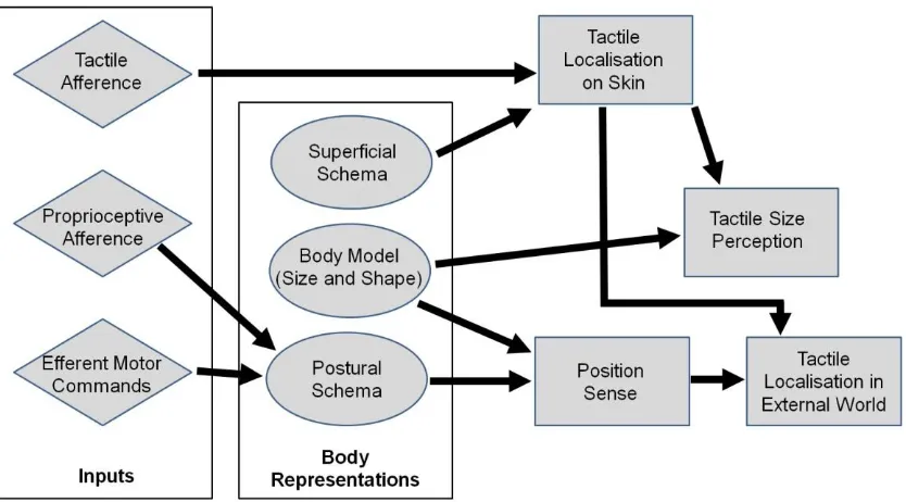
Longo, Azañón, and Haggard (2010) recently proposed a model of somatoperceptual
information processing (shown, in simplified form, in Figure 1) which suggested that
higher-order somatosensory percepts are constructed by combined immediate sensory signals from
the peripheral nerves with stored representations of the body. In addition to the superficial
and postural schemas of Head and Holmes (1911), Longo and colleagues (2010) argued that
several aspects of somatoperception also required a model of the metric properties of the
body (i.e., body size and shape), which they called the ‘body model’. Specifically, they
argued that the body model was required for tactile size perception and position sense. In this
section, I will describe recent results investigating the nature of these body representations
underlying tactile localisation, tactile size perception, and position sense. In particular, I will
discuss various distortions of these representations, and ways in which these appear to reflect
Figure 1: A simplified version of Longo and colleagues’ (2010) model of somatoperceptual
information processing. The key feature of the model is the combination of immediate sensory and motor signals (indicated as diamonds) and body representations (indicated as ovals) in generating high-level percepts (indicated as rectangles). In addition to the classic superficial and postural schemas, first postulated by Head and Holmes (1911), the model proposed that a Body Model providing information about the metric properties of the body (i.e., size and shape) is critical for perceptual abilities including tactile size perception and position sense.
In thinking about the relation between different body representations, it is important
to consider the spatial scale at which they represent the body. The body itself is a volumetric,
three-dimensional (3-D) object in the world, and we consciously experience it as such. In
contrast, somatotopic maps of the body surface in the primary somatosensory cortex (SI) are
two-dimensional (2-D). They are 2-D in the obvious sense that the cortex itself is a 2-D sheet,
but also in a more profound sense. In the case of the hand, for example, separate patches of
cortex represent the glabrous skin of the palmar surface of the hand, and the hairy skin of the
dorsal surface of the hand (e.g., Sur, Merzenich, & Kaas, 1980). Further, cortical
magnification (the relative amount of cortical tissue devoted to representing a given bit of
skin) is substantially higher on the palmar than the dorsal hand surface, reflecting the palm’s
higher tactile sensitivity. Thus, the hand is initially represented in somatosensory cortex as
Do higher-order body representations represent the body as a fragmented collection of
2-D skin surfaces, or as coherent, volumetric 3-D body parts? I will discuss evidence bearing
on this question for each of the representations I describe. Specifically, I will argue that
distortions of representations provide a valuable tool to address this question. If part of the
body is represented as a coherent, volumetric, 3-D object, then distortions should affect all
sides of the body part. For example, distortions of a fully 3-D representation of the hand
should appear in a consistent manner on both the palmar and dorsal surfaces of the hand. In
contrast, if a body part is represented as a fragmented collection of distinct 2-D skin surfaces,
then each surface may very well be distorted in different ways.
Tactile Localisation
The ability to tell where on the body a touch has occurred is among the most
fundamental of sensory abilities. The location of stimulation can even be specified by single
nerve fibres in the periphery (Schady, Torebjörk, & Ochoa, 1983). Head and Holmes (1911)
reported several patients who could accurately report when they had been touched, but were
unable to report where on their body the touch had been applied (for more recent findings,
see Halligan, Hunt, Marshall, & Wade, 1995). Since these patients could still detect that they
had been touched, it couldn’t be just that the relevant location in primary somatopic maps had
been destroyed, which ought to have impaired all processing of the stimulus. Instead, the
initial cortical processing of the stimulus appeared intact, with some other stage of processing
being impaired. On the basis of such results, Head and Holmes proposed the concept of
‘schemas’ mediating the interpretation of sensory signals. This higher-order representation
mediating tactile localisation has come in the literature to be known as the superficial
Longo and colleagues (2010) argued that the well known plasticity of somatosensory
cortex following both physical changes to the body (Merzenich et al., 1984; Pons et al., 1991)
and learning (Elbert, Pantev, Weinbruch, Rockstroh, & Taub, 1995; Pascual-Leone & Torres,
1993) implied that there could be no hard-wired representation between locations in
somatotopic maps and locations on the body. They suggested that tactile localisation required
an additional linking function connecting these locations, which could be thought of as
constituting the superficial schema. A fascinating example of this is a study by Rapp, Hendel,
and Medina (2002), reporting two patients with lesions to the left hemisphere who show
highly structured, but massively distorted, patterns of localisation. On each trial, the patient
was touched somewhere on the hand with eye closed, then opened their eyes and pointing
with their other hand to the perceived location of stimulation, which was recorded on a
drawing of a hand outline. The perceived locations of touch were systematically shifted in
these patients. Critically, the errors these patients made preserved the overall somatotopic
arrangement of skin locations with respect to each other, suggesting that the overall
somatotopic arrangement of skin locations with respect to each other was preserved. Each
point, however, was systematically misplaced. This pattern is strongly suggestive of
preserved somatotopic maps, with an impaired linking function between locations in these
maps and locations on the body, that is, an impaired superficial schema.
A series of studies by Trojan and colleagues have revealed intriguing distortions of
perceptual maps of the body surface (Trojan et al., 2006, 2009). In these studies, radiant heat
was applied to specific locations on the forearm using a CO2 laser, and participants indicated
the perceived location of touch by positioning a pointer connected to a 3-D motion-tracking
system above their arm, without touching the skin. While all participants showed a clearly
somatotopic pattern of responses, there were striking patterns of mislocalisation, with some
axis of the forearm (Trojan et al., 2006). A recent study used this paradigm to have
participants judge the position of electric shocks applied to the forearm showed strong re-test
reliability, suggesting that though the distortions of perceptual maps were idiosyncratic across
individuals, they were nevertheless highly stable within each individual.
A recent study by Mancini and colleagues (Mancini, Longo, Iannetti, & Haggard,
2011) investigated the superficial schema in healthy participants by measuring tactile
localisation on the hand using a very simple paradigm in which participants were touched and
then judged where on their hand they had been touched by clicking a mouse cursor on the
corresponding location on a silhouette of their hand. In contrast to the studies of Trojan and
colleagues on the forearm who found idiosyncratic distortions across indididuals, Mancini
and colleagues found highly consistent patterns of constant errors. On the dorsal hand
surface, there were large distal biases in localisation (i.e., touch was perceived farther
forward on the hand than it had actually been). These biases were highly consistent across
different types of stimulation. For example, nearly identical distal biases were found
following stimulation of mechanoreceptive and thermal afferent fibres. This generality
suggests that these biases emerge from a supramodal representation of hand, abstracting
across categories of stimuli. A recent study by Steenbergen and colleagues (2012), measuring
localisation on the forearm, found similar (though less striking) correspondence between
sensory modalities.
In striking contrast to the large distal biases they observed on the hand dorsum, Mancini
and colleagues (2011) found no such biases on the palmar hand surface. Thus, in contrast to
the generality found across different types of stimulation, the biases were highly specific to
individual skin surfaces. This surface specificity suggests that the superficial schema relies on
fragmented representations of individual skin regions as 2-D surfaces, rather than the body as
Tactile Size Perception
The metric properties of objects, their size and shape, can be perceived through
passive touch in multiple ways. When we hold an object between our thumb and index finger,
we can perceive its size through proprioception, which requires referencing to body
representations for reasons described in the next section. We can also perceive the size of
objects touching a single skin surface. Suppose, for example, that you are touched at two
points on opposite sides of the back of your hand. While each of the afferent volleys
produced by those touches may be sufficient to localise each stimulus (cf. Schady et al.,
1983), there is nothing intrinsic to either of the signals or their combination that specifies how
far apart they are. The problem of perceiving the distance between two objects on either side
of your hand effectively reduces to the problem of knowing how big your hand is.
What sort of representation of body size and shape is used for tactile size perception?
More than a century and a half ago, Weber (1834/1996) found that as he moved two tactile
points across his skin, the distance he perceived between the two points changed.
Specifically, it felt like the points were father apart when they were on a region of relatively
high tactile sensitivity (e.g., the palm of the hand), compared to when they were on a region
of lower tactile sensitivity (e.g., the forearm), an effect now referred to as Weber’s illusion.
Subsequent research has confirmed and extended Weber’s finding, showing systematic
relations between tactile sensitivity and tactile size perception (e.g., Anema, Wolswijk, Ruis,
& Dijkerman, 2008; Cholewiak, 1999; Goudge, 1918; Taylor-Clarke, Jacobsen, & Haggard,
2004). Thus, body representations mediating tactile size perception may preserve distortions
characteristic of primary somatosensory maps (e.g., the ‘Penfield homunculus’, Penfield &
Several studies have found that interventions which alter perceived body size produce
corresponding changes in tactile size perception. For example, Taylor-Clarke and colleagues
(2004) used a video image to provide participants with the visual appearance of their forearm
magnified and hand minified. After this experience, the relative perceived size of touch on
the forearm – compared to the hand – was increased. de Vignemont, Ehrsson, and Haggard
(2005) used a combination of an illusion of body posture and self touch to alter perceived
finger length. By applying vibration to the tendons of the biceps or triceps muscles, they
generated illusions of forearm extension or flexion, respectively (the ‘vibrotactile illusion’;
cf. Goodwin, McCloskey, & Matthews, 1972). By having participants hold the index finger
of their contralateral hand during these postural illusions, they produced the illusion that the
finger was becoming shorter or longer (the ‘Pinocchio illusion’; cf. Lackner, 1988, see
below). The illusion of finger lengthening (though, interestingly, not the illusion of finger
shortening) produced a corresponding change in the perceived size of tactile stimuli applied
to the finger. Similarly, Bruno and Bertamini (2010) showed that using the rubber hand
illusion to create the illusion of increased hand size produced corresponding increases on the
haptic perception of object size. Analogously, Tajadura-Jiménez and colleagues (2012)
manipulated apparent arm length by playing sounds from speakers at varying distances
time-locked to participants’ knocks on the floor. The illusion of arm lengthening increased the
perceived size of touch on the acting arm, compared to the contralateral arm.
In its classical form, Weber’s illusion suggests that the perceived size of sensitive skin
surfaces is overestimated compared to less sensitive surfaces. Longo and Haggard (2011)
applied the same logic to investigate the representation of body shape by comparing the size
of tactile stimuli applied to the body in different orientations. The logic of this approach was
that if the hand is represented as longer and more slender than it actually is, then the distance
overestimated relative to touches applied in the medio-lateral orientation (running across the
hand). Conversely, if the hand is represented as wider and squatter than it actually is, the
opposite pattern should be found, with touches oriented across the hand perceived as bigger
than those along the hand. In fact, Longo and Haggard (2011) found that stimuli running
across the hand dorsum are perceived as approximately 40% larger than those running along
the hand, suggesting that touch is being informed by a fat, squat model of the hand.
Intriguingly, this bias mirrors other known properties of the somatosensory system, including
increased tactile acuity in the across orientation on the limbs (Cody, Garside, Lloyd, &
Poliakoff, 2008; Weber, 1834/1996), and the fact that tactile receptive fields of both spinal
(Brown, Fucks, & Tapper, 1975) and cortical (Alloway, Rosenthal, & Burton, 1989) neurons
are generally oval-shaped (rather than circular), with their long axis running along the length
of the limbs.
Thus, the bias in tactile size perception found on the hairy skin of the hand dorsum
mirrors the geometry of tactile receptive fields in somatosensory cortex. But what about the
glabrous skin of the palm of the hand? Does tactile size perception rely on a 2-D or a 3-D
representation of the body? In contrast to the large anisotropy found on the hairy skin of the
hand dorsum, Longo and Haggard (2011) did not find any apparent bias on the glabrous skin
of the palm. This difference is consistent with results showing that receptive fields on the
palmar hand surface are generally more circular than on the dorsal surface and, when
oval-shaped, the long axis of the oval is distributed more uniformly (DiCarlo & Johnson, 2002;
Vega-Bermudez & Johnson, 1999). Thus, the representations of the dorsal and palmar sides
of the hand appear to be stretched in different ways, a basic violation of 3-D geometry. Thus
suggests that tactile size perception, like tactile localisation, may rely on a set of fragmented,
Position Sense
Position sense refers to the ability to perceive where the different parts of our body
are located in space, even when we can’t see them. Though position sense usually remains in
the background of our mental life, it is critically important for all our everyday behaviours.
The importance of position sense is strikingly evident when it is lost in patients such as I.W.,
who suffered a near total loss of the sensory fibres below the neck at age 19, leaving him
completed deafferented and without position sense (Cole, 1995). Though the fibres carrying
motor information to his body were unimpaired, I.W. was only able to teach himself to walk
again through an intense programme of practice using constant and vigilant visual guidance,
leading the neurologist who worked with him to refer to I.W.’s life as a “daily marathon”
(Cole, 1995).
Several types of afferent signal from the periphery contribute to position sense,
including receptors from joints signalling flexion or extension, from the skin signalling
stretch, and from muscle spindles signalling contraction or lengthening (Proske & Gandevia,
2012). Together with efferent copies of motor comments, these signals provide a
specification of the postural configuration of the body (Burgess, Wei, Clark, & Simon, 1982).
Critically, all of these signals specify joint angles, that is the relative flexion or extension of
each joint. There is no afferent signal, or combination of signals, that function like a global
positioning system (GPS) signal, providing information about the absolute location of body
parts in external space. As a matter of trigonometry, information about joint angles is
insufficient to determine the absolute position in external space of part of the body. As shown
in Figure 2, perceiving the absolute spatial position of the body requires that information
about joint angles, which is specified by immediate proprioceptive afferent signals, be
combined with information about the length of body segments, which is not specified by any
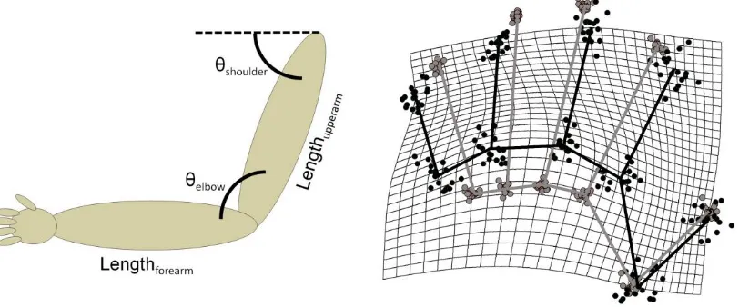
Figure 2: Left Panel: A schematic depiction of the need for stored body
representations in position sense in the case of the arm. Proprioceptive afferent signals specify joint angles, such as those at the shoulder (θshoulder) and elbow (θelbow).
However, determining the absolute spatial position of the hand with respect to the shoulder also requires information about the length of the upper (Lengthupperarm) and forearm (Lengthforearm), which critically is not specified by immediate sensory signals from the body. Right Panel: Results from Longo and Haggard’s (2010) study,
showing implicit perceptual maps of judged locations (in black) put into Procrustes alignment with actual hand shape (in grey) for 18 participants. The black and grey lines connect the knuckle and tip of each finger, as well as adjacent knuckles, to give an overall sense of hand shape. The grid shows how a rectangular grid superimposed on actual hand shape would have to be stretched to transform actual hand shape into represented hand shape. The implicit hand maps clearly overestimate hand width, and underestimate finger length.
While many authors have identified the need for stored metric information about the
size and shape of the body for position sense (e.g., Craske, Kenny, & Keith, 1984; Gurfinkel
& Levick, 1991; Longo et al., 2010; Soechting, 1982; van Beers, Sittig, & Denier van der
Gon, 1998), it has usually been assumed that such information is readily available to the
somatosensory system. This seems like quite a reasonable assumption to make, for several
reasons. Though the size and shape of our body changes substantially over developmental
time, on the everyday time scale the body remains largely constant. The body, moreover, is
ubiquitous in perceptual experience and metric information about the body is available
through vision or self-touch. Further, inaccurate body representations would seem to pose
provided an intriguing anecdotal report that when participants were asked to judge the
location of two parts of their arm, the judgments were closer together than the two points
actually were, suggesting the position sense may in fact rely on a distorted body
representation.
Longo and Haggard (2010) developed a novel procedure to isolate and measure the
representation of body size and shape underlying position sense of the hand. The participant’s
hand was placed palm-down on a table and covered with an occluding board. They were then
asked to judge the location of the knuckles and tips of their fingers by placing the tip of a
long baton on the board, directly above each location. Each judgment was photographed by
an overhead camera. Previous studies of proprioceptive localisation have focused on the
‘error of localisation’, the spatial displacement of judged location from actual location,
measuring bias as the constant error of localisation, and precision as the variable error. In
contrast, Longo and Haggard (2010) focused on the internal configuration of judgments to
each of the landmarks with respect to each other, completely ignoring where judgments were
in relation to the participant’s actual hand. This allowed them to construct perceptual maps of
represented hand form, which they then compared to the actual form of each participant’s
hand.
The resulting hand maps from Longo and Haggard’s (2010) first experiment are
shown in the right panel of Figure 3. Remarkably, these maps were massively distorted in a
highly consistent and stereotyped way across participants. In particular, there were three clear
patterns of distortions: (1) overestimation of hand width, quantified as the distance between
pairs of knuckles; (2) overall underestimation of finger length, quantified as the distance
between the knuckle and tip of each finger; and (3) a radio-ulnar gradient, with
underestimation of finger length increasing systematically from the thumb to the little finger.
cortical maps. For example, the overestimation of hand width compared to length mirrors
anisotropies in RF geometry and tactile size perception described in the previous section.
Similarly, the radial-ulnar gradient of finger size mirrors differences in tactile sensitivity and
cortical magnification of the five fingers (Duncan & Boynton, 2007; Vega-Bermudez &
Johnson, 2001).
In a subsequent study, Longo and Haggard (2012a) investigated the level of spatial
abstraction at which these implicit maps are organised, using the same logic discussed above
for tactile localisation and tactile size perception. If the hand is represented as two distinct
2-D skin surfaces, there may be different distortions on each. If, in contrast, the two sides of the
hand are integrated into a fully 3-D representation of the hand as volumetric object in the
world, then consistent distortions should appear on both sides of the hand, and should be
correlated across people. Longo and Haggard (2012a) found that distortions on the dorsal and
palmar hand surfaces were qualitatively similar, and strongly correlated across participants,
suggesting that the representations of the two skin surfaces are bound into a common
representation, suggesting something more abstract than a purely 2-D representation.
However, the distortions were of different magnitude on the two surfaces, being substantially
reduced on the palmar surface. This is a clear violation of the geometry of 3-D space,
suggesting something less abstract than a fully 3-D volumetric representation. Thus, Longo
and Haggard (2012a) suggested that position sense may rely on something intermediate
between a 2-D representation of distinct skin surfaces and a fully 3-D representation of the
hand as a volumetric object, which they called a 2.5-D representation, in analogy to Marr’s
(1982) ‘2.5-D sketch’ in vision.
In the first part of this paper, I have described several large and highly stereotyped
distortions of body representation underlying perceptual processing. Intuitively, this seems
quite surprising, since for most of us it seems like we have quite an accurate sense of what
our body is like. Surely, if there’s anything we ‘know like the back of our hand’ it would be
the actual back of our hand. Do the distortions I have described also characterise our
conscious experience of our body, our body image? To address this question, Longo and
Haggard (2010) adapted the ‘template matching’ procedure of Gandevia and Phegan (1999)
to measure participant’s conscious experience of their hand. The same participants who
produced the distorted hand maps in Figure 2 were shown arrays of hand images which had
been stretched in various ways, resulting in a range of hand shapes, from very long and
slender to very squat and wide. In contrast to their highly distorted hand maps in the
localisation task, participants on average selected hands quite similar to their actual hand
shape. Thus, the explicit image of the hand is approximately veridical, even as
somatoperception relies on a set of implicit, and highly distorted, representations.
Implicit Body Representations and the Cognitive Unconscious
The dissociation between implicit and explicit body representations fits within a
larger trend in psychology and the cognitive sciences over the past few decades emphasizing
that much of cognitive processing remains inaccessible to conscious awareness as part of the
so-called ‘cognitive unconscious’ (Kihlstrom, 1987). While we are clearly able to introspect
on much of our psychological life, we are also unaware of much of the cognitive machinery
underlying our thoughts, beliefs, and actions (e.g., Nisbett & Wilson, 1977; Tranel &
Damasio, 1985; Tulving & Schacter, 1991).
In the domain of perception, there are numerous clinical reports of preserved ability to
including blindsight (Weiskrantz, 1986), visual object agnosia (Milner & Goodale, 2006),
and numb-sense (Paillard, Michel, & Stelmach, 1983). The research reviewed above is
similar in showing dissociations between the cognitive machinery of perception and
conscious awareness, but also strikingly different in showing that these implicit processes are
highly inaccurate, in contrast to more veridical explicit representations. This parallels
findings of implicit processes producing highly biased results in multiple domains, including
reasoning and decision making (Tversky & Kahneman, 1981; Kahneman, 2011) and attitudes
(Greenwald & Banaji, 1995; Nosek, Hawkins, & Frazier, 2011), even while more deliberate
reflection may produce more rational decisions and more egalitarian attitudes.
A Hierarchy of Body Representations
What, then, is the relationship between our explicit, conscious body image, and
implicit body representations? One possibility is that they reflect entirely distinct
representations emerging from different sensory modalities, the body image arising through
vision and distorted implicit representations through somatosensation. However, there is
strong evidence for bidirectional interactions between the visual body image and
somatosensory processing. For example, cutting off inputs from the peripheral nerves with
cutaneous anaesthesia produces the subjective experience that that body part has gotten
larger, both on the hand (Gandevia & Phegan, 1999) and the mouth (Türker, Yeo, &
Gandevia, 2005). This experience may be familiar to anyone unfortunate enough to have had
dental anaesthesia, in which the gums and teeth begin to feel enormous. Conversely, visual
illusions producing the experience of the body being larger than it actually is produce
corresponding changes in the perceived size of touch, as described above (Bruno &
Bertamini, 2010; Taylor-Clarke et al., 2004). Thus, somatosensory and visual body
Another possibility, which I will defend here, is that implicit and explicit body
representations lie at opposite ends of a continuum of body representations. This continuum
can be thought of in terms of the different spatial scales at which the body is represented,
which I discussed in the first part of the paper. At one end are primary somatosensory maps,
representing the body surface as a mosaic of individual receptive fields, each constituting a
single ‘pixel’. At the other end is our conscious experience of our body as a volumetric object
in the world. In between these extremes may be 2-D maps of individual skin surfaces (such as
I have argued may underlie tactile localisation and tactile size perception), and 2.5-D
representations (such as I have argued underlies position sense).
Intriguingly, there is some evidence that different measures of the conscious body
image may index different points along this continuum. For example, Longo and Haggard
(2012b) compared three different measures of hand representation: (1) the localisation task
measuring implicit body representations underlying position sense, (2) the template matching
task described above, and (3) a ‘line length’ task in which participants judged whether a line
presented on the monitor was shorter or longer than different parts of their hands. As in their
previous study described above, Longo and Haggard (2012b) found that the hand
representation revealed by the localisation task was massively distorted, while that revealed
through template matching was approximately veridical. The line length task, however,
appeared intermediate between the two. Participants in the line length task showed distortions
of perceived hand size and shape qualitatively similar to those found in the localisation task,
but smaller in magnitude. The template matching task, as a purely visual recognition task,
may be a purer measure of the ‘visual’ end of this continuum of body representations, while
the line length task may involve a larger contribution of the ‘somatosensory’ side of body
On this view, body representations emerge from the operation and mutual interactions
of complementary bottom-up and top-down processes. First, from the bottom-up,
somatosensation represents the body surface as a mosaic of discrete receptive fields, which
become progressively agglomerated into larger and larger units of organisation, a process I
call fusion. Second, from the top-down, vision starts out depicting the body as an
undifferentiated whole, which is progressively broken into smaller parts, a process I call
segmentation. Thus, body representation operates from the bottom-up as a process of fusion
of primitive elements into larger complexes, as well as from the top-down as a process of
segmentation of an initially undifferentiated whole into more basic parts.
Implications for Clinical Disorders of Body Representation
While most of the studies I have described have been conducted with healthy
individuals, this research also has potential implications for understanding clinical disorders
involving disrupted body representation. While this connection remains speculative, in this
final part of the paper I will discuss some ways in which the distinction between implicit and
explicit body representations may relate to conditions such as eating disorders.
It has been widely accepted since the classic work of Hilde Bruch (1978) that
anorexia nervosa involves a distorted body image. Indeed, such distortions are strong
predictors of poor prognosis for recovery (Casper et al., 1979) and of relapse following
remission of symptoms (Fairburn et al., 1993; Keel et al., 2005). Could the distortions of the
conscious body image seen in such cases reflect normal distortions of somatosensory body
representations which have risen into conscious awareness, implicit representations which
have become explicit? While the majority of the results I have described have investigated
representations are for the hand to be wider and squatter than it actually is, mirroring the
body image distortions of individuals with eating disorders who experience their body as fat.
Two sets of considerations may seem to make this hypothesis unlikely. First, while
meta-analyses of studies of eating disorders have found clear evidence for distortions of
perceived body size (e.g., Cash & Deagle, 1997; Smeets et al., 1997), these same studies have
found even stronger effects for bodily attitudes, suggesting that perceptual aspects of body
image may be secondary to disrupted attitudes. Indeed, some authors have suggested that
body-size estimates themselves may actually reflect attitudes, rather than perception
(Ben-Tovim, Walker, Murray, & Chin, 1990). Second, eating disorders and distorted body image
are widely linked to the visual depiction of bodies in the Western mass-media (Becker &
Hamburg, 1996; Derenne & Beresin, 2006), making top-down effects of vision seem more
critical than bottom-up effects of somatosensation.
Recently, however, several lines of evidence have suggested that somatosensation,
and potentially implicit body representations, may have a greater role in eating disorders than
previously believed. Intriguingly, recent results have revealed that individuals with anorexia
show evidence for overestimation of body size in implicit action-based tasks (Guardia et al.,
2010, 2012; Keizer et al., 2013). Critically, these studies are less susceptible than overt size
estimates to the critique of implicitly reflecting attitudes towards the body, rather than
distorted body representation per se (cf. Ben-Tovim et al., 1990). Further, and more directly
related to somatosensation, recent results have found that individuals with anorexia show
impaired tactile processing, overestimating the size of tactile stimuli (Keizer et al., 2011,
2012). Intriguingly, this bias, though apparent on the arm as well, was strongest on the
abdomen, and predicted the severity of body dissatisfaction.
Studies using neuroimaging have also produced intriguing findings suggesting that
than healthy individuals. For example, Uher and colleagues (2005) found reduced activations
to visually-presented images of bodies in patients with eating disorders in several visual brain
areas. Similarly, Suchan and colleagues (2010) found reduced grey-matter density within the
extrastriate body area (EBA), a brain area specialised for the visual perception of bodies
(Downing, Jiang, Shuman, & Kanwisher, 2001), in women with anorexia. In a subsequent
study, these authors reported reduced functional connectivity between the EBA and another
region of the ventral visual cortex specialised for body perception, the fusiform body area
(FBA) (Suchan et al., 2013). Consistent with those results, Favaro and colleagues (2012),
analysing resting-state functional connectivity of fMRI data in individuals with anorexia and
healthy controls, found that the patients showed reduced connectivity within the ventral
visual network. Remarkably, these authors also found that anorexia was linked to increased
connectivity within somatosensory cortex.
Thus, in contrast to the long-standing idea that body image distortions may arise from
visual exposure to extreme bodies (Becker & Hamburg, 1996; Derenne & Beresin, 2006),
these results suggest that in some ways individuals with eating disorders may be
paradoxically less sensitive to visually-depicted bodies. Together, these results are consistent
with the hypothesis that individuals with eating disorders may be relatively more reliant on
somatosensory body representations, and less on visual ones. This raises the possibility that
the distortions of implicit body representations underlying several aspects of somatosensation
which I have described here may play a role in producing distortions of the explicit body
References
Alloway, K. D., Rosenthal, P., & Burton, H. (1989). Quantitative measurements of receptive
field changes during antagonism of GABAergic transmission in primary
somatosensory cortex of cats. Experimental Brain Research, 78, 514-532.
Anema, H. A., Wolswijk, V. W., Ruis, C., & Dijkerman, H. C. (2008). Grasping Weber’s
illusion: The effect of receptor density differences on grasping and matching.
Cognitive Neuropsychology, 25, 951-967.
Aslin, R., Pisoni, D., & Jusczyk, P. (1983). Auditory development and speech perception in
infancy. In P. H. Mussen, M. M. Haith, & J. J. Campos (Eds.), Handbook of child
psychology (pp. 573-687). New York: Wiley.
Banks, M. S. (1988). Visual recalibration and the development of contrast and optical flow
perception. In A. Yonas (Ed.), The Minnesota symposia on child psychology (pp.
145-196). Hillsdale, NJ: Erlbaum.
Becker, A. E., & Hamburg, P. (1996). Culture, the media, and eating disorders. Harvard
Review of Psychiatry, 4, 163-167.
Ben-Tovim, D. I., Walker, M. K., Murray, H., & Chin, G. (1990). Body size estimates: Body
image or body attitude measure? International Journal of Eating Disorders, 9, 57-67.
Brown, P. B., Fuchs, J. L., & Tapper, D. N. (1975). Parametric studies of dorsal horn neurons
responding to tactile stimulation. Journal of Neurophysiology, 38, 19-25.
Bruch, H, (1978). The golden cage: The enigma of anorexia nervosa. Cambridge, MA:
Harvard University Press.
Bruno, N., & Bertamini, M. (2010). Haptic perception after a change in hand size.
Neuropsychologia, 48, 1853-1856.
information by peripheral sensory receptors. Annual Review of Neuroscience, 5,
171-187.
Cash, T. F., & Deagle, E. A., III (1997). The nature and extent of body-image disturbances in
anorexia nervosa and bulimia nervosa: A meta-analysis. International Journal of
Eating Disorders, 22, 107–125.
Casper, R. C., Halmi, K. A., Goldberg, S. C., Eckert, E. D., & Davis, J. M. (1979).
Disturbances in body image estimation as related to other characteristics and outcome
in anorexia nervosa. British Journal of Psychiatry, 134, 60-66.
Cholewiak, R. W. (1999). The perception of tactile distance: Influences of body site, space,
and time. Perception, 28, 851-875.
Clifton, R. K., Gwiazda, J., Bauer, J. A., Clarkson, M. G., & Held, R. (1988). Growth in head
size during infancy: Implications for sound localization. Developmental Psychology,
24, 477-483.
Cody, F. W., Garside, R. A., Lloyd, D., & Poliakoff, E. (2008). Tactile spatial acuity varies
with site and axis in the human upper limb. Neuroscience Letters, 433, 103-108.
Cole, J. (1995). Pride and a daily marathon. Cambridge, MA: MIT Press.
Craske, B., Kenny, F. T., & Keith, D. (1984). Modifying an underlying component of
perceived arm length: Adaptation of tactile location induced by spatial discordance.
Journal of Experimental Psychology: Human Perception and Performance, 10,
307-317.
Crick, F., & Koch, C. (1990). Towards a neurobiological theory of consciousness. Seminars
in the Neurosciences, 2, 263-275.
Derenne, J. L., & Beresin, E. V. (2006). Body image, media, and eating disorders. Academic
Psychiatry, 30, 257-261.
perception. Current Biology, 15, 1286-1290.
DiCarlo, J. J., & Johnson, K. O. (2002). Receptive field structure in cortical area 3b of the
alert monkey. Behavioural Brain Research, 135, 167–178.
Downing, P. E., Jiang, Y., Shuman, M., & Kanwisher, N. (2001). A cortical area selective for
visual processing of the human body. Science, 293, 2470-2473.
Duncan, R. O., & Boynton, G. M. (2007). Tactile hyperacuity thresholds correlate with finger
maps in primary somatosensory cortex (S1). Cerebral Cortex, 17, 2878-2891.
Elbert, T., Pantev, C., Weinbruch, C., Rockstroh, B., & Taub, E. (1995). Increased cortical
representation of the fingers of the left hand in string players. Science, 270, 305-307.
Fairburn, C. G., Peveler, R. C., Jones, R., Hope, R. A., & Doll, H. A. (1993). Predictors of
12-month outcome in bulimia nervosa and the influence of attitudes to shape and
weight. Journal of Consulting and Clinical Psychology, 61, 696-698.
Flor, H., Nikolajsen, L., & Staehelin Jensen, T. (2006). Phantom limb pain: A case of
maladaptive CNS plasticity. Nature Reviews Neuroscience, 7, 873-881.
Gandevia, S. C., & Phegan, C. M. (1999). Perceptual distortions of the human body image
produced by local anesthesia, pain and cutaneous stimulation. Journal of Physiology,
514, 609-616.
Goodwin, G. M., McCloskey, D. I., & Matthews, P. B. C. (1972). The contribution of muscle
afferents to kinaesthesia shown by vibration induced illusions of movement and by
the effects of paralysing joint afferents. Brain, 95, 705-748.
Goudge, M. E. (1918). A qualitative and quantitative study of Weber’s illusion. American
Journal of Psychology, 29, 81-119.
Greenwald, A. G., & Banaji, M. R. (1995). Implicit social cognition: Attitudes, self-esteem,
and stereotypes. Psychological Review, 102, 4-27.
Anticipation of body-scaled action is modified in anorexia nervosa.
Neuropsychologia, 48, 3961-3966.
Guardia, D., Conversy, L., Jardri, R., Lafargue, G., Thomas, P., Dodin, V., Cottencin, O., &
Luyat, M. (2012). Imagining one’s own and someone else’s actions: Dissociation in
anorexia nervosa. PLoS One, 7, e43241.
Gurfinkel, V. S., & Levick, Y. S. (1991). Perceptual and automatic aspects of the postural
body scheme. In J. Paillard (Ed.), Brain and space (pp. 147-162). Oxford: Oxford
University Press.
Halligan, P. W., Hunt, M., Marshall, J. C., & Wade, D. T. (1995). Sensory detection without
localization. Neurocase, 1, 259-266.
Head, H., & Holmes, G. (1911). Sensory disturbances from cerebral lesions. Brain, 34, 102-
254.
James, W. (1890). The principles of psychology. New York: Dover.
Kahneman, D. (2011). Thinking, fast and slow. New York: Farrar, Strauss and Giroux.
Keel, P. K., Dorer, D. J., Franko, D. L., Jackson, S. C., & Herzog, D. B. (2005).
Postremission predictors of relapse in women with eating disorders. American
Journal of Psychiatry, 162, 2263-2268.
Keizer, A., Smeets, M. A., Dijkerman, H. C., van den Hout, M., Klugkist, I., van Elburg, A.,
& Postma, A. (2011). Tactile body image disturbance in anorexia nervosa. Psychiatry
Research, 190, 115-120.
Keizer, A., Smeets, M. A., Dijkerman, H. C., van Elburg, A., & Postma, A. (2012). Aberrant
somatosensory perception in anorexia nervosa. Psychiatry Research, 200, 530-537.
Keizer, A., Smeets, M. A., Dijkerman, H. C., Uzunbajakau, S. A., van Elburg, A., & Postma,
A. (2013). Too fat to fit through the door: First evidence for disturbed body-scaled
Kihlstrom, J. F. (1987). The cognitive unconscious. Science, 237, 1445-1452.
Lackner, J. R. (1988). Some proprioceptive influences on the perceptual representation of
body shape and orientation. Brain, 111, 281-297.
Linkenauger, S. A., Ramenzoni, V., & Proffitt, D. R. (2010). Illusory shrinkage and growth:
Body-based rescaling affects the perception of size. Psychological Science, 21,
1318-1325.
Longo, M. R., & Haggard, P. (2010). An implicit body representation underlying human
position sense. Proceedings of the National Academy of Sciences, USA, 107,
11727-11732.
Longo, M. R., & Haggard, P. (2011). Weber's illusion and body shape: Anisotropy of tactile
size perception on the hand. Journal of Experimental Psychology: Human Perception
and Performance, 37, 720-726.
Longo, M. R., & Haggard, P. (2012a). A 2.5-D representation of the human hand. Journal of
Experimental Psychology: Human Perception and Performance, 38, 9-13.
Longo, M. R., & Haggard, P. (2012b). Implicit body representations and the conscious body
image. Acta Psychologica, 141, 164-168.
Longo, M. R., & Lourenco, S. F. (2007). Space perception and body morphology: Extent of
near space scales with arm length. Experimental Brain Research, 177, 285-290.
Longo, M. R., Azañón, E., & Haggard, P. (2010). More than skin deep: Body representation
beyond primary somatosensory cortex. Neuropsychologia, 48, 655-668.
Mancini, F., Longo, M. R., Iannetti, G. D., & Haggard, P. (2011). A supramodal
representation of the body surface. Neuropsychologia, 49, 1194-1201.
Marr, D. (1982). Vision: A computational investigation into the human representation and
processing of visual information. New York: Freeman and Co.
J. M. (1984). Somatosensory cortical map changes following digit amputation in adult
monkeys. Journal of Comparative Neurology, 224, 591-605.
Milner, A. D., & Goodale, M. A. (2006). The visual brain in action, 2nd Ed. Oxford: Oxford
University Press.
Nisbett, R. E., & Wilson, T. D. (1977). Telling more than we can know: Verbal reports on
mental processes. Psychological Review, 84, 231-259.
Nosek, B. A., Hawkins, C. B., & Frazier, R. S. (2011). Implicit social cognition: From
measures to mechanisms. Trends in Cognitive Sciences, 15, 152-159.
Paillard, J., Michel, F., & Stelmach, G. (1983). Localization without content: A tactile
analogue of ‘blind sight’. Archives of Neurology, 40, 548-551.
Pascual-Leone, A., & Torres, F. (1993). Plasticity of the sensorimotor cortex representation
of the reading finger in Braille readers. Brain, 116, 39-52.
Penfield, W., & Boldrey, E. (1937). Somatic motor and sensory representation in the cerebral
cortex of man as studied by electrical stimulation. Brain, 60, 389-443.
Phillips, K. A., Didie, E. R., Feusner, J., & Wilhelm, S. (2008). Body dysmorphic disorder:
Treating an underrecognized disorder. American Journal of Psychiatry, 165,
1111-1118.
Pons, T. P., Garraghty, P. E., Ommaya, A. K., Kaas, J. H., Taub, E., & Mishkin, M. (1991).
Massive cortical reorganization after sensory deafferentation in adult macaques.
Science, 252, 1857-1860.
Proske, U., & Gandevia, S. C. (2012). The proprioceptive senses: Their roles in signaling
body shape, body position and movement, and muscle force. Physiological Reviews,
92, 1651-1697.
Rapp, B., Hendel, S. K., & Medina, J. (2002). Remodeling of somatosensory hand
Schady, W. J., Torebjörk, H. E., & Ochoa, J. L. (1983). Cerebral localisation function from
the input of single mechanoreceptive units in man. Acta Physiologica Scandinavica,
119, 277-285.
Soechting, J. F. (1982). Does position sense at the elbow reflect a sense of elbow joint angle
of one of limb orientation? Brain Research, 248, 392-395.
Steenbergen, P., Buitenweg, J. R., Trojan, J., Klaassen, B., & Veltink, P. H. (2012). Subject-
level differences in reported locations of cutaneous tactile and nociceptive stimuli.
Frontiers in Human Neuroscience, 6, 325.
Steenbergen, P., Buitenweg, J. R., Trojan, J., & Veltink, P. H. (2013). Reproducibility of
somatosensory spatial perceptual maps. Experimental Brain Research, 224, 417-427.
Suchan, B., Busch, M., Schulte, D., Grönemeyer, D., Herpertz, S., & Vocks, S. (2010).
Reduction of gray matter density in the extrastriate body area in women with anorexia
nervosa. Behavioural Brain Research, 206, 63-67.
Suchan, B., Bauser, D. S., Busch, M., Schulte, D., Grönemeyer, D., Herpertz, S., & Vocks, S.
(2013). Reduced connectivity between the left fusiform body area and the extrastriate
body area in anorexia nervosa is associated with body image distortion. Behavioural
Brain Research, 241, 80-85.
Sur, M., Merzenich, M. M., & Kaas, J. H. (1980). Magnification, receptive-field area, and
“hypercolumn” size in areas 3b and 1 of somatosensory cortex in owl monkeys.
Journal of Neurophysiology, 44, 295-311.
Tajadura-Jiménez, A., Väljamäe, A., Toshima, I., Kimura, T., Tsakiris, M., & Kitagawa, N.
(2012). Action sounds recalibrate perceived tactile distance. Current Biology, 22,
R516-R517.
Taylor-Clarke, M., Jacobsen, P., & Haggard, P. (2004). Keeping the world a constant size:
Torebjörk, H. E., Vallbo, Å. B., & Ochoa, J. L. (1987). Intraneural microstimulation in man:
Its relation to specificity of tactile sensations. Brain, 110, 1509-1529.
Tranel, D., & Damasio, A. R. (1985). Knowledge without awareness: An automatic index of
facial recognition in prosopagnosics. Science, 228, 1453-1454.
Treasure, J., Claudino, A. M., & Zucker, N. (2010). Eating disorders. Lancet, 375, 583-593.
Trojan, J., Kleinböhl, D., Stolle, A. M., Andersen, O. K., Hölzl, R., & Arendt-Nielsen, L.
(2006). Psychophysical ‘perceptual maps’ of heat and pain sensations by direct
localization of CO2 laser stimuli on the skin. Brain Research, 1120, 106-113.
Trojan, J., Kleinböhl, D., Stolle, A. M., Andersen, O. K., Hölzl, R., & Arendt-Nielsen, L.
(2009). Independent psychophysical measurement of experimental modulations in the
somatotopy of cutaneous heat-pain stimuli. Somatosensory and Motor Research, 26,
11-17.
Tulving, E., & Schacter, D. L. (1990). Priming and human memory systems. Science, 247,
301-306.
Türker, K. S., Yeo, P. L., & Gandevia, S. C. (2005). Perceptual distortion of face deletion by
local anaesthesia of the human lips and teeth. Experimental Brain Research, 165,
37-43.
Tversky, A., & Kahneman, D. (1981). The framing of decisions and the psychology of
choice. Science, 211, 453-458.
Uher, R., Murphy, T., Friederich, H.-C., Dalgleish, T., Brammer, M. J., et al. (2005).
Functional neuroanatomy of body shape perception in healthy and eating-disordered
women. Biological Psychiatry, 58, 990-997.
van Beers, R. J., Sittig, A. C., Denier van der Gon, J. J. (1998). The precision of
proprioceptive position sense. Experimental Brain Research, 122, 367-377.
variability, and population responses with a probe array. Journal of Neurophysiology,
81, 2701–2710.
Vega-Bermudez, F., & Johnson, K. O. (2001). Differences in spatial acuity between digits.
Neurology, 56, 1389-1391.
Warren, W. H., & Whang, S. (1987). Visual guidance of walking through apertures: Body-
scaled information for affordances. Journal of Experimental Psychology: Human
Perception and Performance, 13, 371-383.
Weber, E. H. (1996). De subtilitate tactus (H. E. Ross, Trans.). In H. E. Ross & D. J. Murray
(Eds.), E. H. Weber on the tactile senses, 2nd ed. Hove, East Sussex: Erlbaum.
(original work published in 1834)
Acknowledgments
This research was supported by a grant from the European Research Council