OSMOLALITY AND ELECTROLYTE COMPOSITION OF
PLEON FLUID IN PORCELLIO SCABER (CRUSTACEA,
ISOPQDA, ONISCIDEA): IMPLICATIONS FOR WATER
VAPOUR ABSORPTION
BY JONATHAN C. WRIGHT
Department of Zoology, University of Toronto, Toronto, Ontario, Canada M5S 1A1
AND MICHAEL J. O'DONNELL
Department of Life Sciences, McMaster University, Hamilton, Ontario, Canada Accepted 14 November 1991
Summary
Active water vapour absorption in Porcellio scaber is associated with the endogenous production of strongly hyperosmotic fluid in the pleoventral chamber (pleoventralraum; PV). Pre-desiccated animals show increased pleon fluid se-cretion within 1-2 min of transfer to suprathreshold humidities (>87 % relative humidity). The conspicuous increase in fluid volume is accompanied by a rise in osmolality from isosmotic (approximately TSOmosmolkg"1) up to as much as 8.2osmol kg~L. Vapour absorption is marked by the onset of metachronal pleopodal ventilation and a subsequent decline in fluid osmolality as uptake fluid approaches equilibrium with ambient water activities. The effects of sealing of the PV by the margins of the depressed pleopods, along with the observation that animals ventilate even when PV fluid activities are somewhat below ambient, suggest that resultant pressure increases may elevate humidity within the PV and thereby augment vapour uptake. Ion-selective microelectrode measurements of Na+, K+, Ca2+ and Cl~ concentrations in nanolitre samples of isolated pleon fluid identify Na+ and Cl~ as the major osmolytes. Possible preadaptions favouring evolution of vapour absorption in the Oniscidea are discussed.
Introduction
A capacity for water vapour absorption (WVA) in three species of Oniscidea has been clearly established by recent gravimetric studies (Wright and Machin, 1990). Subsequent work suggests that WVA is a universal feature of the series Crinocheta, including the large, familiar 'woodlice' or 'sowbugs' and all the mesic-xeric oniscideans (J. C. Wright, unpublished results). Maximum uptake flux (mgh~') in dehydrated animals is proportional to ambient relative humidity
190 J. C. WRIGHT AND M. J. O ' D O N N E L L
(RH) above uptake thresholds of 87-91 % RH. Mass increases of up to 0.5 % body mass per hour are possible in 97% RH. Uptake fluxes are modulated according to an animal's water deficit and rates of passive loss, enabling hydrated animals to offset simultaneous losses from cutaneous transpiration and excretion of maxillary urine (Hoese, 1981).
The segmented body of terrestrial isopods is organised in three major tagmata: the head, the pereion or trunk, and the pleon. Seven pairs of uniramous trunk appendages, the pereiopods, serve in ambulatory locomotion, whilst the five pairs of pleopods retain the primitive biramous condition, and form broad overlapping plates. The pleopodal exopods are oriented superficially, protecting and conceal-ing the thinner-walled endopods. The latter are thus enclosed in a cavity, the pleo-ventral chamber ('pleopleo-ventralraum', PV), bounded by the exopods below and the pleon sternites above. The PV typically contains small quantities of pleon fluid (Verhoeff, 1920).
Wax-blocking of various body surfaces indicates the PV as the site of vapour absorption (Wright and Machin, 1990). This conclusion is reinforced by the demonstration of hyperosmotic fluid in the PV of absorbing animals by means of freezing-point depression studies of frozen sections. The calculated osmolalities of such fluids are sufficient to provide the requisite vapour pressure lowering for the uptake rates and ambient humidities involved. Further support for a pleoventral uptake site is provided by the observation that absorbing animals invariably ventilate the PV by metachronal beating of the pleopods (Wright and Machin, 1990). One likely function of ventilation is to circulate humid air over the uptake fluid.
Previous work suggested that pleon fluid was involved primarily in gas exchange and ammonia excretion (Verhoeff, 1917,1920). The respiratory significance of the pleon is clear from the measurements of oxygen uptake across different surfaces (Edney and Spencer, 1955). These studies also demonstrated the dramatic decline in oxygen uptake when animals are transferred from humid to dry air. This may result from a local restriction in pleon oxygen uptake, perhaps due to sealing of the pleopodal exopodites or resorption of pleon fluid. Subsequent workers have generally assumed that pleon fluid is derived from the maxillary nephridia and provides a permanent, thin film over the endopods, which thereby function in a manner akin to the gills of marine isopods (Hoese, 1981; Little, 1983). The posterior conveyance of maxillary fluid via the elaborate ventrolateral capillary channels of the water transport system or WS (Verhoeff, 1920; Hoese, 1981) is assumed not only to moisten the endopods for gas exchange but also to serve in the volatilisation of ammonia, a process that might be augmented by elevated pH in the pleoventralraum (Weiser, 1972, 1984; Little, 1983).
water transport, and maxillary urine? In addition, we were interested in determining the composition of uptake fluid and the relative contributions of organic and inorganic osmolytes to vapour pressure lowering. A systematic study of the osmotic and ionic relationships of pleon fluid was undertaken to approach these problems.
Materials and methods
Fluid sampling
Porcellio scaber (Latreille) was selected for study as a large and abundant species of introduced woodlouse (Oniscidea, Crinocheta) in the Canadian Great Lakes Basin. Animals were collected in local synanthropic habitats and main-tained in a laboratory culture provided with bark and litter. Humidity in the culture chambers was maintained above 95 % RH with moistened paper towels, allowing animals to maintain normal hydration levels.
The experimental set-up for sampling of pleon fluid is illustrated in Fig. 1. Study animals were taken either directly from the culture or after varying periods of desiccation in the laboratory at 30-40 % RH. They were attached by the median
/ / Collection pipette
TL
Filter paper wick
Saturated solution
192 J. C. WRIGHT AND M. J. O ' D O N N E L L
dorsal surface to a 1 cm diameter polyethylene disc (the lid of a 1 ml Eppendorf tube proved ideal) using molten beeswax. This disc was similarly attached by molten wax to the shaft of an inverted 1 cm scanning electron microscope stub and inclined at about 40° to the horizontal so that the animal's ventral pleon was oriented uppermost. This allowed easy observation of the pleopods for fluid sampling. The stub was positioned in a 2.5 cmx3 cm Perspex vial containing 0.5 ml of a saturated humidifying solution. A filter paper wick served to conduct the humidifying solution throughout the vertical extent of the chamber. The top was largely occluded with 20 mm x 60 mm glass coverslips during sampling, leaving an opening for insertion of the collection pipette. Preliminary studies with a Vaisala Humicap probe (Vaisala Ltd, Finland) and comparable cover showed the humidity to be stable up to 2cm above the humidifying solution but variable higher up, depending on local air movements. Six solutions were used in the vials, providing the nominal humidities (Winston and Bates, 1960) listed in parentheses: water (100%), K2SO4 (98.0%), KNO3 (93.5%), K2Cr04 (88.0%), KCl (85.0%). A
chamber humidity of 30-40 % was obtained by using an open chamber. Accurate determination of chamber humidities at the elevation of the animal was ac-complished by replacing the animal with a sliver of glass coverslip supporting 10-100 nl calibration droplets of 1-5 mol 1~' NaCl. After equilibration, these were transferred to the osmometer and measured osmolality was converted to the corresponding equilibrium humidity. Laboratory temperature remained between 19 and 22°C throughout the course of experiments.
Pleon fluid was sampled from the median or lateral margins of the pleopodal exopodites or between the fifth pair and the underlying uropodal coxopodites. Collection pipettes were constructed from glass capillaries pulled on a vertical electrode puller, then broken back to tip diameters of about 20,14m. These were oriented with a micromanipulator. Fluid was taken up by capillarity, sup-plemented by suction from a hand-held syringe connected to the pipette with polyethylene tubing. When fluid was copious, superficial collections of up to 1 f.i\ could be taken from the margins of the pleopods without causing visible disturbance to the animal. Samples were expelled within 5-10s into light mineral oil held in the central, 1 [i\ well of a flat silver plate (5 mm x5 mm x 1 mm) located on a support of glass tubing beside the animal within the chamber (Fig. 1). Drop volumes ranged between 0.1 and 10.0 nl.
Osmometry
Fluid osmolality was determined from the depression of freezing point. The silver plate was transferred to the cooled stage of a Clifton nanolitre osmometer (Clifton Technical Physics, Wanamassa, NJ). Zinc oxide in mineral oil was used as a conductive sealant, and the stage was covered with a glass slide. Low air-flow and water-flow rates (20-50 cm3 s"1) minimised condensation on the oil surface and sample holder without appreciably restricting cooling. Each sample was cooled rapidly to -40°C until frozen (supercooling often meant that this took a minute or more), then warmed in gradual increments to induce thawing of the droplet. The true melting point was judged as the temperature at which a single small ice crystal remained, pulsing over a stable volume range in synchrony with the cooling cycles of the thermostat, for an extended period (>2min). The osmometer was calibrated before use with NaCl standards and distilled water, and calibrations were checked regularly during use.
Ion analysis
Concentrations of Na+, K+, Ca2+ and Cl~ in nanolitre samples of pleon fluid and urine were determined under light mineral oil using ion-selective microelec-trodes (ISMEs). Micropipettes were fabricated from 15 cm lengths of unfila-mented capillary glass (lmm o.d., 0.67 mm i.d.) which were washed for 5min in nitric acid, rinsed with distilled water, then dried for 20-60min on a hotplate at 200°C. Pipettes were pulled to tip diameters of 1-2//m on a vertical micropipette puller. They were silanized by placing them on a hotplate, where they were covered for 20-60min with an inverted 180mm diameter glass Petri dish, the interior surfaces of which had been wetted with 0.5 ml of dimethyldichlorosilane (Sigma). Micropipettes were left on the hotplate for a further 1-2 h, then stored in a vacuum desiccator over silica gel for up to 2 weeks before use.
194 J. C. WRIGHT AND M. J. O ' D O N N E L L
than 10. Ca2+ microelectrodes were filled with the neutral carrier ETH 129 (Calcium Ionophore II, Cocktail A, Fluka) and backfilled with an internal reference solution consisting of 200 mmol I"1 KC1, 5 mmol P1 CaCl2 and 10 mmol 1~1EGTA to yield a calcium activity of 10~7 mol 1~1. Calibration solutions were (in mmol I"1) 10 CaCl2/180 KC1 and 1 CaCl2/198 KC1. Selectivity for Ca2+
exceeds that for Na+ and K+ by 103.7 and 104, respectively (Schefer et al. 1986). Two types of chloride-selective microelectrodes were used. Liquid membrane microelectrodes were filled with the Corning Cl~ exchanger 477913 (IE-173, World Precision Instruments, Sarasota, FL) with 500 mmol l~l KC1 as the internal reference solution. The reference electrode was filled with 3moll"1 sodium acetate in the tip and shank, and 3 mol I"1 KC1 in the stem. This procedure maintained KC1 in contact with the Ag/AgCl wire inserted into the back of the micropipette and eliminated leakage of Cl~ into the sample drop. Electrodes based on chloride ion exchangers are sensitive to interference from a number of organic anions (Saunders and Brown, 1977), and for this reason we also used solid-state chloride microelectrodes, which are only appreciably sensitive to other halides, presumably present only at low concentrations in biological fluids. Solid-state chloride microelectrodes were fabricated by first abrading a length of 100 jitm diameter silver wire on a carborundum stone to a tip diameter of about 20 fim. The wire was inserted into a glass micropipette broken back to a tip diameter of about 40 jum, and then sealed with a drop of melted paraffin wax, leaving a 50^tm length protruding. At the back of the pipette the wire was sealed with a drop of epoxy cement, leaving a 30 mm length protruding. The tip of the micropipette was then dipped in l m o l P1 HC1, and AgCl was electroplated onto the silver wire with current supplied by a 6 V battery. Both types of chloride electrode were calibrated in 20-200 mmol P1 KC1.
Ion-selective and reference electrodes were connected through chlorided silver wires to the high-impedance input stages (>10l 5Q) of a differential electrometer (Dagan Corp., Minneapolis, MN). Potentials were recorded on a chart recorder and computer-based data acquisition system (AXOTAPE, Axon instruments, Burlingame, CA). The difference in voltage between ion-selective and reference electrodes was recorded after microelectrodes had been positioned in drops of pleon fluid or urine under oil in the silver plate cavity. Fluid samples were diluted so that measured ion concentrations fell between the two calibration concen-trations. Before and after each test measurement, voltages were recorded in the calibration solution. Concentrations of test droplets were determined by interpo-lation. This method assumes that the ion activity coefficients in test and calibration droplets are similar.
Results
distributed via the WS (which remained dry, other than during irregular maxillary urination) and was apparently secreted within the PV. After 10-20 min, animals typically began pleopodal ventilation, at a metachronal cycle frequency of approximately 0.25 Hz and of variable amplitude, and the volume of fluid declined. Initially, animals were sensitive to mechanical disturbance and were often 'irritated' by regular, repeated sampling. This tended to postpone the onset of ventilation. Irritation occasionally prompted the synchronised expulsion of repugnatorial, viscous slime from the dermal glands of one or both uropodal exopodites (see Gorvett, 1956; Gorvett and Taylor, 1960).
The changes in pleoventral fluid volume were accompanied by simultanous changes in fluid osmolality. Relationships between sample time (following transfer to the chamber) and fluid osmolality for pre-desiccated animals are illustrated in Fig. 2A-F for the different chamber humidities. Individual histograms also indicate the mean chamber humidity at the elevation of the isopod as determined from replicate calibration droplets (iV=5-16). Mean haemolymph osmolality was 0.827±0.037osmolkg~' (S.E., N=Y1) for the pooled (hydrated and pre-desic-cated) animals and 0.767±0.021osmolkg~1 (N=14) for hydrated specimens.
When transferred to a chamber of elevated humidity, pre-desiccated isopods responded by producing strongly hyperosmotic fluid within the first few minutes. Following the onset of ventilation and diminution of fluid volume, the osmolality fell to levels close to the determined equilibrium osmolality for the humidity of the chamber (Fig. 2A-B). Sometimes ventilation was interrupted following irritation and the fluid volume declined, to be renewed with a subsequent pulse of more concentrated fluid. This probably accounted for the occasional aberrant high osmolalities at later sampling times. In the 88.1% RH chamber (close to the threshold humidity for vapour absorption of P. scaber: approximately 87% RH, J. C. Wright and J. Machin, unpublished data) animals initially produced copious, highly concentrated fluid, but they never initiated ventilation, and subsequently reduced the fluid volume with the osmolality falling to isosmotic (haemolymph) levels (Fig. 2D). In the lower humidity of the 85% RH chamber, hyperosmotic fluid production was intermittent and unpredictable, most animals maintaining, or soon reverting to, isosmotic fluid (Fig. 2E) and never ventilating. Animals transferred to the open chamber (mean RH=33%) never showed concentrated fluid secretion (Fig. 2F) and maintained very small volumes of near-isosmotic fluid.
Hydrated animals, taken directly from the laboratory culture, reacted similarly to high humidities by secreting strongly hyperosmotic pleon fluid and often initiating ventilation (Fig. 3A,B). This never persisted for more than l h , after which fluid osmolalities fell to values near that of the haemolymph. These results indicate short-term WVA, replenishing modest water debts incurred during transfer to the sampling chamber. In the open, low-RH chamber, no animals produced concentrated fluid (Fig. 3C) and, with ensuing desiccation, fluid volumes became too small to sample reliably.
196
J. C. WRIGHT AND M. J. O'DONNELL10 20 40 60 80 100 120140160 220 10 20 40 60 80 100 120 140
10 20 40 60 80 100 160 220
D 4 2 RH=88.1"
2
I
20 40 60E
16
i
13
In
RH=85.0%
j
il
(
i
10 20 30 40 100 120 5 10 15 20 Fluid sampling time (min)
Fig. 2. ( A - F ) Histograms showing the relationship between mean pleon fluid
osmola-lity (osmolkg"1) and sampling time (min) following transfer to the chamber for
pre-desiccated isopods in different humidities. Standard errors and N values are indicated, the latter indicating the numbers of samples collected (from separate animals) within the time intervals indicated on the abscissa. Ambient equilibrium osmolalities, determined from calibration droplets, are indicated as a bar on the ordinate ( A - C ) or
[image:8.595.102.462.169.613.2]6
5
4
3
2
10 15 20 30 40 50 80 170
B , 5
4 5 :
II
RH=94.9%
>
1
| 10 20 30 40 50
Fluid sampling time (min)
osmo l ity ( "a osmo l Flui d u 5 4' 3 2' 1-n C 4
i
15 Fluid 2i
20 sampling 3I
30 time RH=33.0% 1I
40 (min)Fig. 3. (A-C) Histograms showing the relationship between pleon fluid osmolality (osmolkg"1) and fluid sampling time (min) for hydrated isopods in different chamber humidities. Following brief production of concentrated fluid and water vapour absorption, probably overcoming modest water debts, animals in suprathreshold humidities reverted to maintaining small quantities of isosmotic pleon fluid. Isopods in the open chamber (C) showed no concentrated fluid production. Ambient equilibrium osmolalities, determined from calibration droplets, are indicated on the ordinate (A,B) or exceed 6.0osmolkg~' (C). Further details are given in the legend to Fig. 2.
assessing both evaporative concentration and possible circulation of the pleoven-tral fluid. Circulatory patterns will only be detected during WVA, when dilution of uptake fluid might be expected to generate osmotic gradients within the PV. All samples were therefore taken from ventilating animals. Comparisons of 'deep' and 'superficial' fluid, collected almost simultaneously below the same pleon exopo-dite, indicated no significant difference between osmolalities of the paired samples (P>0.1, Student's t, N=ll). If hyperosmotic fluid were attributable solely to evaporative concentration, superficial fluid would be expected to have a higher osmolality. Similarly, differences in osmolality were not evident between anterior and posterior samples (P>0.1, Student's t, N=7).
198 J. C. WRIGHT AND M. J. O ' D O N N E L L
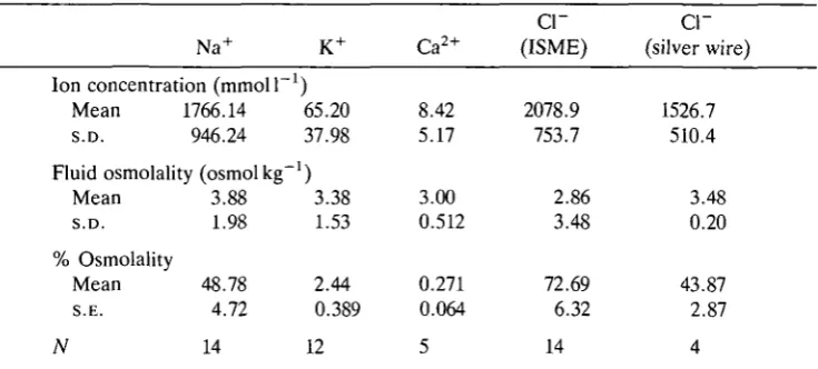
Table 1. Ion concentrations determined from ISME recordings and a solid-state chloride electrode (silver wire)
Na+
Ion concentration (mmol 1 1 Mean 1766.14 S.D. 946.24
Fluid osmolality (osmolkg"1 Mean 3.88 S.D. 1.98 % Osmolality
Mean 48.78 S.E. 4.72
K+ ) 65.20 37.98 ) 3.38 1.53 2.44 0.389 Ca2+ 8.42 5.17 3.00 0.512 0.271 0.064
cr
(ISME) 2078.9 753.7 2.86 3.48 72.69 6.32cr
(silver wire) 1526.7 510.4 3.48 0.20 43.87 2.87N 14 12 5 14 4
Concentrations listed are corrected for the dilution factor employed to bring the fluid sample within the osmotic range of the electrode calibration solutions; this procedure assumes that the ion behaves as an ideal solute (see text). Since ion concentrations are a function of fluid osmolality, which is regulated over a wide range according to uptake rates and ambient humidity, standard deviations rather than standard errors are quoted for these variables.
Sample number (N) refers to the number of fluid samples analysed; ion concentrations in each sample were calculated from multiple impalements.
original ion concentrations were calculated using the dilution factor determined for each test drop. This procedure assumes that each of the ionic species behaves as an ideal solute. Errors arising from this assumption will be small, since osmotic coefficients of typical diluted drop concentrations and original concentrated samples are similar. For a representative test drop concentration of 0.086moll"1 the osmotic coefficient for aqueous NaCl is 0.938, whilst for a concentrated original sample of 2.03 m o l F1 the corresponding osmotic coefficient is 0.971. Errors in the determined original ion concentrations are proportional to the difference between these two osmotic coefficients - about 3.5%. Table 1 shows that Na+ and Cl~ are the predominant electrolytes accounting, respectively, for 48.8 % and 43.9 % (silver wire electrode) of the total osmolality over the full range of uptake fluid concentrations. The somewhat elevated Cl~ concentrations interpolated from ISME recordings using the Corning Cl~ exchanger may be due to interfering anions; the solid-state electrode potentials give more credible Cl~ concentrations, consistent with electrical neutrality of the sample droplets. Potassium and calcium are minor electrolytes, accounting for 2.44 % and 0.271 %, respectively, of total osmolality.
Discussion
secreting a substantial quantity of strongly hyperosmotic uptake fluid into the pleoventralraum. This behaviour was observed even after brief periods of dehydration (<5min in 30-40% RH). In the absence of subsequent irritation, they began pleopodal ventilation within a few minutes and the fluid osmolality fell from the initial high levels (up to S^osmolkg"1) to levels slightly above the equilibrium osmolality of the chamber humidity. The concurrence of this osmotic decline with the onset of ventilation, and the invariable association between ventilation and WVA in gravimetric studies (Wright and Machin, 1990; unpub-lished observations), clearly suggests dilution of the concentrated uptake fluid by absorbed vapour.
Several lines of evidence argue against hyperosmotic uptake fluid being an artefact of evaporative concentration during pre-desiccation. Animals retained in open chambers (laboratory RH) for extended sampling periods maintained a small quantity of near iso-osmotic pleon fluid, with no indication of long-term changes in osmolality. Hyperosmotic uptake fluid was observed only in humidities near or above demonstrated uptake thresholds. Although transfer from laboratory RH to moderately sub-threshold humidities (85%, 88%) sometimes triggered an initial pulse of uptake fluid - the animal perhaps anticipating spatial or temporal proximity to more humid microclimates - secretion was never sustained for more than a few minutes, and fluid osmolalities subsequently declined to haemolymph levels (0.69-0.85 osmol kg"1). More typically, animals in sub-threshold humidities revealed iso-osmotic pleon fluid from the outset, regardless of the degree of pre-desiccation, indicating that sealing of the pleoventralraum against evaporative losses is actually very efficient.
Further evidence against evaporative concentration was the absence of signifi-cant differences in osmolality between deep and superficial samples collected simultaneously. Possible long-term changes in osmolality of the bulk pleon fluid, not identifiable from local osmotic gradients, also seem unlikely. Pre-desiccated animals often had several hours to undergo evaporative concentration but regularly revealed iso-osmotic fluid immediately following transfer to high humidities. By contrast, hydrated animals sometimes produced highly concen-trated fluid following transfer to a humid chamber. These observations suggest that variation in fluid osmolality is under independent physiological control and is not a simple function of previous desiccation history.
200 J. C. WRIGHT AND M. J. O ' D O N N E L L
analysed, owing to evaporative concentration in the WS. During osmometry it was easily distinguished from pleon fluid by the presence of slow-thawing, orthogonal crystals (KC1?) and by the different electrolyte composition. Potassium and calcium were more concentrated, accounting for approximately 10.5 % and 1.0 % of osmotic activity, respectively, whilst Na+ contributed approximately 35 % of osmotic activity. Concentrations of Cl~ and organic osmolytes in urine were not determined.
Pleopodal ventilation may play a role in the circulation of uptake fluid within the pleon as well as the more obvious role of increasing the absorption rate by maintaining a regular cycling of humid air. The latter could be more efficiently achieved, however, by maintaining a continuous-flow rather than a cyclic ventilatory pattern. The intermittent sealing of the pleopodal margins during depression - evident from the marginal displacement of uptake fluid - only serves to reduce the net atmospheric water supply. A possible explanation for intermit-tent sealing is that the pleopods compress an enclosed volume of air to elevate its relative humidity. Such 'pressure cycling' (Corbet, 1988) would not only increase the water activity difference between air and uptake fluid (thereby increasing uptake flux) but would permit WVAin ambient humidities below the equilibrium activity of the uptake fluid. This is suggested by the results from the 91.9% RH chamber: several ventilating animals revealed uptake fluid activities above the mean activity measured for the chamber (Fig. 2C). Although imperfect sealing of the pleoventralraum during ventilation (evident from the marginal displacement of fluid) would reduce pressure elevation during pleopodal depression, even a modest net force of 5mN (a conservative estimate of the depressor force generated by the walking pereiopods) would generate a maximum pressure of 1 kPa assuming a ventral surface area of 5 mm2 for the pleoventralraum. Since this is approximately 1 % of standard atmospheric pressure, and pressure is pro-portional to vapour density and activity, it would elevate the activity of the air by 1 %. For humidities in the region of 90 % this is equivalent to an osmotic saving of about 800mosmolkg~J. As pointed out by Corbet (1988), pressure cycling may offer considerable energetic advantages over secreting hyperosmotic fluid against increasing osmotic gradients.
possible inter-relationships between these variables, only tentative conclusions can presently be drawn. The routine appearance of high-osmolality fluid before initial ventilation suggests that uptake fluid is secreted at a fixed concentration, variation in recorded osmolalities being attributable to osmotic dilution across the permeable endopods or to marginal vapour uptake. The small subsequent activity deficits between uptake fluid and ambient air in ventilating animals further suggest that fluid and air are allowed to approach equilibrium.
During sampling in the present study, it was evident that the fluid content of the pleoventralraum varied greatly according to an animal's water status. Primary secretion of uptake fluid leads to probable saturation of the cavity, with fluid welling conspicuously between the overlapping exopods. Similar conditions accompany urination and persist throughout the period of excretion (anything from a few minutes to several hours). Uptake fluid volume declines with the onset of ventilation but remains copious and accessible to superficial sampling. In non-absorbing animals, fluid is scant and the exopods are tightly apposed to the pleon sternites. Deep sampling from such animals (hydrated or pre-desiccated) usually recovered iso-osmotic fluid, suggesting that endogenous pleon fluid maintains a permanent, thin film over the endopods. The irregularity of urination, and the ability of animals to replace sampled fluid within a few minutes, clearly excludes a maxillary source.
More specific information on the source of endogenous pleon fluid is currently lacking. The elaborate transporting epithelia of the endopods (Kummel, 1981) are not likely candidates since the distribution of mitochondria clearly indicates basal transport. Active secretion of uptake fluid would therefore involve non-physio-logical intracellular concentrations of both Na+ and Cl~ (see Yancey et al. 1982). This could be circumvented by a paracellular pathway, but there is no confluence of the basal infoldings and lateral membranes as is seen, for example, in avian salt glands (Berridge and Oschman, 1972). Our attempts to localise uptake fluid secretion by regional sampling were unsuccessful, implying either a broad distribution of the secretory epithelia or very efficient fluid circulation. The latter possibility is supported by the insignificant differences in fluid concentration between simultaneous superficial and deep samples.
202 J. C. WRIGHT AND M. J. O ' D O N N E L L
exploiting the exchange diffusion of ammonium ions as Na+ is resorbed across the endopods and hence concentrating NH4+ in the pleon fluid. NH3 could then be
volatilised by a rise in pH as suggested by Weiser (1972, 1984) and Little (1983). Elevated rates of Na+ secretion and resorption would accumulate NH4+ more
rapidly and give rise to hyperosmotic pleon fluid. Local absorption of water from high ambient activities would then shift the selection pressure in favour of WVA and increasingly hyperosmotic fluid to depress the uptake threshold. Interrelation-ships between pH, NH4+ and Na+ activity in oniscidean pleon fluid await detailed
investigation.
The authors are grateful for the financial support of the Leverhulme Trust, UK (J.C.W) and NSERC (M.J.O). Thanks are also extended to Dr J. Machin for valuable discussions and loan of the Clifton osmometer.
References
AMMANN, D. AND ANKER, P. (1985). Neutral carrier sodium ion-selective microelectrode for extracellular studies. Neurosci. Lett. 57, 287.
BERRIDGE, M. J. AND OSCHMAN, J. L. (1972). Transporting Epithelia. New York, London: Academic Press.
CORBET, S. A. (1988). Pressure cycles and the water economy of insects. Review. Phil. Trans. R.
Soc. Lond. B 318, 377-407.
EDNEY, E. B. AND SPENCER, J. (1955). Cutaneous respiration in woodlice. J. exp. Biol. 32, 256-269.
EVANS, D. H. (1977). Further evidence for Na+/NH4+ exchange in marine teleost fish. /. exp.
Biol. 70, 213-220.
GORVETT, H. (1956). Tegumental glands and terrestrial life in woodlice. Proc. zool. Soc, Lond. 126, 291-304.
GORVETT, H. AND TAYLOR, J. C. (1960). A further note on the tegumental glands in woodlice.
Proc. zool. Soc, Lond. 133, 653-655.
HOESE, B. (1981). Morphologie und Function des Wasserleitungssystems der Terrestrichen Isopoden (Crustacea, Isopoda, Oniscoidea). Zoomorphology 98, 135-167.
HUNTER, K. C. AND KIRSCHNER, L. B. (1986). Sodium absorption coupled to ammonia excretion in osmoconforming marine invertebrates. Am. J. Physiol. 251, R957-R962.
KUMMEL, G. (1981). Fine structural indications of an osmoregulatory function of the 'gills' in terrestrial isopods (Crustacea, Oniscoidea). Cell Tissue Res. 214, 663-666.
LITTLE, C. (1983). The Colonisation of Land. Cambridge: Cambridge University Press. MANTEL, L. H. AND FARMER, L. L. (1983). Osmotic and ionic regulation. In The Biology of the
Crustacea, vol. 5 (ed. D. E. Bliss), pp. 53-161. New York: Academic Press.
OEHME, M. AND SIMON, W. (1976). Microelectrode for potassium ions based on a neutral carrier and comparison of its characteristics with a cation exchanger sensor. Analyt. chim. Ada 86, 21-25.
SAUNDERS, J. H. AND BROWN, H. M. (1977). Liquid and solid-state CP micro-electrodes: characteristics and application to intracellular Cl~ activity in Balanus photoreceptors. /. gen.
Physiol. 70, 507-530.
SCHEFER, U., AMMANN, D., PRETSCH, E., OESCH, U. AND SIMON, W. (1986). Neutral carrier based Ca2+-selective electrode with detection limit in the sub-nanomolar range. Analyt.
Chem. 58, 2282-2285.
THOMAS, R. C. (1978). Ion-sensitive Intracellular Microelectrodes. How to Make and Use Them. London: Academic Press.
VERHOEFF, K. W. (1920). Uber die Atmung der Landasseln Zugleich ein Beitrag zur Kenntnis der Entstchung der Landteire. Z. wiss. Zool. 118, 365-447.
WEISER, W. (1972). Oxygen consumption and ammonia excretion in Ligia beaudiana M.-E.
Comp. Biochem. Physiol. 43A, 869-876.
WEISER, W. (1984). Ecophysiological adaptations of terrestrial isopods: a brief review. Symp.
zool. Soc, Lond. 53, 247-265.
WINSTON, P. W. AND BATES, D. H. (1960). Saturated solutions for the control of humidity in biological research. Ecology 41, 232-237.
WRIGHT, J. C. AND MACHIN, J. (1990). Water vapour absorption in terrestrial isopods. J. exp.
Biol. 154,13-30.