AJPRHC
Research Article
ENHANCED LIVER DELIVERY AND SUSTAINED RELEASE OF CURCUMIN WITH DRUG LOADED NANOPARTICLES AFTER INTRAVENOUS ADMINISTRATION IN RATS
SURESH KONATHAM1, BONEPALLY REDDY2, JITHAN AUKUNURU2, 3*
For Author affiliations see end of the text
This paper is available online at www.jprhc.in
ABSTRACT:
Liver targeting drug delivery systems can improve the delivery of several drugs useful in the treatment of liver disorders such as cirrhosis and liver cancer. The objective of this study was to prepare the biodegradable nanoparticles containing curcumin, a well-known hepatoprotective agent and further to evaluate the liver targetability and sustained release of curcumin with the developed nanoformulation. Curcumin nanoparticles were prepared by double emulsion (w/o/w) solvent evaporation method using different drug polymer ratios. Poly-ε-caprolactone was used in the preparation. The prepared formulations were evaluated for particles size, surface potential, entrapment efficiency, in vitro release, drug polymer interaction. Four different formulations CNP1, CNP2, CNP3 and CNP4 were prepared. Optimized formulation (CNP3) was evaluated for pharmacokinetics and hepatoprotective activity in CCl4
induced liver toxicity model after i.v. administration. Optimized formulation was selected based on the size, entrapment efficiency and release characteristics. Curcumin i.v. solution and oral suspension form were used as the reference. Particle size of all formulations was in the range of 300-470 nm and the entrapment efficiencies were in the range of 75-85 %. Drug release from the nanoparticles was sustained both in vitroand in
vivo. Nanoparticle formulation tested in vivo
demonstrated better pharmacokinetics and pharmacodynamics compared to the reference. Drug levels in the liver were significantly higher with nanoparticular formulation. Thus, this study successfully prepared a nanoparticular formulation containing curcumin with polycaprolactone as the polymer. With the developed formulation better liver targetability was achieved.
Key words: cirrhosis, liver targeting, nanoparticles, curcumin, pharmacokinetics, sustained drug release, hepatoprotective activity.
INTRODUCTION
Curcumin (diferuloyl methane; 1, 7-Bis-(4-hydroxy-3-methoxyphenyl)-hepta-1, 6-diene-3, 5-dione) is a major constituent found in the spice turmeric, which is derived from the rhizomes of Curcuma longa L. It is commonly used as a dietary spice and colouring agent in cooking and is used as an herb in traditional Indian and Chinese medicine. Curcumin possesses diverse pharmacological effects including anti-inflammatory, antioxidant, anticancer, antidiabetic, antirheumatic, angiogenic, antifertility, antiviral and anti-infectious activities and wound healing properties.1 Despite
curcumin’s multiple medicinal benefits, low oral bioavailability of curcumin continues to be highlighted as a major challenge in developing formulations for clinical efficacy. Lower serum and tissue levels of curcumin are observed irrespective of the route of administration due to extensive intestinal and hepatic metabolism and rapid elimination thus restraining curcumin’s bioavailability.2-4 Curcumin is also found to
be photo-sensitive and requires careful handling. In spite of numerous formulations challenges several formulation strategies like nanoparticles, liposomes, complexation with phospholipids and cyclodextrins, solid dispersions are being developed to improve curcumin’s bioavailability.5-7It is also popularly known
as a hepatoprotective agent.
Liver is the main site of metabolism for many drugs and xenobiotics. The central role played by liver in the clearance and transformation of chemicals also makes it susceptible to drug induced injury. Liver fibrosis and liver cirrhosis are generally the end result of majority of chronic liver insults. The development of fibrosis, and particularly cirrhosis, are associated with a significant morbidity and mortality. The causes of hepatic fibrosis and cirrhosis are multiple and include congenital metabolic, inflammatory, and toxic liver diseases. In all circumstances, the composition of the hepatic scar is similar.8 Many chemicals damage mitochondria, an
AJPRHC
Research Article
in the cytochrome P-450 system such as CYP2E1 also leads to oxidative stress.9This suggests that increase in
oxidative stress is the center to the progression of these liver disorders. Further, injury to hepatocyte and bile duct cells lead to accumulation of bile acid inside liver. This promotes further liver damage and enhances inflammation.10Non-parenchymal cells such as Kupffer
cells, fat storing stellate cells, sinusoidal endothelial cells and leukocytes (i.e., neutrophils and monocytes) have a significant role in the mechanism.11In all these
conditions, curcumin which is both antioxidant as well as antiinflammatory should demonstrate significant benefit in liver toxicity. This was demonstrated by showing the hepatoprotective activity of curcumin. Interestingly, Kupffer cells in the liver are exposed to systemic circulation and are phagocytic in nature.12
Sinusoidal endothelial cells also exposed to systemic circulation can actively take up particles by endocytosis.13Thus, curcumin can be targeted to liver
cells involved in fibrosis and cirrhosis using particulate approach in the way of passive targeting to improve the treatment of these liver disorders. Previous reports clearly indicate that smaller size particles are avidly taken up by liver cells compared to microparticles.13
Further, so as to target the liver, the size of the microparticles that can be injected into the systemic circulation is restricted to 6 µm only as the greater size particles are known to reach the lungs and gets lodged in the pulmonary tissues.14Also, the amount of the drug
released in the cells of interest with nanoparticles after cellular entrapment is far greater than compared to the release from the microparticles after cellular entrapment because of the greater surface area of the nanoparticles.15 This could result in better
pharmacodynamic activity with nanoparticles compared to the microparticles. Thus, the objective of this study was to prepare the biodegradable nanoparticles containing curcumin, a well-known hepatoprotective agent and further to evaluate the liver targetability and sustained release of curcumin with the help of developed nanoformulation.
1. Materials and Methods
1.1. Materials
Curcumin was procured from Yucca Enterprises, Mumbai. Poly-ε-caprolactone was procured from Sigma Aldrich, Germany. Poly vinyl alcohol was procured from Qualikems Fine Chemicals Pvt Ltd, New Delhi. Chloroform was procured from Finar chemicals, Ahmedabad, India. HPLC solvents were procured from Merck specialities, Mumbai, India. SGOT and SGPT
kits were purchased from Coral Clinical Systems, Goa, India.
1.2. Methods
1.2.1. Preparation of curcumin nanoparticles
Double emulsion (W/O/W) solvent evaporation method was employed in the preparation of curcumin nanoparticles.16 Briefly, Curcumin and poly-ε
-caprolactone previously dissolved in 10 ml of chloroform was emulsified with 5 ml of 3% PVA solution under homogenization for 10min to form a W/O emulsion. This primary emulsion was thereafter poured into the 30 ml of PVA aqueous solution (3 % W/V) and homogenized in a homogenizer (Homogenizer 150 VT, M/S Biologics, Inc –USA) for 15 min to form a W/O/W emulsions.17, 18This multiple
emulsion was stirred on a magnetic stirrer till complete evaporation of chloroform. The nanoparticles were obtained by centrifuging the resulted nanoparticular suspension at 10,000 rpm for 45 min. The obtained nanoparticles were washed with phosphate buffer saline (PBS) and dried.
1.2.2. Characterization of the curcumin nanoparticles
Determination of the particle size and the surface potential
The prepared nanoparticles were evaluated for their particle size, polydispersity index(PDI) of size distribution and surface charge potential, by photon correlation spectroscopy (PCS) using Zetasizer 3000 HAS (Malvern Instruments, Malvern, UK). The formulations were diluted 1:1000 with the aqueous phase of the formulation to get a suitable kilo counts per second (kcps). Analysis was performed at 25°C with an angle of detection of 90°.Each sample was measured in triplicate.
Encapsulation efficiency
To determine the encapsulation efficiency, the drug from nanoparticles was extracted with acetonitrile, the obtained solution was evaporated to dryness and reconstituted with 100µl of mobile phase consisting of acetonitrile/methanol/water/acetic acid (40:23:36:1, v/v/v/v)(water and acetic acid solvents whose pH was adjusted to 2.8 prior to the preparation of mobile phase). The column used was C18ODS column and the size of
AJPRHC
Research Article
into HPLC (Cyber Labs, USA) column and analysis was carried out a spectrophotometer at 230 nm. 19
In vitro release study
In vitro release of Curcumin nanoparticles was performed by taking weighed amount of nanoparticles into a dialysis bag(D-70, MWCO 12000-2000, nanoparticles suspended in 1 ml of distilled water), suspended in 50 ml of Phosphate Buffer 7.4 (release medium).20 This entire system was kept at 370C on
magnetic stirrer with 50 rpm. Aliquots were withdrawn at predetermined time intervals and the receptor compartment was replenished with same volume of fresh dialysing medium. The samples were analyzed at 425 nm using UV-Visible spectrophotometer (Elico SL 164 Double beam). The release study was terminated when no more drug is released for more than 48 hours in the release conditions. Data obtained from in vitro release studies were fitted to various kinetic equations to find out the mechanism of curcumin release from the nanoparticles.
Drug- polymer interactions
Drug – Polymer interaction was investigated using FTIR. FTIR spectra of drug, polymer, placebo nanoparticles and drug loaded particles were taken using a Thermo Nicolet Nexus 670 Spectrophotometer with KBr pellets.
ANIMAL STUDIES
IN VIVO DRUG RELEASE AND HEPATO
PROTECTIVE ACTIVITY OF THE
FORMULATION
In Vivodrug release was investigated in male wistar rats. All the animal experiments were conducted according to the rules and guidelines of Committee for the Purpose of Control and Supervision of Experiments on Animals (CPCSEA), Chennai, India. The study was approved by Institutional Animal Ethical Committee of Vaagdevi College of Pharmacy, Warangal, registered under CPCSEA, India (Registration No: 1047/ac/07/CPCSEA). The rats were acclimatized with 12 hour dark and 12 hour light cycle at a temperature of 20º C at a humidity of 60% and were fed on standard diet for 10 days prior to the commencement of the experiment. The conditions continued during the next 10 days of experimentation. Rats were divided into three groups,
each group containing six rats. Group 1 received 1ml of curcumin solution (15mg/ml DMSO solution) intravenously. Group 2 received 1ml of curcumin nanoparticular suspension (equivalent to 15mg of curcumin) by intravenous route. Group 3 received 1ml curcumin oral suspension (15mg). After administration of the formulations blood samples were collected at 0.5, 1, 2, 3, 6, 12, 24 hours and 3, 6 and 9 days and plasma was separated. The drug was extracted from the plasma by adding 500 µl of acetonitrile. Curcumin in these samples were estimated using a HPLC (Cyberlab, USA) at 230nm. In case of liver, kidney, colon and brain, the tissue was homogenized and then the drug was extracted with acetonitrile. A HPLC standard curve for the drug in the plasma was generated. The mobile phase consisted of acetonitrile/methanol/water/acetic acid at a ratio of 40:23:36:1, v/v/v/v.
Hepatoprotective activity
Carbon tetrachloride induced liver damage model was used in the evaluation of hepatoprotective activity of the prepared formulations. Male wistar rats (150-180g) were divided into 5 groups containing six rats each. Group 1 received normal saline (1 ml/rat) daily for 9 days. Group 2 received carbon tetrachloride (0.7 ml/kg), administered intraperitoneally on the 3rd, 6th
and 9thday consisting of a mixture of CCl4 and olive oil
(25:75). Group 3 received curcumin oral suspension (100 mg/kg) daily for 9 days. Group 4 received curcumin solution (100mg/kg) intravenously, daily for 9 days. Group 5 received curcumin nanoparticular suspension (equivalent to 100mg/kg of curcumin) intravenously at day one. All groups received CCl4at 3rd, 6thand 9thday
of the study except normal control. Hepatoprotective activity was quantified by the SGOT (serum glutamate oxaloacetate transaminase), SGPT (serum glutamate pyruvate transaminase) levels and histological studies following a previously published report.21
Statistical analysis
Results are expressed as means ± S.E.M. of six rats per treatment group. Data were analyzed using a one-way analysis of variance (ANOVA) followed by Dunnett’s test. Differences were considered significant at p≤0.05.
RESULTS
AJPRHC
Research Article
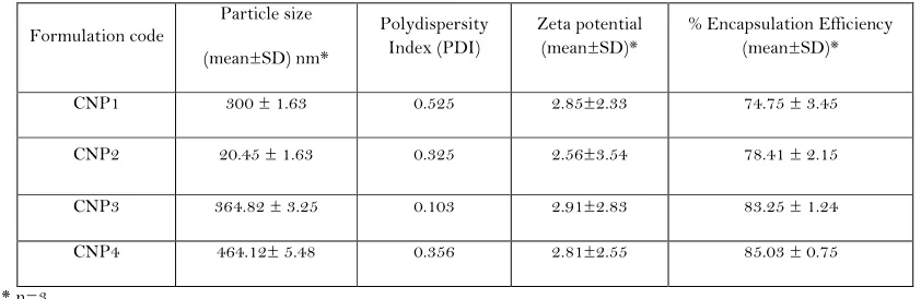
Curcumin nanoparticles were successfully prepared by double emulsion solvent evaporation method. Four formulations were prepared by taking different drug to polymer ratios like 1:1, 1:2, 1:3 and 1:4. Compositions of nanoparticles are described in Table 1. The results for particle size, zeta potential and encapsulation efficiency are shown in Table 2. Drug polymer interactions were studied by FTIR; from the spectra we observed that there is no interaction between the drug and polymer. The in vitro release studies from curcumin nanoparticles were fitted into various release kinetic models (Table 3). From release profiles (Fig. 1.) it was observed that increase in polymer concentration results in decrease of drug release rate. From all four formulations, the drug release was up to 20% during the first 3 days. The percent cumulative drug release observed was 80%, 86%, 90% and 92% and in vitro drug release was sustained up to 6, 7, 9 and 12 days for CNP1, CNP2, CNP3 and CNP4, respectively. Different kinetic release patterns were evaluated. The log percent cumulative drug released was plotted as a function of log time and the slope of the curves was determined as the values of diffusional release exponent (n). The values of diffusional release exponent (n) from the straight lines were noted to be 0.395, 0.410, 0.421 and 0.422 for CNP1, CNP2, CNP3 and CNP4 respectively, which indicated that the release of drug from all formulations followed a Fickian pattern.22
IN VIVO DRUG RELEASE AND THE
HEPATOPROTECTIVE ACTIVITY
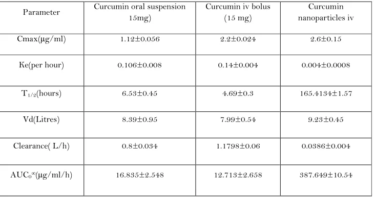
Drug levels in the plasma and the tissues were determined using HPLC. The retention time of the drug was 9.6 min. Plasma profile of the drug after nanoparticular, oral and i.v. solution form administration was plotted (Fig. 2.). From the figure it clearly indicates that nanoparticular formulation sustains the drug release up to 9 days reflecting in an increased area under curve. Peak serum concentration of 2.2µg/ml was observed within 30 min when curcumin solution was given intravenously. Peak serum concentration of 1.12µg/ml was observed within 6 hours when curcumin suspension was given orally. Peak serum concentration of 2.6µg/ml was observed within 1 hour when curcumin nanoparticles (CNP3) suspension was given intravenously. Pharmacokinetic parameters were showed in Table 4. Data obtained from hepatoprotective study is shown in Table 5. Nanoparticular formulation significantly reduces the elevated enzymes levels. From the table it was observed that all formulations were having significant hepatoprotective activity when compared to CCl4
treated group (p <0.05). Among all the formulations studied, curcumin nanoparticles showed highest hepatoprotective activity (Fig. 3.). The drug levels in various tissues like liver, brain, kidney, and colon were estimated and shown in Table 6. Histopathology of liver is shown in Fig. 4.
DISCUSSIONS
Particle size and entrapment efficiency of the curcumin nanoparticles were increased with increasing the polymer content up to 300 mg. This is may be due to high amount of polymer available for coating the drug. Upon increasing the polymer amount, number of layers was increased; this resulted in the increase in particle size and entrapment efficiency. Further increase in the polymer content to 400 mg did not increase the entrapment presumably due to the less availability of the drug to be incorporated. The in vitrodrug release was fitted into various release models. From the release data we observed that increase in the polymer content delays the drug release due to increased particle size and reduced surface area available for drug release. From all the four formulations studied, formulation CNP3 was selected as optimized formulation because of less PDI, high entrapment efficiency and since the release was prolonged up to 9 days, the study period we selected for determining hepatoprotective activity. From plasma profile it was observed that the drug levels in plasma was lower in case of curcumin when given orally compared to that of curcumin and CNP3 given intravenously. This could be because of lower oral bioavailability of curcumin and significant degradation of free curcumin in the plasma. With the nanoparticular formulation the drug release was sustained up to 10 days. This could be due to reduction in the elimination and metabolism. From the liver enzyme studies we observed that SGOT and SGPT levels were more in case of CCl4 treated animals because of tissue damage
caused by CCl4 which releases the enzymes in to the
blood stream. A fatty layer was observed in the histopathology of liver. Upon administration of curcumin formulations the enzyme levels was decreased due to the fibrosis caused by the CCl4 was reversibly
AJPRHC
Research Article
distribution studies it was observed that drug was more concentrated in the liver compared to colon, kidney, and brain. This may be due to nanosize of the particles, which were easily taken up by the RES of liver compared to the other tissues. The drug accumulation order in the different tissues is as follows: liver, colon, kidney and brain. The drug levels in brain were not detected and may due to the presence of blood brain barrier (BBB) which could hinder the transport of curcumin across the barrier. In case of nanoparticles small amount of drug was detected in the brain. This may be due to nanosize of the particles, which are accessible through the BBB.
The results of this study clearly indicate that a nanoparticular formulation containing curcumin is
better in pharmacokinetic properties and sustained plasma drug levels when compared to higher cumulative doses of curcumin administered via i.v. and oral administrations. The reversal of biochemical end points in a CCl4 hepatotoxic model is better with
nanoparticular curcumin compared to oral and i.v. curcumin. This suggests that this formulation may be of potential use in the treatment of cirrhosis and fibrosis with curcumin. The results can be extrapolated to other drugs suggesting the significant benefits of nanoparticular passive targeting of drugs to the liver. Similar formulations could not only be used in fibrosis and cirrhosis but also could be used in the liver cancers and several other liver diseases with additional benefits
Table 1. Compositions of curcumin nanoparticles.
Formulation code CNP1 CNP2 CNP3 CNP4 Curcumin (mg) 100 100 100 100 Poly-ε-caprolactone (mg) 100 200 300 400
Table 2. The particle size, zeta potential and encapsulation efficiencies of curcumin nanoparticles.
Formulation code
Particle size (mean±SD) nm*
Polydispersity Index (PDI)
Zeta potential (mean±SD)*
% Encapsulation Efficiency (mean±SD)* CNP1 300 ± 1.63 0.525 2.85±2.33 74.75 ± 3.45 CNP2 20.45 ± 1.63 0.325 2.56±3.54 78.41 ± 2.15 CNP3 364.82 ± 3.25 0.103 2.91±2.83 83.25 ± 1.24 CNP4 464.12± 5.48 0.356 2.81±2.55 85.03 ± 0.75
* n=3
Table 3. The in vitro release studies from curcumin nanoparticles were fitted into various release kinetic models.
Formulation code
Zero order First order Higuchi model Korsemeyer - Peppas model R2 R2 R2 R2 N
AJPRHC
Research Article
CNP2 0.9533 0.9872 0.9947 0.9922 0.410 CNP3 0.9511 0.9909 0.9956 0.9928 0.421 CNP4 0.9223 0.9978 0.9923 0.9918 0.422
Table 4. Pharmacokinetic parameters obtained after administration of curcumin formulations.
Parameter Curcumin oral suspension 15mg)
Curcumin iv bolus (15 mg)
Curcumin nanoparticles iv Cmax(µg/ml) 1.12±0.056 2.2±0.024 2.6±0.15
Ke(per hour) 0.106±0.008 0.14±0.004 0.004±0.0008
T1/2(hours) 6.53±0.45 4.69±0.3 165.4134±1.57
Vd(Litres) 8.39±0.95 7.99±0.54 9.23±0.45 Clearance( L/h) 0.8±0.034 1.1798±0.06 0.0386±0.004
AUC0∞(µg/ml/h) 16.835±2.548 12.713±2.658 387.649±10.54 * Values indicate mean ± standard error mean (S.E.M). n=6
Table 5. Effect of curcumin formulations on enzyme levels in rats with CCl4induced hepatotoxicity (Mean ± SEM; n=3)
Groups SGOT( U/L) SGPT( U/L) Normal control( saline ) 16.09±1.306 10.73±2.359 CCl4treatment 73.39±2.45b 43.79±3.485b
CCl4+ Curcumin oral(100mg/kg) 41.44±3.611a 26.16±2.955a
CCl4+ Curcumin i.v (10mg/kg) 33.07±2.571a 20.82±2.767a
CCl4+ Curcumin nanoparticles (100mg/kg) 21.02±1.01a 14.81±2.881a.
Note: a p < 0.05 vs CCl4treated, b p < 0.05 vs normal control Table 6.Curcumin levels in various tissues (µg/mg).
AJPRHC
Research Article
AJPRHC
Research Article
CONCLUSION
The nanoparticular formulations of curcumin were successfully prepared using double emulsion (W/O/W) solvent evaporation method. The drug release from the formulations was sustained up to 10 days. The in vivo
performance of the formulation was better compared to the respective oral and i.v formulations. From the distribution studies it was concluded that better liver targeting was achieved by the nanoformulations compared to oral and i.v. formulations.
ACKNOWLEDGMENTS
The authors work would like to acknowledge the management of Vaagdevi College of Pharmacy, Warangal, for providing facilities for this work. One of
the Authors, Dr. Aukunuru Jithan would like to acknowledge the Department of Science and Technology (DST), Government of India. This project is partly funded under a SERC-DST Fast Track Proposal for Young Scientists awarded to Dr. Aukunuru Jithan.
Declaration of interest: The authors report no conflicts of interest.
REFERENCES
AJPRHC
Research Article
2. P. Anand, A. B. Kunnumakkara, R.A. Newman, and B.B. Aggarwal, Bioavailability of curcumin: problems and promises, Mol. Phar. 4, 807(2007).
3. M. H. Pan, T. M. Huang, and Lin JK, Biotransformation of curcumin through reduction and glucuronidation in mice, Drug Metab. Dispos.27, 486 (1999).
4. R. A. Sharma, W. P. Steward, and A. J. Gescher, Pharmacokinetics and pharmacodynamics of curcumin, Adv. Exp. Med. Biol.595, 453 (2007).
5. C. Karikar, A. Maitra, S. Bisht, G. Feldmann, S. Soni, and R. Ravi, Polymeric nanoparticle-encapsulated curcumin (“nanocurcumin”): a novel strategy for human cancer therapy. J. Nanobiotechnol. Available
at: http://www.jnanobiotechnology.com/cont ent/5/1/3. (2007).
6. K. Maiti, K. Mukherjee, A. Gantait, B. P. Saha, and P. K. Mukherjee, Curcumin–phospholipid complex: Preparation, therapeutic evaluation and pharmacokinetic study in rats, Int. J. Pharm.330, 155
(2007).
7. W. Tiyaboonchai, W. Tungpradit, and P. Plianbangchang, Formulation and characterization of curcuminoids loaded solid lipid nanoparticles, Int. J. Pharm.337, 299(2007).
8. Manoj Kumar and S. K. Sarin, Is cirrhosis of the liver reversible?, Indian J. Pediatr.74,393 (2007).
9. H. Jaeschke, G. J. Gores, and A. I. Cederbaum, Mechanisms of hepatotoxicity, Toxicol. Sci. 65, 166
(2002).
10. T. Patel, L. R. Roberts, B. A. Jones, and G. J. Gores,
Dysregulation of apoptosis as a mechanism of liver disease: An overview, Semin. Liver Dis.18, 105 (1998). 11. P. J. De Bleser, T. Niki, V. Rogiers, and A. Geerts,
Transforming growth factor-beta gene expression in normal and fibrotic rat liver, J. Hepatol. 26, 886
(1997).
12. S. Nagayama, K. Ogawara, Y. Fukuoka, K. Higaki, and T. Kimura, Time-dependent changes in opsonin amount associated on nanoparticles alter their hepatic uptake charactereistics, Int. J. Phar. 342, 215(2007). 13. K. Ogawara, M. Yoshida, K. Higaki, T. Kimura, K.
Shiraishi, M. Nishikawa, Y. Takakura, and M. Hashida, Hepatic uptake of polystyrene microspheres in rats: effect of particle size on intrahepatic distribution, J. Control. Rel.59, 15 (1999).
14. P. Chao, M. Deshmukh, H. L. Kutscher, D. Gao, S. S. Rajan, P. Hu, D. L. Laskin, S. Stein, and P. J. Sinko,
Pulmonary targeting microparticulate camptothecin delivery system: anticancer evaluation in a rat orthotopic lung cancer model, Anticancer Drugs.21:65
(2010).
15. J. V. Aukunuru and U. B. Kompella, In vitro delivery of nano- and micro-particles to human retinal pigment epithelial cells (ARPE-19). Drug Delivery Technology.
Vol. 2 No. 2 March/April 2002, Posted On: 3/28/2008.
16. F. Tewes, E. Munnier, B. Antoon, L.N. Okassa, S. Cohen-Jonathan, H. Marchais, L. Douziech-Eyrolles, M. Soucé, P. Dubois, and I. Chourpa, Comparative study of doxorubicin-loaded poly (lactide-co-glycolide) nanoparticles prepared by single and double emulsion methods, Eur. J. Pharm. Biopharm. 66, 488(2007).
17. N. Lamprecht, M. Ubrich, C. M. Hombreiro Perez, M. Lehr, and M. P. Hoffman, Biodegradable monodispersed nanoparticles prepared by pressure homogenization-emulsification, Int. J. Pharm. 184, 97(1999).
18. Y. Y. Yang, T. S. Chung, X. L. Bai, and W. K. Chan, Effect of preparation conditions on morphology and release profiles of biodegradable polymeric microspheres containing protein fabricated by double-emulsion method, Chem. Eng. Sci.55, 2223 (2000).
19. V. Guduri, J. Aukunuru, and V. M. Reddy, Development of new delivery strategies to increase bioavailability of curcumin, Int. J. Pharm. Sci. and Nanotech.1, 335(2009).
20. C. A. Anuradha and J. Aukunuru, Preparation, characterisation and in vivo evaluation of bis-demethoxy curcumin analogue (BDMCA) nanoparticles, Trop. J. Pharm. Res,9,51 (2010). 21. C. R. Bonepally, J. V. Aukunuru, N. R. Yellu, and M.
R. Vanga, Fabrication and investigations on hepatoprotective activity of sustained release biodegradable piperine microspheres, Int. J. Pharm. Sci. and Nanotech.1, 87 (2008).
AJPRHC
Research Article
AUTHORS AFFILIATIONS AND ADDRESS FOR CORRESPONDENCE
1 Formulation Research and Development, Global Oncology, Dr. Reddys Research Laboratories, Hyderabad, AP, India.
2Vaagdevi College of Pharmacy (Affiliated to Kakatiya
University), Ramnagar, Warangal, AP, India, 506001.
3 Mother Teresa College of Pharmacy (Affiliated to
Osmania University), NFC Nagar, RR Dist, AP, India-501301