Vol -4, Issue-4, October-December 2015
ISSN: 2278-7496
RESEARCH ARTICLE
FORMULATION DEVELOPMENT OF MICROBALLOONS FOR THE MANAGEMENT OF HYPERACIDITY USING RANITIDINE HYDROCHLORIDE
Sandeep Yadav*, Jitendra Banweer, N.P.S. Sengar, P.D. Mehta
Sagar Institute of Research Technology & Science-Pharmacy, Bhopal (M.P.) 462041
Article Received on
10-Aug-2015
Accepted on
05-Sept-2015
Abstract:
The aim of the present research is to develop multiple unit dosage form as microballoons of a drug meant for management of hyperacidity using ranitidine hydrochloride employing poly vinyl alcohol (PVA), and
eudragit RS 100 as polymers by Quasi-emulsion solvent diffusion technique. Different batches of microballoons (F1 to F6) were prepared by varying the polymer ratios. With the increase in eudragit concentration entrapment efficiency were increased which may be due to extended release property of polymer. The formulation F6 was selected as an ideal formulation based on entrapment efficiency and in vitro drug release tests. In vitro drug release was carried out in simulated gastric fluid (50ml of 0.1N HCl) for 6h by dialysis technique. The shape of microspheres demonstrated by scanning electron microscopy and found to be spherical. The drug release from the ideal formulation (F6) followed Higuchi model than the zero order kinetic models.
Key words: Ranitidine hydrochloride, PVA, Eudragit RS 100, scanning electron microscopy.
*Correspondence for
Author:
Mr. Sandeep Yadav*
Sagar institute of research
technology & science-pharmacy Bhopal (M.P.) 462041
AJPER October - December 2015, Vol 4, Issue 4 (81-94) INTRODUCTION:
Controlled drug delivery technique presents front line part of today’s developed
technique, in this includes many scientific approaches, serving for individual care.1 The drug
deliverance technique having abundant advantages than existing conventional type of
dosage, it involves enhanced effectiveness, minimized poisoning, enhanced consumer
conformity also ease.2-3 This type of drug deliverance technique utilizes micro molecules, for
caring drugs. As the varieties of forms for dosage are invented like microparticle as well as
nanoparticles shown more significance.4-6
An ideal and advanced oral drug delivery system is that, which exactly controls
speed, time as well as site of release of medicament separately of normal physiological
variables such as gastrointestinal tract pH, digestive condition of the gastrointestinal tract,
peristalsis movement and circadian rhythm. Advance in polymer science and technology
outcome in pick up the pace research and developmental activity in the design of drug
delivery devices.7-9
Ranitidine is a histamine H2-receptor antagonist that inhibits stomach acid
production. It is commonly used in treatment of peptic ulcer disease and gastroesophageal
reflux disease.10-12
Ranitidine is a competitive, reversible inhibitor of the action of histamine at the
histamine H2-receptors found in gastric parietal cells. This results in decreased gastric acid
secretion and gastric volume, and reduced hydrogen ion concentration.7,13
H2-receptor antagonists are widely prescribed in gastric ulcers, duodenal ulcers,
Zollinger-Ellison syndrome and gastroesophageal reflux disease. In the management of
benign gastric and duodenal ulceration the dose of famotidine 20mg by oral twice daily for 6
to 12 weeks, where gastroesophageal reflux disease is associated with esophageal ulceration,
the recommended dosage is 40mg twice daily for a similar period. For the short term
symptomatic relief of heartburn or non ulcer dysopsia a dose of 10mg up to twice daily is
suggested.12-16
The purpose of this research was to develop a controlled delivery system containing
drug Ranitidine with different ratio of synthetic hydrophilic polymers. Hydrophilic polymers
are widely used in the formulation of modified release oral dosage forms.17-18 Their
AJPER October - December 2015, Vol 4, Issue 4 (81-94)
hydrophilic polymer matrix system offers several additional advantages over other
technologies for controlled release drug delivery.19-20
METERIAL AND METHOD:
Ranitidine Hydrochloride obtained as gift sample from Torrent Pharmaceutical
private limited, Ahmedabad.
PVA from Fisher scientific, eudragit RS 100 were purchased from Finar scientific.
Methanol and other chemical was purchase from Loba Chemical Private Limited Mumbai.
Method:
Preparation of microballoons containing Ranitidine Hcl:
Inner phase
To prepare the inner phase, Eudragit RS 100 was dissolved in 3 ml of methanol and
triethylcitrate (TEC) was added at an amount of 20% of the polymer in order to facilitate the
plasticity. The drug was then added to the solution and dissolved under ultrasonication at
35°C.
Outer phase
To prepare the inner phase PVA dissolved in 200 ml of water in a seprate container.
Mixing step
The inner phase was poured into the PVA solution in 200 ml of water (outer phase). The
resultant mixture was stirred for 60 min, and filtered to separate the microballoons. The
microballoons were washed with distilled water and dried at 40°C for 24h.
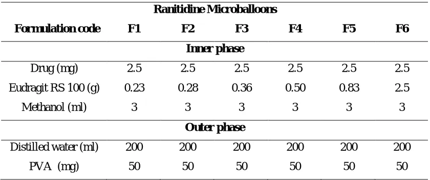
Table 1: Composition of microballoons containing Ranitidine Hcl
Ranitidine Microballoons
Formulation code F1 F2 F3 F4 F5 F6
Inner phase
Drug (mg) 2.5 2.5 2.5 2.5 2.5 2.5
Eudragit RS 100 (g) 0.23 0.28 0.36 0.50 0.83 2.5
Methanol (ml) 3 3 3 3 3 3
Outer phase
Distilled water (ml) 200 200 200 200 200 200
AJPER October - December 2015, Vol 4, Issue 4 (81-94) Evaluation of Microballoons :
Determination of Production Yield and Loading Efficiency
The production yield of the microparticles was determined by calculating accurately
the initial weight of the raw materials and the last weight of the microballoonsobtained.
The loading efficiency (%) of the microballoons can be calculated according to the
following equation:
Particle Size Analysis
Particle size analysis of prepared microballoons was carried by using Malvern
Particle Size Analyzer Hydro 2000 MU (A). Microballoons were dispersed in double
distilled water before running sample in the instrument, to ensure that the light scattering
signal, as indicated by particles count per second, was within instrument’s sensitivity range.
During the measurement, particles are passed through a focused laser beam. These
particles scatter light at an angle that is inversely proportional to their size. The angular
intensity of the scattered light is then measured by a series of photosensitive detectors. The
map of scattering intensity versus angle is the primary source of information used to
calculate the particle size. The scattering of particles is accurately predicted by the Mie
scattering model. The Mastersizer 2000 software, allows accurate sizing across the widest
possible dynamic range.
Scanning Electron Microscopy
For morphology and surface topography, prepared microballoons were coated with
platinum at room temperature so that the surface morphology of the microballoons could be
studied by SEM.
The SEM, a member of the same family of imaging is the most widely used of all
electron beam tools (Goldstein J. I., 2003). The SEM employs a focused beam of electrons,
with energies typically in the range from a few hundred eV to about 30 keV, which is across
AJPER October - December 2015, Vol 4, Issue 4 (81-94)
irradiation are collected, amplified, and then used to modulate the brightness of a suitable
display device which is being scanned in synchronism with probe beam.
Infrared Spectroscopy
FTIR spectroscopy was conducted using Perkin Elmer, Spectrum 100 FT-IR spectrometer.
Spectrum was recorded in the wavelength region of 4000 to 400 cm-1. The procedure
consisted of dispersing a sample in excess of potassium bromide nearly at the ratio 1:100,
mixed well, after which the mixture was kept into the sample holder for analysis.
Differential Scanning Calorimetry (DSC)
Thermal analysis is an important evaluation technique to find any possible interaction
between the drug and used polymers. Any of such interaction may reduce the drug
entrapment efficiency of the polymer and may also alter the efficacy of the drug. Such
interaction can be identified by any change in thermogram.
In-vitro Release Study of Microballoons
Accurately weighed loaded microballoons (5 mg) were placed in 50 ml of
ethanol/methanol in 100 ml glass bottles. The later were horizontally shaken at 37°C at
predetermined time intervals. Aliquot samples were withdrawn (replaced with fresh
medium) and analysed UV spectrophotometrically at 350 nm for ranitidine. The contents
of drugs were calculated at different time intervals up to 6hrs.
Results and Discussion:
Yield of the product
Percentage Yield
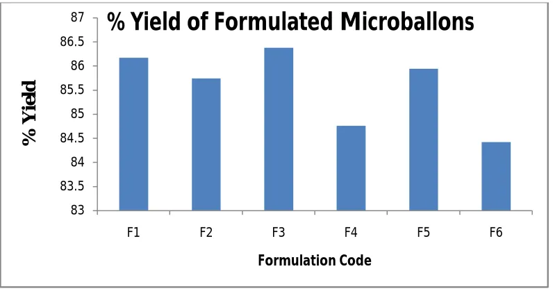
Table 2. Percentage yield of formulated microballoons
Formulation code Production yield (%)
F1 86.17±1.23
F2 85.74±2.28
F3 86.38±2.06
F4 84.76±1.52
F5 85.94±2.04
F6 84.42±1.24
AJPER October - December 2015, Vol 4, Issue 4 (81-94) Figure 1: Percentage yield of ranitidine microballoons formulations
Production yield of Ranitidine microballoons were between 84.42 to 87.73%. In case
of Eudragit RS 100 microballoons , it was revealed that, by increasing drug: polymer ratio
there is increase in the production yield of the microballoons .
Drug Loading Efficiency
Table 3: Drug loading efficiency of Salicylic acid microballoons formulations
Formulation code Drug Loading efficiency (%)
F1 86.17±1.13
F2 85.74±0.18
F3 84.38±1.24
F4 86.76±2.03
F5 87.94±1.28
F6 85.42±0.68
*Each value is average of three separate determinations ±SD
The loading efficiency was found to be high i.e. 84.38 to 88.73 % in ranitidine microballoons
it was found that as drug: polymer ratio increases, drug loading efficiency also increases.
83 83.5 84 84.5 85 85.5 86 86.5 87
F1 F2 F3 F4 F5 F6
%
Y
ie
ld
Formulation Code
AJPER October - December 2015, Vol 4, Issue 4 (81-94)
Figure 2: Loading efficiency of ranitidine microballoons formulations
Particle Size Analysis
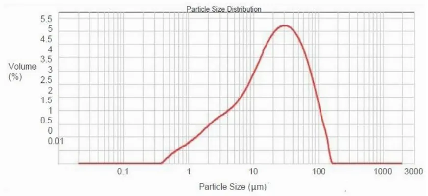
Figure 3: Particle size distribution of ranitidine microballoons (Mean particle size 39.92μm)
Free-flowing powders with fine aesthetic attributes are possible to obtain by controlling the
size of particles during both the polymerization methods. The mean particle size of ranitidine
microballoons found to be 39.92μm. 82
83 84 85 86 87 88 89
F1 F2 F3 F4 F5 F6
Lo
ad
in
g
Ef
fi
ci
en
cy
(
%
)
Formulation Code
AJPER October - December 2015, Vol 4, Issue 4 (81-94) Scanning Electron Microscopy
The morphology of the microballoons prepared by entrapment method and
quasi-emulsion solvent diffusion method were investigated by SEM. The representative SEM
photographs of the microballoons are shown in Figure.
SEM images showed that microballoons prepared by liquid-liquid suspension
polymerization method were finely spherical and uniform; no entire drug crystals were
observed visually.
Figure 4: SEM Photographs of Ranitidine Microballoons
Infrared Spectroscopy
FTIR spectra of ranitidine microballoons
AJPER October - December 2015, Vol 4, Issue 4 (81-94)
All characteristic peaks of drugs in the IR spectra of F 6 f o r m u l a t i o n were observed
to be concordant with respective pure drugs.
Differential Scanning Calorimetry (DSC)
The results of DSC were observed for the integrity of the drug in microballoons formulation
prepared by the entrapment process. In the DSC curve of selected F6 formulation, the
endothermic melting peak concerning ranitidine. According to this data, there was no
interaction between drug and Eudragit RS 100 in microballoons results showed that there
was no interaction between the drugs and the polymer.
AJPER October - December 2015, Vol 4, Issue 4 (81-94)
In-vitro Release Study of Microballoons
Table 4: In-vitro release study of ranitidine microballoons
Time
(Min) Cumulative % drug release
F1 F2 F3 F4 F5 F6
0 0 0 0 0 0 0
15 15.62±0.12 20.34±1.11 28.21±1.32 13.41±1.54 15.83±1.99 16.82±0.86
30 25.35±0.45 34.45±0.41 34.1±0.19 23.19±1.52 25.72±2.03 27.05±1.38
45 44.81±0.37 49.32±1.14 38.32±1.13 38.86±1.93 39.42±1.96 36.31±1.96
60 55.67±0.16 54.92±0.53 44.41±1.17 52.68±1.57 56.81±2.31 44.52±1.56
120 64.58±0.42 60.12±0.17 60.73±0.16 61.21±1.05 65.21±2.08 52.14±0.87
180 74.73±0.76 66.34±1.47 68.84±0.14 70.35±1.48 72.47±1.00 61.32±1.37
240 80.98±1.18 76.81±0.25 76.72±1.81 75.54±1.55 78.52±0.99 70.54±1.23
300 85.97±0.87 82.16±0.48 80.22±1.04 81.14±1.56 82.85±1.52 76.25±0.85
360 89.34±1.02 87.84±0.72 88.41±0.18 84.85±1.52 83.69±1.02 83.62±0.74
*Each value is average of three separate determinations ±SD
AJPER October - December 2015, Vol 4, Issue 4 (81-94)
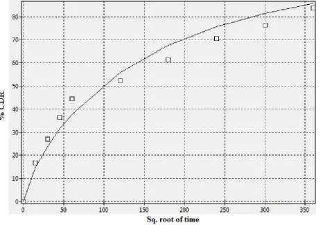
The drug release profiles of the ranitidine microballoons formulations are illustrated
in Table and Figure. Drug release from ranitidine microballoons was found to range from
81.32 % to 89.34 % from all formulations.
From the results it was found that, as concentration of polymer increases, percentage
of drug released decreases. The initial high drug release could be due to two reasons: first,
the drug near or on the surface of the microballoons and second, well known porous nature
of microballoons , the pores providing a channel for release of the drug (Mandal T. K.,
2001).
The microballoons differ from regular microspheres with their highly porous surface.
This characteristic gives property to release the drug at a faster rate through the pores.
Kawashima reported that microballoons having a more porous internal structure, exhibited
a faster drug release rate than that of rigid microspheres (Kawashima Y., 1992). Release
from F6 formulation has Higuchi release pattern followed zero order reaction kinetics (R2=
0.948, 0.965 and 0.983).
AJPER October - December 2015, Vol 4, Issue 4 (81-94) Conclusion:
From the overall investigation, one can conclude that the optimized microballoons of
ranitidine using both polymers can meet ideal requirements for microballoons. The relatively
high percentage yield and loading efficiency of microballoons indicated that the method is
suitable for preparing the microballon formulations. Quasi-emulsion solvent diffusion
method is simple, less time consuming and involves use of safer ingredients than free radical
polymerization and hence more preferred.
The microballoons differ from regular microspheres with their highly porous surface.
This characteristic gives property to release the drug at a faster rate through the pores. Due to
smaller pore diameter, the Eudragit Rs 100 microballoons showed less and slower drug
release in the in-vitro release studies. Release from all the microballoons followed zero order
reaction kinetics.
In future, the formulations can used as controlled release dosage form for better
bioavailability and improved patient compliance.
Acknowledgement
The authors are thankful to Sagar institute of research technology & science-pharmacy Bhopal, for providing necessary facilities to carry out the work.
Reference:
1. Jinuk K, Jinyoung K, Dongmyung P, Haksoo H. A novel synthesis method for an
open-cell microsponge polyimide for heat insulation. Polymer. 2015;56: 68-72
2. Jelvehgari M, Siahi-Shadbad MR, Azarmi S, Martin GP and Nokhodchi A. The
microsponge delivery system of benzoyl peroxide: Preparation, characterization and
release studies. International Journal of Pharmaceutics. 2006;308(1–2): 124-132.
3. Iwai S, Sawa Y, Ichikawa H, Taketani S, Uchimura E, Chen G,Hara M, Miyake J and
Matsuda H. Biodegradable polymer with collagen microsponge serves as a new
bioengineered cardiovascular prosthesis. The Journal of Thoracic and Cardiovascular
Surgery. 2004;128(3): 472-479.
4. Siepmann J and Siepmann F. Microparticles Used as Drug Delivery Systems.
Program Colloid PolymerSci. 2006;133: 15–21.
5. Majeti N and Ravi Kumar V. Nano and Microparticles as Controlled Drug Delivery
AJPER October - December 2015, Vol 4, Issue 4 (81-94)
6. Dey NS, Majumdar S and Rao MEB. Multiparticulate Drug Delivery Systems for
Controlled Release. 2009; 1826-1837. Available online at http://www.tjpr.org.
7. Reddy JR, Gnanaprakash K, Badarinath AV, Madhusudhanachetty C. Formulation
and Evaluation of Microparticles of MetronidazoleIn J. Pharm. Sci. & Res. 2009;
1:131-136.
8. Padalkar AN, Shahi1 SR and Thube MW. Microparticles: An Approach For
Betterment of Drug Delivery System. 2011; ijprd//pub/arti/vov-3/issue-1/march/012.
9. Bhadke SE. Formulation and Development of Repaglinide Microparticles by
Ionotropic Gelation Technique. Thesis at Dept of Pharmaceutical sciences Hubli.
2007; 1-129.
10.Duane Birnbaum T and Brannon-Peppas L. Microparticle Drug Delivery Systems,
Drug Delivery Systems in Cancer Therapy Edited by: D. M. Brown © Humana Press
Inc., Totowa, NJ117-136/Brown.Ch. 2003;06: 1- 117.
11.Bansode SS, Banarjee SK, Gaikwad DD, Jadhav SL and Thorat RM.
Microencapsulation: A Review International Journal of Pharmaceutical Sciences
Review and Research. 2010;1(2):8-15.
12.Yadav AV, Shete AS, Dabke AP and Shinde VR. Formulation and In-vitro
Evaluation of Aceclofenac Microcapsules, International Journal of PharmTech
Research. 2009;1(2):135-138.
13.Ofokansi KC and Adikwu MU. Formulation and Evaluation of Microspheres Based
on Gelatin-Mucin Admixtures for the Rectal Delivery of Cefuroxime Sodium.
Tropical Journal of Pharmaceutical Research. 2007; 6(4): 825-832.
14.Simon B. Microencapsulation: Methods and industrial applications, 2nd ed. Drugs
Pharmaceutical Sci., Marcel Dekker, Inc. N.Y. 2006; 158: 1-55.
15.Xun LD and Dong Soo KJO. Development of Nifedipine-loaded coated gelatin
microcapsule as a long acting oral delivery. Refdocest UN service.
2009;32(1):127-132.
16.Nokhodchi A and Farid D. Microencapsulation of Paracetamol by Various Emulsion
Techniques Using Cellulose Acetate Phthalate. Pharmaceutical Technology. 2002;6:
54-60.
17. Chowdary KPR and Nagarajan M. Microencapsulation of Nifedipine-MCC solvent
deposited system for sustained release. Indian Journal of Pharmaceutical Sciences.
AJPER October - December 2015, Vol 4, Issue 4 (81-94)
18.Chowdary KPR, Mohapatra P and Murali Krishna MN. Pharmacokinetic Evaluation
of Natural Resin Coated Microcapsules of Nifedipine. Asian Journal of Chemistry.
2009;21(6): 4199-4204.
19.Deore BV, Mahajan HS and Deore UV. Development and characterization of
sustained release microspheres by quasi emulsion solvent diffusion method.
International Journal of Chem Tech Research. 2009;1(3): 634-642.
20.Cilurzo F, Mangetti P, Caseraghi A and Montanari L. Characterization of Nifedipine
Solid Dispersions. Int.J. of Pharmaceutics. 2002;242:313-317.
21.Arias MJ, Gines JM, Moyano JR and Rabasco AM. Dissolution properties and invivo
behavior of triamterene in solid dispersions with poyethelyne glycols. Pharmaceutica
Acta Helvetiae 1996;7:229-235.
22.Umamahesh B, Lavanya N, Kumar KP and Guggilla SR. Design and evaluation of
gelatin microspheres containing diclofenac sodium, IJPDT. 2012; 2(1),11-14.
23.Mankala SK. Nagamalli NK, Raprla R and Komulla R. Preparation and
characterization of mucoadhesive microcapsule of glicazide with natural gums.
Stamford journal of Pharmaceutical sciences. 2004;4(1): 38-48.
24.Pandey V and Bhadoria S. Formulation, Development & Optimization of
Pioglitazone HCl Microsphere using ionotropic gelation technique. Pharmacia. 2011;
I(1): 67-74.
25.Sivanarayana V, Kishore SP and Kumar J. Effect of crosslinking agent and polymer
on the characteristics of Diltiazem HCl loaded mucoadhesive microsphere. American
Journal of Pharmatech Research. 2012; 2(1), 398-410.
26.Rasala TM, Kale VV, Bhalekar MR and Avari JG. Formulation and evaluation of
mucoadhesive microcapsule of Diltiazem HCl and diclofenac sodium by orifice ionic
gelation method. IJPI’S Journal of Pharmaceutics and Cosmetology. 2010; 1(1): 1-8.
27.Sambathkumar R, Venkateswaramurthy N, Vijayabaskaran M, Perumal P,
Formulation of clarythromycin loaded mucoadhesive microsphere by emulsion
internal gelation technique for antihelicobacter pylori therapy. International Journal